- 1Department of Ultrasonography, Maternity and Child Health Care of Guangxi Zhuang Autonomous Region, Nanning, China
- 2Graduate School, Guangxi University of Chinese Medicine, Nanning, China
- 3Birth Defects Prevention and Control Institute of Guangxi Zhuang Autonomous Region, Nanning, China
- 4Maternity and Child Health Care of Guangxi Zhuang Autonomous Region, Nanning, China
Objective: to evaluate the research status and development hotspots of fetal α-thalassemia by quantitatively analyzing the diagnostic status, key areas, related management measures and prospects of the disease by bibliometrics.
Methods: The global literature on fetal α-thalassemia and severe α-thalassemia from 2009–2023 in the Web of Science Core Collection (WOSCC) was visually analyzed by VOSviewer and CiteSpace.
Results: (1) The examination of the quantity of publications concerning fetal α-thalassemia indicates a rising tendency prior to 2018, followed by a decrease after 2018. (2)The United States, China, Italy, Thailand have published more papers, and the United States has more collaborating countries such as Italy and China. (3) Chiang Mai University and Harvard University are the top two institutions with the highest contribution. However, Chiang Mai University's H index (12) and citation frequency per article (8.05) are relatively low and the NC (6,342), H index (33) and citations per article (75.42) of Harvard University are higher than those of the other institutions. (4) Tongsong T, Gambari R and Fucharoen S are the top three prolific authors. Fucharoen S emerges as the most frequently cited author with 738 citations, excluding self-citations. (5) HEMOGLOBIN leading with 87 published papers (NC:601,IF: 0.82, H-index: 13), followed by BLOOD(58 papers, Nc: 3755, IF: 25.48, H-index: 40) and BLOOD CELLS MOLECULES AND DISEASES(39 papers, Nc: 729, IF: 2.37, H-index: 16). (6) The most cited article was published in science and the second and third cited articles were featured in the Proceedings of the National Academy of Sciences; the top 3 clusters of co-cited literature are “gene editing”, “polymorphisms”, “hydroxyurea”. (7) Keywords analysis showe that the top two categories of keyword cluster focus on the prenatal diagnosis and the current treatment strategy of the disease, which remain the research hotspots.
Conclusions: Recent research on this topic has primarily focused on prenatal diagnosis and treatment strategies. A particular area of interest is the ongoing research on gene therapy.The advances in non-invasive diagnosis and therapeutic methods will change the current management approaches for fetal severe α-thalassemia in the future.
1 Introduction
Alpha-thalassemia(α-thalassemia) is a notable issue in global health (1). Severe α-thalassemia, also called Hemoglobin Bart's hydrops fetalis syndrome, is a hemoglobinopathy resulted from inactivating or deleting all four α-globin alleles. This syndrome is commonly found in Southeast Asia and is characterized by a high incidence of fetal edema. Hemoglobin Bart's (γ4, Hb Bart's) has an unusually strong affinity for oxygen and cannot effectively deliver oxygen to fetal tissue (2, 3). The fetus may present with hypoxia, anemia, edema, death, or stillbirth. Furthermore, continued pregnancy increases the risk of severe complications in pregnant women, including preeclampsia, dystocia, and postpartum hemorrhage, and others (4). The prognosis of the disease is unfavorable, and the current clinical approach involves early prenatal diagnosis and induction of labor. Quantitative analysis of the status quo, critical aspects, associated management strategies, and prospects of fetal α-thalassemia holds substantial importance for efficient management.
Bibliometrics is a method of quantifying and evaluating the numbers and contents in a publication (5, 6). The bibliometric index include numbers of publications (Np), numbers of citations (Nc) which serve as measures of the annual global citation frequency of a publication, the H-index which combines publication frequency and citation frequency to create a commonly used influence index (7, 8), and the impact factor (IF) which is a valuable tool for assessing the influence and quality of journals. Based on the analysis of the characteristics of data and literature, this paper evaluates and predicts the development trend and hot research direction in the research field, guides the experimental strategy and fund decision-making, and provides effective evidence (9). Presently, bibliometric methods have been utilized to summarize the research on low-intensity ultrasound, breast cancer radiotherapy, polycystic ovary syndrome (10–12), and so on. However, bibliometric studies on α-thalassemia have not yet been carried out. The aim of this study was to explore the researches on fetal α-thalassemia, and to evaluate the research status and development hotspots in this field.
2 Materials and methods
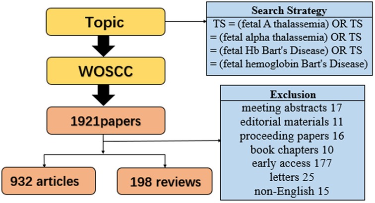
In this research, the Web of Science Core Collection (WOSCC) with Citation Index (CI) was utilized for literature retrieval. To mitigate potential bias stemming from the rapid database updates, literature retrieval was performed on the same day to capture the comprehensive global literature on fetal α-thalassemia and severe α-thalassemia types spanning from January 2009 to December 2023, encompassing a 15-year period. The search strategy employed was as follows: TS = (fetal A thalassemia) OR TS = (fetal alpha thalassemia) OR TS = (fetal Hb Bart's Disease) OR TS = (fetal hemoglobin Bart's Disease). The data was exported as a plain text file, with CiteSpace utilized for time span clustering analysis and literature citation burst analysis. The Java program VOS viewer was employed for literature co-citation analysis, keyword co-occurrence network, and author cooperation analysis. When using VOS viewer analysis, we set the minimum number of documents or citations of each analysis content, so that the threshold reaches the maximum value less than 200.
3 Results
3.1 Trend of annual publication number of fetal α-thalassemia
The final screening of 1,130 valid literatures included only works and reviews written in English from the retrieved publications. The detailed process is illustrated in Figure 1. Figure 2A depicts the variation in the Np pertaining to fetal α-thalassemia and the change curve of citation frequency over the course of the last 15 years. The examination of the quantity of publications concerning fetal α-thalassemia indicates a rising tendency prior to 2018, followed by a decrease after 2018. The number of publications escalated from 67 in 2009 to 110 in 2018, subsequently dwindling to 39 in 2023. The citation frequency continued to rise, peaked in 2021, and then showed a slight downward trend. The correlation coefficient between the annual quantity of publications and the multiple fitting curve of the publication year is 0.5716, as demonstrated in Figure 2B.

Figure 2. Publications and citations of each year in the past 15 years (A). Fits the relationship curve between Np and publication year (R2 = 0.5716) (B).
3.2 Trend analysis of national and regional publications
A comprehensive review of the literature indicates that 69 countries have contributed articles in this field between 2009 and 2023. The United States emerged as the leading contributor with 334 papers, representing 29.56% of all publications; the following countries are China, Italy, Thailand, and others, as shown in Table 1. Figure 3A shows the regional distribution of articles published in this field. The combination of Figure 3A,B illustrates the correlation between the quantity of publications and the size of nodes, as well as the extensive level of collaboration among nations. Notably, the United States, China, and Italy are represented by larger nodes, with the United States demonstrating a higher overall level of connectivity. Furthermore, the United States engages in collaborative efforts with 46 countries, including particularly close partnerships with Italy, China, and the United Kingdom. Figure 3C shows the changes in the annual number of documents issued by each country intuitively. A citation bursts analysis of Citespace reveals that Pakistan has experienced the most rapid growth in terms of publications since 2018 and is the country with the most significant increase in influence. However, the Netherlands is the country with the highest incidence of sudden citation, which was in an outbreak state from 2012–2014, as presented in Figure 3D.

Table 1. The top 10 countries with the highest number of publications in the field of fetal α-thalassemia research.
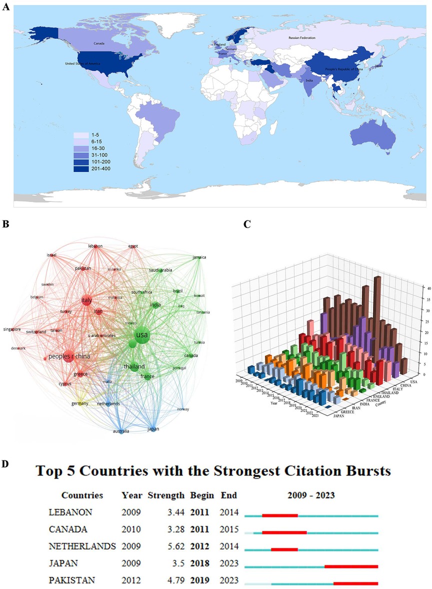
Figure 3. Global geographical distribution map of countries in the field of fetal α-thalassemia research (A). National co-citation analysis map of fetal α-thalassemia research (B). A 3D histogram of the number of papers published by countries in the study of fetal alpha-thalassemia in the past 15 years (C). Analysis map of national emergent value in the field of α-thalassemia research (D).
3.3 Analysis of affiliation relationship
The top 10 institutions were summarized according to the statistics of the affiliated institutions of the published literature related to fetal α-thalassemia in the past 15 years (Table 2). Among the institutions, Chiang Mai University in Thailand stands out as having published the most researches (55). However, its H index (12) and citation frequency per article (8.05) are relatively low. The institutions with the most papers were predominantly from the USA (70%, 7/10). The NC (6,342), H index (33), and citations per article (75.42) of Harvard University are relatively higher than those of the other institutions. The collaboration among institutions is characterized by a close relationship, delineated into nine clusters as depicted in Figure 4A. When considering the temporal distribution of institutional publications (Figure 4B) and the frequency heat map of institutional publications (Figure 4C), it is evident that CHIANG MAI UNIVERSITY and MAHIDOL UNIVERSITY in Thailand initiated research activities at an earlier stage, as indicated by a concentrated red area on the heat map. In contrast, GUANGZHOU MEDICAL UNIVERSITY in China commenced research activities at a later stage, also reflected by a concentrated red area on the heat map.

Table 2. The top 10 institutions with the highest number of publications in the field of fetal α-thalassemia research.
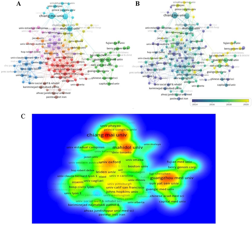
Figure 4. Co-occurrence analysis map of publishing institutions (A). Time map of co-occurrence analysis of publishing institutions (B). Density map of co-occurrence analysis of publishing institutions (C).
3.4 Analysis of authors of published literature
The data presented in Table 3 reveals the top ten authors with the highest Np in the field over the last 15 years. Tongsong T, Gambari R and Fucharoen S are the top three prolific authors. Fucharoen S emerges as the most frequently cited author with 738 citations, excluding self-citations. The following closely are Gambari R and Borgatti M, with 534 and 525 citations respectively. As depicted in Figure 5A,B, the author and the author's cited visual analysis of keyword clustering suggest that these individuals serve as central figures within their respective organizations. Moreover, their academic pursuits in the field began in the years 2014, 2015, and 2015 respectively, with the most intense correlation between their nodes, suggesting a higher level of collaboration and highlighting their crucial role in the research of fetal α-thalassemia. Among the top 10 authors with the most citation bursts, Tongprasert F, Nakamura Y, and Srisupundit K are the most prominent, with Nakamura, Y experiencing a notable increase in citations since 2018, as illustrated in Figure 5C.

Table 3. The top 10 authors with the highest number of publications in the field of fetal α-thalassemia research.
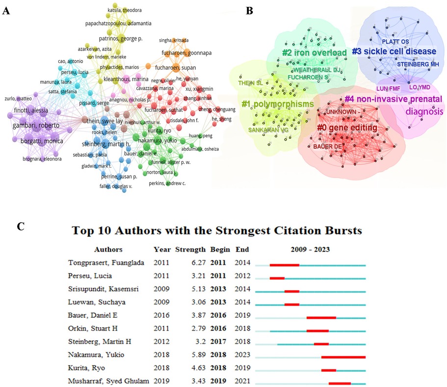
Figure 5. Co-citation analysis map of authors in the field of fetal α-thalassemia research. (A) Cluster diagram of cited keywords of authors (B) The author's emergent value analysis map in the field of fetal α-thalassemia research (C).
3.5 Published journal analysis
This field has been covered in 200 journals over the past 15 years. The analysis focused on the ten most productive journals (Table 4), with HEMOGLOBIN leading with 87 published papers (IF: 0.82, H-index: 13), followed by BLOOD and BLOOD CELLS MOLECULES AND DISEASES. However, BLOOD displayed the highest non-self-cited literature citation frequency (Nc: 3755, IF: 25.48, H-index: 40), and average number of citations (66.09). Following closely behind was the BRITISH JOURNAL OF HAEMATOLOGY. The data presented in Figure 6A,B show the changes in the annual Np of the top five journals. Specifically, HEMOGLOBIN stands out with a significantly higher Np compared to other top five journals in 2016. During the period from 2011–2016, most of the journals experienced a peak in publications, suggesting a heightened focus on the respective fields. The primary subjects covered by these journals include hematology, cell biology, sports science, parasitology, and obstetrics and gynecology. Figure 6C illustrates the clustering of journals based on subject matter, providing researchers with guidance on selecting appropriate publications for their work.
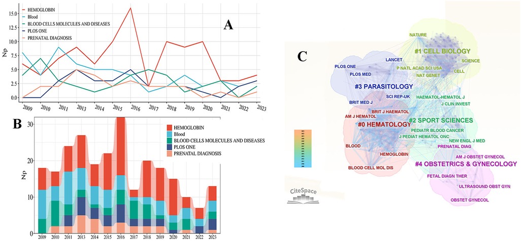
Figure 6. The annual growth curve of the number of publications of the top 5 journals (A). The annual publication volume stack diagram of the top 5 journals (B). Journal subject category clustering diagram of published articles (C).
3.6 Literature co-citation cluster analysis
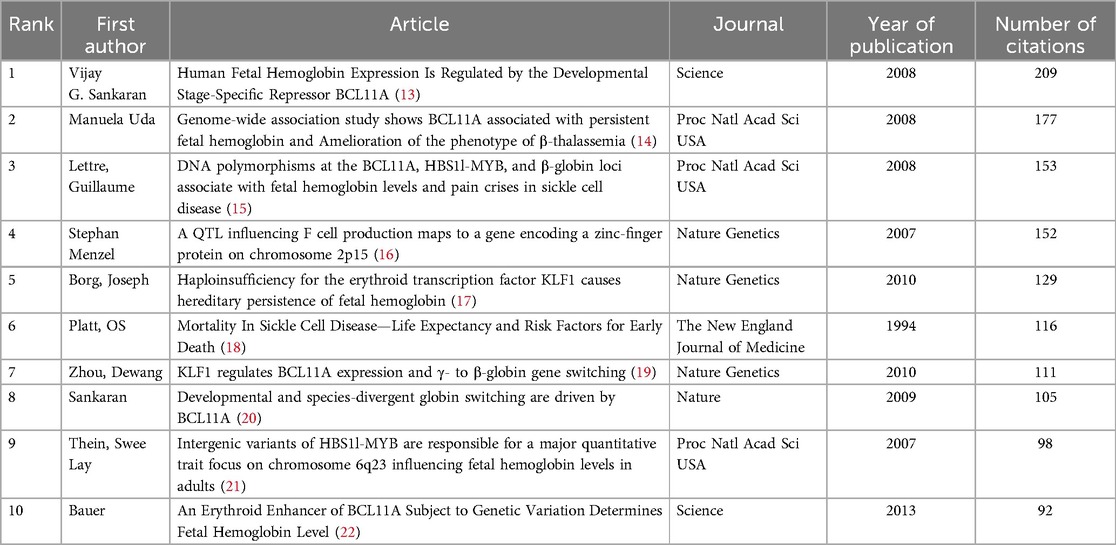
An analysis of the referenced literature spanning the last 15 years indicates that the majority of the top ten cited articles were published between 2007 and 2010, as shown in Table 5. The foremost article (13), published in Science, explores the role of BCL11A as a potential regulator of HbF expression, offering a new target for the treatment of thalassemia. It's primary role is to stimulate HbF reactivation and elevate fetal hemoglobin (HbF) levels through modulation of F cell production. The second and third most cited articles were featured in the Proceedings of the National Academy of Sciences (PNAS) (14, 15). The citation burst observed in Figure 7A highlights the scholarly works that are of particular interest to researchers during the specified period. Article (23) published in Nature Genetics has experienced a notable surge in influence within the field since 2019, characterized by a period of exponential growth. Figure 7B shows that the top 7 clusters of co-cited literature are “gene editing”, “polymorphisms”, “hydroxyurea”, “erythroid differentiation”, “hemoglobin switching”, “klf1”, “sirolimus”.
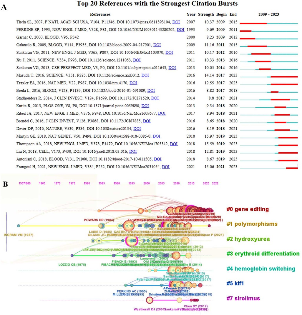
Figure 7. Representative top 20 papers with the strongest burst citations (A). Co-cited literature keyword clustering visualization map (B).
3.7 Keyword cluster analysis and time series analysis
A keyword cluster analysis and a time series analysis were conducted on the frequency of citation of keywords in the field of fetal α-thalassemia research spanning the last 15 years. The analysis, presented in Table 6 and Figure 8A, identified the top ten most cited keywords as “fetal hemoglobin”, “sickle cell disease”, “transcription”, “gene therapy”, “expression”, “α-thalassemia”, “hydroxyurea”, “anemia”, “prevalence”, and “severity”. In Figure 8B, keywords categorized by their average publication year (APY), with red indicating more recent keywords from 2018 such as “classification”, “prognosis”, and “survival”. The heat map in Figure 8C illustrates the average frequency of keywords, with red indicating keywords that appear more frequently in the literature. Based on the examination of the top ten keywords (Figure 8D), it was determined that “fetal hemoglobin production” exhibited the highest intensity of outbreak, while “gene therapy” demonstrated the longest duration of outbreak until the year 2023. Additionally, the keyword co-citation time series analysis map (Figure 8E) indicated that cluster 0, focusing on prenatal diagnosis, is currently the primary area of research interest. Cluster 1 pertains to fetal hemoglobin, cluster 2 to erythrocyte differentiation, cluster 3 to bcl11a, cluster 4 to diagnosis, and cluster 5 to iron chelation. Combined with the above analysis, the predominant studies have focused on this domain, encompassing maternal plasma composition, gene therapy, erythrocyte differentiation, genetic modifiers, case reports, and current treatment strategies. In addition, researchers are encouraged to investigate potential treatment strategies for fetal α-thalassemia through the utilization of animal experiments.
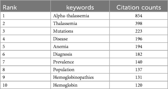
Table 6. The top 10 keywords with the highest number of publications in the field of fetal α-thalassemia research.
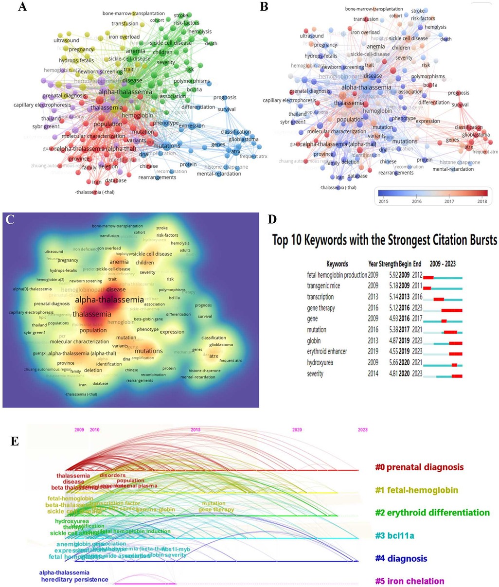
Figure 8. Keyword analysis map of α-thalassemia in fetus (A). Keyword visualization based on APY (B). Heatmap of keywords according to the average frequency of occurrence (C). Top 10 keywords with the most vigorous citation bursts (D) timeline distribution of keyword clustering in the field of fetal α-thalassemia research (E).
4 Discussion
This study presents a summary of the current research status of fetal α-thalassemia integrating bibliometrics and visual analysis. It delineates the emerging trends and identifies the most promising research avenues in this field. A quantitative analysis of global publication volume reveals a notable increase in the number of publications prior to 2018. This growth in annual publication volume is inextricably linked to the advancement of basic research and clinical trials. Based on the analysis of publications by national researchers, the countries such as the United States, China, Italy, and Thailand, where the most publications were published are largely congruent with the distribution of thalassemia diseases, were concentrated in tropical and subtropical regions. The United States exhibits the highest H-index (67) and Nc values (14,200). The majority of the top ten institutions with the most papers were predominantly from the USA, particularly HARVARD UNIVERSITY, suggesting the country's significant depth of academic research and strong scholarly capabilities in this field. Furthermore, the United States' extensive collaborative partnerships with other countries contribute to its elevated H-index and overall scholarly influence. Thailand is a country with a high prevalence of α-thalassemia and has the most published institutions CHIANG MAI UNIVERSITY and outstanding professional researchers, such as Tongsong T, Fucharoen S and Luewan S. However, its H index and Nc are low, and attention should be paid to improving the cooperation among countries. In this field, China stands out as one of the leading contributors, with numerous papers and extensive research activities, and GUANGZHOU MEDICAL UNIVERSITY being a recent contributor. Despite the large volume of publications in a brief period of time, particularly in the field of genetic molecular science, the H-index and Nc metrics of China are lower than those of Italy, which Np are lower than China. It suggests that the quality and impact of Chinese research outputs require enhancement (24, 25).
Among the journals with numerous publications, nine have higher impact factors (greater than 1). With the most articles published, HEMOGLOBIN has a lower IF (0.82), while the second-ranked BLOOD has the highest IF (25.48) and H index (40). The influences of these journals are so high, that it suggests that researchers interested in this field should pay attention to these journals and relevant authors. By examining the pertinent literature published in the journal, it is evident that BLOOD and HEMOGLOBIN are publications associated with hematology, while CELL and P NATL ACAD SCI USA are journals focused on cell biology, and PRENATAL DIAG is a journal specializing in obstetrics and gynecology. Therefore, scholars could contribute to their respective scientific disciplines, engage in discussions, and share knowledge with fellow academics.
The co-cited authors are categorized into 9 distinct clusters, allowing researchers to identify authors whose work aligns with their own research focus through the network of connections. By integrating the keyword clustering of cited authors, researchers can identify authors specializing in specific research areas, such as Bauer in the realm of gene editing, LUN F in the realm of non-invasive prenatal diagnosis. Although Fucharoen S may not have the highest number of published papers, his H-index (16) and Nc (738) are the highest in their field, establishing him as a scientist of significant influence. Similarly, Nakamura Y stands out for having the highest citation intensity and longest duration since 2018. By examining the achievements of these authors, researchers can gain valuable insights to develop new ideas and methods, ultimately fostering innovation in the field. The literature co-citation cluster analysis reveals that most highly cited papers (60%) pertained to the fundamental research of BCL11 A. Furthermore, the top ten articles with the strongest citation burst were published after 2016, indicating that the related topics have received considerable attention in recent years. It is noteworthy that the majority of these publications pertain to the expression of genes that affect fetal hemoglobin (23, 26–27).
Keyword clustering analysis is the key content of this paper. The first category of keyword cluster analysis focuses on prenatal diagnosis of the disease, which appears earliest and remains a hotspot for research. In the current system, pregnant women are tested for their hemoglobin levels and their red blood cell levels. If positive, further genetic testing is performed. If they are diagnosed as thalassemia gene carriers, their spouses should also take this exam. If both are carriers of the same type of thalassemia gene, they are high-risk couples, that is, their offspring may have severe type of thalassemia. Then DNA detection technology is necessary to identify fetal genotypes and gene mutation types (28–30). Srisupundit K has authored numerous scholarly articles in the discipline of hematology (31–33). Invasive methods such as villus biopsy, amniocentesis and umbilical cord puncture are used for sampling and then testing (34). The primary method for molecular diagnostics is polymerase chain reaction (PCR) (35–38). Nevertheless, conventional molecular diagnostic methods are intricate, offer restricted probe coverage, and are time-consuming. China's contributions to this area have advanced the utilization of Next-Generation Sequencing (NGS) technology for genetic screening of thalassemia (39–41). Several studies have suggested that third-generation sequencing (TGS) technology offers a substantial enhancement in the detection rate of thalassemia genes with Mediterranean gene variation and new structural variation when compared to NGS technology (42, 43). In recent years, there has been a growing interest among scholars in the advancement of high-throughput sequencing technology, which has facilitated quicker and more comprehensive diagnosis of thalassemia.
Non-invasive prenatal diagnosis (NIPD) encompasses noninvasive genetic diagnosis, noninvasive ultrasound prenatal diagnosis and maternal serological indicators. The non-invasive genetic diagnosis technology detects thalassemia genes by analyzing fetal nucleated red blood cells (FNR-BC) and cell-free fetal DNA (cffDNA) in maternal blood (44–48). However, the accuracy of this technology is constrained by the intricate nature of the procedure and the scarcity of fetal cells in the peripheral blood of pregnant individuals, leading to its limited adoption in clinical settings. Since the signs of increased fetal blood flow, enlarged heart, thickened placenta appear in severe α-thalassemia fetuses can be observed by ultrasound before the onset of hydrops symptoms, these markers can be served as predicting markers for them in high-risk pregnancies; and it has been proven that this non-invasive ultrasound markers are both cost-effective and efficient (49–51). Fetal cardiothoracic ratio (CTR) has been recognized as a highly effective predictor, with a commonly utilized threshold range of 0.50–0.52 (52, 53). Researches conducted in China have demonstrated that the Z score of fetal heart size may serve as a novel and sensitive indicator for identifying severe α-thalassemia (54, 55). Another study also found that the Z value of fetal cardiac volume (CV) to predict this disease may be better than CTR (56). For maternal serological indicators, Tongprasert F and other researchers have identified maternal serum alpha fetoprotein (AFP), placental like growth factor (PlGF), inhibin-A, and other biomarkers as potentially effective in predicting fetal α-thalassemia major during the second trimester (57–60).
The second category of keyword clustering focuses on the current treatment strategy of the disease. Fetuses diagnosed with severe α-thalassemia generally need to terminate in time due to poor prognosis and high incidence of complications in pregnant women. With the progress of modern medicine, the possible treatment options include intrauterine transfusion (IUT) and postnatal bone marrow transplantation. While some rare cases have been effectively managed prenatally (61–63), some of them may experience complications such as chronic hypoxemia, postnatal respiratory failure, and pulmonary hypertension (63). Hematopoietic stem cell transplantation (HSCT) for severe α-thalassemia is practical option with a high cure rate (64, 65). However, due to factors such as rejection and donor sources, intrauterine HSC transplantation has not yet been successful (66). Choosing the appropriate HSC source, optimizing the scheme, and timing of intrauterine transplantation are still challenging. Another potentially promising therapy is gene therapy such as gene editing (67); virus vector; small RNA interference, etc, which is more promising to intervene in fetus. Constrained by donor availability and limited funding, current research and development efforts in gene therapy primarily concentrate on β-thalassemia (68). Although the progress of gene therapy for severe fetal α-thalassemia is slow, clinical trials are gradually being carried out (69). A studiy have found the possibility of applying protein replacement therapy to α-thalassemic (HBH) by PTD technology to recombine α-globin chains (70). Although there have not been any reported cases of fetal α-thalassemia treated with gene therapy to date, we believe that the advances in genetic technology may have the potential to revolutionize current management approaches for fetal severe α-thalassemia in the future (69, 71).
This study utilized bibliometric analysis to explore the theme of fetal α thalassemia (72), focusing solely on SCI-extended English articles and reviews. However, this approach may exclude recently published high-quality articles with limited citations, thereby introducing certain limitations. Citespace focuses on individual publication analysis and Vosviewer emphasizes cluster analysis (73), both software tools are unable to analyze the complete text of articles, potentially leading to information omission and delays.
5 Conclusion
Our findings suggest that research on fetal α-thalassemia has developed steadily, with a concentration of literature in papers. Fucharoen S is identified as the author with the highest citation frequency of non-self-cited literature. The United States leads in both publications and citations in this field, with HARVARD UNIVERSITY having the highest H index. The journal BLOOD stands out for its high citation frequency of non-self-cited literature. The primary areas of focus in fetal α-thalassemia research include prenatal diagnosis and disease management, epidemiological studies, genetic investigations, molecular biology and intervention, and current treatment modalities. A particular area of interest is the ongoing research on treatment strategies, with gene therapy representing current frontier research in the field. The advances in non-invasive diagnosis and therapeutic methods will change current management approaches for fetal severe α-thalassemia in the future.
Data availability statement
The original contributions presented in the study are included in the article/Supplementary Material, further inquiries can be directed to the corresponding author.
Author contributions
QL: Data curation, Visualization, Writing – original draft. XL: Funding acquisition, Writing – review & editing, Supervision. SH: Writing – review & editing, Supervision. JL: Writing – review & editing, Supervision.
Funding
The author(s) declare financial support was received for the research, authorship, and/or publication of this article. The study was supported by the National Natural Science Foundation of China (Grant/Award No. 82060322), the Natural Science Foundation of Guangxi Zhuang Autonomous Region (Nos. 2020GXNSFAA297066), and the open topic of Guangxi Key Laboratory of Birth Defects and Stem Cell Biobank (Maternal and Child Health Hospital of Guangxi Zhuang Autonomous Region) (GXWCHZDKF-2023-08).
Acknowledgment
We sincerely thank Wang Linlin of the Maternal and Child Health Hospital of Guangxi Zhuang Autonomous Region for her valuable modification suggestions for this study.
Conflict of interest
The authors declare that the research was conducted in the absence of any commercial or financial relationships that could be construed as a potential conflict of interest.
Publisher's note
All claims expressed in this article are solely those of the authors and do not necessarily represent those of their affiliated organizations, or those of the publisher, the editors and the reviewers. Any product that may be evaluated in this article, or claim that may be made by its manufacturer, is not guaranteed or endorsed by the publisher.
References
1. Musallam KM, Lombard L, Kistler KD, Arregui M, Gilroy KS, Chamberlain C, et al. Epidemiology of clinically significant forms of alpha- and beta-thalassemia: a global map of evidence and gaps. Am J Hematol. (2023) 98(9):1436–1451. doi: 10.1002/ajh.27006
2. Laosombat V, Viprakasit V, Chotsampancharoen T, Wongchanchailert M, Khodchawan S, Chinchang W, et al. Clinical features and molecular analysis in Thai patients with HbH disease. Ann Hematol. (2009) 88(12):1185–92. doi: 10.1007/s00277-009-0743-5
3. Hanprasertpong T, Kor-anantakul O, Leetanaporn R, Suntharasaj T, Suwanrath C, Pruksanusak N, et al. Pregnancy outcomes amongst thalassemia traits. Arch Gynecol Obstet. (2013) 288(5):1051–4. doi: 10.1007/s00404-013-2886-9
4. Jatavan P, Chattipakorn N, Tongsong T. Fetal hemoglobin Bart’s hydrops fetalis: pathophysiology, prenatal diagnosis and possibility of intrauterine treatment. J Matern Fetal Neonatal Med. (2018) 31(7):946–57. doi: 10.1080/14767058.2017.1301423
5. Wallin JA. Bibliometric methods: pitfalls and possibilities. Basic Clin Pharmacol Toxicol. (2005) 97(5):261–75. doi: 10.1111/j.1742-7843.2005.pto_139.x
6. Dong Y, Chen S, Wang Z, Ma Y, Chen J, Li G, et al. Trends in research of prenatal stress from 2011 to 2021: a bibliometric study. Front Pediatr. (2022) 10:846560. doi: 10.3389/fped.2022.846560
7. Hu G, Wang L, Ni R, Liu W. Which h-index? An exploration within the web of science. Scientometrics. (2020) 123(3):1225–33. doi: 10.1007/s11192-020-03425-5
8. Gianoli E, Molina-Montenegro MA. Insights into the relationship between the h-index and self-citations. J Am Soc Inf Sci Technol. (2009) 60(6):1283–5. doi: 10.1002/asi.21042
9. Chen P, Lin X, Chen B, Zheng K, Lin C, Yu B, et al. The global state of research and trends in osteomyelitis from 2010 to 2019: a 10-year bibliometric analysis. Ann Palliat Med. (2021) 10(4):3726–3738. doi: 10.21037/apm-20-1978
10. Jia B, Lim D, Zhang Y, Dong C, Feng Z. Global research trends in radiotherapy for breast cancer: a systematic bibliometric analysis. Jpn J Radiol. (2023) 41(6):648–59. doi: 10.1007/s11604-022-01383-x
11. Chang H, Wang Q, Liu T, Chen L, Hong J, Liu K, et al. A bibliometric analysis for low-intensity ultrasound study over the past three decades. J Ultrasound Med. (2023) 42(10):2215–2232. doi: 10.1002/jum.16245
12. Xu Y, Cao Z, Chen T, Ren J. Trends in metabolic dysfunction in polycystic ovary syndrome: a bibliometric analysis. Front Endocrinol. (2023) 14:1245719. doi: 10.3389/fendo.2023.1245719
13. Sankaran VG, Menne TF, Xu J, Akie TE, Lettre G, Van Handel B, et al. Human fetal hemoglobin expression is regulated by the developmental stage-specific repressor BCL11A. Science. (2008) 322(5909):1839–1842. doi: 10.1126/science.1165409
14. Uda M, Galanello R, Sanna S, Lettre G, Sankaran VG, Chen W, et al. Genome-wide association study shows BCL11A associated with persistent fetal hemoglobin and amelioration of the phenotype of beta-thalassemia. Proc Natl Acad Sci U S A. (2008) 105(5):1620–1625. doi: 10.1073/pnas.0711566105
15. Lettre G, Sankaran VG, Bezerra MA, Araújo AS, Uda M, Sanna S, et al. DNA polymorphisms at the BCL11A, HBS1l-MYB, and beta-globin loci associate with fetal hemoglobin levels and pain crises in sickle cell disease. Proc Natl Acad Sci U S A. (2008) 105(33):11869–11874. doi: 10.1073/pnas.0804799105
16. Menzel S, Garner C, Gut I, Matsuda F, Yamaguchi M, Heath S, et al. A QTL influencing F cell production maps to a gene encoding a zinc-finger protein on chromosome 2p15. Nat Genet. (2007) 39(10):1197–1199. doi: 10.1038/ng2108
17. Borg J, Papadopoulos P, Georgitsi M, Gutiérrez L, Grech G, Fanis P, et al. Haploinsufficiency for the erythroid transcription factor KLF1 causes hereditary persistence of fetal hemoglobin. Nat Genet. (2010) 42(9):801–805. doi: 10.1038/ng.630
18. Platt OS, Brambilla DJ, Rosse WF, Milner PF, Castro O, Steinberg MH, et al. Mortality in sickle cell disease. Life expectancy and risk factors for early death. N Engl J Med. (1994) 330(23):1639–1644. doi: 10.1056/NEJM199406093302303
19. Zhou D, Liu K, Sun CW, Pawlik KM, Townes TM. KLF1 Regulates BCL11A expression and γ- to β-globin gene switching. Nat Genet. (2010) 42(9):742–4. doi: 10.1038/ng.637
20. Sankaran VG, Xu J, Ragoczy T, Ippolito GC, Walkley CR, Maika SD, et al. Developmental and species-divergent globin switching are driven by BCL11A. Nature. (2009) 460(7259):1093–1097. doi: 10.1038/nature08243
21. Thein SL, Menzel S, Peng X, Best S, Jiang J, Close J, et al. Intergenic variants of HBS1l-MYB are responsible for a major quantitative trait locus on chromosome 6q23 influencing fetal hemoglobin levels in adults. Proc Natl Acad Sci U S A. (2007) 104(27):11346–11351. doi: 10.1073/pnas.0611393104
22. Bauer DE, Kamran SC, Lessard S, Xu J, Fujiwara Y, Lin C, et al. An erythroid enhancer of BCL11A subject to genetic variation determines fetal hemoglobin level. Science. (2013) 342(6155):253–257. doi: 10.1126/science.1242088
23. Martyn GE, Wienert B, Yang L, Shah M, Norton LJ, Burdach J, et al. Natural regulatory mutations elevate the fetal globin gene via disruption of BCL11A or ZBTB7A binding. Nat Genet. (2018) 50(4):498–503. doi: 10.1038/s41588-018-0085-0
24. Hu F, Zhou DH, Gale RP, Lai YR, Yao HX, Li C, et al. Thalassaemia in China. Blood Rev. (2023) 60:101074. doi: 10.1016/j.blre.2023.101074
25. Lai K, Huang G, Su L, He Y. The prevalence of thalassemia in mainland China: evidence from epidemiological surveys. Sci Rep. (2017) 7(1):920. doi: 10.1038/s41598-017-00967-2
26. Liu N, Hargreaves VV, Zhu Q, Kurland JV, Hong J, Kim W, et al. Direct promoter repression by BCL11A controls the fetal to adult hemoglobin switch. Cell. (2018) 173(2):430–442.e17. doi: 10.1016/j.cell.2018.03.016
27. Masuda T, Wang X, Maeda M, Canver MC, Sher F, Funnell AP, et al. Transcription factors LRF and BCL11A independently repress expression of fetal hemoglobin. Science. (2016) 351(6270):285–289. doi: 10.1126/science.aad3312
28. Dündar Yenilmez E, Tuli A. Cord blood hematological parameters of fetuses detected different thalassemia genotypes in the second trimester of pregnancy. Balk Med J. (2023) 40(4):279–86. doi: 10.4274/balkanmedj.galenos.2023.2023-1-86
29. Harteveld CL, Achour A, Arkesteijn SJG, Ter Huurne J, Verschuren M, Bhagwandien-Bisoen S, et al. The hemoglobinopathies, molecular disease mechanisms and diagnostics. Int J Lab Hematol. (2022) 44(Suppl 1):28–36. doi: 10.1111/ijlh.13885
30. Gupta V, Sharma P, Jora R, Amandeep M, Kumar A. Screening for thalassemia carrier status in pregnancy and pre-natal diagnosis. Indian Pediatr. (2015) 52(9):808–9.26519723
31. Srisupundit K, Piyamongkol W, Tongsong T. Comparison of red blood cell hematology among normal, alpha-thalassemia-1 trait, and hemoglobin Bart’s fetuses at mid-pregnancy. Am J Hematol. (2008) 83(12):908–10. doi: 10.1002/ajh.21287
32. Pranpanus S, Sirichotiyakul S, Srisupundit K, Tongsong T. Sensitivity and specificity of mean corpuscular hemoglobin (MCH): for screening alpha-thalassemia-1 trait and beta-thalassemia trait. J Med Assoc Thai. (2009) 92(6):739–43.19530577
33. Srisupundit K, Wanapirak C, Sirichotiyakul S, Tongprasert F, Luewan S, Traisrisilp K, et al. Fetal red blood cell hematology at mid-pregnancy among fetuses at risk of homozygous β-thalassemia disease. J Pediatr Hematol Oncol. (2013) 35(8):628–630. doi: 10.1097/MPH.0b013e3182a2717a
34. Yang Y, Li DZ. A survey of pregnancies with Hb Bart’s disease in Mainland China. Hemoglobin. (2009) 33(2):132–6. doi: 10.1080/03630260902817313
35. Yang X, Ye Y, Fan D, et al. Non-invasive prenatal diagnosis of thalassemia through multiplex PCR, target capture and next-generation sequencing. Mol Med Rep. (2020) 22(2):1547–57. doi: 10.3892/mmr.2020.11234
36. Wang J, Ma Y, Guo J, Li R, Zhou C, Xu Y. A comprehensive preimplantation genetic testing approach for SEA-type α-thalassemia by fluorescent gap-polymerase chain reaction combined with haplotype analysis. Front Genet. (2023) 14:1248358. doi: 10.3389/fgene.2023.1248358
37. Xu L, Chen M, Zheng J, Zhang S, Zhang M, Chen L, et al. Identification of a novel 91.5 kb-deletion (αα)FJ in the α-globin gene cluster using single-molecule real-time (SMRT) sequencing. J Matern Fetal Neonatal Med. (2023) 36(2):2254890. doi: 10.1080/14767058.2023.2254890
38. Chen DM, Ma S, Tang XL, Yang JY, Yang ZL. Diagnosis of the accurate genotype of HKαα carriers in patients with thalassemia using multiplex ligation-dependent probe amplification combined with nested polymerase chain reaction. Chin Med J. (2020) 133(10):1175–81. doi: 10.1097/CM9.0000000000000768
39. Shang X, Peng Z, Ye Y, Asan, Zhang X, Chen Y, et al. Rapid targeted next-generation sequencing platform for molecular screening and clinical genotyping in subjects with hemoglobinopathies. EBioMedicine. (2017) 23:150–159. doi: 10.1016/j.ebiom.2017.08.015
40. Li Z, Shang X, Luo S, Zhu F, Wei X, Zhou W, et al. Characterization of two novel alu element-mediated α-globin gene cluster deletions causing α0-thalassemia by targeted next-generation sequencing. Mol Genet Genomics. (2020) 295(2):505–514. doi: 10.1007/s00438-019-01637-w
41. He J, Song W, Yang J, Lu S, Yuan Y, Guo J, et al. Next-generation sequencing improves thalassemia carrier screening among premarital adults in a high prevalence population: the Dai nationality, China. Genet Med. (2017) 19(9):1022–1031. doi: 10.1038/gim.2016.218
42. Liang Q, He J, Li Q, Zhou Y, Liu Y, Li Y, et al. Evaluating the clinical utility of a long-read sequencing-based approach in prenatal diagnosis of thalassemia. Clin Chem. (2023) 69(3):239–250. doi: 10.1093/clinchem/hvac200
43. Long J, Sun L, Gong F, Zhang C, Mao A, Lu Y, et al. Third-generation sequencing: a novel tool detects complex variants in the α-thalassemia gene. Gene. (2022) 822:146332. doi: 10.1016/j.gene.2022.146332
44. Lo YM, Corbetta N, Chamberlain PF, Rai V, Sargent IL, Redman CW, et al. Presence of fetal DNA in maternal plasma and serum. Lancet. (1997) 350(9076):485–487. doi: 10.1016/S0140-6736(97)02174-0
45. Jomoui W. Non-invasive prenatal testing for hemoglobin Bart’s Hydrops Fetalis syndrome (SEA deletion) using cell-free fetal DNA in maternal plasma: systematic review and meta-analysis. Int J Human Genet. (2018) 18(04):292–300. doi: 10.31901/24566330.2018/18.04.708
46. Hudecova I, Chiu RWK. Non-invasive prenatal diagnosis of thalassemias using maternal plasma cell free DNA. Best Pract Res Clin Obstet Gynaecol. (2017) 39:63–73. doi: 10.1016/j.bpobgyn.2016.10.016
47. Li X, Yang T, Li CS, Jin L, Lou H, Song Y. Prenatal detection of thalassemia by cell-free fetal DNA (cffDNA) in maternal plasma using surface enhanced Raman spectroscopy combined with PCR. Biomed Opt Express. (2018) 9(7):3167–76. doi: 10.1364/BOE.9.003167
48. Lau ET, Kwok YK, Luo HY, Leung KY, Lee CP, Lam YH, et al. Simple non-invasive prenatal detection of hb bart’s disease by analysis of fetal erythrocytes in maternal blood. Prenat Diagn. (2005) 25(2):123–128. doi: 10.1002/pd.1096
49. Li X, Zhou Q, Zhang M, Tian X, Zhao Y. Sonographic markers of fetal α-thalassemia major. J Ultrasound Med. (2015) 34(2):197–206. doi: 10.7863/ultra.34.2.197
50. Thammavong K, Luewan S, Wanapirak C, Tongsong T. Ultrasound features of fetal anemia lessons from hemoglobin Bart disease. J Ultrasound Med. (2021) 40(4):659–74. doi: 10.1002/jum.15436
51. Lee HHL, Mak ASL, Poon CF, Leung KY. Prenatal ultrasound monitoring of homozygous α-thalassemia-induced fetal anemia0. Best Pract Res Cl Ob. (2017) 39:53–62. doi: 10.1016/j.bpobgyn.2016.10.014
52. Zhen L, Pan M, Han J, Yang X, Ou YM, Liao C, et al. Non-invasive prenatal detection of haemoglobin bart’s disease by cardiothoracic ratio during the first trimester. Eur J Obstet Gynecol Reprod Biol. (2015) 193:92–95. doi: 10.1016/j.ejogrb.2015.07.006
53. Chankhunaphas W, Tongsong T, Tongprasert F, Srisupundit K, Luewan S, Traisrisilp K, et al. Comparison of the performances of middle cerebral artery peak systolic velocity and cardiothoracic diameter ratio in predicting fetal Anemia: using fetal hemoglobin bart’s disease as a study model. Fetal Diagn Ther. (2021) 48(10):738–745. doi: 10.1159/000519543
54. Li X, Qiu X, Huang H, Zhao Y, Li X, Li M, et al. Fetal heart size measurements as new predictors of homozygous α-thalassemia-1 in mid-pregnancy. Congenit Heart Dis. (2018) 13(2):282–287. doi: 10.1111/chd.12568
55. Li X, Zhou Q, Huang H, Tian X, Peng Q. Z-score reference ranges for normal fetal heart sizes throughout pregnancy derived from fetal echocardiography. Prenat Diagn. (2015) 35(2):117–24. doi: 10.1002/pd.4498
56. Thammavong K, Luewan S, Tongsong T. Performance of fetal cardiac volume derived from VOCAL (virtual organ computer-aided analysis) in predicting hemoglobin (Hb) Bart’s disease. J Clin Med. (2021) 10(20):4651. doi: 10.3390/jcm10204651
57. Tongprasert F, Srisupundit K, Luewan S, Tongsong T. Second trimester maternal serum markers and a predictive model for predicting fetal hemoglobin Bart’s disease. J Matern Fetal Neonatal Med. (2013) 26(2):146–9. doi: 10.3109/14767058.2012.730583
58. Tongprasert F, Srisupundit K, Luewan S, Tongsong T. Comparison of maternal serum PlGF and sFlt-1 between pregnancies with and without fetal hemoglobin Bart’s disease. Prenat Diagn. (2013) 33(13):1272–5. doi: 10.1002/pd.4246
59. Tongprasert F, Srisupundit K, Luewan S, Tongsong T. Second trimester maternal serum inhibin-A in fetal anemia secondary to hemoglobin Bart’s disease. J Matern Fetal Neonatal Med. (2014) 27(10):1005–9. doi: 10.3109/14767058.2013.852532
60. Tongprasert F, Srisupundit K, Luewan S, Tongsong T. Second trimester maternal serum alpha-fetoprotein (MSAFP) as predictor of fetal hemoglobin Bart’s disease. Prenat Diagn. (2014) 34(13):1277–82. doi: 10.1002/pd.4465
61. Hui PW, Pang P, Tang MHY. 20 Years review of antenatal diagnosis of haemoglobin Bart’s disease and treatment with intrauterine transfusion. Prenat Diagn. (2022) 42(9):1155–61. doi: 10.1002/pd.6125
62. Şavkli AÖ, Çetin BA, Acar Z, Özköse Z, Behram M, Çaypinar SS, et al. Perinatal outcomes of intrauterine transfusion for foetal anaemia due to red blood cell alloimmunisation. J Obstet Gynaecol. (2020) 40(5):649–653. doi: 10.1080/01443615.2019.1647521
63. Schwab ME, Lianoglou BR, Gano D, Gonzalez Velez J, Allen IE, Arvon R, et al. The impact of in utero transfusions on perinatal outcomes in patients with alpha thalassemia major: the UCSF registry. Blood Adv. (2023) 7(2):269–279. doi: 10.1182/bloodadvances.2022007823
64. Songdej D, Babbs C, Higgs DR, BHFS International Consortium. An international registry of survivors with Hb Bart’s hydrops fetalis syndrome. Blood. (2017) 129(10):1251–9. doi: 10.1182/blood-2016-08-697110
65. Chan WYK, Lee PPW, Lee V, Chan GCF, Leung W, Ha SY, et al. Outcomes of allogeneic transplantation for hemoglobin bart’s hydrops fetalis syndrome in Hong Kong. Pediatr Transplant. (2021) 25(6):e14037. doi: 10.1111/petr.14037
66. Li T, Luo C, Zhang J, et al. Efficacy and safety of mesenchymal stem cells co-infusion in allogeneic hematopoietic stem cell transplantation: a systematic review and meta-analysis. Stem Cell Res Ther. (2021) 12(1):246. doi: 10.1186/s13287-021-02304-x
67. Li L, Yi H, Liu Z, Long P, Pan T, Huang Y, et al. Genetic correction of concurrent α- and β-thalassemia patient-derived pluripotent stem cells by the CRISPR-Cas9 technology. Stem Cell Res Ther. (2022) 13(1):102. doi: 10.1186/s13287-022-02768-5
68. Thompson AA, Walters MC, Kwiatkowski J, Rasko JEJ, Ribeil JA, Hongeng S, et al. Gene therapy in patients with transfusion-dependent β-thalassemia. N Engl J Med. (2018) 378(16):1479–1493. doi: 10.1056/NEJMoa1705342
69. Ikawa Y, Miccio A, Magrin E, Kwiatkowski JL, Rivella S, Cavazzana M. Gene therapy of hemoglobinopathies: progress and future challenges. Hum Mol Genet. (2019) 28(R1):R24–30. doi: 10.1093/hmg/ddz172
70. Miliotou AN, Papagiannopoulou D, Vlachaki E, Samiotaki M, Laspa D, Theodoridou S, et al. PTD-mediated delivery of α-globin chain into Κ-562 erythroleukemia cells and α-thalassemic (HBH) patients’ RBCs ex vivo in the frame of protein replacement therapy. J Biol Res (Thessalon). (2021) 28(1):16. doi: 10.1186/s40709-021-00148-3
71. Zheng X, Bao Y, Wu Q, et al. Genetic epidemiology of thalassemia in couples of childbearing age: over 6 years of a thalassemia intervention project. Mol Biol Rep. (2024) 51(1):138. doi: 10.1007/s11033-023-09091-z
72. Traag VA, Van Dooren P, Nesterov Y. Narrow scope for resolution-limit-free community detection. Physical Review E. (2011) 84(1 Pt 2):016114. doi: 10.1103/PhysRevE.84.016114
Keywords: α-thalassemia, bibliometrics, visual analysis, Citespace, VOSviewer
Citation: Li Q, Li X, He S and Li J (2024) Hotspots and status of Fetal Alpha-Thalassemia from 2009 to 2023: a bibliometric analysis. Front. Pediatr. 12:1467760. doi: 10.3389/fped.2024.1467760
Received: 20 July 2024; Accepted: 19 November 2024;
Published: 11 December 2024.
Edited by:
Andrew S Day, University of Otago, New ZealandReviewed by:
Mehmet Ali Ergün, Gazi University, TürkiyeJing He, The First People's Hospital of Yunnan Province, China
Copyright: © 2024 Li, Li, He and Li. This is an open-access article distributed under the terms of the Creative Commons Attribution License (CC BY). The use, distribution or reproduction in other forums is permitted, provided the original author(s) and the copyright owner(s) are credited and that the original publication in this journal is cited, in accordance with accepted academic practice. No use, distribution or reproduction is permitted which does not comply with these terms.
*Correspondence: Xinyan Li, MTcxMDYwMjI3QHFxLmNvbQ==
†These authors have contributed equally to this work
 Qiuying Li
 Xinyan Li1*†
 Sheng He