 Mengmeng Ding
 Shanshan Shi
 Shuyan Qie
 Jinglu Li
94% of researchers rate our articles as excellent or good
Learn more about the work of our research integrity team to safeguard the quality of each article we publish.
Find out more
ORIGINAL RESEARCH article
Front. Pediatr., 04 July 2023
Sec. Children and Health
Volume 11 - 2023 | https://doi.org/10.3389/fped.2023.1169733
Background: Studies have found that toxic heavy metals exposure could induce the generation of reactive oxygen species (ROS), and is of epigenetic effect, which might be associated with the occurrence of Autistic Disorder (ASD). This systematic review and meta-analysis aims to elucidate the association between exposure to 4 heavy metals, cadmium (Cd), lead (Pb), arsenic(As), and mercury (Hg), and the occurrence of ASD in children.
Methods: We searched PubMed, Web of Science, Embase, and Cochrane Library, from their inception to October 2022, for epidemiological investigations that explore the association between exposure to Cd, Pb, As, or Hg and the occurrence of child ASD.
Results: A total of 53 studies were included, involving 5,054 individuals aged less than 18 (2,533 ASD patients and 2,521 healthy controls). Compared with the healthy controls, in hair and blood tests, concentrations of the 4 heavy metals were significantly higher in the ASD group than in the healthy control group, and the differences in Pb, arsenic and Hg were statistically significant (P < 0.05). In the urine test, concentrations of arsenic and Hg were significantly higher in the ASD group than in the healthy control group (P < 0.05), while the results of Cd and Pb were opposite to those of arsenic and Hg (P > 0.05). Subgroup analysis for geographic regions showed that ASD patients in Asia and Europe had higher concentrations of the 4 heavy metals, compared with the healthy controls, in which the differences in Pb, arsenic, and Hg were statistically significant (P < 0.05), while in North America, the healthy controls had higher Cd, arsenic, and Hg concentrations (P > 0.05).
Conclusion: Compared with the healthy control group, the ASD group had higher concentrations of Cd, Pb, arsenic, and Hg. These 4 heavy metals play different roles in the occurrence and progression of ASD. Moreover, there is significant heterogeneity among the included studies due to controversies about the study results among different countries and regions and different sources of detection materials. The results of this study firmly support the policies to limit heavy metals exposure, especially among pregnant women and young children, so as to help reduce the incidence of ASD.
Autistic Disorder (ASD) refers to a complex neurodevelopmental disorder affecting approximately 78 million people worldwide. The morbidity of ASD has been rapidly increasing during the past decades, and its overall estimated prevalence ranges from 1.5% to 2% (1, 2). Despite the rapid increase in its morbidity, there remains a poor understanding of the pathogenesis, risk factors, and prognosis of ASD. There are neither biomarkers that can be applied for disease screening and diagnosing, nor effective therapeutic agents (1). ASD presents to be a lifelong disease bringing a heavy burden on both the patients’ family and social health care system, making the prophylaxis of ASD an important public health problem.
Recent studies have shown that the pathogenesis of ASD would be multi-factorial, with genetic, biophysiological, and environmental factors (such as heavy metals exposure) jointly involved. Environmental factors (including neurotoxic heavy metals exposure) play a critical role in the occurrence and progression of ASD. Several studies discover that among the risk factors for ASD, environmental factors are more important than genetic factors (3). Mohamed et al. have found that exposure to heavy metals during pregnancy, consumption of chemically-contaminated foods, resident near petrol stations, and use of aluminum-made pots are associated with the occurrence of ASD (4). Dickerson et al, have found that urban populations residing near industrial facilities that discharge air pollutants and heavy metals (arsenic, Pb, and Hg) have a higher incidence of ASD (5). In addition, multiple studies also indicate an association between heavy metal-related biomarkers and ASD occurrence (3, 6–8).
Heavy metals and their compounds exist widely in the natural environment. These substances are difficult to be metabolized due to their stable chemical properties, and can accumulate in the food chain. Previous epidemiological investigations show that heavy metal exposure plays a significant role in the pathogenesis of ASD. Exposure to these toxic substances induces the release of cytotoxic substances, immune response, neuronal inflammation, and generation of reactive oxygen species (ROS), and subsequently causes irreversible damage to the brain development of ASD individuals (9–11). However, there are some other studies that have observed no significant differences in the severity of heavy metals exposure between ASD patients and healthy populations. The association between heavy metals exposure and ASD occurrence remains controversial. This study, through a comprehensive literature search, selects 4 heavy metals (Cd, Pb, arsenic, Hg) that are well-studied and cause wide controversies in the field of ASD to perform a systematic review and meta-analysis.
There is just 1 meta-analysis regarding the association between heavy metal exposure and ASD (12), which included 22 studies, 18 microelements, and 2,013 participants, while the study design was simple and the literature search was uncomprehensive. We selected 4 controversial heavy metals, included 53 studies and 5,054 participants, and performed meta-analysis based on case-control studies, so as to identify the association between heavy metals exposure and ASD occurrence and provide a reference for the prevention of child ASD.
PubMed, Web of Science, Embase, and Cochrane Library were searched, from their inception to October 2022, for studies regarding the association of Cd, Pb, arsenic, and Hg exposure with the occurrence of ASD. Search items mainly included: “Autistic Disorder” OR “Autism” AND “Cadmium” OR “Lead” OR “arsenic” OR “mercury”. The detailed search strategy is provided in Supplementary Files S1.
• ASD patients aged less than 18.
• Case-control study taking ASD children as observation group and healthy populations as control.
• Tested types of heavy metals should include at least one of the above-mentioned heavy metals.
• Letter, conference summary, case report, literature review, animal experiment, in-vitro or laboratory study, and studies published in non-English languages.
• Study with the data of full-text unavailable.
After searching all the databases, data extraction was performed on the included articles, including publication time, number of included participants, geographical location of included population, types of heavy metals for detection, sources of detection substances, detection methods, mean and standard deviation of heavy metal concentrations.
We adopted the New Castle-Ottawa Scale (NOS) to assess the methodological quality of included studies, which contains 3 domains: Selection, Comparability, and Outcome. Each study can be scored for at most 9, studies scored over 6 would be graded as of high quality (13). Two researchers independently conducted the assessment, and disagreements were settled by discussion.
Statistical analyses were performed using Stata 15.0 (StataCorp, College Station, TX, USA). Heterogeneity among the included studies was assessed by the Cochrane Q test and I2 statistic. If an I2 was less than 50%, a fixed-effects model was applied to estimate the effect size and the 95% confidence interval (95%CI); otherwise, a random-effects model would be used. Continuous data were expressed as Standardized Mean Difference (SMD). Since the included literature was from different countries and regions, and different researchers have not uniformly selected the detection substances for detection of heavy metals, we conducted subgroup analyses on the participants' countries and sources for heavy metal detection, to further explore the reasons for the difference in heavy metal concentration between the ASD group and the control group. Sensitivity analysis was performed to assess the robustness of the results, by removing the included studies one by one to detect potential outliers. Publication bias was assessed using Egger’s and Begg’s tests. A P-value greater than 0.05 indicated no evident publication bias; otherwise, the Trim-and-Fill method would be used to detect the small-sample effect. A P-value less than 0.05 would be considered statistically significant.
A total of 1,374 articles were retrieved (372 from PubMed, 341 from Embase, 645 from Web of Science, and 16 from Cochrane), and 574 duplicates were removed. Guideline (15 studies), case report (28 studies), letter (20 studies), review (110 studies), animal experiments (44 studies), meta-analysis (29 studies), irrelevant to trace elements and ASD (465 studies), full text not available (7 studies), and unretrievable data (29 studies) were excluded through browsing titles and abstracts. Finally, 53 studies (14–64) were included (Table 1 and Figure 1), involving 5,054 participants (2,533 ASD children and 2,521 healthy controls). Among the included studies, 12 studies were conducted in North America, 19 were conducted in Asia, 20 in Europe, 1 in Australia, and 1 in Africa.
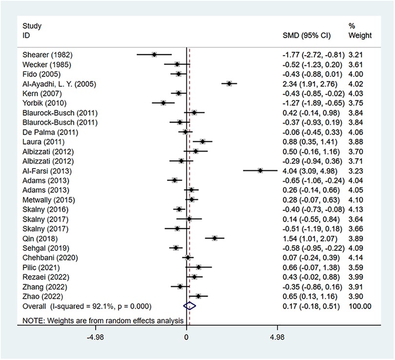
Figure 1. Flow chart of literature screening.
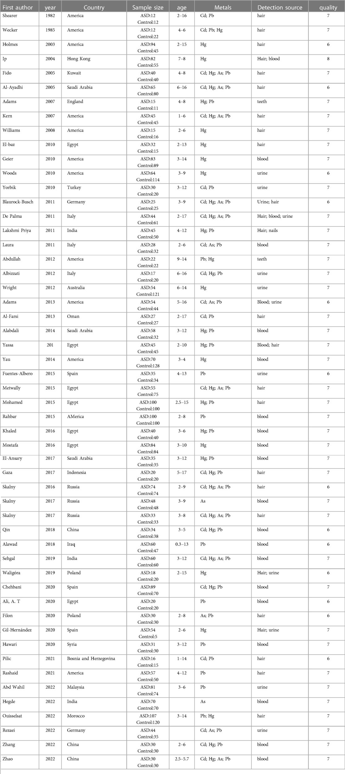
Table 1. Basic information of the literature was included.
The NOS scores of all the included studies were greater than 6, with an average score of 7.1(Table 1), which indicated no studies of low quality were included.
There were 22 studies reporting the difference in Cd concentration between the ASD group and the healthy control group. A random-effects model was applied for meta-analysis (I2 = 92.1%, P < 0.001). ASD group had higher Cd concentration compared to the healthy control group [SMD = 0.17, 95%CI (−0.18, 0.51), P > 0.05] (Figure 2). Subgroup analysis based on geographical regions of the participants and the testing sources showed that compared with the healthy controls, ASD patients had higher Cd concentration in hair [SMD = 0.22, 95%CI (−0.33, 0.77), P > 0.05] and blood [SMD = 0.21, 95%CI (−0.36, 0.77), P > 0.05], while had lower Cd concentration in urine [SMD = −0.05, 95%CI (−0.69, 0.59), P > 0.05]. As for geographical regions, ASD patients in Asia [SMD = 0.61, 95%CI (−0.18, 1.40), P > 0.05] and in Europe [SMD = 0.12, 95%CI (−0.18, 0.42), P > 0.05] had higher Cd concentration than the healthy controls in these regions, while patients in Australia [SMD = −0.52, 95% (−1.04, −0.01), P < 0.05] had a lower concentration (Table 2). Sensitivity analysis showed that the removal of the included studies one by one did not reverse the results, suggesting the results of the meta-analysis were robust. No significant publication bias was observed (P = 0.507 in Begg’s test and P = 0.67 in Egger’s test) (Figure 3).
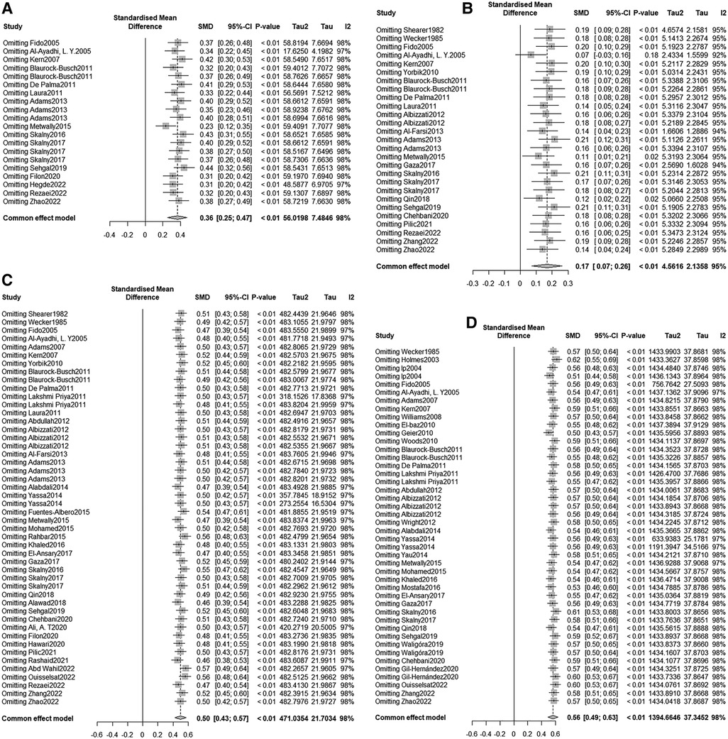
Figure 2. Cd meta-analysis forest plot.
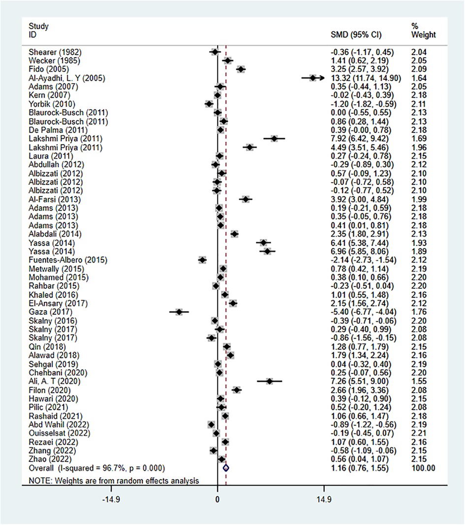
Figure 3. Sensitivity analysis (A) As (B) Cd (C) Hg (D) Pb.
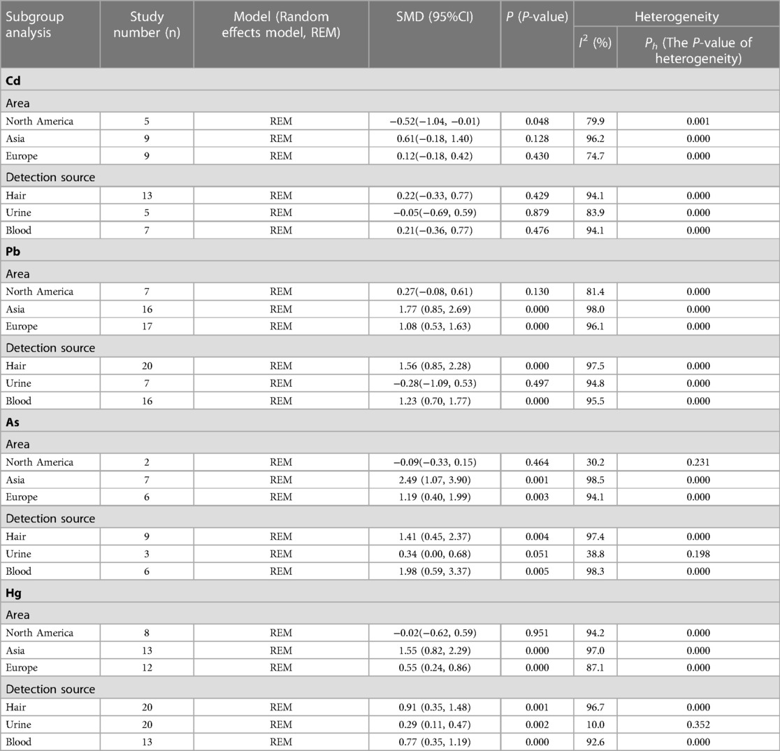
Table 2. Subgroup analysis.
There were 41 studies reporting the difference in Pb concentration between the ASD group and the healthy control group. A random-effects model was applied for meta-analysis (I2 = 96.7%, P < 0.001). ASD group had higher Pb concentration compared to the healthy control group [SMD = 1.16, 95%CI (0.76, 1.55), P < 0.001] (Figure 4). Subgroup analysis based on geographical regions of the participants and the testing sources showed that compared with the healthy controls, ASD patients had higher Pb concentration in hair [SMD = 1.56, 95%CI (0.85, 2.28), P < 0.001], fingernails [SMD = 4.49, 95%CI (3.51, 5.46), P < 0.05], and blood [SMD = 1.23, 95%CI (0.70, 1.77), P < 0.001], while had lower Pb concentration in urine [SMD = −0.28, 95%CI (−1.09, 0.53), P > 0.05] and teeth [SMD = −0.03, 95%CI (−0.64, 0.59), P > 0.05]. As for geographical regions, ASD patients in North America [SMD = 0.27, 95%CI (−0.08, 0.61), P > 0.05], Asia [SMD = 1.77, 95%CI (0.85, 2.69), P < 0.001], and Europe [SMD = 1.08, 95%CI (0.53, 1.63), P < 0.001] had higher Pb concentration than the healthy controls in these regions (Table 2). Sensitivity analysis showed that the removal of the included studies one by one did not reverse the results, suggesting the results of the meta-analysis were robust. Significant publication bias was observed (P = 0.002 in Begg’s test and P = 0.000 in Egger’s test) (Figure 3). Trim-and-Fill method showed no newly-added studies, and no small-sample effect. Publication bias did not affect the results (Supplementary Files S2).
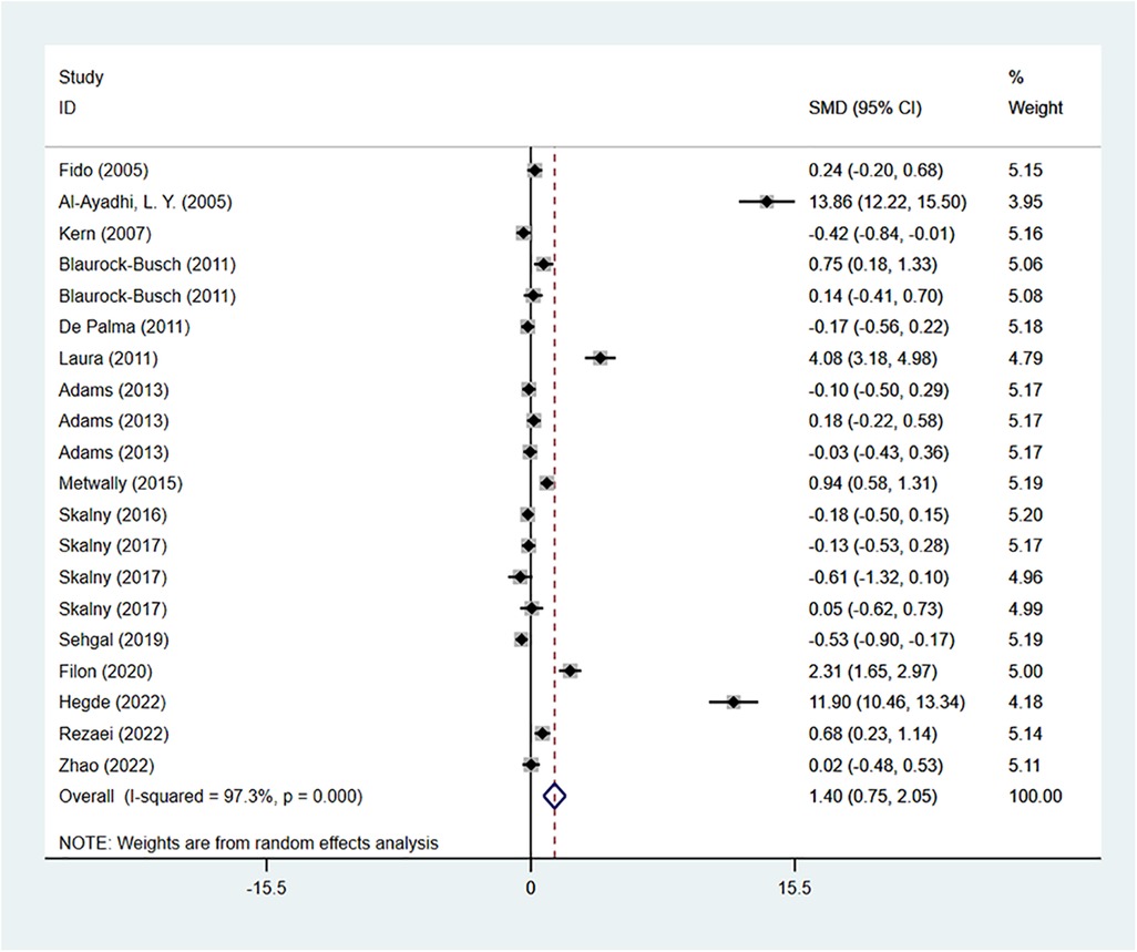
Figure 4. Pb meta-analysis forest plot.
There were 16 studies reporting the difference in arsenic concentration between the ASD group and the healthy control group. A random-effects model was applied for meta-analysis (I2 = 97.3%, P < 0.001). ASD group had higher arsenic concentration compared to the healthy control group [SMD = 1.40, 95%CI (0.75, 2.05), P < 0.001] (Figure 5). Subgroup analysis based on geographical regions of the participants and the testing sources showed that compared with the healthy controls, ASD patients had higher arsenic concentration in hair [SMD = 1.41, 95%CI (0.45, 2.37), P < 0.05], urine [SMD = 0.34, 95%CI (0.00, 0.68), P > 0.05], and blood [SMD = 1.98, 95%CI (0.59, 3.37), P < 0.05]. As for geographical regions, ASD patients in Asia [SMD = 2.49, 95%CI (1.07, 3.90), P < 0.05] and Europe [SMD = 1.19, 95%CI (0.40, 1.99), P < 0.05] had higher arsenic concentration than the healthy controls in these regions, while patients in North America [SMD = −0.09, 95%CI (0.40, 1.99), P < 0.05] had a lower arsenic oncentration (Table 2). Sensitivity analysis showed that the removal of the included studies one by one did not reverse the results, suggesting the results of the meta-analysis were robust. Significant publication bias was observed (P = 0.003 in Begg’s test and P = 0.000 in Egger’s test) (Figure 3). Trim-and-Fill method showed no newly-added studies, and no small-sample effect. Publication bias did not affect the results (Supplementary Files S3).
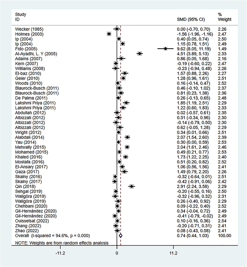
Figure 5. As meta-analysis forest plot.
There were 34 studies reporting the difference in As concentration between the ASD group and the healthy control group. A random-effects model was applied for meta-analysis (I2 = 94.6%, P < 0.001). ASD group had higher Hg concentration compared to the healthy control group [SMD = 0.74, 95%CI (0.44, 1.03), P < 0.001] (Figure 6). Subgroup analysis based on geographical regions of the participants and the testing sources showed that compared with the healthy controls, ASD patients had higher Hg concentration in hair [SMD = 0.91, 95%CI (0.35, 1.48), P < 0.05], blood [SMD = 0.77, 95%CI (0.35, 1.19), P < 0.001], urine [SMD = 0.29, 95%CI (0.11, 0.47), P < 0.05], fingernails [SMD = 1.22, 95%CI (0.60, 1.83), P < 0.05], and teeth [SMD = 0.40, 95%CI (−0.42, 1.21), P > 0.05]. As for geographical regions, ASD patients in Asia [SMD = 1.55, 95%CI (0.82, 2.29), P < 0.001], Europe [SMD = 0.55, 95%CI (0.24, 0.86), P < 0.001], and Africa [SMD = 0.10, 95%CI (−0.16, 0.36), P > 0.05] had higher Hg concentration than the healthy controls in these regions, while patients in North America [SMD = −0.02, 95%CI (−0.62, 0.59), P > 0.05] had a lower Hg concentration(Table 2). Sensitivity analysis showed that the removal of the included studies one by one did not reverse the results, suggesting the results of the meta-analysis were robust. Significant publication bias was observed (P = 0.002 in Begg's test and P = 0.008 in Egger’s test) (Figure 3). Trim-and-Fill method showed no newly-added studies, and no small-sample effect. Publication bias did not affect the results (Supplementary Files S4).
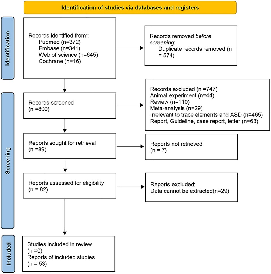
Figure 6. Hg meta-analysis forest plot.
Among the included 53 studies, Atomic Absorption Spectrometry (AAS), Inductively Coupled Plasma Mass-spectrometry (ICPMS), Flame Atomic Absorption Spectrometry (FAAS), Automatic Biochemical Analysis (ABA), and Inductively Coupled Plasma Emission Spectrum Analysis (ICPOES) were applied to detect microelements in the samples. The results showed that the ASD group had significantly higher Cd, Pb, arsenic, and Hg concentrations than the healthy control group. Subgroup analysis of testing methods indicated that concentrations of the 4 heavy metals tested by different methods were all higher in the ASD group, which suggested that testing methods were not the source of heterogeneity. Subgroup analysis of testing sources showed that compared with the heavy controls, ASD patients had higher hair and urine concentrations of the 4 heavy metals but had lower urine concentrations of Pb and arsenic. Based on previous studies, this might be associated with the patients’ reduced ability to excrete heavy metals. Subgroup analysis of geographical regions showed that ASD patients in Asian and European countries had higher concentrations of the 4 heavy metals. However, those in North America (especially the United States) had lower concentrations of Cd, arsenic, and Hg. The specific mechanisms remain unclear, leading to heterogeneity among the studies. In general, the results of different testing sources and geographical regions were controversial, which could be the source of heterogeneity.
The mechanisms of heavy metals exposure causing ASD might be related to the following aspects: Heavy metals are believed to play a role in the pathogenesis of ASD through epigenetic mechanisms. Heavy metals exposure during the growth of children could be of potential epigenetic effects on DNA methylation by mediating the dysregulation of methyltransferase (65, 66). In addition, exposure to air pollutants could cause oxidative stress and inflammatory response because heavy metals disrupt the enzyme function and cell signaling processes, produce ROS, and mediate autoimmune responses. ASD patients often have deficiencies in the defense system against ROS and impaired REDOX homeostasis, which increases susceptibility to oxidative stress, leading to consequences such as heavy metal poisoning. This might be related to altered glutathione (GSH) synthesis and impaired antioxidant defense system (6, 67, 68). Lastly, studies have shown that heavy metals could accumulate within the central neuronal system of ASD patients due to the reduced ability to excrete these substances, leading to neurotoxicity (69, 70).
Thea S Skogheim et al, have found that individuals exposed to Cd could increase the risk for ASD in their offspring (71). In this meta-analysis, the ASD group had evidently higher Cd concentration than the healthy control group (P > 0.05). Subgroup analysis showed that ASD patients had evidently higher Cd concentrations in their hair and blood compared to the healthy controls (P > 0.05), while they had a lower urine concentration (P > 0.05), which is consistent with the conclusion that ASD patients have a reduced ability of heavy metals excretion. Asian and European patients had higher Cd concentration (P > 0.05), whereas North American patients had an opposite result (P < 0.05), indicating geographical regions could cause differences in Cd concentration among ASD patients. The specific reason is still unclear and further research is needed in the future.
Cd is a kind of pollutant introduced into the environment due to the rapid development of industry and modern technology. People are mainly exposed to Cd from water, food, and air pollutants through respiratory absorption, and this substance could accumulate within the body, with a half-life period of 25–30 years (72). Cd is ranked seventh in the Substance Priority List released by the U.S. Agency for Toxic Substances and Disease Registry (ATSDR) (73), indicating its adverse impact on the human body. The kidney and liver would be the biggest victims, and 50% of the absorbed Cd is deposited in these organs. Therefore, they are the most susceptible to the adverse impact of Cd exposure (74). Cd damage to the central nervous system, especially the immature brain, could lead to neurodysplasia, memory deterioration, and hypophrenia (75). 70% of ASD patients have intellectual disability. Its association with early exposure to Cd still needs to be further discussed. The findings of this study support the need to reduce the consumption of Cd, especially in children whose brains are still growing.
Pb is a kind of non-ferrous heavy metal element that naturally exists in the earth crust. It is abundant in the environment. Even a very low concentration of Pb could be of irreversible neurotoxicity. Frequent human exposure to Pb has been a severe global environmental health problem (59). In this meta-analysis, the ASD group had a significantly higher Pb concentration than the healthy control group (P < 0.001). In the subgroup analysis of geographical regions, the ASD group also had a higher Pb concentration than the healthy control group, in which the differences between Asia and Europe were statistically significant (P < 0.001). Subgroup analysis of testing sources found that ASD patients had significantly higher hair and blood Pb concentrations than the healthy controls (P < 0.001), while had a lower urine concentration (P > 0.05), which is consistent with the study results by Nakhaee S et al. Their findings also support that ASD patients have a declined heavy metals (including Pb) excretion ability (16, 69, 70, 76, 77). The reason for this declined ability might be, according to previous studies, that ASD patients typically have reduced levels of anti-oxidants, and that the excretion of calcium ions (Ca2+) could competitively inhibit that Pb excretion, leading to reduced binding of Pb to the anti-oxidants and subsequently a low urine Pb excretion rate (21). Unlike other heavy metals, Pb could directly damage human brain cells, especially the neural plate of fetus. Fetal Pb exposure obscures the cerebral sulci and causes wide neurodysplasia and hypophrenia (78). ASD is a neurodevelopmental disorder, characterized by extensively abnormal neural network connections. The results of this study support that Pb is associated with the occurrence of ASD. Given the neurotoxicity of Pb, our findings would be of directive significance for clinical practice. Daily exposure to Pb should be avoided, especially for pregnant women and children.
Arsenic is a prevalent toxic substance that widely exists in groundwater all over the world. It is mainly absorbed through the digestive tract and respiratory system. Arsenic compounds are capable of inhibiting the activity of more than 200 enzymes within the human body, causing damage to the structure and function of the central nervous system. Moreover, arsenic could disrupt cellular metabolism (54, 79). Extensive exposure to inorganic arsenic could occur through drinking polluted water, industrial production, consumption of rice-based foods, and cigarettes and tobacco (80, 81). Emerging epidemiological investigations support that arsenic exposure especially during the critical period of neurodevelopment could be an environmental risk factor for ASD occurrence (82, 83).
This meta-analysis found that the ASD group had a higher arsenic concentration than the healthy control group (P < 0.001). Subgroup analysis of testing sources showed that compared with the healthy controls, ASD children had higher arsenic concentrations in their hair, urine, and blood (P < 0.05), which further validated the hypothesis that arsenic exposure might be associated with the occurrence of ASD. ASD was once considered to be a metabolic disease, characterized by a metabolic disorder of glucose, lipid, and amino acid (84, 85). Arsenic could affect the cellular metabolic processes (86).
The results of this study support the importance of reducing arsenic exposure. It remains to be further explored the specific mechanisms of the arsenic concentration increase in ASD patients and the association between arsenic exposure and the metabolic disturbance of ASD patients.
Hg is a toxic heavy metal substance, which is methylated in the air or water, accumulates in animal tissues, and deposits in the human body through the food chain. High levels of Hg exposure could induce irreversible neuronal and renal injury (73). Meta-analysis showed that ASD children had a significantly higher Hg concentration than the healthy controls (P < 0.001). Subgroup analysis indicated that ASD children had significantly higher Hg concentrations in their hair, blood, and urine, compared with the healthy control group (P < 0.05). The patients in Asia and Europe had a higher Hg concentration than the healthy controls (P < 0.001), while those in North America had a lower concentration (P > 0.05). A possible reason for the higher Hg concentration in ASD patients is that they take antibiotics more frequently than normal people. An animal experiment has shown that antibiotics can block the excretion of Hg, reduce the number of intestinal flora by demethylating methylmercury, and possibly increase the number of yeasts and escherichia coli by methylating inorganic mercury, thereby promoting mercury uptake and lowering mercury excretion (87). On the other hand, a lower level of glutathione and higher level of oxidative stress in ASD patients can also compromise mercury excretion, resulting in a heavier burden on the body (20). The present study demonstrates that it is necessary to reduce exposure to Hg, especially for pregnant women and children in brain-developing.
The strength of this study lies in that: Firstly, we selected 4 heavy metals that are the most controversial and the most studied, and performed a comprehensive search for the published studies. The results of this study are reported in accordance with the PRISMA guideline, and most of the included studies are the latest published, with high quality. Secondly, our statistical analysis strategy is relatively conservative, including sensitivity analysis, subgroup analysis, and publication bias assessment, to assess the potential effects of different geographical regions and different testing sources on the results. We have observed that due to the declined heavy metal excretion ability in ASD patients, the results are different in their urine specimens compared to their hair and blood samples. In addition, the patients in North America have different results from those in Asia and Europe, leading to the heterogeneity of the results.
This study has several limitations. Firstly, the included studies are case-control design so that a specific causality could not be determined between heavy metals exposure and ASD occurrence. Secondly, all the study results are directly from the level of case group and control group, so the effects of confounding factors (age, gender, BMI, etc.) on the results could not be assessed.
Compared with the healthy control group, the ASD group had higher concentrations of Cd, Pb, arsenic, and Hg, and the differences in Pb, arsenic, and Hg were statistically significant. Subgroup analysis indicated the results could be of regional difference. Studies in Asia and Europe showed that ASD children had higher concentrations of Cd, Pb, arsenic, and Hg than the healthy controls, while studies in North America yielded the opposite results regarding Cd, arsenic, and Hg. The reason remains unclear. In addition, ASD children had higher concentrations of the 4 heavy metals in their hair and blood, while had lower concentrations of Cd and Pb in urine, which could be attributed to their declined heavy metal excretion ability. Future studies need to focus on: (1) Biological mechanisms of heavy metals exposure inducing ASD. (2) The regional difference of this association. In general, reducing heavy metals exposure and keeping a good diet would be beneficial to ASD prevention.
The original contributions presented in the study are included in the article/Supplementary Material, further inquiries can be directed to the corresponding authors.
MD and SQ contributed to conception and design of the study. ZL and JL organized the database. XX and CQ performed the statistical analysis. MD and ZL wrote the first draft of the manuscript. SQ and JL wrote sections of the manuscript. All authors contributed to manuscript revision, read, and approved the submitted version. All authors contributed to the article and approved the submitted version.
We would like to thank the researchers and study participants for their contributions.
The authors declare that the research was conducted in the absence of any commercial or financial relationships that could be construed as a potential conflict of interest.
All claims expressed in this article are solely those of the authors and do not necessarily represent those of their affiliated organizations, or those of the publisher, the editors and the reviewers. Any product that may be evaluated in this article, or claim that may be made by its manufacturer, is not guaranteed or endorsed by the publisher.
The Supplementary Material for this article can be found online at: https://www.frontiersin.org/articles/10.3389/fped.2023.1169733/full#supplementary-material
SUPPLEMENTARY FILES 1
Search strategy.
SUPPLEMENTARY FILES 2
Pb published offset analysis.
SUPPLEMENTARY FILES 3
Pb published offset analysis.
SUPPLEMENTARY FILES 4
Hg published offset analysis.
1. Jiang CC, Lin LS, Long S, Ke XY, Fukunaga K, Lu YM, et al. Signalling pathways in autism spectrum disorder: mechanisms and therapeutic implications. Signal Transduct Target Ther. (2022) 7(1):229. doi: 10.1038/s41392-022-01081-0
2. Zeidan J, Fombonne E, Scorah J, Ibrahim A, Durkin MS, Saxena S, et al. Global prevalence of autism: a systematic review update. Autism Res. (2022) 15(5):778–90. doi: 10.1002/aur.2696
3. Rossignol DA, Genuis SJ, Frye RE. Environmental toxicants and autism spectrum disorders: a systematic review. Transl Psychiatry. (2014) 4(2):e360. doi: 10.1038/tp.2014.4
4. Mohamed Fel B, Zaky EA, El-Sayed AB, Elhossieny RM, Zahra SS, Salah Eldin W, et al. Assessment of hair aluminum, lead, and mercury in a sample of autistic Egyptian children: environmental risk factors of heavy metals in autism. Behav Neurol. (2015) 2015:545674. doi: 10.1155/2015/545674
5. Dickerson AS, Rahbar MH, Han I, Bakian AV, Bilder DA, Harrington RA, et al. Autism spectrum disorder prevalence and proximity to industrial facilities releasing arsenic, lead or mercury. Sci Total Environ. (2015) 536:245–51. doi: 10.1016/j.scitotenv.2015.07.024
6. Kern JK, Geier DA, Sykes LK, Haley BE, Geier MR. The relationship between mercury and autism: a comprehensive review and discussion. J Trace Elem Med Biol. (2016) 37:8–24. doi: 10.1016/j.jtemb.2016.06.002
7. Özbaran B. Do environmental factors have influence on autism spectrum disorder. J Pediatr Res. (2014) 1(4):170–3. doi: 10.4274/jpr.44153
8. Geier DA, Kern JK, King PG, Sykes LK, Geier MR. Hair toxic metal concentrations and autism spectrum disorder severity in young children. Int J Environ Res Public Health. (2012) 9(12):4486–97. doi: 10.3390/ijerph9124486
9. Pugsley K, Scherer SW, Bellgrove MA, Hawi Z. Environmental exposures associated with elevated risk for autism spectrum disorder may augment the burden of deleterious de novo mutations among probands. Mol Psychiatry. (2022) 27(1):710–30. doi: 10.1038/s41380-021-01142-w
10. Kaur I, Behl T, Aleya L, Rahman MH, Kumar A, Arora S, et al. Role of metallic pollutants in neurodegeneration: effects of aluminum, lead, mercury, and arsenic in mediating brain impairment events and autism spectrum disorder. Environ Sci Pollut Res Int. (2021) 28(8):8989–9001. doi: 10.1007/s11356-020-12255-0
11. Amadi CN, Orish CN, Frazzoli C, Orisakwe OE. Association of autism with toxic metals: a systematic review of case-control studies. Pharmacol Biochem Behav. (2022) 212:173313. doi: 10.1016/j.pbb.2021.173313
12. Zhang J, Li X, Shen L, Khan NU, Zhang X, Chen L, et al. Trace elements in children with autism spectrum disorder: a meta-analysis based on case-control studies. J Trace Elem Med Biol. (2021) 67:126782. doi: 10.1016/j.jtemb.2021.126782
13. Lo CK, Mertz D, Loeb M. Newcastle-Ottawa Scale: comparing reviewers’ to authors’ assessments. BMC Med Res Methodol. (2014) 14:45. doi: 10.1186/1471-2288-14-45
14. Shearer TR, Larson K, Neuschwander J, Gedney B. Minerals in the hair and nutrient intake of autistic children. J Autism Dev Disord. (1982) 12(1):25–34. doi: 10.1007/BF01531671
15. Wecker L, Miller SB, Cochran SR, Dugger DL, Johnson WD. Trace element concentrations in hair from autistic children. J Ment Defic Res. (1985) 29(Pt 1):15–22. doi: 10.1111/j.1365-2788.1985.tb00303.x
16. Holmes AS, Blaxill MF, Haley BE. Reduced levels of mercury in first baby haircuts of autistic children. Int J Toxicol. (2003) 22(4):277–85. doi: 10.1080/10915810305120
17. Ip P, Wong V, Ho M, Lee J, Wong W. Mercury exposure in children with autistic spectrum disorder: case-control study. J Child Neurol. (2004) 19(6):431–4. doi: 10.1177/088307380401900606
18. Al-Ayadhi LY. Heavy metals and trace elements in hair samples of autistic children in central Saudi Arabia. Neurosciences (Riyadh, Saudi Arabia). (2005) 10(3):213–8. PMID: 22473261
19. Fido A, Al-Saad S. Toxic trace elements in the hair of children with autism. Autism. (2005) 9(3):290–8. doi: 10.1177/1362361305053255
20. Adams JB, Romdalvik J, Ramanujam VM, Legator MS. Mercury, lead, and zinc in baby teeth of children with autism versus controls. J Toxicol Environ Health Part A. (2007) 70(12):1046–51. doi: 10.1080/15287390601172080
21. Kern JK, Grannemann BD, Trivedi MH, Adams JB. Sulfhydryl-reactive metals in autism. J Toxicol Environ Health Part A. (2007) 70(8):715–21. doi: 10.1080/15287390601188060
22. Williams PG, Hersh JH, Allard A, Sears LL. A controlled study of mercury levels in hair samples of children with autism as compared to their typically developing siblings. Res Autism Spectr Disord. (2008) 2(1):170–5. doi: 10.1016/j.rasd.2007.05.001
23. El-baz F, Elhossiny RM, Elsayed AB, Gaber GM. Hair mercury measurement in Egyptian autistic children. Egypt J Med Hum Genet. (2010) 11(2):135–41. doi: 10.1016/j.ejmhg.2010.10.007
24. Geier DA, Audhya T, Kern JK, Geier MR. Blood mercury levels in autism spectrum disorder: is there a threshold level? Acta Neurobiol Exp (Wars). (2010) 70(2):177–86. PMID: 20628441
25. Woods JS, Armel SE, Fulton DI, Allen J, Wessels K, Simmonds PL, et al. Urinary porphyrin excretion in neurotypical and autistic children. Environ Health Perspect. (2010) 118(10):1450–7. doi: 10.1289/ehp.0901713
26. Yorbik O, Kurt I, Haşimi A, Oztürk O. Chromium, cadmium, and lead levels in urine of children with autism and typically developing controls. Biol Trace Elem Res. (2010) 135(1-3):10–5. doi: 10.1007/s12011-009-8494-7
27. Blaurock-Busch E, Amin OR, Rabah T. Heavy metals and trace elements in hair and urine of a sample of arab children with autistic spectrum disorder. Maedica (Buchar). (2011) 6(4):247–57. PMID: 22879836
28. De Palma G, Catalani S, Franco A, Brighenti M, Apostoli P. Lack of correlation between metallic elements analyzed in hair by ICP-MS and autism. J Autism Dev Disord. (2012) 42(3):342–53. doi: 10.1007/s10803-011-1245-6
29. Lakshmi Priya MD, Geetha A. Level of trace elements (copper, zinc, magnesium and selenium) and toxic elements (lead and mercury) in the hair and nail of children with autism. Biol Trace Elem Res. (2011) 142(2):148–58. doi: 10.1007/s12011-010-8766-2
30. Vergani L, Cristina L, Paola R, Luisa AM, Shyti G, Edvige V, et al. Metals, metallothioneins and oxidative stress in blood of autistic children. Res Autism Spectr Disord. (2011) 5(1):286–93. doi: 10.1016/j.rasd.2010.04.010
31. Abdullah MM, Ly AR, Goldberg WA, Clarke-Stewart KA, Dudgeon JV, Mull CG, et al. Heavy metal in children’s tooth enamel: related to autism and disruptive behaviors? J Autism Dev Disord. (2012) 42(6):929–36. doi: 10.1007/s10803-011-1318-6
32. Albizzati A, Morè L, Di Candia D, Saccani M, Lenti C. Normal concentrations of heavy metals in autistic spectrum disorders. Minerva Pediatr. (2012) 64(1):27–31. PMID: 22350041
33. Wright B, Pearce H, Allgar V, Miles J, Whitton C, Leon I, et al. A comparison of urinary mercury between children with autism spectrum disorders and control children. PLoS One. (2012) 7(2):e29547. doi: 10.1371/journal.pone.0029547
34. Adams JB, Audhya T, McDonough-Means S, Rubin RA, Quig D, Geis E, et al. Toxicological status of children with autism vs. neurotypical children and the association with autism severity. Biol Trace Elem Res. (2013) 151(2):171–80. doi: 10.1007/s12011-012-9551-1
35. Al-Farsi YM, Waly MI, Al-Sharbati MM, Al-Shafaee MA, Al-Farsi OA, Al-Khaduri MM, et al. Levels of heavy metals and essential minerals in hair samples of children with autism in Oman: a case-control study. Biol Trace Elem Res. (2013) 151(2):181–6. doi: 10.1007/s12011-012-9553-z
36. Alabdali A, Al-Ayadhi L, El-Ansary A. A key role for an impaired detoxification mechanism in the etiology and severity of autism spectrum disorders. Behav Brain Funct. (2014) 10:14. doi: 10.1186/1744-9081-10-14
37. Yassa HA. Autism: a form of lead and mercury toxicity. Environ Toxicol Pharmacol. (2014) 38(3):1016–24. doi: 10.1016/j.etap.2014.10.005
38. Yau VM, Green PG, Alaimo CP, Yoshida CK, Lutsky M, Windham GC, et al. Prenatal and neonatal peripheral blood mercury levels and autism spectrum disorders. Environ Res. (2014) 133:294–303. doi: 10.1016/j.envres.2014.04.034
39. Fuentes-Albero M, Puig-Alcaraz C, Cauli O. Lead excretion in spanish children with autism spectrum disorder. Brain Sci. (2015) 5(1):58–68. doi: 10.3390/brainsci5010058
40. Khaled EM, Meguid NA, Bjørklund G, Gouda A, Bahary MH, Hashish A, et al. Altered urinary porphyrins and mercury exposure as biomarkers for autism severity in Egyptian children with autism spectrum disorder. Metab Brain Dis. (2016) 31(6):1419–26. doi: 10.1007/s11011-016-9870-6
41. Rahbar MH, Samms-Vaughan M, Dickerson AS, Loveland KA, Ardjomand-Hessabi M, Bressler J, et al. Blood lead concentrations in Jamaican children with and without autism spectrum disorder. Int J Environ Res Public Health. (2014) 12(1):83–105. doi: 10.3390/ijerph120100083
42. Mostafa GA, Bjørklund G, Urbina MA, Al-Ayadhi LY. The levels of blood mercury and inflammatory-related neuropeptides in the serum are correlated in children with autism spectrum disorder. Metab Brain Dis. (2016) 31(3):593–9. doi: 10.1007/s11011-015-9784-8
43. El-Ansary A, Bjørklund G, Tinkov AA, Skalny AV, Al Dera H. Relationship between selenium, lead, and mercury in red blood cells of Saudi autistic children. Metab Brain Dis. (2017) 32(4):1073–80. doi: 10.1007/s11011-017-9996-1
44. Gaza MA, Hakim L, Sabarudin A, Sumitro S. Evaluation on mercury, cadmium, and lead in the hair sample as an indicator of autism for children. Int J Pharm Clin Res. (2017) 9(12):710–5.
45. Skalny AV, Simashkova NV, Klyushnik TP, Grabeklis AR, Bjørklund G, Skalnaya MG, et al. Hair toxic and essential trace elements in children with autism spectrum disorder. Metab Brain Dis. (2017) 32(1):195–202. doi: 10.1007/s11011-016-9899-6
46. Skalny AV, Simashkova NV, Klyushnik TP, Grabeklis AR, Radysh IV, Skalnaya MG, et al. Assessment of serum trace elements and electrolytes in children with childhood and atypical autism. J Trace Elem Med Biol. (2017) 43:9–14. doi: 10.1016/j.jtemb.2016.09.009
47. Skalny AV, Simashkova NV, Klyushnik TP, Grabeklis AR, Radysh IV, Skalnaya MG, et al. Analysis of hair trace elements in children with autism spectrum disorders and communication disorders. Biol Trace Elem Res. (2017) 177(2):215–23. doi: 10.1007/s12011-016-0878-x
48. Qin YY, Jian B, Wu C, Jiang CZ, Kang Y, Zhou JX, et al. A comparison of blood metal levels in autism spectrum disorder and unaffected children in Shenzhen of China and factors involved in bioaccumulation of metals. Environ Sci Pollut Res Int. (2018) 25(18):17950–6. doi: 10.1007/s11356-018-1957-7
49. Alawad ZM, Al-Jobouri SM, Majid AY. Lead among children with autism in Iraq. Is it a potential factor? J Clin Anal Med. (2019) 10(2):215–9. doi: 10.4328/JCAM.5908
50. Sehgal R, Gulati S, Gupta YK, Sapra S, Mehta M, Pandey RM, et al. Blood heavy metal levels in children with autism spectrum disorder: a cross-sectional study from northern India. J Nepal Paediatr Soc. (2019) 39(1):6–14. doi: 10.3126/jnps.v39i1.19905
51. Waligóra S, Waligóra A, Damasiewicz-Bodzek A, Gorczyca P, Tyrpień-Golder K. Exposure of children with autism spectrum disorders to mercury and polycyclic aromatic hydrocarbons. Adv Psychiatry Neurol. (2019) 28(3):176–83. doi: 10.5114/ppn.2019.89128
52. Ali AT. Autism. What does it mean and what are the reasons for its occurrence and conduct a study to compare children exposed to lead and not exposed and its effect on autism. Int J Pharm Res. (2020) 12:4524–32. doi: 10.31838/ijpr/2020.SP2.566
53. Chehbani F, Gallello G, Brahim T, Ouanes S, Douki W, Gaddour N, et al. The status of chemical elements in the blood plasma of children with autism spectrum disorder in Tunisia: a case-control study. Environ Sci Pollut Res Int. (2020) 27(28):35738–49. doi: 10.1007/s11356-020-09819-5
54. Fiłon J, Ustymowicz-Farbiszewska J, Krajewska-Kułak E. Analysis of lead, arsenic and calcium content in the hair of children with autism spectrum disorder. BMC Public Health. (2020) 20(1):383. doi: 10.1186/s12889-020-08496-w
55. Gil-Hernández F, Gómez-Fernández AR, la Torre-Aguilar MJ, Pérez-Navero JL, Flores-Rojas K, Martín-Borreguero P, et al. Neurotoxicity by mercury is not associated with autism spectrum disorders in Spanish children. Ital J Pediatr. (2020) 46(1):19. doi: 10.1186/s13052-020-0780-1
56. Hawari I, Eskandar MB, Alzeer S. The role of lead, manganese, and zinc in autism spectrum disorders (ASDs) and attention-deficient hyperactivity disorder (ADHD): a case-control study on syrian children affected by the syrian crisis. Biol Trace Elem Res. (2020) 197(1):107–14. doi: 10.1007/s12011-020-02146-3
57. Pilić S, Kalić E, Selović A. The content of essential and toxic metals in the hair of children with autistic Spectrum disorders. Glasnik Hemicara i Tehnologa Bosne i Hercegovine. (2021) 56:13–20. doi: 10.35666/2232-7266.2021.56.03
58. Rashaid AHB, Nusair SD, Alqhazo MT, Adams JB, Abu-Dalo MA, Bashtawi MA. Heavy metals and trace elements in scalp hair samples of children with severe autism spectrum disorder: a case-control study on Jordanian children. J Trace Elem Med Biol. (2021) 67:126790. doi: 10.1016/j.jtemb.2021.126790
59. Abd Wahil MS, Ja’afar MH, Md Isa Z. Assessment of urinary lead (pb) and essential trace elements in autism spectrum disorder: a case-control study among preschool children in Malaysia. Biol Trace Elem Res. (2022) 200(1):97–121. doi: 10.1007/s12011-021-02654-w
60. Hegde R, Hegde S, Kulkarni S, Kulkarni SS, Pandurangi A, Kariduraganavar MY, et al. Total reflection x-ray fluorescence analysis of plasma elements in autistic children from India. Biol Trace Elem Res. (2023) 201(2):644–54. doi: 10.1007/s12011-022-03199-2
61. Ouisselsat M, Maidoumi S, Elmaouaki A, Lekouch N, Pineau A, Sedki A. Hair trace elements and mineral content in moroccan children with autism spectrum disorder: a case-control study. Biol Trace Elem Res. (2022) 201(6):2701–10. doi: 10.1007/s12011-022-03365-6
62. Rezaei M, Rezaei A, Esmaeili A, Nakhaee S, Azadi NA, Mansouri B. A case-control study on the relationship between urine trace element levels and autism spectrum disorder among Iranian children. Environ Sci Pollut Res Int. (2022) 29(38):57287–95. doi: 10.1007/s11356-022-19933-1
63. Zhang J, Lin J, Zhao X, Yao F, Feng C, He Z, et al. Trace element changes in the plasma of autism spectrum disorder children and the positive correlation between chromium and vanadium. Biol Trace Elem Res. (2022) 200(12):4924–35. doi: 10.1007/s12011-021-03082-6
64. Zhao G, Liu SJ, Gan XY, Li JR, Wu XX, Liu SY, et al. Analysis of whole blood and urine trace elements in children with autism spectrum disorders and autistic behaviors. Biol Trace Elem Res. (2023) 201(2):627–35. doi: 10.1007/s12011-022-03197-4
65. Yasuda H, Yasuda Y, Tsutsui T. Estimation of autistic children by metallomics analysis. Sci Rep. (2013) 3:1199. doi: 10.1038/srep01199
66. Schneider JS, Kidd SK, Anderson DW. Influence of developmental lead exposure on expression of DNA methyltransferases and methyl cytosine-binding proteins in hippocampus. Toxicol Lett. (2013) 217(1):75–81. doi: 10.1016/j.toxlet.2012.12.004
67. Jafari T, Rostampour N, Fallah AA, Hesami A. The association between mercury levels and autism spectrum disorders: a systematic review and meta-analysis. J Trace Elem Med Biol. (2017) 44:289–97. doi: 10.1016/j.jtemb.2017.09.002
68. Zaky E. Toxic heavy metals and autism spectrum disorder; is there a link. J Child Adolesc Behav. (2017) 5(2):1–2.
69. Cekici H, Sanlier N. Current nutritional approaches in managing autism spectrum disorder: a review. Nutr Neurosci. (2019) 22(3):145–55. doi: 10.1080/1028415X.2017.1358481
70. Baj J, Flieger W, Flieger M, Forma A, Sitarz E, Skórzyńska-Dziduszko K, et al. Autism spectrum disorder: trace elements imbalances and the pathogenesis and severity of autistic symptoms. Neurosci Biobehav Rev. (2021) 129:117–32. doi: 10.1016/j.neubiorev.2021.07.029
71. Skogheim TS, Weyde KVF, Engel SM, Aase H, Surén P, Øie MG, et al. Metal and essential element concentrations during pregnancy and associations with autism spectrum disorder and attention-deficit/hyperactivity disorder in children. Environ Int. (2021) 152:106468. doi: 10.1016/j.envint.2021.106468
72. Genchi G, Sinicropi MS, Lauria G, Carocci A, Catalano A. The effects of cadmium toxicity. Int J Environ Res Public Health. (2020) 17(11):3782. doi: 10.3390/ijerph17113782
73. Sulaiman R, Wang M, Ren X. Exposure to aluminum, cadmium, and mercury and autism spectrum disorder in children: a systematic review and meta-analysis. Chem Res Toxicol. (2020) 33(11):2699–718. doi: 10.1021/acs.chemrestox.0c00167
74. Jalili C, Kazemi M, Cheng H, Mohammadi H, Babaei A, Taheri E, et al. Associations between exposure to heavy metals and the risk of chronic kidney disease: a systematic review and meta-analysis. Crit Rev Toxicol. (2021) 51(2):165–82. doi: 10.1080/10408444.2021.1891196
75. Pulido G, Treviño S, Brambila E, Vazquez-Roque R, Moreno-Rodriguez A, Peña Rosas U, et al. The administration of cadmium for 2, 3 and 4 months causes a loss of recognition memory, promotes neuronal hypotrophy and apoptosis in the hippocampus of rats. Neurochem Res. (2019) 44(2):485–97. doi: 10.1007/s11064-018-02703-2
76. Nakhaee S, Amirabadizadeh A, Farnia V, Ali Azadi N, Mansouri B, Radmehr F. Association between biological lead concentrations and autism Spectrum disorder (ASD) in children: a systematic review and meta-analysis. Biol Trace Elem Res. (2022) 201(4):1567–81. doi: 10.1007/s12011-022-03265-9
77. Bradstreet J, Geier DA, Kartzinel JJ, Adams JB, Geier MR. A case-control study of mercury burden in children with autistic spectrum disorders. J Am Phys Surg. (2003) 8(3):76–9.
78. Canfield RL, Henderson CR Jr, Cory-Slechta DA, Cox C, Jusko TA, Lanphear BP. Intellectual impairment in children with blood lead concentrations below 10 μg per deciliter. N Engl J Med. (2003) 348(16):1517–26. doi: 10.1056/NEJMoa022848
79. Zhang X, Mei D, Li Y, You M, Wang D, Yao D, et al. Arsenic exposure via drinking water during pregnancy and lactation induces autism-like behaviors in male offspring mice. Chemosphere. (2022) 290:133338. doi: 10.1016/j.chemosphere.2021.133338
80. Tripathi MK, Kartawy M, Ginzburg S, Amal H. Arsenic alters nitric oxide signaling similar to autism spectrum disorder and Alzheimer’s disease-associated mutations. Transl Psychiatry. (2022) 12(1):127. doi: 10.1038/s41398-022-01890-5
81. Khan MI, Ahmad MF, Ahmad I, Ashfaq F, Wahab S, Alsayegh AA, et al. Arsenic exposure through dietary intake and associated health hazards in the Middle East. Nutrients. (2022) 14(10):2136. doi: 10.3390/nu14102136
82. Rodríguez-Barranco M, Gil F, Hernández AF, Alguacil J, Lorca A, Mendoza R, et al. Postnatal arsenic exposure and attention impairment in school children. Cortex. (2016) 74:370–82. doi: 10.1016/j.cortex.2014.12.018
83. Zuo Z, Liu Z, Gao T, Yin Y, Wang Z, Hou Y, et al. Prolonged inorganic arsenic exposure via drinking water impairs brown adipose tissue function in mice. Sci Total Environ. (2019) 668:310–7. doi: 10.1016/j.scitotenv.2019.03.008
84. Gevezova M, Minchev D, Pacheva I, Sbirkov Y, Yordanova R, Timova E, et al. Cellular bioenergetic and metabolic changes in patients with autism spectrum disorder. Curr Top Med Chem. (2021) 21(11):985–94. doi: 10.2174/1568026621666210521142131
85. Du C, Liu WJ, Yang J, Zhao SS, Liu HX. The role of branched-chain amino acids and branched-chain α-keto acid dehydrogenase kinase in metabolic disorders. Front Nutr. (2022) 9:932670. doi: 10.3389/fnut.2022.932670
86. Medda N, De SK, Maiti S. Different mechanisms of arsenic related signaling in cellular proliferation, apoptosis and neo-plastic transformation. Ecotoxicol Environ Saf. (2021) 208:111752. doi: 10.1016/j.ecoenv.2020.111752
Keywords: autism spectrum disorder, trace elements, cadmium, lead, arsenic, mercury
Citation: Ding M, Shi S, Qie S, Li J and Xi X (2023) Association between heavy metals exposure (cadmium, lead, arsenic, mercury) and child autistic disorder: a systematic review and meta-analysis. Front. Pediatr. 11:1169733. doi: 10.3389/fped.2023.1169733
Received: 20 February 2023; Accepted: 23 June 2023;
Published: 4 July 2023.
Edited by:
Andreas Martin Grabrucker, University of Limerick, IrelandReviewed by:
Prasunpriya Nayak, All India Institute of Medical Sciences Jodhpur, India© 2023 Ding, Shi, Qie, Li and Xi. This is an open-access article distributed under the terms of the Creative Commons Attribution License (CC BY). The use, distribution or reproduction in other forums is permitted, provided the original author(s) and the copyright owner(s) are credited and that the original publication in this journal is cited, in accordance with accepted academic practice. No use, distribution or reproduction is permitted which does not comply with these terms.
*Correspondence: Mengmeng Ding ZG1tNTEyQG1haWwuY2NtdS5lZHUuY24= Shanshan Shi c3Vubnk0MzNAY2NtdS5lZHUuY24= Shuyan Qie U2h1eWFucGJAbWFpbC5jY211LmVkdS5jbg==
Disclaimer: All claims expressed in this article are solely those of the authors and do not necessarily represent those of their affiliated organizations, or those of the publisher, the editors and the reviewers. Any product that may be evaluated in this article or claim that may be made by its manufacturer is not guaranteed or endorsed by the publisher.
Research integrity at Frontiers
Learn more about the work of our research integrity team to safeguard the quality of each article we publish.