95% of researchers rate our articles as excellent or good
Learn more about the work of our research integrity team to safeguard the quality of each article we publish.
Find out more
ORIGINAL RESEARCH article
Front. Oncol. , 25 March 2025
Sec. Cancer Epidemiology and Prevention
Volume 15 - 2025 | https://doi.org/10.3389/fonc.2025.1530339
This article is part of the Research Topic Advancing Personalized Cancer Care: Novel Genetic and Statistical Approaches View all 5 articles
 Jinhe Xu1†
 Wenting Zhang1
 Feilai Xie2
 Chenxi Wang1
 Feng Cheng1
 Ruiying Rao3
 Ying Chen4
 Lei Zhang4
 Wen Wen4
 Zhongquan Zhao4
 Jialing Yuan4
 Yuqin Zheng4
 Zongyang Yu4*
Purpose: The aim of this study was to investigate the prognostic value of the abnormal expression of anaplastic lymphoma kinase (ALK) protein in patients with small cell lung cancer (SCLC) based on 9-year data from our center.
Methods: A retrospective cohort study was conducted to assess the clinical outcomes of patients with ALK-positive SCLC diagnosed in our hospital over the past 9 years. We used public databases to analyze the expression of ALK in pan-cancer and its prognostic value and analyzed the correlation between ALK and SCLC prognosis-related genes.
Results: A total of 685 patients diagnosed with SCLC underwent ALK testing, and 59 patients were identified to have abnormal expression of the ALK protein, with 10 cases showing strong expression, 14 cases displaying moderate expression, and 35 cases exhibiting weak expression. The median age of the ALK-positive cohort was 64 years (range: 58–70 years), 91.5% (54/59) were male, 61.0% (36/59) were smokers, and the median overall survival (mOS) was 7.0 months (95% CI: 4.5–9.5 months). Within this cohort, the mOS for the ALK (+) subgroup was 4.0 months (95% CI: 2.9–5.1 months), the mOS for the ALK (++) subgroup was 10.0 months (95% CI: 4.9–15.1 months), and the mOS for the ALK (+++) subgroup was 12.0 months (95% CI: 7.4–16.6 months). Kaplan–Meier revealed that the mOS of the ALKLow group was significantly worse than that of the ALKHigh group [mOS: 4.0 months (95% CI: 2.9–5.1 months) versus 11.0 months (95% CI: 8.3–13.7 months), p = 0.009]. Following covariate adjustment using a Cox regression model, it was indicated that the level of abnormal expression of the ALK protein was an independent prognostic factor for patients with SCLC (HR: 0.486, 95% CI: 0.271–0.871, p = 0.015).
Conclusion: The prognosis for patients with SCLC with strong abnormal expression of the ALK protein was significantly better than those with weak expression.
Lung cancer ranks among the malignancies with the highest incidence and mortality rates both nationally and globally (1), of which small cell lung cancer (SCLC) accounts for 13%–15%, which has a poor prognosis, is highly invasive, and is related to smoking status (2, 3). SCLC is primarily classified into limited-stage and extensive-stage disease (4). Early prognostic assessment of SCLC could facilitate the development of an appropriate treatment strategy, thereby enabling proactive intervention to enhance quality of life and extend survival. Over the past three decades, there has been significant progress in the development of targeted therapies aimed at specific driver genes that have markedly extended the survival of patients with advanced non-small cell lung cancer (NSCLC) who test positive for these genetic alterations (5). Anaplastic lymphoma kinase (ALK) is present in 5%–6% of NSCLC cases, with common aberrations including ALK fusions, mutations, and amplifications (6). The ALK fusion gene is colloquially referred to as a “masonry mutation” due to its low prevalence in NSCLC; however, patients with this mutation can derive substantial clinical benefits from ALK-tyrosine kinase inhibitors (ALK-TKIs), potentially achieving prolonged survival (6). Because of the rarity of targeted gene mutations in SCLC and its insensitivity to targeted drugs, routine testing for gene mutations in SCLC is currently not recommended clinically (7). Nonetheless, some studies suggested that aberrant ALK protein expression may be associated with the prognosis of some tumors. In a study that examined the prognosis of gastric cancer, it was observed that an increase in the intensity of ALK protein expression correlated with a higher proportion of tumor signet ring cells and a poorer prognosis (8). ALK protein expression has been shown to predict micrometastasis and unfavorable outcomes in patients with hepatocellular carcinoma (9). Conversely, elevated ALK protein expression is associated with prolonged survival in patients with Merkel cell carcinoma (MCC) and anaplastic large cell lymphoma (ALCL) (10, 11). ALK immunohistochemistry (IHC) serves as a standard clinical marker for SCLC. Previous research has identified that the protein expression rate of ALK in SCLC can reach 11% (12). However, there is no research that investigated the prognostic significance of aberrant ALK protein expression in SCLC. This study undertook a retrospective analysis of 685 patients with SCLC and their tissue samples collected over the past 9 years. The objectives are to examine the clinical characteristics associated with positive abnormal ALK protein expression and to evaluate the prognostic implications of ALK protein expression levels in SCLC.
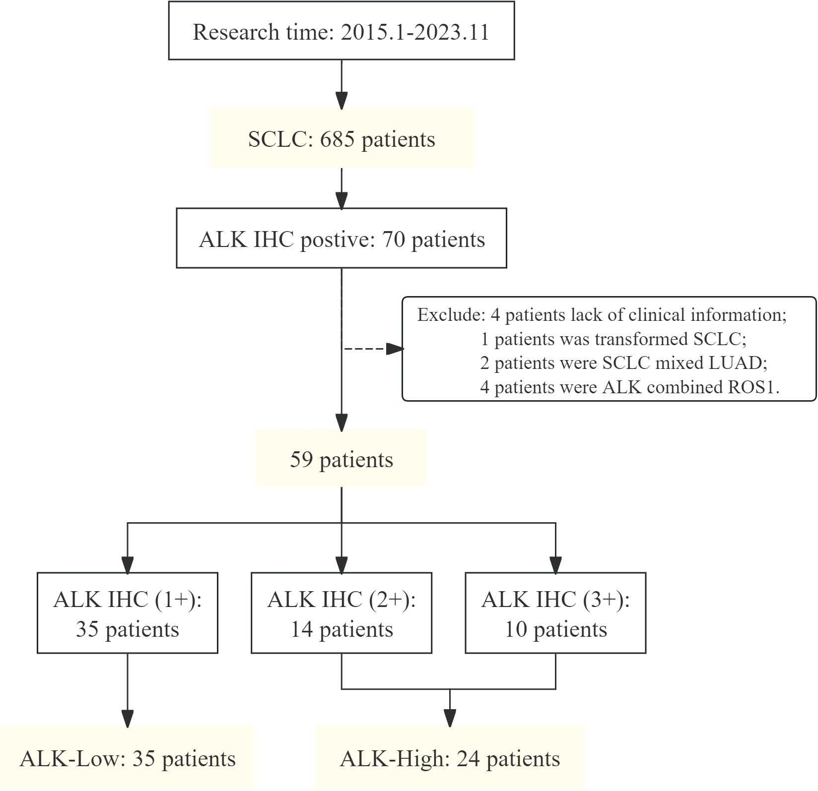
A total of 685 eligible patients with de novo SCLC who were subjected to ALK IHC for analysis at the 900th Hospital of the Joint Logistic Support Force of China (Fujian, China) between January 2015 and November 2023 were enrolled in this study. All patients had a de novo diagnosis of SCLC excluding the histological transformation types. Among them, 35 patients with SCLC with weak positive ALK IHC (1+) were classified as ALKLow, while 24 patients with strong positive ALK IHC (2+, 3+) were categorized as ALKHigh (Figure 1). In this series, all samples were detected by ALK IHC at the initial biopsy. Histologic diagnosis of SCLC was based on the standard criteria defined by the WHO classification 2015 version (13). All procedures performed in this study involving human participants were in accordance with the Declaration of Helsinki (as revised in 2013). This study was approved by the Ethics Committee of the 900th Hospital of the Joint Logistic Support Force of China.

Figure 1. Flowchart of patient enrollment. SCLC, small cell lung cancer; ALK, anaplastic lymphoma kinase; IHC, immunohistochemistry; LUAD, lung adenocarcinoma; ROS1, ROS proto-oncogene 1, receptor tyrosine kinase.
The following information was retrospectively collected from the medical records of the patients: patient demographics such as sex, age, smoking history, disease stage, anatomy type, histological type, systemic therapies, tumor imaging, and tumor response to therapies. Tumor response was assessed in accordance with the Response Evaluation Criteria in Solid Tumors (RECIST version 1.1) (14). Overall survival (OS) was defined as the time from initial therapy to death, and the last follow-up was on 15 September 2024.
IHC stains were conducted on an automated immunostainer machine. IHC staining was performed on 4-μm sections obtained from formalin-fixed and paraffin-embedded tissue blocks and then mounted on charged slides. After deparaffinization and rehydration, antigen retrieval was performed with Cell Condition Solution-1 (CC1) for 64 min at 95°C. ALK IHC assay was performed using Ventana anti-ALK rabbit monoclonal primary antibody (clone D5F3, Cell Signaling Technology). The expression level of ALK was expressed using IHC scoring, and the expression of all samples was scored by two experienced pathologists (Table 1). When using the four-tiered scoring system, cases showing no staining were considered as negative, cases with 2+/3+ staining were considered as positive/moderate and strong, and cases with 1+ intensity expression were considered as weak. The representative images of immunohistochemical markers are shown in Figure 2.

Figure 2. ALK immunohistochemistry staining pattern. Examples of negative and positive (1+, 2+, and 3+) staining patterns of ALK immunohistochemistry are shown (hematoxylin and eosin stain, ×400). neg, negative.
Continuous variables were analyzed as the median and interquartile range (IQR) or mean and standard deviation (mean ± SD), and differences were compared using t-test. Categorical data were calculated as the frequency (percentage) and composition ratio (%), and non-parametric methods such as the χ2 test were used to compare differences. Survival analysis was performed using the Kaplan–Meier method, and the log-rank test was used to determine the significance of differences between two subgroups of patients. Single-factor and multi-factor Cox proportional hazards regression models were used to analyze the relationship between survival time and survival status and factors. Among them, dead and alive are used as dependent variables, and other variables are used as independent variables. IBM SPSS Statistics (Armonk, NY), version 26 was used in statistical analyses. A p-value < 0.05 was considered significant, while all tests were two-sided.
A total of 685 eligible patients diagnosed with SCLC at our institution were included in our study; 70 cases showed positive ALK protein expression in IHC test results, with an incidence rate of 10.22%. The following cases were excluded from the study: four cases with incomplete information, two cases of mixed tissue type, four cases with other mutations, and one case of transformed SCLC. Finally, 59 patients were included in the follow-up study. The median age was 64 years (range: 58–70 years), 54 patients (91.5%) were men, and 36 patients (61.0%) were smokers. Among the cohort, 35 patients exhibited weak ALK protein expression (+), 14 patients demonstrated moderate expression (++), and 10 patients showed strong expression (+++). Comparative analysis of the different ALK protein expression groups revealed no statistically significant differences in age, gender, smoking history, TNM stage, primary lesion site, or Ki-67 expression. Regarding first-line treatment, aside from those detailed in Table 2, two additional patients received only chest radiotherapy (ALK 1+), and one patient underwent alectinib targeted therapy (ALK 2+). No statistically significant differences were observed in the first-line treatment outcomes in different ALK protein groups.
The heterogeneity of SCLCs is substantial, and they are not simply a result of combining multiple subtypes or the presence of different mutant molecules. Therefore, we examined whether there is a discernible histological inclination between different ALK protein expression levels in 59 patients with ALK IHC-positive SCLC using IHC. Results of the IHC analysis are shown in Table 3. The results of IHC were interpreted using semiquantitative criteria; Ki-67 results determined the positive rate of tumor cell nuclei. The expression of CD56 was statistically significant in different ALK IHC expression groups (p < 0.05), and the difference was mainly in the ALK IHC (2+) group. However, the expression of CK7, CgA, Syn, TTF-1, and Ki-67, and the proportion of patients with marker 0, 1, 2, 3, and 4 did not differ significantly among the groups, which means the IHC characteristics of different ALK IHC expression groups were similar in general.
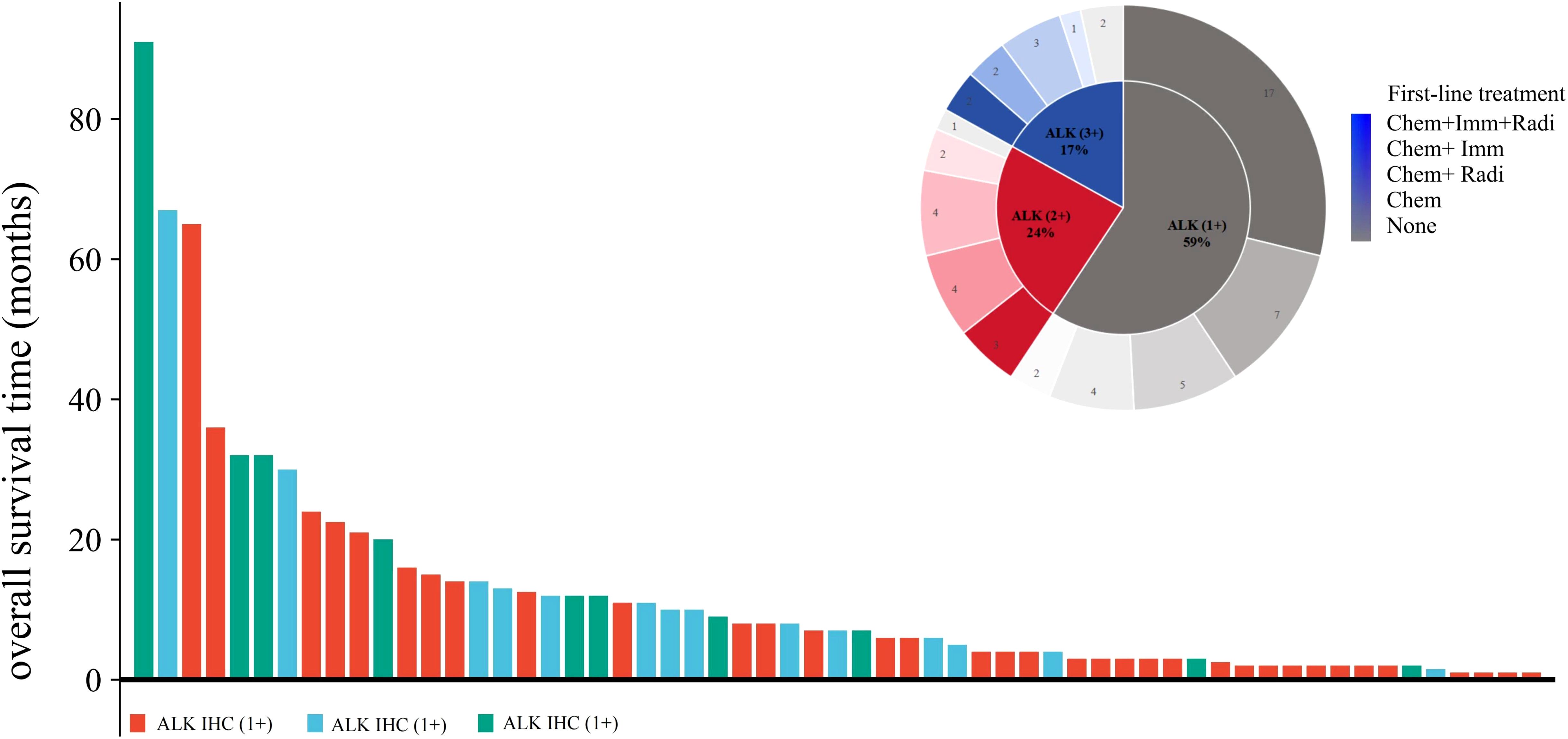
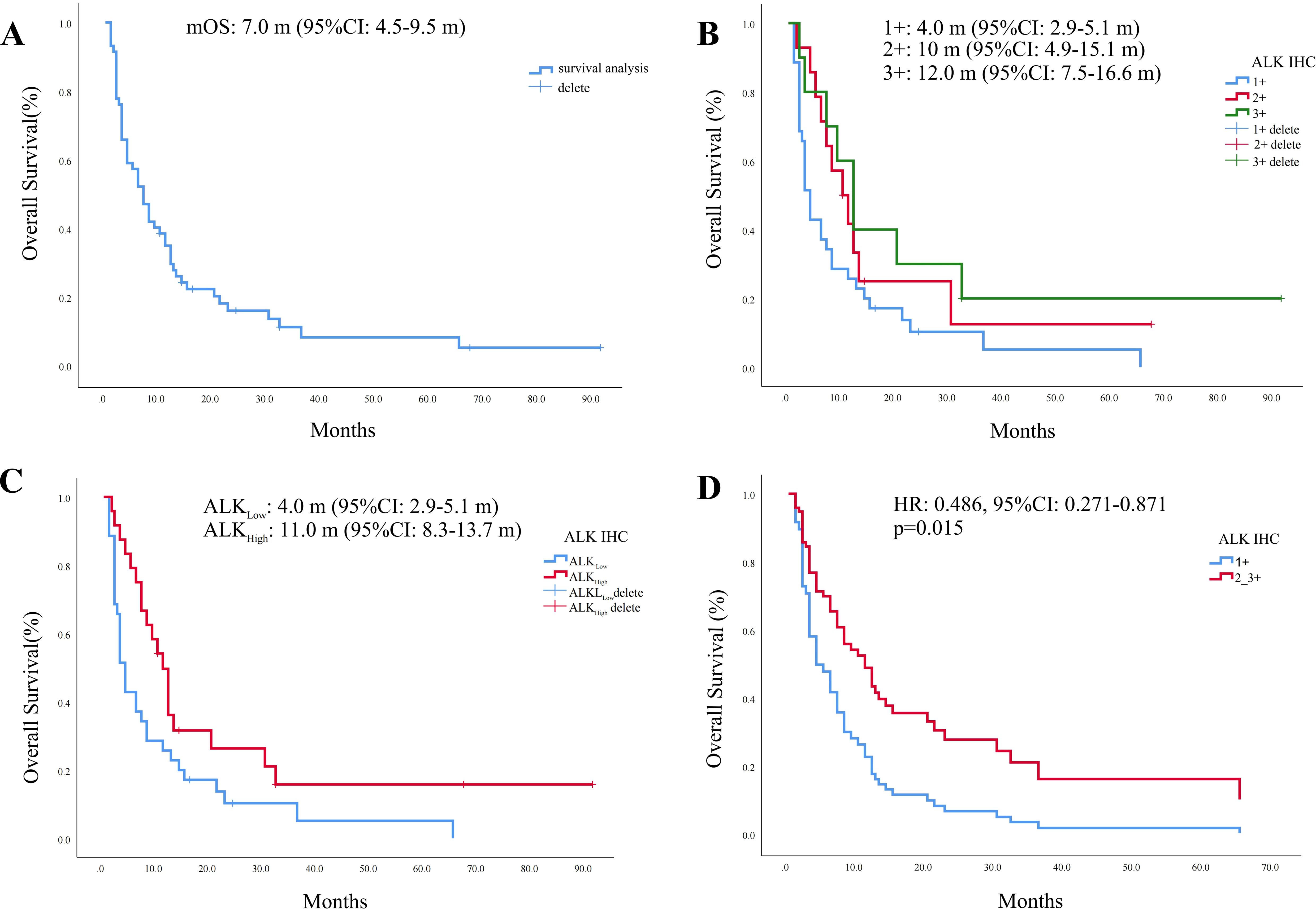
In our study cohort, 88% (52/59) of the patients had OS. Here, we used a swim lane diagram to show the specific survival situation of the patients in the study cohort (Figure 3). Kaplan–Meier analysis showed that the overall population median OS (mOS) was 7.0 months [95% confidence interval (CI): 4.5–9.5 months] (Figure 4A). Among them, the mOS of the ALK (+) group was 4.0 months (95% CI: 2.9–5.1 months), the mOS of the ALK (++) group was 10.0 months (95% CI: 4.9–15.1 months), and the mOS of the ALK (+++) group was 12.0 months (95% CI: 7.5–16.6 months). Kaplan–Meier analysis showed that there were significant differences in prognosis between groups with different levels of ALK protein expression (p = 0.031) (Figure 4B).

Figure 3. Patient survival swim lane diagram of this study cohort. Chem, chemotherapy; Radi, radiotherapy; Imm, immunotherapy; None, no treatment.

Figure 4. Kaplan–Meier survival curve. (A) Kaplan–Meier curve of the overall population. (B) Kaplan–Meier curve of different ALK protein expression groups. (C) Survival analysis of ALKLow and ALKHigh groups. (D) ALK protein Cox survival curve after adjusting for clinical parameter covariates.
Since the number of cases with moderate to strong and weak expression of ALK protein in our study cohort was relatively small, the cohort population was regrouped into two groups—the weak expression group (ALK IHC 1+, ALKLow) and the moderate to strong expression group (ALK IHC 2+_3+, ALKHigh)—for comparison. General clinical characteristics showed that there were still no statistical differences between the two groups in terms of age, gender, smoking history, TNM stage, primary tumor site, and Ki-67 expression (Table 4). Survival analysis showed that the mOS of the ALKLow group was significantly worse than that of ALKHigh [mOS: 4.0 months (95% CI: 2.9–5.1 months) vs. 11.0 months (95% CI: 8.3–13.7 months), p = 0.009] (Figure 4C). After covariate adjustment analysis of different clinical parameters used the Cox regression model, the results showed that ALK protein expression level was an independent factor that affected the prognosis of patients with SCLC (HR: 0.486, 95% CI: 0.271–0.871, p = 0.015) (Figure 4D).
ALK fusion is a significant molecular event observed in approximately 5% of NSCLC cases, particularly in adenocarcinoma (15). Identifying patients with ALK fusion is crucial, as they derive significant benefits from ALK-TKIs, which have demonstrated remarkable efficacy in this patient group (16–18). Different from NSCLC, owing to rapid growth and early metastatic spread, SCLC does not benefit from low-dose computed tomography (CT) screening (19, 20). OS for the extended disease is extremely poor (<10% at 5 years) (21). The standard approach has involved platinum-based chemotherapy, resulting in rapid and profound responses but rarely achieving long-term durability (21). Recent translational research has begun to challenge this paradigm, fueling increasing interest in the molecular subtypes of SCLC and their potential implications for therapeutic strategies (22, 23).
The ALK (D5F3) IHC assay, while not a molecular test, demonstrates high sensitivity and specificity in detecting ALK fusions, making it a valuable diagnostic tool in clinical settings as companion diagnostics (24–27). Among the companion diagnostics for detecting ALK fusion, fluorescence in situ hybridization (FISH) was the first method to be clinically validated (28). Another molecular method for detecting ALK fusion is next-generation sequencing (NGS), which allows the parallel examination of millions or billions of deoxyribonucleic acid (DNA) strands. NGS has the advantage of simultaneously detecting multiple driver gene mutations across a wide range. Previous studies have demonstrated that NGS exhibits high sensitivity and specificity for ALK fusion detection (29). However, the turnaround time for NGS is over 2 weeks, which is longer than that of other tests, and it is more expensive, presenting certain limitations. The Ventana ALK (D5F3)-approved ALK IHC assay provides high analytical sensitivity and specificity, high concordance with FISH, and very high interobserver reproducibility (30, 31). The D5F3 clone detects the carboxyl terminus of the ALK protein, and numerous studies have demonstrated its excellent performance (24). In a series of 142 consecutive cases of treatment-naïve SCLC, 11% (n = 16) showed expression of ALK in IHC (12). The expression rate of ALK IHC in our cohort was 10.22%, which is similar to that in this study. Compared with the diffuse high-intensity expression generally shown in NSCLC, ALK expression was focal and of low intensity: 59.3% were weakly expressed (+), 23.7% were moderately expressed (++), and 16.9% were strongly expressed (+++).
ALK gene alterations are gaining more attention as pan-cancer markers in both solid and hematological malignancies (32). ALK genomic alterations are found in ~3.3% of patients with cancer, though ALK fusions/rearrangements are less common (33). In large-scale analyses of genomes, ALK fusions/rearrangements are detected in ~0.5%–0.8% of cancers (33, 34). In order to explore the expression of ALK in other tumors and its impact on prognosis, we downloaded the unified normalized pan-cancer dataset (TCGA, TARGET, and GTEx) from the UCSC (https://xenabrowser.net/) database, from which we extracted ENSG00000171094 (ALK) gene expression data in various samples. It was found that the expression rate of ALK in cancer patients was 0.2%–10.3%, significant upregulation of ALK expression was observed in 16 tumor types (GBM, LGG, UCEC, KIPAN, KIRC, LIHC, WT, SKCM, THCA, OV, PAAD, UCS, LAML, PCPG, KICH, and CHOL), and significant downregulation was observed in 11 tumor types (BRCA, ESCA, STES, COAD, COADREAD, PRAD, STAD, BLCA, READ, TGCT, and ACC) (Figure 1). In addition, we analyzed the prognostic relationship between ALK expression and each tumor by an established Cox proportional hazards regression model, and we used the log-rank test for statistical testing to obtain prognostic significance. The results showed that ALK expression was negatively correlated with the prognosis of LAML [p < 0.001, HR = 1.10 (1.05, 1.16)], STAD [p = 9.8e-3, HR = 1.10 (1.02, 1.18)], NB [p = 0.04, HR = 1.17 (1.01, 1.36)], and UVM [p = 8.4e-3, HR = 1.37 (1.08, 1.74)], and positively correlated with the prognosis of CESC [p = 0.01, HR = 0.90 (0.83, 0.98)], KIRP [p = 0.02, HR = 0.89 (0.81, 0.99)], SKCM [p = 8.2e-3, HR = 0.91 (0.85,0.98)], and DLBC [p = 0.04, HR = 0.72 (0.53, 0.97)] (Figure 2).
A review of previously published studies found that ALK expression is indeed associated with improved prognosis in some tumors. Previous studies have found that inflammatory myelofibrotic tumors (IMTs) are associated with ALK gene rearrangement in approximately 50% of cases (35). ALK-overexpressing IMTs may have a better prognosis than ALK non-expressing tumors (36, 37). In 2015, Chou et al. detected ALK translocation in 2.2% of patients with papillary thyroid cancer and believed that it was associated with a good prognosis of thyroid cancer (38). Actually, ALK gene rearrangements/fusions are more commonly found in hematological malignancies such as ALCL (32). Earlier studies have shown that ALK-expressing ALCL has a much better 5-year OS rate (70%–80% vs. 15%–45%) and 5-year failure-free survival (FFS: 60% vs. 36%; p = 0.015) than non-ALK-expressing ALCL (11, 39) and found that in high-grade serous ovarian cancer (HGSOC), HGSOC harboring activating ALK mutations might be associated with a better survival, while ALK overexpression and ALK amplification do not impact the prognosis (40). Similarly, in MCC, the presence of ALK and phosphorylated ALK (p-ALK) was determined by immunohistochemistry, with almost half of the analyzed MCC tumors displaying ALK phosphorylation (47.8%). Survival analysis showed that p-ALK in MCC was associated with longer survival, with intermediate/high ALK and p-ALK tumor expression having better survival (10). In addition, a study that explored prognosis-related clinical and molecular factors in malignant pleural mesothelioma found that the median survival time (MST) of EML4-ALK-positive patients was longer than that of negative ones (19.6 months vs. 9.57 months), although no statistical significance was seen (p = 0.159), but the authors considered it to be related to the small sample size (41).
In the past 9 years, we found that 70 patients were identified as having abnormal expression of the ALK protein in SCLC among the 685 patients diagnosed with SCLC. In our study, we used the results of ALK IHC to reclassify SCLC and evaluated the correlation between the nonspecific expression of the ALK protein and the prognosis of patients with SCLC. We divided the patients into an ALK weak expression group (ALKLow) and an ALK moderate to strong expression group (ALKHigh) according to the ALK protein expression level on IHC. Survival analysis showed that the prognosis of the ALKLow group was significantly worse than that of the ALKHigh group [mOS: 4.0 months (95% CI: 2.9–5.1 months) vs. 11.0 months (95% CI: 8.3–13.7 months), p = 0.009], which suggested that high expression of the ALK protein is a better prognostic marker for SCLC. To our knowledge, this was the first clinical study on the prognostic significance and value of ALK protein expression directly evaluated by IHC in SCLC. Indeed, differential gene expression in the different molecular subtypes and during the disease course might influence sensitivity and resistance to several therapeutic agents (42). In recent years, our understanding of molecular profiling for SCLC has been steadily expanding, revealing potential genetic alterations that can be identified via various molecular biology techniques and panels despite the absence of approved targeted treatments for the disease (43). Previous studies have found that epidermal growth factor receptor (EGFR)-mutated patients with SCLC had longer OS than EGFR wild type even though they were not treated with EGFR-TKI, suggesting a potential favorable prognostic role of EGFR mutations in SCLC (7). Moreover, the expression of KRAS G12C, BRAF, NF1, NEUROD1, ASCL1, SOX2, CYP1B1, SLIT2, CDK6, GCLC, and NFYA genes is also associated with improved prognosis of SCLC (7, 42, 44–49). Our study suggests that identifying ALK protein expression in SCLC has significant prognostic value. Through correlation network analysis, we found that the expression of ALK was positively correlated with KRAS, BRAF, NF1, ASCL1, SOX2, CYP1B1, SLIT2, CDK6, and GCLC (Figure 3), suggesting that the mechanism by which high expression of ALK improves the prognosis of SCLC may be related to them.
The limited experiences of targeted therapies for the treatment of SCLC regard almost exclusively the use of EGFR and ALK inhibitors in the presence of specific molecular alterations of the corresponding genes. EGFR mutations and ALK rearrangements have been described in cases of histological transformation as a mechanism of resistance to TKIs, but de novo alterations of these genes in SCLC are unusual (50–53). As mentioned in a previous study, a recent large-scale NGS analysis was performed on 3,600 real-world SCLC cases. Besides EGFR mutations, ALK (n = 5), RET (n = 5), ROS1 (n = 3), and NTRK1 (n = 1) oncogenic molecular alterations were also detected (54). The characteristics in this study cohort showed a similar distribution based on sex; most patients had a smoking history (96.5%) and exhibited advanced-stage disease at the time of initial diagnosis (82.6%). mOS for the cohort was 8.0 months (95% CI: 7.3–9.0 months). However, in our study cohort, most patients were men (91.53%), and 61.02% (36/59) were smokers. The mOS of the cohort was 7.0 months (95% CI: 4.5–9.5 months). Subgroup analysis of our cohort suggested that high ALK IHC expression was associated with increased OS in patients with SCLC (HR: 0.486, 95% CI: 0.271–0.871, p = 0.015).
Furthermore, as regards the possibility of using ALK-TKI as targeted therapy in selected cases of SCLC, a clinical case reported rapid partial response (PR) to alectinib combined with irinotecan as second-line treatment for a 26-year-old patient with SCLC, and the progression-free survival (PFS) reached 6 months (55). A 38-year-old male SCLC patient with EML4-ALK fusion confirmed by IHC, FISH, and direct sequencing received crizotinib as first-line treatment and achieved PR, but the PFS and OS were not reported in the article (56). In one case involving a patient with SCLC, NGS detected a novel pleckstrin homology and RUN domain containing M2 (PLEKHM2)-ALK fusion; the patient experienced long-lasting clinical benefit after treatment with a combination of standard chemotherapy and crizotinib, achieving an OS of more than 27 months (57). However, in our cohort, one patient with ALK (++) experienced rapid progression after first-line targeted therapy with alectinib, with an OS of only 5 months. In a study that retested SCLC with ALK IHC expression using FISH and polymerase chain reaction (PCR), no activating alterations (rearrangements, point mutations, or amplification) were detected, and in the cases where copy number gains were shown (4/12), they were mild, accounting for three to five copy increases (12). The absence of activating mutations behind the expression of the protein suggests that only a normal form of ALK is expressed. Thus, ALK expression should not be considered as a surrogate for the presence of a molecular target in SCLC (58). Based on these experiences, we propose that patients with ALK-positive SCLC may benefit from chemotherapy combined with ALK-TKI rather than ALK-TKI targeted therapy alone.
In conclusion, our study innovatively evaluated the prognostic value of ALK abnormal protein expression levels in patients with SCLC when ALK IHC was positive. Interestingly, elevated expression levels of the ALK protein were significantly correlated with favorable prognosis in patients with SCLC. It is important to acknowledge that this investigation was conducted as a single-center retrospective study, which may introduce potential biases and limit the representativeness of the sample. To address these limitations and validate the findings, our center is spearheading a multicenter retrospective study aimed at further assessing the reliability of these conclusions. Additionally, our analysis revealed that the intensity ALK protein expression did not exhibit any significant association with various clinical and histopathological characteristics. Future studies are needed to elucidate the association between ALK expression and ALK gene status and to investigate disease progression, especially the oncogenesis of ALK-positive SCLC.
The original contributions presented in the study are included in the article/Supplementary Material. Further inquiries can be directed to the corresponding author.
The studies involving humans were approved by the Ethics Committee of the 900th Hospital of the Joint Logistic Support Force of China. The studies were conducted in accordance with the local legislation and institutional requirements. The participants provided their written informed consent to participate in this study. Written informed consent was obtained from the individual(s) for the publication of any potentially identifiable images or data included in this article.
JX: Conceptualization, Data curation, Formal Analysis, Investigation, Methodology, Project administration, Software, Validation, Visualization, Writing – original draft, Writing – review & editing. WZ: Data curation, Investigation, Writing – original draft. FX: Data curation, Investigation, Writing – review & editing. CW: Data curation, Formal Analysis, Writing – review & editing. FC: Data curation, Formal Analysis, Visualization, Writing – review & editing. RR: Data curation, Writing – review & editing. YC: Data curation, Writing – review & editing. LZ: Data curation, Writing – review & editing. WW: Data curation, Writing – review & editing. ZZ: Data curation, Writing – review & editing. JY: Data curation, Writing – review & editing. YZ: Data curation, Writing – review & editing. ZY: Conceptualization, Formal Analysis, Funding acquisition, Project administration, Supervision, Writing – review & editing.
The author(s) declare that financial support was received for the research and/or publication of this article. The present work was supported by the 900th Hospital of the Joint Logistic Support Force of China: Youth Incubation Special Program [grant number 2023QN04], the Fujian Medical University: Set sail Fund Project [grant number 2022QH1333], the External Cooperation of Science and Technology Program of Fujian Province [grant number 202210034], and the 900th Hospital of the Joint Logistic Support Force of China: National Science and Technology Fund Incubation Special Program [grant number 2023GK04].
We give our sincere thanks to the participating patients for supporting the publication of this study.
The authors declare that the research was conducted in the absence of any commercial or financial relationships that could be construed as a potential conflict of interest.
The author(s) declare that no Generative AI was used in the creation of this manuscript.
All claims expressed in this article are solely those of the authors and do not necessarily represent those of their affiliated organizations, or those of the publisher, the editors and the reviewers. Any product that may be evaluated in this article, or claim that may be made by its manufacturer, is not guaranteed or endorsed by the publisher.
The Supplementary Material for this article can be found online at: https://www.frontiersin.org/articles/10.3389/fonc.2025.1530339/full#supplementary-material
1. Bray F, Laversanne M, Sung H, Ferlay J, Siegel RL, Soerjomataram I, et al. Global cancer statistics 2022: GLOBOCAN estimates of incidence and mortality worldwide for 36 cancers in 185 countries. CA: Cancer J Clin. (2024) 74:229–63. doi: 10.3322/caac.21834
2. Solta A, Ernhofer B, Boettiger K, Megyesfalvi Z, Heeke S, Hoda MA, et al. Small cells - big issues: biological implications and preclinical advancements in small cell lung cancer. Mol Cancer. (2024) 23:41. doi: 10.1186/s12943-024-01953-9
3. Sen T, Takahashi N, Chakraborty S, Takebe N, Nassar AH, Karim NA, et al. Emerging advances in defining the molecular and therapeutic landscape of small-cell lung cancer. Nat Rev Clin Oncol. (2024) 21:610–27. doi: 10.1038/s41571-024-00914-x
4. Megyesfalvi Z, Gay CM, Popper H, Pirker R, Ostoros G, Heeke S, et al. Clinical insights into small cell lung cancer: Tumor heterogeneity, diagnosis, therapy, and future directions. CA: Cancer J Clin. (2023) 73:620–52. doi: 10.3322/caac.21785
5. Makarem M, Jänne PA. Top advances of the year: Targeted therapy for lung cancer. Cancer. (2024) 130:3239–50. doi: 10.1002/cncr.35423
6. Schneider JL, Lin JJ, Shaw AT. ALK-positive lung cancer: a moving target. Nat Cancer. (2023) 4:330–43. doi: 10.1038/s43018-023-00515-0
7. Xie X, Qiu G, Chen Z, Liu T, Yang Y, You Z, et al. Characteristics and prognosis of EGFR mutations in small cell lung cancer patients in the NGS era. Clin Trans Oncol. (2024) 26:434–45. doi: 10.1007/s12094-023-03263-w
8. Chon HJ, Kim HR, Shin E, Kim C, Heo SJ, Lee CK, et al. The clinicopathologic features and prognostic impact of ALK positivity in patients with resected gastric cancer. Ann Surg Oncol. (2015) 22:3938–45. doi: 10.1245/s10434-015-4376-8
9. Liu J, Jin H, Tian H, Lian G, Chen S, Li J, et al. Anaplastic lymphoma kinase protein expression predicts micrometastases and prognosis for patients with hepatocellular carcinoma. Oncol Lett. (2016) 11:213–23. doi: 10.3892/ol.2015.3859
10. Jaatinen J, Veija T, Salmikangas M, Böhling T, Sihto H, Koljonen V. ALK is frequently phosphorylated in Merkel cell carcinoma and associates with longer survival. PloS One. (2021) 16:e0252099. doi: 10.1371/journal.pone.0252099
11. Savage KJ, Harris NL, Vose JM, Ullrich F, Jaffe ES, Connors JM, et al. ALK- anaplastic large-cell lymphoma is clinically and immunophenotypically different from both ALK+ ALCL and peripheral T-cell lymphoma, not otherwise specified: report from the International Peripheral T-Cell Lymphoma Project. Blood. (2008) 111:5496–504. doi: 10.1182/blood-2008-01-134270
12. Kondoh C, Horio Y, Hayashi Y, Ebi H, Hida T, Hasegawa Y, et al. Anaplastic lymphoma kinase expression in small-cell lung cancer. Histopathology. (2019) 75:20–8. doi: 10.1111/his.13842
13. Travis WD, Brambilla E, Burke AP, Marx A, Nicholson AG. Introduction to the 2015 world health organization classification of tumors of the lung, pleura, thymus, and heart. J Thorac Oncol. (2015) 10:1240–2. doi: 10.1097/jto.0000000000000663
14. Eisenhauer EA, Therasse P, Bogaerts J, Schwartz LH, Sargent D, Ford R, et al. New response evaluation criteria in solid tumours: revised RECIST guideline (version 1.1). Eur J Cancer (Oxford England: 1990). (2009) 45:228–47. doi: 10.1016/j.ejca.2008.10.026
15. Elshatlawy M, Sampson J, Clarke K, Bayliss R. EML4-ALK biology and drug resistance in non-small cell lung cancer: a new phase of discoveries. Mol Oncol. (2023) 17:950–63. doi: 10.1002/1878-0261.13446
16. Camidge DR, Dziadziuszko R, Peters S, Mok T, Noe J, Nowicka M, et al. Updated efficacy and safety data and impact of the EML4-ALK fusion variant on the efficacy of alectinib in untreated ALK-positive advanced non-small cell lung cancer in the global phase III ALEX study. J Thorac Oncol. (2019) 14:1233–43. doi: 10.1016/j.jtho.2019.03.007
17. Solomon BJ, Mok T, Kim DW, Wu YL, Nakagawa K, Mekhail T, et al. First-line crizotinib versus chemotherapy in ALK-positive lung cancer. New Engl J Med. (2014) 371:2167–77. doi: 10.1056/NEJMoa1408440
18. Peng Y, Zhao Q, Liao Z, Ma Y, Ma D. Efficacy and safety of first-line treatments for patients with advanced anaplastic lymphoma kinase mutated, non-small cell cancer: A systematic review and network meta-analysis. Cancer. (2023) 129:1261–75. doi: 10.1002/cncr.34664
19. Aberle DR, DeMello S, Berg CD, Black WC, Brewer B, Church TR, et al. Results of the two incidence screenings in the National Lung Screening Trial. New Engl J Med. (2013) 369:920–31. doi: 10.1056/NEJMoa1208962
20. Silva M, Galeone C, Sverzellati N, Marchianò A, Calareso G, Sestini S, et al. Screening with low-dose computed tomography does not improve survival of small cell lung cancer. J Thorac Oncol. (2016) 11:187–93. doi: 10.1016/j.jtho.2015.10.014
21. Dingemans AC, Früh M, Ardizzoni A, Besse B, Faivre-Finn C, Hendriks LE, et al. Small-cell lung cancer: ESMO Clinical Practice Guidelines for diagnosis, treatment and follow-up(☆). Ann Oncol. (2021) 32:839–53. doi: 10.1016/j.annonc.2021.03.207
22. Qu S, Fetsch P, Thomas A, Pommier Y, Schrump DS, Miettinen MM, et al. Molecular subtypes of primary SCLC tumors and their associations with neuroendocrine and therapeutic markers. J Thorac Oncol. (2022) 17:141–53. doi: 10.1016/j.jtho.2021.08.763
23. Paz-Ares L, Champiat S, Lai WV, Izumi H, Govindan R, Boyer M, et al. Tarlatamab, a first-in-class DLL3-targeted bispecific T-cell engager, in recurrent small-cell lung cancer: an open-label, phase I study. J Clin Oncol. (2023) 41:2893–903. doi: 10.1200/jco.22.02823
24. Uruga H, Mino-Kenudson M. ALK (D5F3) CDx: an immunohistochemistry assay to identify ALK-positive NSCLC patients. Pharmacogenomics Personalized Med. (2018) 11:147–55. doi: 10.2147/pgpm.S156672
25. Lindeman NI, Cagle PT, Aisner DL, Arcila ME, Beasley MB, Bernicker EH, et al. Updated molecular testing guideline for the selection of lung cancer patients for treatment with targeted tyrosine kinase inhibitors: guideline from the college of American pathologists, the international association for the study of lung cancer, and the association for molecular pathology. J Mol Diagnostics: JMD. (2018) 20:129–59. doi: 10.1016/j.jmoldx.2017.11.004
26. Minca EC, Portier BP, Wang Z, Lanigan C, Farver CF, Feng Y, et al. ALK status testing in non-small cell lung carcinoma: correlation between ultrasensitive IHC and FISH. J Mol Diagnostics: JMD. (2013) 15:341–6. doi: 10.1016/j.jmoldx.2013.01.004
27. Wang Y, Wang S, Xu S, Qu J, Liu B. Clinicopathologic features of patients with non-small cell lung cancer harboring the EML4-ALK fusion gene: a meta-analysis. PloS One. (2014) 9:e110617. doi: 10.1371/journal.pone.0110617
28. Camidge DR, Kono SA, Flacco A, Tan AC, Doebele RC, Zhou Q, et al. Optimizing the detection of lung cancer patients harboring anaplastic lymphoma kinase (ALK) gene rearrangements potentially suitable for ALK inhibitor treatment. Clin Cancer Res. (2010) 16:5581–90. doi: 10.1158/1078-0432.Ccr-10-0851
29. Letovanec I, Finn S, Zygoura P, Smyth P, Soltermann A, Bubendorf L, et al. Evaluation of NGS and RT-PCR methods for ALK rearrangement in European NSCLC patients: results from the European thoracic oncology platform lungscape project. J Thorac Oncol. (2018) 13:413–25. doi: 10.1016/j.jtho.2017.11.117
30. Thorne-Nuzzo T, Williams C, Catallini A, Clements J, Singh S, Amberson J, et al. A sensitive ALK immunohistochemistry companion diagnostic test identifies patients eligible for treatment with crizotinib. J Thorac Oncol. (2017) 12:804–13. doi: 10.1016/j.jtho.2017.01.020
31. Wakuda K, Morita M, Sekikawa M, Morikawa N, Miura K, Doshita K, et al. Concordance of ALK fusion gene-rearrangement between immunohistochemistry and next-generation sequencing. Int J Clin Oncol. (2024) 29:96–102. doi: 10.1007/s10147-023-02451-6
32. Shreenivas A, Janku F, Gouda MA, Chen HZ, George B, Kato S, et al. ALK fusions in the pan-cancer setting: another tumor-agnostic target? NPJ Precis Oncol. (2023) 7:101. doi: 10.1038/s41698-023-00449-x
33. Project Genie AACR. Powering precision medicine through an international consortium. Cancer Discovery. (2017) 7:818–31. doi: 10.1158/2159-8290.Cd-17-0151
34. Ross JS, Ali SM, Fasan O, Block J, Pal S, Elvin JA, et al. ALK fusions in a wide variety of tumor types respond to anti-ALK targeted therapy. Oncologist. (2017) 22:1444–50. doi: 10.1634/theoncologist.2016-0488
35. Butrynski JE, D’Adamo DR, Hornick JL, Dal Cin P, Antonescu CR, Jhanwar SC, et al. Crizotinib in ALK-rearranged inflammatory myofibroblastic tumor. New Engl J Med. (2010) 363:1727–33. doi: 10.1056/NEJMoa1007056
36. Chun YS, Wang L, Nascimento AG, Moir CR, Rodeberg DA. Pediatric inflammatory myofibroblastic tumor: anaplastic lymphoma kinase (ALK) expression and prognosis. Pediatr Blood Cancer. (2005) 45:796–801. doi: 10.1002/pbc.20294
37. Coffin CM, Patel A, Perkins S, Elenitoba-Johnson KS, Perlman E, Griffin CA. ALK1 and p80 expression and chromosomal rearrangements involving 2p23 in inflammatory myofibroblastic tumor. Modern Pathol. (2001) 14:569–76. doi: 10.1038/modpathol.3880352
38. Chou A, Fraser S, Toon CW, Clarkson A, Sioson L, Farzin M, et al. A detailed clinicopathologic study of ALK-translocated papillary thyroid carcinoma. Am J Surg Pathol. (2015) 39:652–9. doi: 10.1097/pas.0000000000000368
39. Cao Z, Gao Q, Fu M, Ni N, Pei Y, Ou WB. Anaplastic lymphoma kinase fusions: Roles in cancer and therapeutic perspectives. Oncol Lett. (2019) 17:2020–30. doi: 10.3892/ol.2018.9856
40. Gorczyński A, Miszewski K, Gager Y, Koch S, Pötschke J, Ugrinovski D, et al. Prognostic value of ALK overexpression and molecular abnormalities in high-grade serous ovarian carcinoma. Cancer Biomarkers: section A Dis Markers. (2023) 38:17–26. doi: 10.3233/cbm-230117
41. Wang YY, Zhang H, Bai H, Wang SH, Wu MN, An TT, et al. Prognosis related clinical and molecular factors in Malignant pleural mesothelioma. Zhonghua jie he he hu xi za zhi = Zhonghua jiehe he huxi zazhi = Chin J Tuberculosis Respir Dis. (2013) 36:162–8. doi: 10.3760/cma.j.issn.1001-0939.2013.03.002
42. Baine MK, Hsieh MS, Lai WV, Egger JV, Jungbluth AA, Daneshbod Y, et al. SCLC subtypes defined by ASCL1, NEUROD1, POU2F3, and YAP1: A comprehensive immunohistochemical and histopathologic characterization. J Thorac Oncol. (2020) 15:1823–35. doi: 10.1016/j.jtho.2020.09.009
43. Zullo L, Dall’Olio FG, Rossi G, Dellepiane C, Barletta G, Bennicelli E, et al. Molecular and genetic advances in small cell lung cancer landscape: from homogeneity to diversity. Int J Mol Sci. (2023) 25:224. doi: 10.3390/ijms25010224
44. Rudin CM, Durinck S, Stawiski EW, Poirier JT, Modrusan Z, Shames DS, et al. Comprehensive genomic analysis identifies SOX2 as a frequently amplified gene in small-cell lung cancer. Nat Genet. (2012) 44:1111–6. doi: 10.1038/ng.2405
45. Li GS, Huang ZG, Li DM, Tang YL, Zheng JH, Yang L, et al. CDK6 is a novel predictive and prognosis biomarker correlated with immune infiltrates in multiple human neoplasms, including small cell lung carcinoma. Funct Integr Genomics. (2023) 23:332. doi: 10.1007/s10142-023-01253-3
46. Ito T, Oi I, Saito Z, Imakita T, Kanai O, Fujita K, et al. De novo SCLC transformation from KRAS G12C-mutated lung adenocarcinoma with excellent response to sotorasib: A case report. JTO Clin Res Rep. (2023) 4:100510. doi: 10.1016/j.jtocrr.2023.100510
47. Lu HY, Qin J, Han N, Lei L, Xie F, Li C. EGFR, et al. PTEN, and PIK3CA mutation in plasma of small cell lung cancer patients. OncoTargets Ther. (2018) 11:2217–26. doi: 10.2147/ott.S159612
48. Liu L, Shao X, Gao W, Bai J, Wang R, Huang P, et al. The role of human epidermal growth factor receptor 2 as a prognostic factor in lung cancer: a meta-analysis of published data. J Thorac Oncol. (2010) 5:1922–32. doi: 10.1097/jto.0b013e3181f26266
49. Asiedu MK, Thomas CF Jr., Dong J, Schulte SC, Khadka P, Sun Z, et al. Pathways impacted by genomic alterations in pulmonary carcinoid tumors. Clin Cancer Res. (2018) 24:1691–704. doi: 10.1158/1078-0432.Ccr-17-0252
50. Zakowski MF, Ladanyi M, Kris MG. EGFR mutations in small-cell lung cancers in patients who have never smoked. New Engl J Med. (2006) 355:213–5. doi: 10.1056/NEJMc053610
51. Offin M, Chan JM, Tenet M, Rizvi HA, Shen R, Riely GJ, et al. Concurrent RB1 and TP53 Alterations Define a Subset of EGFR-Mutant Lung Cancers at risk for Histologic Transformation and Inferior Clinical Outcomes. J Thorac Oncol. (2019) 14:1784–93. doi: 10.1016/j.jtho.2019.06.002
52. Leonetti A, Sharma S, Minari R, Perego P, Giovannetti E, Tiseo M. Resistance mechanisms to osimertinib in EGFR-mutated non-small cell lung cancer. Br J Cancer. (2019) 121:725–37. doi: 10.1038/s41416-019-0573-8
53. Cooper AJ, Sequist LV, Lin JJ. Third-generation EGFR and ALK inhibitors: mechanisms of resistance and management. Nat Rev Clin Oncol. (2022) 19:499–514. doi: 10.1038/s41571-022-00639-9
54. Sivakumar S, Moore JA, Montesion M, Sharaf R, Lin DI, Colón CI, et al. Integrative analysis of a large real-world cohort of small cell lung cancer identifies distinct genetic subtypes and insights into histologic transformation. Cancer Discovery. (2023) 13:1572–91. doi: 10.1158/2159-8290.Cd-22-0620
55. Sun N, Zhuang Y, Zhang J, Chen S, Dai Y, Guo R. ALK rearrangement in small-cell lung cancer and durable response to alectinib: A case report. OncoTargets Ther. (2021) 14:5161–6. doi: 10.2147/ott.S323700
56. Shen Q, Kalyani FS, Qu J, Chen Z, Zhang J, Zhou J. A case of small cell lung carcinoma harboring an EML4-ALK fusion with partial response to crizotinib. Clin Lung Cancer. (2021) 22:e799–803. doi: 10.1016/j.cllc.2021.03.012
57. Li T, Zhang F, Wu Z, Cui L, Zhao X, Wang J, et al. PLEKHM2-ALK: A novel fusion in small-cell lung cancer and durable response to ALK inhibitors. Lung Cancer (Amsterdam Netherlands). (2020) 139:146–50. doi: 10.1016/j.lungcan.2019.11.002
Keywords: small cell lung cancer, ALK, immunohistochemistry, 5D3F, prognosis
Citation: Xu J, Zhang W, Xie F, Wang C, Cheng F, Rao R, Chen Y, Zhang L, Wen W, Zhao Z, Yuan J, Zheng Y and Yu Z (2025) Prognostic significance of ALK high expression in SCLC: a 9-year cohort analysis. Front. Oncol. 15:1530339. doi: 10.3389/fonc.2025.1530339
Received: 18 November 2024; Accepted: 04 March 2025;
Published: 25 March 2025.
Edited by:
Gisella Figlioli, Humanitas University, ItalyReviewed by:
Xingguang Luo, Yale University, United StatesCopyright © 2025 Xu, Zhang, Xie, Wang, Cheng, Rao, Chen, Zhang, Wen, Zhao, Yuan, Zheng and Yu. This is an open-access article distributed under the terms of the Creative Commons Attribution License (CC BY). The use, distribution or reproduction in other forums is permitted, provided the original author(s) and the copyright owner(s) are credited and that the original publication in this journal is cited, in accordance with accepted academic practice. No use, distribution or reproduction is permitted which does not comply with these terms.
*Correspondence: Zongyang Yu, eXV6eTUyN0BzaW5hLmNvbQ==
†ORCID: Jinhe Xu, orcid.org/0000-0003-2681-9893
Disclaimer: All claims expressed in this article are solely those of the authors and do not necessarily represent those of their affiliated organizations, or those of the publisher, the editors and the reviewers. Any product that may be evaluated in this article or claim that may be made by its manufacturer is not guaranteed or endorsed by the publisher.
Research integrity at Frontiers

Learn more about the work of our research integrity team to safeguard the quality of each article we publish.