94% of researchers rate our articles as excellent or good
Learn more about the work of our research integrity team to safeguard the quality of each article we publish.
Find out more
BRIEF RESEARCH REPORT article
Front. Oncol., 13 June 2024
Sec. Pediatric Oncology
Volume 14 - 2024 | https://doi.org/10.3389/fonc.2024.1426638
This article is part of the Research TopicGlobal Approaches to Molecular Diagnostics for Pediatric CancerView all 8 articles
 Julie M. Gastier-Foster1,2,3,4*
 Fredrick Lutwama5
 Olive Mbabazi5
 Steven Mlenga6
 Kennedy Ulaya6
 Ruth Namazzi7
 E. Faith Hollingsworth4
 Dolores Lopez-Terrada2,4
 Kevin E. Fisher2,4
 Angshumoy Roy2,4
 Carl E. Allen1,3
 David G. Poplack1,3
 Rizine Mzikamanda6
 Nmazuo Ozuah1,3
 Peter Wasswa1,3
Risk stratification and molecular targeting have been key to increasing cure rates for pediatric cancers in high-income countries. In contrast, precise diagnosis in low-resource settings is hindered by insufficient pathology infrastructure. The Global HOPE program aims to improve outcomes for pediatric cancer in Sub-Saharan Africa (SSA) by building local clinical care and diagnostic capacity. This study aimed to assess the feasibility of implementing molecular assays to improve leukemia diagnoses in SSA. Custom NanoString nCounter gene fusion assays, previously validated in the US, were used to test samples from suspected leukemia patients. The NanoString platform was chosen due to relatively low cost, minimal technical and bioinformatics expertise required, ability to test sub-optimal RNA, and rapid turnaround time. Fusion results were analyzed blindly, then compared to morphology and flow cytometry results. Of 117 leukemia samples, 74 were fusion-positive, 30 were negative, 7 were not interpretable, and 6 failed RNA quality. Nine additional samples were negative for leukemia by flow cytometry and negative for gene fusions. All 74 gene fusions aligned with the immunophenotype determined by flow cytometry. Fourteen samples had additional information available to further confirm the accuracy of the gene fusion results. The testing provided a more precise diagnosis in >60% of cases, and 9 cases were identified that could be treated with an available tyrosine kinase inhibitor, if detected at diagnosis. As risk-stratified and targeted therapies become more available in SSA, implementing this testing in real-time will enable the treatment of pediatric cancer to move toward incorporating risk stratification for optimized therapy.
In high-resource countries, modern treatment approaches have improved the cure rate for childhood cancers to approximately 85% (1). Achieving these excellent cure rates depends on therapeutic regimens tailored to the specific histological or molecular characteristics of each cancer. Unfortunately, the majority (~80%) of children with cancer live in low- and middle-income countries (LMICs), where there is limited access to pathology services, and cure rates are closer to 20% (1–5). The gold standard of modern cancer diagnosis in Western countries incorporates molecular diagnosis, specifically genomic analysis of the cancer sample. Genomic analysis can be used to determine a precise diagnosis, suggest prognosis to guide risk-adapted treatment, and identify cases that may benefit from targeted therapy. The pediatric cancer treatment protocols used in sub-Saharan Africa (SSA) are typically one treatment regimen per cancer type. This approach means many African children are undertreated and fail to achieve cure, while others are potentially over-treated and unnecessarily exposed to toxic chemotherapy agents.
The Texas Children’s Hospital Global HOPE (Hematology Oncology Pediatric Excellence) program launched in February 2016 as a capacity-building initiative, strengthening local healthcare infrastructure in SSA to effectively provide the multi-disciplinary care that is needed to optimally care for children with cancer, sickle cell disease and other blood disorders (6). The program, based at Texas Children’s Hospital and Baylor College of Medicine, partners with Ministries of Health and local hospitals in SSA. Global HOPE supported the establishment of the first accredited specialist training program for childhood cancer in the East African region (7). More than 30 pediatric hematologists/oncologists have graduated from the program. To improve patient outcomes, it is imperative to develop improved but sustainable diagnostic capacity alongside specialized clinical care for pediatric cancer.
Leukemia is the most common pediatric cancer, accounting for approximately 30% of all cases globally (2), and a similar rate has been observed at the Global HOPE-affiliated sites in Africa. Immunophenotyping is a critical diagnostic method for hematologic cancers in Western settings, and Global HOPE is working to ensure flow cytometry is incorporated at all its affiliated sites in SSA (8). The current approach to leukemia diagnosis in SSA includes morphology review and immunophenotype by flow cytometry (if available). Cytogenetic or molecular genetic analysis is rarely available. Diagnoses based on these limited evaluations often lack the specificity necessary to inform the application of effective, contemporary risk-adapted treatment regimens that incorporate molecular and genomic data. The genomics of pediatric leukemias have been well-studied in high resource settings (9–11). Gene fusions are estimated to occur in >50% of pediatric leukemias (12–15), with an additional 25% due to aneuploidy. The presence of a gene fusion in pediatric leukemia can be prognostic: examples include a favorable prognosis for B-acute lymphoblastic leukemia (B-ALL) with a ETV6-RUNX1 fusion or unfavorable prognosis of an acute myeloid leukemia (AML) patient with CBFA2T3-GLIS1 fusion. Specific leukemia fusions can also inform specific therapeutic option such as retinoic acid treatment for AML with a PML-RARA fusion, or a tyrosine kinase inhibitor for BCR-ABL1 or Ph-like B-ALL. Without the addition of a tyrosine kinase inhibitor, the outcome for these B-ALL patients can be as low as 30%, even in high-income countries (16, 17). Given that the highest frequency of genomic changes in pediatric leukemias is due to gene fusions, this class of abnormalities was a logical choice for the introduction of molecular testing in SSA, with the intent to address copy number changes in another round of development.
This project aimed to establish molecular methods to accurately diagnose pediatric cancers in a manner that is sustainable and standardized across multiple sites in the LMICs of SSA. Cost-effective methods for proper diagnosis are a key component to reach this goal and reduce the inequalities of available care. While next-generation sequencing (NGS) analysis has become standard for cancer care in high-income countries, SSA is not yet prepared to utilize NGS due to prohibitive cost, need for highly trained personnel, and intensive bioinformatics requirements. To address the current pathology infrastructure limitations in SSA, it is critical to innovate strategies that consider cost, turnaround time, personnel expertise required, small quantity of tumor available, and means to support interpretation and troubleshooting. Novel high-throughput molecular techniques have been developed with capabilities to detect multiple gene fusions associated with various pediatric cancers. One such platform, the NanoString nCounter analysis system, uses nucleic acid hybridization probes to simultaneously analyze hundreds of gene fusions in a single reaction. This technique requires neither nucleic acid amplification steps, library preparation, nor a bioinformatics pipeline, reducing the need for highly skilled technical or bioinformatics expertise. It is also tolerant of poor nucleic acid quality, making it amenable to a broad range of sample types. This method of fusion analysis requires less than 1 hour of hands-on time for the laboratory technologist, approximately 30 minutes on two successive days. Total time from RNA extraction to results interpretation can be as little as 48 hours, with 12 samples processed per batch. This methodology has been used to develop fusion panels for targeted tumor types (18–22). Given the high frequency of gene fusions in pediatric leukemias and the potential impact to improve outcome via customized therapy, the Global HOPE team tested the feasibility of implementing a custom gene fusion panel at the Global HOPE-affiliated sites in Uganda and Malawi. It was anticipated that the gene fusion results, in combination with the existing morphology and flow cytometry analyses, would be a first step towards refining diagnostic accuracy and eventually improving survival for children with leukemia in SSA.
Literature review, clinical laboratory results and public databases were used to obtain exact breakpoint sequence information for gene fusions associated with pediatric and adolescent cancers. The hematologic malignancy panel was designed to detect 439 breakpoints for 223 non-IGH/TCR fusions reported in ALL, AML, CML, lymphomas, and histiocytosis. The panel was purposely designed to be very broad, incorporating both common and rare gene fusions given the scarcity of knowledge of the genomic landscape of pediatric tumors in SSA. Due to the large size of the panel and limitation of probe sets per multiplexed tube, the Hematologic custom gene fusion panel consisted of two tubes: Heme 1 and Heme 2 with the intent to combine the most relevant probe sets for SSA into a single tube in a future iteration. The fusions in the panel, number of probe sets per fusion, and the number of samples positive for the fusion included in the validation set are shown in Table 1. The panels were validated in the Department of Pathology at Texas Children’s Hospital using residual, deidentified pediatric cancer samples with known fusion status determined by previous clinical testing, as well as commercially available RNA controls (Invivoscribe, San Diego, CA, USA and SeraCare Life Sciences, Milford, MA, USA). The validation design included samples to test a variety of fusions, different breakpoints for the same fusion, robustness between technicians, precision across runs, sensitivity of detection, and ease of performance. The NanoString results yielded a 99% accuracy after follow-up sequencing of any potential false negative/positive results (Supplementary Table 1).
During the validation, 48 clinical samples were used to determine fusion positivity cutoffs. The NanoString nCounter background counts for each probe set were averaged for all samples negative for that fusion. A sample was considered positive for a fusion probe set if the counts for the sample were more than 5 standard deviations (SD) above the negative sample average, based on the validation data to allow detection of all known positives, without false positives. Samples with counts more than 3 SD above the average were marked for further evaluation and potential for repeat analysis with more or less RNA.
Bone marrow or peripheral blood from patients with suspected new or relapsed leukemia were ficoll-separated using SepMate™-15 tubes (StemCell Technologies, Vancouver CA) and Lymphoprep™ (StemCell Technologies, Vancouver, CA) onsite in the local laboratories in Uganda or Malawi. An aliquot of up to 107 cells was used for extraction using the AllPrep DNA/RNA AllPrep mini kit (Qiagen, Hilden, Germany) with nucleic acids quantified on a NanoDrop™, Thermo Fisher Scientific, Waltham, MA, USA).
NanoString nCounter Sprint instruments were installed in Uganda and Malawi by a local distributor. Technologists (OM, SM, KU) were trained on maintenance of the instrument and performance of the assays. The gene fusion analysis was performed in-country following manufacturer’s instructions. Briefly, 100 ng of RNA Heme 1 or Heme 2 probes sets (Integrated DNA Technologies, Coralville, IA, US), Elements Core and Extension TagSets (NanoString Technologies, Seattle, WA, USA), and Hybridization Buffer (NanoString Technologies, Seattle, WA, USA) were mixed and hybridized at 67°C for 20 hours. Samples were loaded onto a NanoString Sprint cartridge and processed on the NanoString Sprint instrument (run time 6 hours). Data were analyzed using the nSolver 4.0 software (approximately 5 minutes), followed by pasting of the data into the interpretation template (see below). The 12 samples can then be interpreted in 15–30 minutes. Total time from RNA extraction to result can be as little as 48 hours with batching of samples (12 per run) most cost effective.
Data from each NanoString Sprint run (12 samples) was viewable in a spreadsheet containing approximately 250 rows and 15 columns. Given the challenge to review that volume of data quickly and accurately, analysis templates were created to allow data to be easily and consistently analyzed at the SSA sites. The templates were color coded to indicate fusion probe sets highly suggestive of a positive result (>5 SD above the average background for the probe set) or those that may require further review or repeat testing (>3 SD above the average background). For each sample, quality of the assay and RNA were assessed using positive and negative assays controls included in the NanoString hybridization reagents and housekeeping genes included in the probe set design. Selected sample data from a Heme 1 gene fusion run in Malawi are shown in Table 2.
Samples for the initial SSA studies included 126 suspected leukemia samples that had adequate quantity of RNA to perform the gene fusion testing (at least 50 ng of RNA for the assay, although 100 ng was used whenever possible). Most samples were run on both Heme 1 and Heme 2 custom gene fusion panels and analyzed blindly, without knowledge of flow cytometry results. In more recent runs, a more cost-effective means of reflex testing was utilized, where the Heme 2 panel was tested only if the Heme 1 panel was negative. Samples were called negative for fusions only if RNA quality was adequate (at least 3 of 5 housekeeping genes above the cutoff). Repeat testing was performed using more RNA (if the sample was negative but failed RNA quality using 100 ng) or less RNA (if the sample had high background with positives across numerous probe sets), as needed.
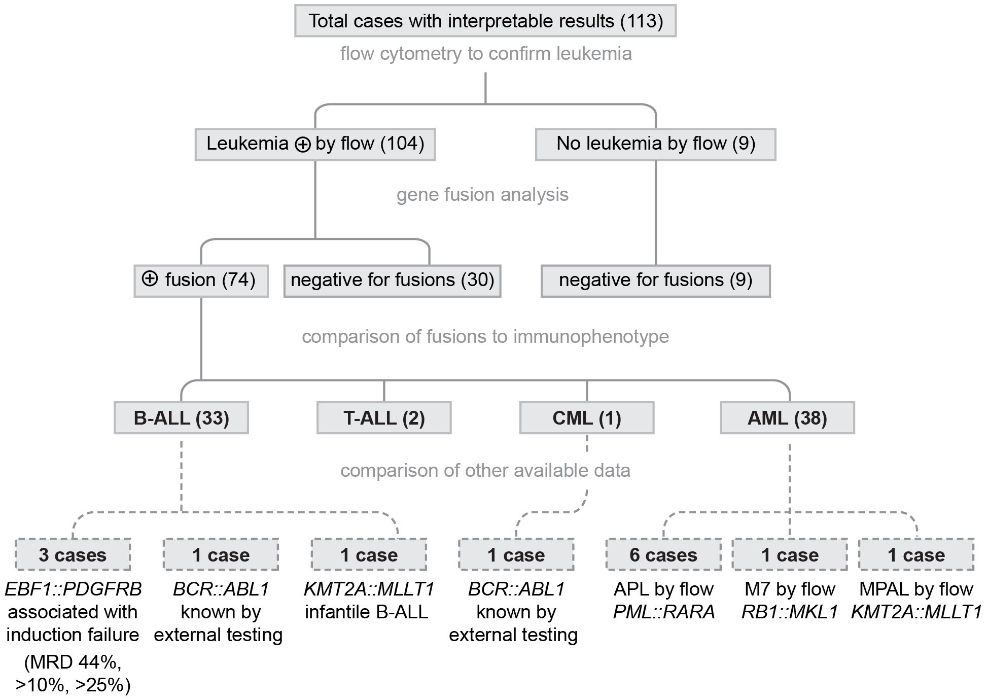
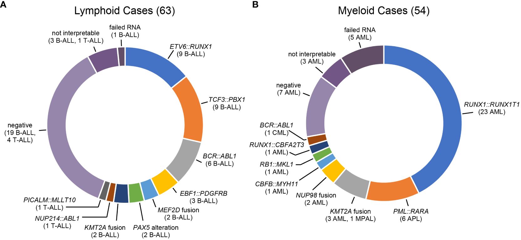
Very few leukemia cases in SSA are characterized by cytogenetic or molecular genetic testing, so genomic results were not available for comparison in most cases. To assess the accuracy of the results, the gene fusion calls were compared to flow cytometry results, including presence or absence of leukemic cells and immunophenotype (Figure 1). As a first step to confirming the accuracy of the fusion results, the identified fusions were compared to the fusions associated with the immunophenotype based on flow cytometry. There is some overlap in the fusions found in ALL and AML (e.g. KMTA rearrangements) and the BCR: ABL1 fusion is observed in both pediatric B-ALL and CML. However, most fusions are characteristic for a subtype of leukemia (B-ALL, T-ALL, AML, or CML). All fusion results were consistent with the flow cytometry immunophenotype. To further assess accuracy of the gene fusion testing, additional clinical or laboratory information was available for 14 cases. Three B-ALL cases were positive for the EBF1::PDGFRB fusion. This fusion is associated with Ph-like ALL and characterized by induction failure (23). The end of induction flow cytometry minimal residual disease results for these cases confirmed 44%, >10%, and >25% blasts, consistent with induction failure. One case was an infant with B-ALL that was positive for the KMT2A::MLLT1 fusion commonly found in infantile B-ALL. Two cases (1 B-ALL and 1 CML) were positive for the BCR::ABL1 fusion by NanoString analysis. Both cases had prior external molecular analysis consistent with the BCR::ABL1 fusion. Eight cases had a flow immunophenotype consistent with a more specific subtype. Six had a PML::RARA fusion, consistent with their diagnosis of acute promyelocytic leukemia (APL). One case was an infantile AML was consistent with an AML M7 megakaryoblastic leukemia immunophenotype (CD61+, CD34-, HLA-DR-) and a characteristic RB15::MKL1 fusion was identified (24). One case was considered to be mixed-phenotype acute leukemia (MPAL) for flow cytometry and showed a KMT2A::MLLT1 gene fusion. MPAL with KMT2A rearrangement is now described as a distinct entity for the World Health Organization (25). Thus, additional information available for these 14 cases further supported the accuracy of the gene fusion testing. The fusion results for 117 confirmed leukemia cases included 74 positive for a gene fusion, 26 negative cases, 7 cases with results that could not be interpreted, and 6 cases that failed RNA quality (Figure 2). As noted previously, little has been published about the leukemia subtypes in SSA. Although case numbers in this study were relatively low, and did not represent a consecutive series of cases, the general trends of fusions identified (relatively high frequency of RUNX1::RUNX1T1, PML::RARA, and BCR::ABL1) were consistent with comparisons of African American and white patients in a study of AML subtypes in the United States (26).

Figure 1 Evidence for Accuracy of the SSA Gene Fusion Results. All samples underwent molecular testing without knowledge of flow cytometry results. Nine cases were negative for leukemia by flow cytometry (all were negative for fusions). 104 leukemia cases were characterized as B-ALL (33), T-ALL (2), CML (1), or AML (38). After unblinding the flow cytometry results, all fusions results were consistent with the immunophenotype. Additional data were available for 14 cases and all data supported the accuracy of the fusion data.

Figure 2 Gene Fusion Testing Results for SSA Leukemic Samples. (A) Lymphoid leukemias. KMT2A partners: MLLT1 (1 case), MLLT3 (1 case); MEF2D partners: BCL9 (1 case), HRNPUL1 (1 case); PAX5 alterations: PAX5 internal tandem duplication (1 case), PAX5::ZCCHC7 (1 case) (B) Myeloid leukemias. KMT2A partners: AFF1 (1 MPAL case), AFDN (1 case), MLLT10 (1 case), USP2 (1 case); NUP98 partners: KDM5A (1 case), NSD1 (1 case).
This initial study supports implementation of gene fusion testing in real-time for new or relapsed pediatric leukemia cases in SSA using the NanoString platform and custom gene fusion assays. This testing would impact more than 60% of newly diagnosed leukemia patients’ diagnoses, a significant percentage as a first step into molecular testing. All testing for this study was performed by trained technologists in Uganda and Malawi, and standard operating procedures have been implemented for all steps of the process. Review of fusion results will occur with knowledge of leukemic content and immunotype available from flow cytometry results of the sample. Positivity cutoffs were modified for in-country results, as both instruments in SSA had lower background counts than the instrument used for validation testing in the US. Prospective testing in SSA will allow for implementation of risk adapted and/or targeted therapy, to maximize chances of cure. Gene fusion results obtained at the SSA sites during the initial years of patient testing will be evaluated to identify the most common fusions in the SSA pediatric cancer population and better understand the genomic landscape of SSA pediatric leukemias. This analysis will also allow reduction of the hematologic gene fusion assay to a smaller number of fusions, making the assay more cost-effective (approximately $150 vs. $250 currently) without decreasing detection rate. These reagent costs remain lower than a targeted RNA fusion panel via next-generation sequencing (approximately $500 for library preparation reagents), although only known fusions can be detected with the NanoString panels. A project with collaborators in Malawi allows for low-pass RNA sequencing using the Nanopore Minion for approximately $100 in reagent cost, but the results are best for classification of leukemias by gene expression and not fusion identification. Documented fusion results are a requirement for access to some targeted drugs including imatinib at the sites in SSA. The NanoString fusion assay is only intended for diagnostic samples and not for monitoring MRD. The sites in SSA are using flow cytometry for MRD analysis, a much more sensitive test compared to NanoString (due to direct measurement of RNA fusion molecules without an amplification step in the NanoString assay).
In addition to fusion analysis, the Global HOPE team is initiating studies of copy number detection, including gene amplification, on the NanoString nCounter Sprint platform using the NanoString Cancer CNV 2.0 assay with additional probes added based on relevance to pediatrics tumors. Gains and losses of whole chromosomes (a critical next step for leukemia diagnosis) can be detected using the CNV probes, but interpretation needs to be optimized to address frequent aneuploidy observed in pediatric tumors. A pediatric solid tumor custom fusion panel has also been designed and validated. Implementation in SSA will allow for more rapid and precise diagnosis. Histopathologic review of cases often requires weeks to months for a diagnosis in SSA and often does not include immunohistochemistry due to limited resources and patient lack of ability to pay. Identification of a specific gene fusion, e.g. PAX3::FOXO1 for rhabdomyosarcoma, would lead to a precise diagnosis without the need for extensive immunohistochemistry. In addition, the copy number analysis will allow for the rapid detection of gene amplification in specific pediatric tumors (e.g. MYCN in neuroblastoma). Initial proof of principle experiments confirmed that it will be straightforward to detect gene amplification using copy number variation (CNV) probes.
The possible uses of the NanoString nCounter as a cost-effective diagnostic method in low-resourced setting is starting to be realized due to its ability to be used for clinical and research purposes in numerous disease categories. An increasing number of studies in LMICs describe applications to adult and pediatric cancers including lung (27), breast (28), gastric (29), colon (30) medulloblastoma (31, 32), and Ph-like ALL (33).
The biggest challenges around diagnostic testing in SSA are reagent cost and access, instrument service availability, and shipping of cold chain reagents. Most Western companies utilize local distributors in SSA to supply reagents and instrument service. While allowing for more rapid response time on comparable time zones, these arrangements often lead to reagent stock-outs and extended instrument downtime. In addition, costs are typically elevated to account for secondary distribution. Most testing in pathology is not covered by government funding and therefore requires patients to pay (usually well outside their ability) or philanthropic support. To address these issues, Global HOPE has been purchasing reagents in the US, and shipping to the sites in SSA. Shipping at multiple temperature (room temperature, 4 °C, and frozen) greatly increases the cost of the reagents and creates funding challenge and is not sustainable in the long term. Global HOPE continues to work with diagnostic companies to emphasize the needs and challenge of high quality diagnostics in low-resource settings and to work to create access programs for compassionate pricing. Such partnerships are critical to reducing the extreme disparities in patient care and outcomes.
Current pediatric cancer diagnostics in Sub-Saharan Africa (SSA) lack the specificity necessary to inform the application of modern curative treatment regimens, such as distinguishing between subtypes of pediatric ALL or AML. Validated custom gene fusion panels were implemented at two Global HOPE-affiliated sites in SSA. The testing required minimal technical time, was easy to perform, and results were consistent with other known data such as flow cytometry. Implementing the diagnostic use of this platform to prospectively test patients in real time will greatly impact the ability to treat pediatric cancer in SSA by providing a more precise diagnosis. Global HOPE’s partnerships with Teva and Direct Relief have allowed SSA access to an increasing number of therapeutic options (34). As the available drugs expand to include more targeted therapies, molecular genomic analysis will be critical to identify the appropriate therapy for each patient. Thus, the effect of implementing molecular diagnosis will continue to grow with time. This work will also allow establishing training in molecular and pediatric pathology in low resource settings, a critical need for long-term sustainability and reducing inequities of pediatric cancer care.
The original contributions presented in the study are included in the article/Supplementary Material. Further inquiries can be directed to the corresponding author.
The studies involving humans were approved by Baylor College of Medicine Institutional Review Board. The studies were conducted in accordance with the local legislation and institutional requirements. Written informed consent for participation in this study was provided by the participants’ legal guardians/next of kin.
JG-F: Writing – review & editing, Writing – original draft, Visualization, Validation, Supervision, Project administration, Methodology, Investigation, Funding acquisition, Formal analysis, Data curation, Conceptualization. FL: Writing – review & editing, Validation, Methodology, Investigation. OM: Writing – review & editing, Methodology, Investigation, Data curation. SM: Writing – review & editing, Methodology, Investigation, Data curation. KU: Writing – review & editing, Methodology, Investigation, Data curation. RN: Writing – review & editing, Investigation, Funding acquisition. EH: Writing – review & editing, Validation, Methodology, Investigation. DL-T: Validation, Writing – review & editing, Resources, Investigation. KF: Writing – review & editing, Validation, Resources, Investigation. AR: Writing – review & editing, Validation, Resources, Investigation. CA: Writing – review & editing, Resources, Funding acquisition. DP: Validation, Writing – review & editing, Resources, Project administration, Funding acquisition. RM: Writing – review & editing, Project administration, Investigation, Funding acquisition, Data curation. NO: Writing – review & editing, Resources, Investigation, Funding acquisition, Data curation. PW: Writing – review & editing, Investigation, Funding acquisition, Data curation.
The author(s) declare financial support was received for the research, authorship, and/or publication of this article. Funding for this study was provided by St. Baldrick’s Foundation (International Scholar Award to FL), AACR/AORTIC Beginning Investigator Grant for Catalytic Research (BIG Cat to RM), Alex’s Lemonade Stand Foundation for Childhood Cancer (DGP), Lions Club International Foundation (DGP), Bristol Myers Squibb Foundation (DGP), and a U54 grant from the National Cancer Institute (1U54CA254569, CEA PI).
The authors thank Ronald Naitala (Uganda) and Samuel Makuti (Malawi) for their support of clinical data queries.
The authors declare that the research was conducted in the absence of any commercial or financial relationships that could be construed as a potential conflict of interest.
All claims expressed in this article are solely those of the authors and do not necessarily represent those of their affiliated organizations, or those of the publisher, the editors and the reviewers. Any product that may be evaluated in this article, or claim that may be made by its manufacturer, is not guaranteed or endorsed by the publisher.
The Supplementary Material for this article can be found online at: https://www.frontiersin.org/articles/10.3389/fonc.2024.1426638/full#supplementary-material
1. Lubega J, Kimutai RL, Chintagumpala MM. Global health disparities in childhood cancers. Curr Opin Pediatr. (2021) 33:33–9. doi: 10.1097/MOP.0000000000000984
2. GBD 2017 Childhood Cancer Collaborators. The global burden of childhood and adolescent cancer in 2017: an analysis of the Global Burden of Disease Study 2017. Lancet Oncol. (2019) 20:1211–25. doi: 10.1016/S1470–2045(19)30339–0
3. Kruger M, Hendricks M, Davidson A, Stefan CD, van Eyssen AL, Uys R, et al. Childhood cancer in Africa. Pediatr Blood Cancer. (2014) 61:587–92. doi: 10.1002/pbc.24845
4. Ngwa W, Addai BW, Adewole I, Ainsworth V, Alaro J, Alatise OI, et al. Cancer in sub-Saharan Africa: a lancet oncology commission. Lancet Oncol. (2022) 23:e251–2312. doi: 10.1016/S1470–2045(21)00720–8
5. Fleming KA, Horton S, Wilson ML, Atun R, DeStigter K, Flanigan J, et al. The Lancet Commission on diagnostics: transforming access to diagnostics. Lancet. (2021) 398:1997–2050. doi: 10.1016/S0140–6736(21)00673–5
6. Lubega J, Chirande L, Atwine B, Davidson A, Kashaigili HJ, Kanyamuhunga A, et al. Addressing the childhood cancer crisis in sub-Saharan Africa. Lancet Oncol. (2023) 24:729–32. doi: 10.1016/S1470–2045(23)00173–0
7. Lubega J, Airewele G, Frugé E, Wilson-Lewis K, Ishigami E, Cubbage M, et al. Capacity building: a novel pediatric hematology-oncology fellowship program in sub-Saharan Africa. Blood Adv. (2018) 2:11–3. doi: 10.1182/bloodadvances.2018GS112742
8. Namazzi R, Gaikwad A, Wasswa P, Cubbage M, Kambugu JB, Kasirye P, et al. Improving diagnosis and treatment of acute childhood leukemia in Uganda: impact of flow cytometry. Blood Adv. (2018) 2:21–3. doi: 10.1182/bloodadvances.2018GS112620
9. Brady SW, Roberts KG, Gu Z, Shi L, Pounds S, Pei D, et al. The genomic landscape of pediatric acute lymphoblastic leukemia. Nat Genet. (2022) 54:1376–89. doi: 10.1038/s41588–022-01159-z
10. Bolouri H, Farrar JE, Triche T Jr, Ries RE, Lim EL, Alonzo TA, et al. The molecular landscape of pediatric acute myeloid leukemia reveals recurrent structural alterations and age-specific mutational interactions. Nat Med. (2018) 24:103–12. doi: 10.1038/nm.4439
11. Tazi Y, Arango-Ossa JE, Zhou Y, Bernard E, Thomas I, Gilkes A, et al. Unified classification and risk-stratification in Acute Myeloid Leukemia. Nat Commun. (2022) 13:4622. doi: 10.1038/s41467–022-32103–8
12. Angione SDA, Akalu AY, Gartrell J, Fletcher EP, Burckart GJ, Reaman GH, et al. Fusion oncoproteins in childhood cancers: potential role in targeted therapy. J Pediatr Pharmacol Ther. (2021) 26:541–55. doi: 10.5863/1551–6776-26.6.541
13. Liu Y, Klein J, Bajpai R, Dong L, Tran Q, Kolekar P, et al. Etiology of oncogenic fusions in 5,190 childhood cancers and its clinical and therapeutic implication. Nat Commun. (2023) 14:1739. doi: 10.1038/s41467–023-37438–4
14. Chen X, Wang F, Zhang Y, Ma X, Cao P, Yuan L, et al. Fusion gene map of acute leukemia revealed by transcriptome sequencing of a consecutive cohort of 1000 cases in a single center. Blood Cancer J. (2021) 11:112. doi: 10.1038/s41408–021-00504–5
15. Chang F, Lin F, Cao K, Surrey LF, Aplenc R, Bagatell R, et al. Development and clinical validation of a large fusion gene panel for pediatric cancers. J Mol Diagn. (2019) 21:873–83. doi: 10.1016/j.jmoldx.2019.05.006
16. Tasian SK, Loh ML, Hunger SP. Philadelphia chromosome-like acute lymphoblastic leukemia. Blood. (2017) 130:2064–72. doi: 10.1182/blood-2017–06-743252
17. Hunger SP, Mullighan CG. Redefining ALL classification: toward detecting high-risk ALL and implementing precision medicine. Blood. (2015) 125:3977–87. doi: 10.1182/blood-2015–02-580043
18. Zhong Y, Beimnet K, Alli Z, Arnoldo A, Kowalski PE, Somers GR, et al. Multiplexed digital detection of B-cell acute lymphoblastic leukemia fusion transcripts using the nanoString nCounter system. J Mol Diagn. (2020) 22:72–80. doi: 10.1016/j.jmoldx.2019.08.008
19. Hu D, Zhou W, Wang F, Shu SM, Fan LL, He J, et al. Development of a NanoString assay to detect leukemogenic fusion transcripts in acute myeloid leukemia. Int J Lab Hematol. (2016) 38:663–73. doi: 10.1111/ijlh.12555
20. Chang KTE, Goytain A, Tucker T, Karsan A, Lee CH, Nielsen TO, et al. Development and evaluation of a pan-sarcoma fusion gene detection assay using the nanoString nCounter platform. J Mol Diagn. (2018) 20:63–77. doi: 10.1016/j.jmoldx.2017.09.007
21. Sheth J, Arnoldo A, Zhong Y, Marrano P, Pereira C, Ryall S, et al. Sarcoma subgrouping by detection of fusion transcripts using nanoString nCounter technology. Pediatr Dev Pathol. (2019) 22:205–13. doi: 10.1177/1093526618790747
22. Song W, Platteel I, Suurmeijer AJH, van Kempen LC. Diagnostic yield of NanoString nCounter FusionPlex profiling in soft tissue tumors. Genes Chromosomes Cancer. (2020) 59:318–24. doi: 10.1002/gcc.22834
23. Schwab C, Ryan SL, Chilton L, Elliott A, Murray J, Richardson S, et al. EBF1-PDGFRB fusion in pediatric B-cell precursor acute lymphoblastic leukemia (BCP-ALL): genetic profile and clinical implications. Blood. (2016) 127:2214–8. doi: 10.1182/blood-2015–09-670166
24. Ma Z, Morris SW, Valentine V, Li M, Herbrick JA, Cui X, et al. Fusion of two novel genes, RBM15 and MKL1, in the t(1;22)(p13;q13) of acute megakaryoblastic leukemia. Nat Genet. (2001) 28:220–1. doi: 10.1038/90054
25. Patkar N, Harris MH, Hodge JC, Orgel E, Greipp PT. Mixed-phenotype acute leukaemia with KMT2A rearrangement. In: WHO Classification of Tumours Editorial Board. Haematolymphoid tumours. International Agency for Research on Cancer; Forthcoming, Lyon (France (2024).
26. Sekeres MA, Peterson B, Dodge RK, Mayer RJ, Moore JO, Lee EJ, et al. Differences in prognostic factors and outcomes in African Americans and whites with acute myeloid leukemia. Blood. (2004) 103:4036–42. doi: 10.1182/blood-2003–09-3118
27. Lira ME, Choi YL, Lim SM, Deng S, Huang D, Ozeck M, et al. A single-tube multiplexed assay for detecting ALK, ROS1, and RET fusions in lung cancer. J Mol Diagn. (2014) 16:229–43. doi: 10.1016/j.jmoldx.2013.11.007
28. Hyeon J, Cho SY, Hong ME, Kang SY, Do I, Im YH, et al. NanoString nCounter Approach in breast cancer: A comparative analysis with quantitative real-time polymerase chain reaction, in situ hybridization, and immunohistochemistry. J Breast Cancer. (2017) 20:286–96. doi: 10.4048/jbc.2017.20.3.286
29. Kim ST, Do IG, Lee J, Sohn I, Kim KM, Kang WK. The NanoString-based multigene assay as a novel platform to screen EGFR, HER2, and MET in patients with advanced gastric cancer. Clin Transl Oncol. (2015) 17:462–8. doi: 10.1007/s12094–014-1258–7
30. Ariyannur PS, Joy RA, Menon V, Paulose RR, Pavithran K, Vasudevan DM. Pilot Nanostring PanCancer pathway analysis of colon adenocarcinoma in a tertiary healthcare centre in Kerala, India. Ecancermedicalscience. (2021) 15:1302. doi: 10.3332/ecancer.2021.1302
31. Kaur K, Jha P, Pathak P, Suri V, Sharma MC, Garg A, et al. Approach to molecular subgrouping of medulloblastomas: Comparison of NanoString nCounter assay versus combination of immunohistochemistry and fluorescence in-situ hybridization in resource constrained centres. J Neurooncol. (2019) 143:393–403. doi: 10.1007/s11060-019-03187-y
32. Leal LF, Evangelista AF, de Paula FE, Caravina Almeida G, Carloni AC, Saggioro F, et al. Reproducibility of the NanoString 22-gene molecular subgroup assay for improved prognostic prediction of medulloblastoma. Neuropathology. (2018) 38:475–83. doi: 10.1111/neup.12508
33. Gupta DG, Varma N, Abdulkadir SA, Sreedharanunni S, Sachdeva MUS, Naseem S, et al. A surrogate molecular approach for the detection of Philadelphia chromosome-like B-acute lymphoblastic leukemia. Cancer. (2024) 130:713–26. doi: 10.1002/cncr.35051
34. Teva Pharmaceuticals. Available online at: https://www.tevapharm.com/news-and-media/feature-stories/teva-partnership-brings-critical-medicines-to-children-with-cancer/ (Accessed April 22, 2024)
Keywords: pediatric leukemia, childhood leukemia, Africa, LMIC, molecular diagnosis, gene fusion, nanostring nCounter, genomics
Citation: Gastier-Foster JM, Lutwama F, Mbabazi O, Mlenga S, Ulaya K, Namazzi R, Hollingsworth EF, Lopez-Terrada D, Fisher KE, Roy A, Allen CE, Poplack DG, Mzikamanda R, Ozuah N and Wasswa P (2024) Rapid gene fusion testing using the NanoString nCounter platform to improve pediatric leukemia diagnoses in Sub-Saharan Africa. Front. Oncol. 14:1426638. doi: 10.3389/fonc.2024.1426638
Received: 01 May 2024; Accepted: 31 May 2024;
Published: 13 June 2024.
Edited by:
Jeremy Wang, University of North Carolina at Chapel Hill, United StatesReviewed by:
Sarra Ryan, Newcastle University, United KingdomCopyright © 2024 Gastier-Foster, Lutwama, Mbabazi, Mlenga, Ulaya, Namazzi, Hollingsworth, Lopez-Terrada, Fisher, Roy, Allen, Poplack, Mzikamanda, Ozuah and Wasswa. This is an open-access article distributed under the terms of the Creative Commons Attribution License (CC BY). The use, distribution or reproduction in other forums is permitted, provided the original author(s) and the copyright owner(s) are credited and that the original publication in this journal is cited, in accordance with accepted academic practice. No use, distribution or reproduction is permitted which does not comply with these terms.
*Correspondence: Julie M. Gastier-Foster, anhnYXN0aWVAdGV4YXNjaGlsZHJlbnMub3Jn
Disclaimer: All claims expressed in this article are solely those of the authors and do not necessarily represent those of their affiliated organizations, or those of the publisher, the editors and the reviewers. Any product that may be evaluated in this article or claim that may be made by its manufacturer is not guaranteed or endorsed by the publisher.
Research integrity at Frontiers

Learn more about the work of our research integrity team to safeguard the quality of each article we publish.