95% of researchers rate our articles as excellent or good
Learn more about the work of our research integrity team to safeguard the quality of each article we publish.
Find out more
CLINICAL CASE STUDY article
Front. Oncol. , 24 April 2013
Sec. Cancer Imaging and Image-directed Interventions
Volume 3 - 2013 | https://doi.org/10.3389/fonc.2013.00103
We present the case of a 56-year-old male with a history of recurrent follicular lymphoma undergoing chemotherapy with multiple 18F-FDG PET-CT studies at an outside facility. He developed a painful erythematous, pruritic rash in the left back requiring a visit to the emergency room. He was diagnosed and treated for Varicella zoster infection. He then presented to our imaging center 2 months later for a follow up 18F-FDG PET/CT study. Imaging demonstrated a cutaneous band of increased metabolic activity in the upper back following a dermatomal distribution. This was confirmed to be in the same area as the treated Varicella zoster eruption. A subsequent follow up 18F-FDG PET-CT scan 4 months later to confirm tumor resolution demonstrated the abnormal band of uptake in the back had resolved. This case illustrates the significance of being aware of this entity and to distinguish it from metastasis, especially in patients with a known history of malignancy.
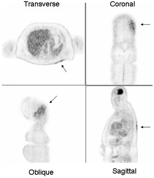
A 56-year-old male with a history of stage IIIA follicular lymphoma initially diagnosed 2 years prior to arriving at our imaging facility as a follow up to treatment. He was initially treated with an R-CVP (Rituximab, Cyclophosphamide, Vincristine, and Prednisone) chemotherapy regimen and attributed a complete response. However, he had evidence of relapse a year later. He was then placed on a MOPP-R regimen (Mustargen, Oncovin, Procarbazine, Prednisone, and Rituximab) and had multiple 18F-FDG PET-CT studies at an outside facility. The most recent PET-CT from 6 months earlier showed no evidence of malignancy. Since the patient had a history of recurrence, a repeat PET-CT scan to document stability was performed at our facility which demonstrated a superficial band of increased metabolic activity in the left mid back following a dermatomal distribution and associated mild skin thickening (see Figure 1). The managing oncologist informed us that the patient had gone to the emergency room 2 months prior to the PET-CT. He presented there with a painful erythematous, pruritic rash in the left mid back following the T10-12 dermatome. The emergency room physician diagnosed the patient with Varicella zoster and treated him with pain medication and valacyclovir 1000 mg orally three times per day for 7 days.

Figure 1. 18FDG PET images demonstrating a dermatomal distribution of increased metabolic activity in the left upper back (arrows). These findings were consistent with active Varicella zoster infection clinically.
Two weeks after the emergency room visit, he returned to his oncologist for a follow up appointment and physical examination found crusted lesions in the area of the rash. The patient continued to have neuropathic pain in the area and was taking oxycodone for relief. He had been prescribed valacyclovir 500 mg orally twice daily as prophylaxis prior to the outbreak, but he admit to not being adherent. The patient returned to our imaging facility 4 months after the previous PET-CT and the superficial band of increased metabolic activity in the left mid back had resolved. A month later the patient returned to his oncologist with no signs of a rash.
Varicella zoster is a common but serious infection in immunocompromised patients, especially in those with deficient cell-mediated immunity. However, it is often effectively prophylaxed with antivirals in these patients. The infection typically manifests as a painful rash often limited to a skin dermatome but can spread causing more serious complications and become life threatening. Active infection will show increased metabolic activity in the inflammatory cells on FDG PET images.
Active Varicella zoster infection is not routinely seen on PET-CT in our experience. There are only a few case reports describing this uncommon entity (Joyce and Carlos, 2006; Nair and Al Shemmari, 2011). However, none of these cases demonstrate a dermatomal distribution, as seen in our case, which may help in differentiating it from cutaneous metastasis. Valacyclovir is commonly used in the treatment of infection in immunocompromised patients and has been found to reduce the incidence of herpesviridae infection (Anderson et al., 1984; Lee et al., 2012).
Active infection with Varicella zoster virus may display increased metabolic activity in the inflammatory cells on PET. This case illustrates the significance of being aware of this entity and to distinguish it from metastasis, especially in patients with known malignancy (Castellucci et al., 2005).
The authors declare that the research was conducted in the absence of any commercial or financial relationships that could be construed as a potential conflict of interest.
Anderson, H., Scarffe, J. H., Sutton, R. N. P., Hickmott, E., Bridgen, D., and Burke, C. (1984). Oral acyclovir prophylaxis against herpes simplex virus in non-Hodgkin lymphoma and acute lymphoblastic leukemia patients receiving remission induction chemotherapy. A randomized double blind, placebo controlled trial. Br. J. Cancer 50, 45–49.
Castellucci, P., Nanni, C., Farsad, M., Alinari, L., Zinzani, P., Stefoni, V., et al. (2005). Potential pitfalls of in a large series of patients treated for malignant lymphoma: prevalence and scan interpretation. Nucl. Med. Commun. 26, 689–694.
Joyce, J., and Carlos, T. (2006). Herpes zoster mimicking recurrence of lymphoma on PET/CT. Clin. Nucl. Med. 31, 104–105.
Lee, H. S., Park, J. Y., Shin, S. H., Kim, S. B., Lee, J. S., Lee, A., et al. (2012). Herpesviridae viral infections after chemotherapy without antiviral prophylaxis in patients with malignant lymphoma incidence and risk factors. Am. J. Clin. Oncol. 35, 146–150.
Keywords: Varicella zoster, shingles, herpes zoster, dermatome, PET/CT
Citation: Muzaffar R, Fesler M and Osman MM (2013) Active shingles infection as detected on 18F-FDG PET/CT. Front. Oncol. 3:103. doi: 10.3389/fonc.2013.00103
Received: 19 March 2013; Accepted: 11 April 2013;
Published online: 24 April 2013.
Edited by:
Georgios Limouris, Athens University Medical Faculty, GreeceReviewed by:
Catherine Foss, Johns Hopkins University, USACopyright: © 2013 Muzaffar, Fesler and Osman. This is an open-access article distributed under the terms of the Creative Commons Attribution License, which permits use, distribution and reproduction in other forums, provided the original authors and source are credited and subject to any copyright notices concerning any third-party graphics etc.
*Correspondence: Medhat M. Osman, Division of Nuclear Medicine, Department of Radiology, Saint Louis University, 3635 Vista Avenue, 2-DT, St. Louis, MO 63110, USA. e-mail:bW9zbWFuQHNsdS5lZHU=
Disclaimer: All claims expressed in this article are solely those of the authors and do not necessarily represent those of their affiliated organizations, or those of the publisher, the editors and the reviewers. Any product that may be evaluated in this article or claim that may be made by its manufacturer is not guaranteed or endorsed by the publisher.
Research integrity at Frontiers

Learn more about the work of our research integrity team to safeguard the quality of each article we publish.