
95% of researchers rate our articles as excellent or good
Learn more about the work of our research integrity team to safeguard the quality of each article we publish.
Find out more
ORIGINAL RESEARCH article
Front. Neurosci. , 04 May 2022
Sec. Neurodegeneration
Volume 16 - 2022 | https://doi.org/10.3389/fnins.2022.869137
This article is part of the Research Topic Neurodegeneration and Neuroprotection in Retinal Disease, Volume II View all 10 articles
Purpose: Early-stage glaucoma diagnosis has been a challenging problem in ophthalmology. The current state-of-the-art glaucoma diagnosis techniques do not completely leverage the functional measures' such as electroretinogram's immense potential; instead, focus is on structural measures like optical coherence tomography. The current study aims to take a foundational step toward the development of a novel and reliable predictive framework for early detection of glaucoma using machine-learning-based algorithm capable of leveraging medically relevant information that ERG signals contain.
Methods: ERG signals from 60 eyes of DBA/2 mice were grouped for binary classification based on age. The signals were also grouped based on intraocular pressure (IOP) for multiclass classification. Statistical and wavelet-based features were engineered and extracted. Important predictors (ERG tests and features) were determined, and the performance of five machine learning-based methods were evaluated.
Results: Random forest (bagged trees) ensemble classifier provided the best performance in both binary and multiclass classification of ERG signals. An accuracy of 91.7 and 80% was achieved for binary and multiclass classification, respectively, suggesting that machine-learning-based models can detect subtle changes in ERG signals if trained using advanced features such as those based on wavelet analyses.
Conclusions: The present study describes a novel, machine-learning-based method to analyze ERG signals providing additional information that may be used to detect early-stage glaucoma. Based on promising performance metrics obtained using the proposed machine-learning-based framework leveraging an established ERG data set, we conclude that the novel framework allows for detection of functional deficits of early/various stages of glaucoma in mice.
Glaucoma, a chronic neurodegenerative disease affecting the retina and optic nerve, and a leading cause of blindness, is characterized by a progressive, irreversible loss of vision. As currently available treatment paradigms focus primarily on a predisposing factor, elevated intraocular pressure (IOP), and do not allow for repair of the retina and optic nerve once the disease has progressed and damage has occurred, technologies enabling an early diagnosis of glaucoma are needed urgently. Consequently, such new diagnostic modalities enabling early therapeutic intervention would significantly improve treatment outcomes. Current methods of glaucoma diagnosis are based on psychophysical techniques and the assessment of structural changes to the retina and optic nerve (Bussel et al., 2014). Standard automated perimetry testing, including the widely used Humphrey visual field testing, currently represents the most commonly utilized technique for glaucoma diagnosis and monitoring of disease progression and therapy outcomes (Ernest et al., 2012; Fidalgo et al., 2015). Recent efforts to employ machine-learning (ML) approaches to improve the analysis of behavioral psychophysical testing approaches produced moderate improvements over conventional analysis algorithms (Saeedi et al., 2021). However, significant damage to the retina and optic nerve, including loss of retinal ganglion cells (RGCs) has often already occurred before changes can be detected with standard automated perimetry testing (Turalba and Grosskreutz, 2010).
Recently, automated retinal image analysis (ARIA) systems have been developed for the diagnosis of complex diseases such as diabetic retinopathy and glaucoma (Sim et al., 2015; Lee et al., 2017). The development of these ARIA systems involved ML-based methods to detect structural changes determined with optical coherence tomography (OCT) imaging resulting in high analytical accuracy in automatically classifying disease phenotypes based on structural characteristics (Zhu et al., 2014; Asaoka et al., 2016; An et al., 2019). Despite such significant progress, early detection of glaucoma is still a challenge (Brandao et al., 2017), given the highly significant limitations of early detection of glaucoma based on structural methods. Systems employing the analysis of structural changes for glaucoma diagnosis are based on measuring retinal nerve fiber layer (RNFL) thickness in OCT images of the retina, which is highly variable and weakly correlated with RGC counts despite RNFL thickness being a surrogate marker of RGC degeneration and optic nerve fiber loss, hallmarks of glaucoma pathogenesis (Ledolter et al., 2015). Further, RGC loss often occurs early during pathogenesis in the absence of measurable RNFL thinning, prompting an urgent clinical need for methods with higher sensitivity, such as functional measures including ERG (Harwerth et al., 2002; Fortune et al., 2003; Takagi et al., 2012; Ledolter et al., 2015; Brandao et al., 2017). In contrast, functional measures such as visual field and ERG are sensitive to subtle changes in RGC function and RGC damage, which suggest a significant potential to enable early detection of glaucoma, even in the absence of elevated IOP, as seen in patients with normotensive glaucoma (Fortune et al., 2003; Aldebasi et al., 2004; Brandao et al., 2017). Therefore, this study aims to investigate such potential considering ERG signals.
Consequently, interventions could be initiated before irreversible damage occurs, allowing for the optimization of treatment strategies based on the improvement of RGC function (Ventura and Porciatti, 2006). This is of high clinical importance as determining the efficacy of therapies aimed at lowering IOP in open-angle glaucoma (Palmberg, 2002; Leske et al., 2007) requires early validation of therapy success (An et al., 2019), but will also be of importance for the development of novel alternative and complementary glaucoma therapies based on neuroprotective strategies (Rohowetz et al., 2018). Recently, in a study conducted by Tang et al. (2020) photopic negative response (PhNR) was used to assess the short-term changes in inner retinal function following intraocular pressure (IOP) decrease in glaucoma using eyedrops. Hui et al. (2020) showed that Nicotinamide supplementation helps improve the function of the inner retina in glaucoma.
Recent advances in the acquisition of complex neuroscience data have a significant innovative potential to progress toward more effective diagnostic systems (Kononenko, 2001). The adequate, timely, and clinically relevant analysis of such data has potentially high clinical impact (Lisboa, 2002). However, while such data sets can be readily acquired and technologies to further improve and simplify data acquisition continue to emerge (McPadden et al., 2019), critical barriers to implement the effective use of such novel data in clinical diagnostics and therapy delivery remain (Lee and Yoon, 2017). While the analysis of complex biomedical data is often part of medical diagnostics, current expert analysis standards and algorithms are limited by pattern recognition in few dimensions, which results in less than optimal identification or even exclusion of potentially relevant diagnostic features (Hannun et al., 2019). Machine learning could significantly augment medical diagnostics and increase their efficacy by analyzing aspects of complex and multi-dimensional biomedical data that are either not being considered adequately or that are not accessible to current analysis methods (Holzinger, 2014). Such machine-learning based diagnostic approaches have been developed and are being actively used for the detection of cardiovascular diseases (Al'Aref et al., 2019), and cancer (Cruz and Wishart, 2006).
ERG data are one such type of complex and multi-dimensional biomedical data that are potentially relevant to the diagnosis of glaucoma, but are currently not considered during routine clinical practice or in clinical research. Historically, this is due to multiple barriers related to clinical ERG data acquisition, such as limitations in reproducibility, high costs of both equipment and of individual tests, long test duration and complex test administration resulting in reduced patient acceptance and compliance, and the need for highly trained experts to administer tests. With the advent of novel ERG technologies, most of these barriers related to clinical ERG data acquisition have been removed (Nakamura et al., 2016; Asakawa et al., 2017; Kato et al., 2017; Hobby et al., 2018; Liu et al., 2018; Man et al., 2020), opening up the possibility to effectively use ERG data for glaucoma diagnostics, calling the necessity for the development of novel approaches (e.g., M-L-based ones) that is capable to quickly and thoroughly analyze such data.
Machine learning is based on statistical techniques to learn from data and develop predictive models (Jordan and Mitchell, 2015). Recently, there has been a surge of interest in machine learning as significant advancements in computational hardware (Shi et al., 2016) facilitate the development of novel machine learning approaches as solutions to problems in various disciplines from financial forecasting to public transportation and healthcare (Trafalis and Ince, 2000; Omrani, 2015; Ahmad et al., 2018). There are several predictive techniques in machine learning with various complexities, ranging from simple linear models to advanced non-linear models such as those based on deep learning algorithms (Shailaja et al., 2018; Khan et al., 2021; Saxe et al., 2021). Currently, available ERG analysis methods, such as those developed by Hood et al. (2000); Ventura and Porciatti (2006), have contributed to a significantly improved understanding of the relationship between ERG signals and vision loss. These methods are limited to frequency domain analysis (Miguel-Jiménez et al., 2010; Luo et al., 2011; Palmowski-Wolfe et al., 2011; Ledolter et al., 2013) and the analyses of differences in amplitude and latency of ERG (Fortune et al., 2002; Thienprasiddhi et al., 2003; Stiefelmeyer et al., 2004; Chu et al., 2007; Todorova and Palmowski-Wolfe, 2011; Ho et al., 2012; Hori et al., 2012). In addition, these methods are often time-consuming, labor-intensive, and focused on parameters developed to address a small subset of mostly genetic diseases of the eye affecting predominantly pediatric patient populations (Frishman et al., 2000; Graham et al., 2000; Dale et al., 2010). To achieve higher accuracy and a more detailed understanding of disease progression and of the impact of therapeutic intervention, more sophisticated features such as those obtained from wavelet analysis are required (Forte et al., 2008; Barraco et al., 2014). Additionally, currently available methods are often not suitable for analyzing large data sets and databases, rendering them incapable of taking advantage of complex and rich datasets (Consejo et al., 2019; Armstrong and Lorch, 2020). These drawbacks prompted others (Bowd et al., 2014; Yousefi et al., 2015; Atalay et al., 2016; Verma et al., 2017) and us to design and develop novel methods capable of handling complex and large datasets and ultimately to provide a unique approach for diagnosing early-stage glaucoma. However, it should be noted that early detection of glaucoma is not possible with currently available techniques during the early stages of glaucoma pathogenesis, when cellular changes occur that do not result in structural damage or visual impairment yet. Such early-onset factors predisposing to glaucoma development include processes preceding the onset of ocular hypertension, for example, the onset of iris pigment dispersion preceding IOP elevation in the DBA/2 mouse model. However, and more importantly, we identified cellular changes resulting in altered ERG signals, such as changes in oscillatory potentials, that currently cannot be detected with other functional or structural measures.
Boquete and colleagues developed a method to automate glaucoma diagnosis based on ERG signals using neural networks and structural pattern analysis (Boquete et al., 2012). They utilized thirteen features (morphological and transitional characteristics) for training the model and achieved a testing accuracy of 80.7% (Boquete et al., 2012). This study was limited to basic morphological characteristics of mfERG recordings (Boquete et al., 2012). Miguel-Jiménez et al. (2015) also employed neural networks for ERG-based glaucoma diagnosis but used continuous wavelet transformed coefficients and achieved a binary classification accuracy of 86.90% (Miguel-Jiménez et al., 2015). Although a higher accuracy was achieved, this analysis was limited to wavelet features only (Miguel-Jiménez et al., 2015). Nevertheless, both studies showed that machine learning-based methods trained even on compact data sets provide powerful tools to analyze ERG signals and provide potentially new information relevant for the early detection of glaucoma. Sarossy and colleagues investigated the relationship between a compact set of features and glaucoma that can be analyzed with machine learning approaches; however, the study was limited to the analysis of the photopic negative response (PhNR) and five additional features (Sarossy et al., 2021).
The goal of the present study was to comprehensively assess the capability of machine-learning-based methods to detect early-stage glaucoma using time-series ERG signals. In particular, the following points are addressed during method development:
1. Develop a framework to extract and identify important predictors (features) from ERG signals.
2. Compare the predictive capability of statistical and wavelet-based features for binary and multiclass classification.
3. Develop a robust ML-based model to diagnose glaucoma (binary classification).
4. Develop a robust ML-based model capable of distinguishing various stages of glaucoma progression (multiclass classification).
5. Develop a robust ML-based model to provide a quantitative assessment of visual function by predicting retinal ganglion cell count from ERG signals for the first time.
ML based algorithms have been applied to Electrocardiogram (ECG) signals in order to develop predictive models for diagnosing heart diseases (Li et al., 2014; Al'Aref et al., 2019). Recently machine learning-based Artificial Neural Networks (ANN) have been applied to ERG signals for obesity diagnosis (Yapici et al., 2021). However, to date, machine learning-based methods have not been applied systematically to analyze ERG signals for glaucoma detection. Therefore, the potential of ERG signals in glaucoma diagnosis has not been fully utilized. The present work aims to develop a predictive model for early glaucoma diagnosis based on machine-learning algorithms by utilizing advanced features from ERG signals as predictors. The steps involved in developing a machine-learning-based predictive model for ERG analysis are shown in Figure 1. Each of these steps is explained in detail below.

Figure 1. Machine learning workflow using ERG signals. ERG Database: the ERG database contains the input ERG data used to train the predictive model. Pre-processing of data: this step ensures data quality by transforming the data to a common baseline, accounting for missing data, and handling outliers. Feature extraction: mathematical operations are performed on the data to extract features/parameters that indicate functional deficits in the eye. Predictive Model Development: algorithms can determine trends and patterns in data from statistical analysis of extracted features during training; these models can predict either class or value from the input data are called classifier and regression models, respectively. Deployment of Model into medical devices: successful predictive models can be included with ERG testing devices to provide real-time prognosis and diagnosis.
Electroretinography measures the electrical responses of different types of cells in the retina, such as ganglion cells. These signals are usually measured in microvolts. Oscillatory Potential (OP) and Scotopic Threshold Response (STR) represent important ERG components indicative of RGC cell function (Saszik et al., 2002; Dong et al., 2004; Hancock and Kraft, 2004; Lei et al., 2006). OPs are small rhythmic wavelets superimposed on the ascending b-wave of the ERG and STR are negative corneal deflection elicited in the fully dark-adapted eye to dim stimuli. An International Society for Clinical Electrophysiology of Vision (ISCEV) standardized ERG protocol (Marmor et al., 2009) included several tests to measure the function of various retinal cell types, including the rod response, standard rod-cone response, Hi-intensity rods, and cones response, cone response, Hi-intensity cone response, flicker, and Hi flicker (Grillo et al., 2018). A visualization of nine ERG signals resulting from two ERG components (OP and STR) and seven ERG test responses is provided in Figure 2. The dynamics of ERG signals vary in people with various conditions and can therefore aid in differentiating individuals with glaucoma (Grillo et al., 2018), schizophrenia (Demmin et al., 2018), obesity (Yapici et al., 2021), and bipolar disorder (Hébert et al., 2020). ERG can also help in evaluating the effectiveness of new or existing drugs and therapy modalities (Lai et al., 2006, 2009; Nebbioso et al., 2009; da Silva et al., 2020).
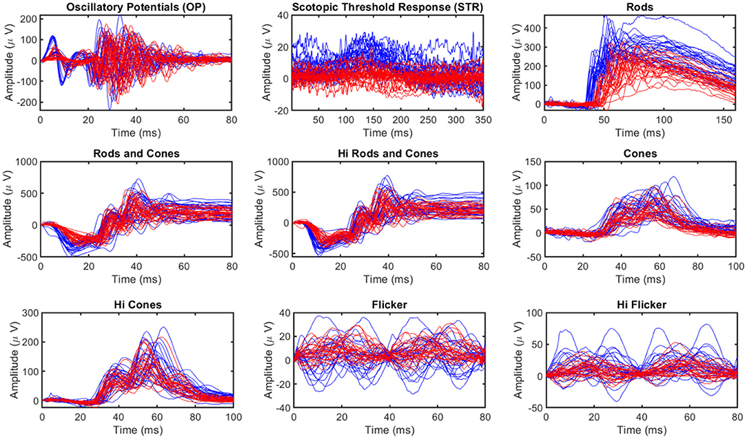
Figure 2. Visualization of ERG Signals manifesting their complex nature. The blue lines correspond to healthy and red lines correspond to glaucomatous Signals. Signals resulting from ERG tests include OP (Oscillatory Potential: small rhythmic wavelets superimposed on the ascending b-wave of the ERG), STR (Scotopic Threshold Response: negative corneal deflection elicited in the fully dark-adapted eye to dim stimuli), Rods (rod response), Rods and cones (standard rod-cone response), Hi Rods and cones (Hi-intensity rods and cones response), Cones (cone response), Hi cones (Hi-intensity cone response), Flicker (Flicker response), and Hi flicker (Hi-intensity flicker response).
The development of pigmentary glaucomatous optic neuropathy in the DBA/2 mouse model had several similarities to glaucoma pathogenesis in human patients, including loss of vision and RGC (McKinnon et al., 2009; Burroughs et al., 2011; Grillo et al., 2013; de Lara et al., 2014; Kaja et al., 2014; Grillo and Koulen, 2015; Montgomery et al., 2016). The Ganzfeld flash electroretinography (fERG) procedures in mice were conducted under dim red light that was followed by an overnight dark adaptation (>12 h). Isoflurane at 3 and 1.5% was used respectively, to anesthetize mice and maintain anesthesia. The pupils were dilated using 1 drop of 1% tropicamide and were allowed to dilate for 10 min. Rectal temperature was monitored and maintained at 37°C using a heating pad. Silver-embedded thread electrodes were placed over the cornea in 1% methylcellulose with mini-contact lenses fitted preventing the corneal dehydration (Ocuscience LLC, Henderson, NV). The head was placed inside the Ganzfeld dome, and fERG with 2 recording channels was performed using an HMsERG system (Ocuscience LLC) equipped with an amplifier with a band pass from 0.3 to 300 Hz. Mice were subjected to the International Society for Clinical Electrophysiology of Vision (ISCEV) standardized ERG protocol [29], whose implementation is described in detail in Marmor et al. (2009). ERGView 4.380V software (OcuScience LLC) was used to perform statistical analyses including averaging multiple flashes recorded at each intensity and stored for further analysis. Additionally, mice were tested using a scotopic flash intensity series in the range of −4.5 to 1.5 log cd s/m2. Further, a 1:1,000 neutral density filter (ND3) was used to control the 7 lowest flash intensities; data were averaged from 10 flashes (−4.5 to −3.5 log cd s/m2), 4 flashes (−3 to 0.5 log cd s/m2) at the lower intensities or measured from 1 flash at the 2 highest intensities (1 to 1.5 log cd s/m2). Following the light adaptation (1.5 log cd s/m2 for 10 min), responses from a photopic series (−2 to 1.5 log cd s/m2; 32 flashes per intensity) were recorded in a separate fashion. Further details about data acquisition can be found in Grillo et al. (2018).
Ganzfeld fERG tests were performed on 4 months old (n = 15) and 11 months old (n = 15) male DBA/2 mice. Each animal had two sets of test data, one for each eye. Therefore, a total of 60 data sets for individual eyes were included in this study. Each data set comprised of nine different ERG signals (OP, STR, and seven signals from ERG testing protocols), as shown in Figure 2 (OPs are small rhythmic wavelets superimposed on the ascending b-wave of the ERG and STR are negative corneal deflection elicited in the fully dark-adapted eye to dim stimuli). Therefore, 540 recordings were utilized in this study. Intraocular pressure (IOP) and retinal ganglion cell (RGC) count measurements were also utilized in this study. Although IOP data was available for all animals, RGC counts were only available for 10 (20 eyes). The animals were grouped in a binary group (healthy and glaucomatous) based on age and multiclass group based on IOP as (normal, <12 mm Hg; high, [≥12 mm Hg <17 mm Hg]; glaucomatous, ≥17 mm Hg). All the data used in this study was well-balanced for respective groups.
ERG raw data may contain several anomalies such as different start times, missing data, different sampling frequencies, noise, and unequal lengths of the signal recordings. In Machine learning-based modeling, the quality of the training data can significantly impact the model performance. Therefore, pre-processing (data preparation and screening) is crucial to ensure the quality of the training dataset (Jambukia et al., 2015). Pre-processing steps considered in the present study include,
1. Baseline adjustment
2. Feature extraction
3. Handling missing data
4. Handling outliers
5. Feature scaling
6. Feature selection
The signal's baseline (start time) can be different for different animals and testing protocols. Therefore, all the measurements were brought to a common baseline (start time was offset to zero) during baseline adjustment (Jambukia et al., 2015). Feature extraction involves computing a reduced set of values from a high-dimensional signal capable of summarizing most of the information contained in the signal (Khalid et al., 2014). The missing data were replaced with mean values (Graham et al., 2013). For handing outliers, values more than three scaled median absolute deviations (MAD) away from the median were detected as outliers and replaced with threshold values used in outlier detection (Aguinis et al., 2013). The feature's values vary widely, even by orders of magnitude. Therefore, it is important to bring the feature values to a similar range (feature scaling), especially when using distance-based machine learning algorithms (Wan, 2019). Feature selection is further dimensionality reduction from the extracted features. It is performed to reduce the computational cost of modeling, to achieve a better generalized, high-performance model that is simple and easy to understand (Aha and Bankert, 1996). Feature extraction and selection are explained in detail in the following sections.
ERG signals are complex high-dimensional data, and training a model with many variables requires significant computational resources. Feature extraction reduces the dimensionality of the data by computing a reduced set of values from a high-dimensional signal capable of summarizing most of the information contained in the signal (Guyon et al., 2008). In the present study, feature extraction was performed in two phases. First, common statistical features were extracted from the signal, followed by the extraction of advanced wavelet-based features. Figure 3 provides an overview of the feature extraction process and is explained below.
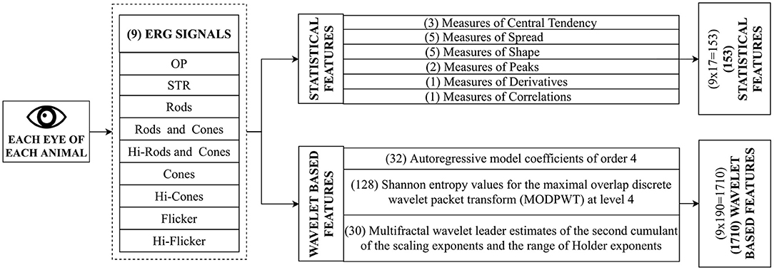
Figure 3. Feature Extraction. During this process, mathematical operations are performed on the data to extract features. This step is crucial for discovering features indicative of functional deficits in the eye. ERG test on each eye leads to nine signals, as shown in Figure 2. Two sets of features (Standard features and advanced features) are extracted from each of the nine signals. The standard set of features include common statistical features such as mean, quartiles, and entropies. In contrast, the advanced set of features include sophisticated features such as autoregressive coefficients, Shannon entropy, and wavelet features.
A total of 17 Statistical features capable of describing the general behavior of ERG signals were extracted from the signal. These features were grouped as follows.
1. Measures of Central Tendency
2. Measures of Spread
3. Measures of Shape
4. Measures of Peaks
5. Measures of Derivatives
6. Measures of Correlation
Measures of central tendency included mean, median, trimmed mean. Measures of spread included range, standard deviation, variance, mean absolute deviation, and interquartile range. Measures of the shape include skewness, kurtosis, central moments of the second and third-order, and aspect ratio. Measures of peaks included the number of peaks and troughs in the signal. Measures of derivatives include the first-order derivative of the signal with respect to time. Measures of correlation included the correlation coefficient of the signal with respect to time. The equations for the computation of these quantities can be found in Asgharzadeh-Bonab et al. (2020); Yapici et al. (2021).
Advanced features capable of capturing subtle changes were extracted from the signal. Each signal was split into 32 blocks (~ 2000 samples/block) to further capture subtle changes in the signal (Martis et al., 2014). Daubechies least-asymmetric wavelet with four vanishing moments (Symlets 4) was used as mother wavelet to derive the wavelet coefficients (Daubechies, 1992). The following features (190 features in total as shown in Figure 3) were extracted from each block of the signal:
AR coefficients: The signal x[n] at time instant n in an AR process of order p can be described as a linear combination of p earlier values of the same signal. The procedure is modeled as follows:
where a[i] is the AR model's ith coefficient, e[n] denotes white noise with mean zero, and p denotes the AR order. The AR coefficients for each block were estimated using the Burg method (Zhao and Zhang, 2005); the order was determined using the ARfit model order selection method (Neumaier and Schneider, 2001) as 4th order. Therefore a 4-order AR model is chosen to represent each of the ERG signal components.
Wavelet based Shannon Entropy: The Shannon entropy is an information-theoretic measure of a signal. Shannon entropy (denoted as SE) values for the maximal overlap discrete wavelet packet transform (MOD- PWT) using four-level wavelet decomposition was computed on the terminal nodes of the wavelet (Li and Zhou, 2016). Mathematical expression for Shannon entropy using wavelet packet transform is as follows:
where N is the number of coefficients in the jth node and pj,k are the normalized squares of the wavelet packet coefficients in the jth terminal node of the wavelet.
Multifractal wavelet leader estimates and multiscale wavelet variance estimates: The multifractal measure of the ERG signal was obtained using two wavelet methods (wavelet leader and cumulant of the scaling exponents). Wavelet leaders are time/space-localized suprema of the discrete wavelet coefficients' absolute value. These suprema are used to calculate the Holder exponents, which characterize the local regularity. In addition, second cumulant of the scaling exponents were obtained. Scaling exponents are scale-dependent exponents that describe the signal's power-law behavior at various resolutions. The second cumulant basically depicts the scaling exponents' divergence from linearity (Leonarduzzi et al., 2010). Wavelet variance of ERG signals were also obtained as features. Wavelet variance quantifies the degree of variability in a signal by scale, or more precisely, the degree of variability in a signal between octave-band frequency intervals (Maharaj and Alonso, 2014).
Feature extraction discussed previously was performed in order to reduce the dimensionality of the signals; however, the resulting number of features was still higher than the number of training data. Therefore, further reduction in the dimensionality of the data was performed using the feature selection method to identify relevant features for classification and regression. It should be noted that feature selection was necessary to reduce the computational cost of modeling, prevent the generation of a complex and over-fitted model with high generalization error, and generate a high-performance model that is simple and easy to understand (Saeys et al., 2007). In particular, the Minimum Redundancy Maximum Relevance (MRMR) sequential feature selection algorithm was used in the present study because this algorithm is specifically designed to drop redundant features [see (Darbellay and Vajda, 1999; Ding and Peng, 2005) for mathematical details/formulations], which was required to design a compact and efficient machine-learning-based model (Zhao et al., 2019). It is worth noting that other available dimensionality reduction techniques such as Principal component analysis (PCA) were not considered in this study as such techniques do not allow for direct tracing and understanding the relevance of each feature (Aha and Bankert, 1996).
ML models are mathematical algorithms that provide predictions based on an inference derived from the generalizable predictive patterns of the training data (Bzdok et al., 2018). Several machine learning models were employed and evaluated in order to identify the best one to classify the ERG signals. These included decision trees, discriminant, support vector machine, nearest neighbor, and ensemble classifiers. Most of these models can perform both classification and regression. Decision tree-based models predict the target variable by learning simple decision rules (Navada et al., 2011). Discriminant classifiers are based on the assumption that each class has different Gaussian distributions of data, and the classification is performed based on Gaussian distribution parameters estimated by the fitting function (Cawley and Talbot, 2003). Support vector machine (SVM) is based on Vapnik–Chervonenkis theory, where a hyperplane separating the classes is determined. SVMs are efficient algorithms suitable for compact datasets (Noble, 2006). The nearest neighbor algorithm is based on the assumption that similar things exist nearby. It is a simple yet versatile model with high computational cost (Zhang and Zhou, 2007). Ensemble methods such as bagged trees (or random forest) combine the predictions of several learning algorithms with improving generalization. Although these methods are also computationally expensive, they are unlikely to over-fit (Dietterich, 2000). Regression analysis based on the above techniques was also performed alongside classification.
Various performance evaluation metrics were utilized to compare different machine learning algorithms. The metrics used in this study include accuracy, sensitivity, specificity, precision, recall, f-score, root mean squared error, and their corresponding mathematical formulations are given below.
The abbreviations used in the following expressions include True Positive (TP) which are the cases the model correctly predicted the positive (glaucomatous) class. True Negative (TN) are the cases the model correctly predicted the negative (non-glaucomatous) class. False Positive (FP) are the cases the model incorrectly predicted the positive (glaucomatous) class. False Negative (FN) are the cases the model incorrectly predicted the negative (non-glaucomatous) class.
Accuracy is the percentage of correctly classified observations, as shown below.
Sensitivity/Recall estimates the proportion of actual positives (e.g., actual glaucomatous) was identified correctly.
Recall estimates the model's ability to correctly reject healthy patients without a Glaucoma.
Precision estimates the proportion of positive predictions (e.g., glaucomatous predictions) that was actually correct.
The F-Score estimates the harmonic mean of the precision and recall.
The Root Mean Square Error (RMSE) was used as the performance evaluation metric for regression analysis. RSME is the standard deviation of the prediction errors (residuals).
Where N is the number of observations.
A machine learning-based approach was developed and trained using the balanced ERG data previously published by Grillo et al. (2018). Although a compact dataset of 60 observations and 540 signals was used in this study, the current framework was able to consistently detect features (Figures 6, 9) that are known to be medically relevant such as OP, STR, Flicker reported in various studies (Tyler, 1981; Saszik et al., 2002; de Lara et al., 2014, 2015; Porciatti, 2015; Grillo et al., 2018). In particular, studies conducted by Wilsey and Fortune (2016); Hermas (2019); Beykin et al. (2021) investigating the variability of PhNR in glaucomatous and healthy subjects in PERG and fERG have found that PhNR to be an important biomarkers for detection of glaucoma. It is worth noting that in fERG analysis (ERG protocol for this study), pSTR, nSTR, PhNR are extracted from STR.
Therefore, we were able to demonstrate that the proposed framework for early-stage glaucoma diagnosis can be reproducibly evaluated and validated even on such a compact database. Furthermore, we would like to note that there are other investigations that successfully applied ML-based method in different fields, including biomedical (Seo et al., 2020) and material science (Zhang and Ling, 2018) using compact datasets. The procedure employed for the development of the predictive modeling framework is summarized below.
• Data Split: Hold out (80% training, k-fold cross-validation, 20% testing).
• Dimensionality reduction: Feature Extraction.
• Feature selection: MRMR.
• Hyper-parameter tuning: k-fold cross-validation (k = 10).
• Model Evaluation: Performance metrics evaluated on the unseen testing set.
The dataset was divided into two parts; 80% of the data was used for training and validation, and the remaining 20% was set aside for testing. The hold-out testing strategy ensured that the test data was never a part of the training process (Yadav and Shukla, 2016). Dimensionality reduction was performed using feature extraction and feature selection. MRMR feature selection algorithm was used to identify the important predictors. K-fold (K = 10) cross-validation was used for training and hyper-parameter tuning (Duan et al., 2003). The cross-validation technique significantly reduces bias when working with small datasets (Varma and Simon, 2006). The loss function was the objective minimization function for both classification regressions during hyper-parameter optimization. The hyper-parameters associated with corresponding ML algorithms (Feurer and Hutter, 2019), as shown in Table 1, were optimized through nested cross-validation. Next, the trained model with optimized hyper-parameters was evaluated using test data that was not a part of training. To further ensure that the machine learning models compared in this investigation were not over-fitted, given the compact dataset used in the present study, the behavior of training and testing error vs. training cycles was monitored. Different techniques, including Tree, Discriminant, SVM, Naive Bayes, Tree Ensemble, and KNN, were applied, and their performances were assessed. The performance of each technique was assessed based on the accuracy (discussed in section 2.9) is tabulated in Table 2. Considering binary and multiclass classifications, it can be seen that the Ensemble-based technique (bagged tree) was consistently outperforming other techniques. Additionally, other performance metrics for ensemble bagged trees (discussed in section 2.9) are summarized in Table 3.
For binary classification (classifying animals with/without glaucoma) based on statistical features, the correlation of cones, mean of flicker, median, and skewness of Hi Rods and cones, and standard deviation of cones were identified as important among the statistical features as shown in Figure 4. Moreover, the box plot demonstrates variations of each feature for each class (with/without glaucoma), respectively. Several models, including SVM and ensemble-based classifiers were used for training, and their performances were assessed. It turned out that the SVM and ensemble bagged tree provide the best performance with a testing accuracy of 83.33%, as shown in Table 2.
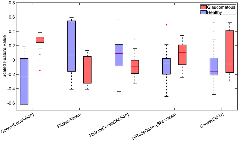
Figure 4. Boxplot of statistical features selected by Minimum Redundancy and Maximum Relevance (MRMR) feature selection algorithm for binary classification (Std D, Standard Deviation). On each box, the central mark indicates the median, and the bottom and top edges of the box indicate the 25th and 75th percentiles, respectively. The whiskers extend to the most extreme data points not considered outliers, and the outliers are plotted individually using the “+” marker symbol.
Next, the binary classification was performed using wavelet-based features. Among the extracted wavelet features, Shannon Entropy Values for Maximal Overlap Discrete Wavelet Packet Transform (MOD-PWT) were identified as important features from Rods and cones, Rods, STR, and OP, as shown in Figure 5. The utilization of the selected advanced features improved the accuracy to 91.70% by the ensemble bagged tree algorithm.
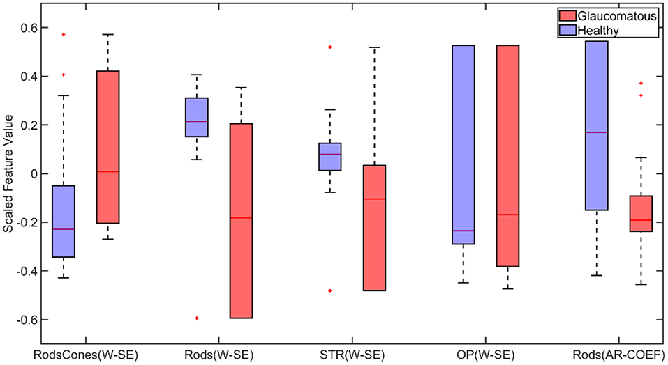
Figure 5. Box plot of wavelet-based features selected by Minimum Redundancy and Maximum Relevance (MRMR) feature selection algorithm for binary classification (W-SE, Wavelet based Shannon Entropy; AR-COEF, Autoregressive Coefficient). On each box, the central mark indicates the median, and the bottom and top edges of the box indicate the 25th and 75th percentiles, respectively. The whiskers extend to the most extreme data points not considered outliers, and the outliers are plotted individually using the “+” marker symbol.
It should be noted that the MRMR method selects features based on statistical relevance while dropping redundant features and thus, is computationally efficient (Darbellay and Vajda, 1999; Ding and Peng, 2005). Figure 6 demonstrates this for binary classification. It can be observed that correlation feature from cones, Moment of order three and trimmed mean feature from Oscillatory Potentials (OP) and Range and aspect ratio from Scotopic Threshold Response (STR) are highly correlated; Therefore, only the feature cones correlation was picked by the MRMR algorithm as inclusion of the other three did not increase/decrease the models predictability.
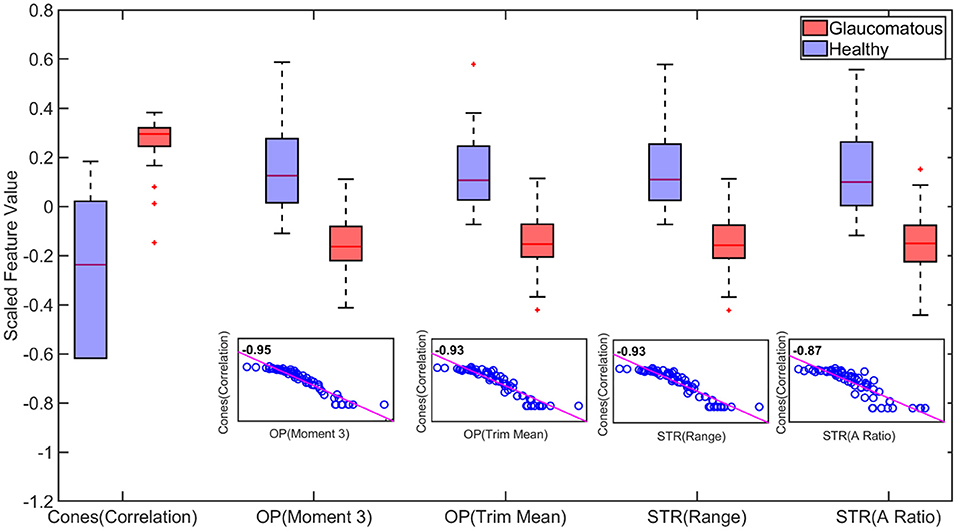
Figure 6. Boxplot of statistically important features for binary classification. The important features capable of distinguishing healthy and glaucomatous are correlated feature from Cones, third order Moment and trimmed mean feature from Oscillatory Potentials (OP) and Range and aspect ratio from Scotopic Threshold Response (STR). However, the high similarity between these features quantified by the correlation scores in the scatter plot create redundancy (inclusion cones(correlation) feature alone vs inclusion all five features does not improve accuracy). Therefore, utilizing the cones correlation feature alone captures the behavior of the other four features. This dropping of redundant features and choosing Cones (correlation) feature alone is achieved by using Minimum Redundancy and Maximum Relevance (MRMR) algorithm (On each box, the central mark indicates the median, and the bottom and top edges of the box indicate the 25th and 75th percentiles, respectively. The whiskers extend to the most extreme data points not considered outliers, and the outliers are plotted individually using the “+” marker symbol.).
Figure 7 compares the predictive importance scores obtained based on the statistical and wavelet-based features. Predictive importance scores describe the predictive capability of selected features (Kuhn and Johnson, 2013). It can be observed that wavelet-based features can distinguish healthy and glaucomatous animals suggesting that they are more sensitive to subtle changes in ERG signals due to glaucoma. It should be noted that the feature selection algorithm MRMR (Maximum Relevance and Minimum Redundancy) ignores highly correlated features for model simplicity. Therefore, only uncorrelated sets of features that improved predictability across the animals were chosen, i.e., for a set of correlated features, one representing the correlated set gets picked by the algorithm. Figure 6 demonstrates the list of important but highly correlated features that were dropped. The scatter plot inside the Figure 6 shows the correlation coefficients confirming the high degree of the correlation between them.

Figure 7. Comparison of predictive importance scores for binary classification using (A) statistical features and (B) wavelet-based features. This bar chart illustrates the superior predictive capability of wavelet-based features. Std D, Standard Deviation; W-SE, Wavelet based Shannon entropy; AR-COEF, Autoregressive coefficient.
For multiclass classification (classifying animals to different stages, normal, high, and glaucomatous as mentioned in section 2.4) based on statistical features, the correlation of cones, number of troughs in Hi cones, kurtosis of STR and mean of flicker were identified as important among the statistical features as shown in Figure 8. Several models, including SVM and ensemble-based classifiers, were used for training, and their performances were assessed. It turned out that the ensemble-based classifiers, specifically the bagged trees model, provided the best performance with a testing accuracy of 53.33%, as shown in Table 2.
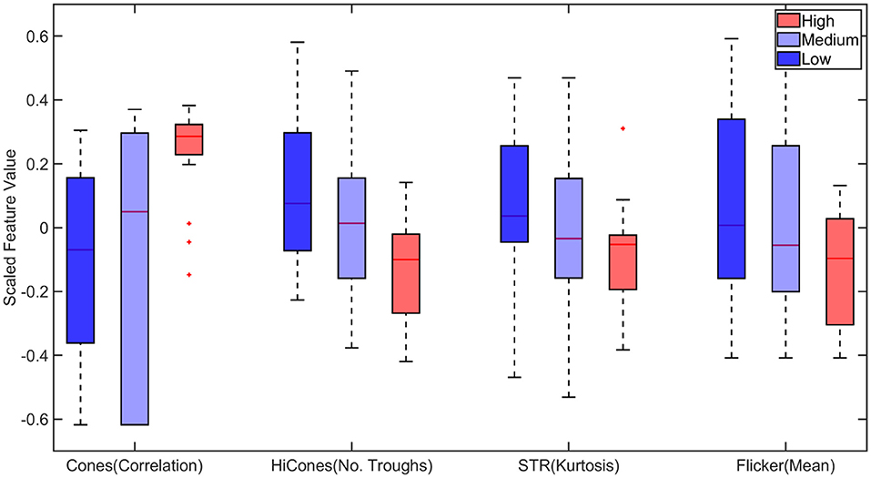
Figure 8. Boxplot of statistical features selected by Minimum Redundancy and Maximum Relevance (MRMR) feature selection algorithm for multiclass classification. STR, Scotopic Threshold Response. On each box, the central mark corresponds to the median, and the bottom and top edges of the box correspond to the 25th and 75th percentiles, respectively. The dashed lines (whiskers) extend to the most extreme data points not considered outliers, and the outliers are plotted individually using the “+” marker symbol.
Next, the multiclass classification was performed using wavelet-based features. Among the extracted wavelet features, Wavelet variance of rods and Shannon Entropy Values and AR coefficients for Maximal Overlap Discrete Wavelet Packet Transform (MOD-PWT) were identified as important features from Hi-Flicker, Flicker, Hi-cones, and STR as shown in Figure 9. The identification of flicker as an important distinguishing feature in diagnosing early-stage glaucoma was consistent with previous studies (Tyler, 1981; Lachenmayr and Drance, 1992; Horn et al., 1997; Yoshiyama and Johnson, 1997). In fact, flicker measurements in eyes with early-stage glaucoma exhibited a loss in sensitivity around 30–40 Hz (Tyler, 1981). It is worth noting that the flicker measurements used in this study were recorded using flashes at 30 Hz. The identification of the flicker ERG test and the corresponding features, among other tests, reconfirmed the capability of the current approach in identifying the relevant features. Training the ensemble bagged trees model, utilizing the selected advanced features, improved the multiclass classification accuracy to 80%, as shown in Table 2. This improvement in accuracy indicated that wavelet-based features can distinguish healthy and glaucomatous animals suggesting that they are more sensitive to subtle changes in ERG signals due to glaucoma. The multiclass classification ability of this framework reaffirmed the rich and complex nature of ERG signals in assessing the disease progression.
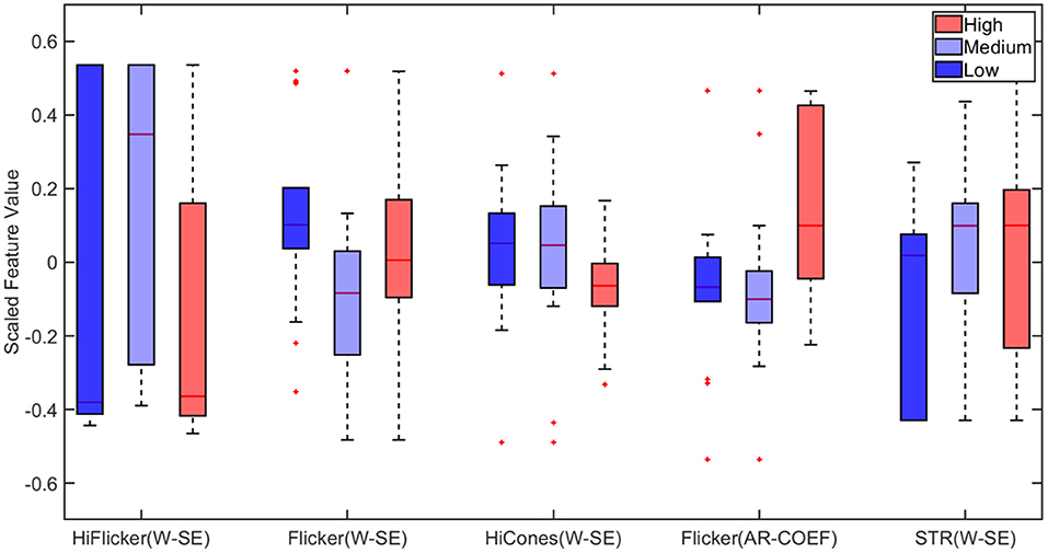
Figure 9. Boxplot of wavelet-based features selected by Minimum Redundancy and Maximum Relevance (MRMR) feature selection algorithm for multiclass classification. STR, Scotopic Threshold Response; W-SE, Wavelet based Shannon Entropy; AR-COEF, Autoregressive Coefficient. On each box, the central mark indicates the median, and the bottom and top edges of the box indicate the 25th and 75th percentiles, respectively. The whiskers extend to the most extreme data points not considered outliers, and the outliers are plotted individually using the “+” marker symbol.
Regression analysis was performed to predict retinal ganglion cell count from ERG signals. Feature selection for regression was performed using MRMR sequential feature selection. RGC values of the animals ranged between 8 and 120. RSME for RGC regression was 15.64 and 11.20 for models trained with statistical features and wavelet-based features, respectively. Regression results using wavelet-based features are shown in Figure 10. The results in Grillo et al. (2018) indicate that RGC counts had a strong correlation with STR and OPs. The dominant features selected for RGC regression (from STR and OP) were in agreement with the findings in Grillo et al. (2018). Table 4 compares performance of various ML based regression models in predicting retinal ganglion cells (RGCs) counts: The higher error (RSME) with statistical features compared with the wavelet-based advanced features emphasized the need for sophisticated features to predict RGC count accurately. SVM- and GPR-based models provided the most accurate prediction of RGC numbers from ERG signals. Specifically, squared exponential and rational quadratic models of GPR provided the least error.
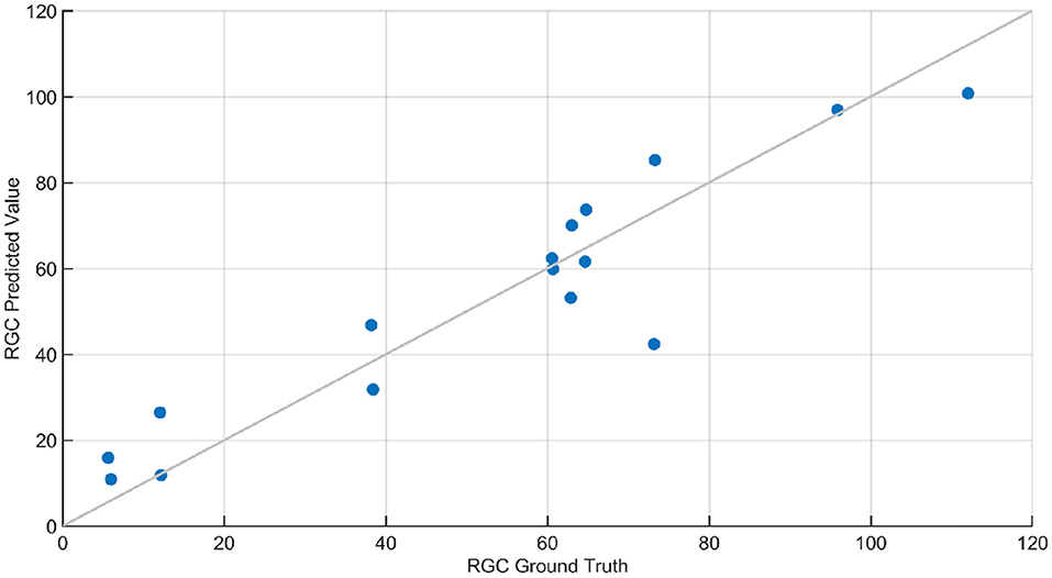
Figure 10. RGC count regression plot. This plot contains the ground truth and predicted response of RGC count predicted using Gaussian Process Regression (GPR). The squared exponential GPR model was trained using both standard and advanced features. The RGC count of the animals ranged between 8 and 120, and the root mean squared error in the prediction of RGC was 11.2. The line in this plot denotes when the predicted values are equal to ground truth values.
Our goal was to determine the feasibility of applying ML-based methods to the analysis of ERG signals for glaucoma detection at different stages of the disease. In the present study, we systematically applied machine-learning-based methods for the first time to detect glaucoma and predict RGC loss based on ERG signals. The present study utilized ERGs measured in mice rather than from human patients, because the use of data from a preclinical model allowed us to validate “ground truth” data sets with a range of complimentary and alternative experimental strategies, which is not possible in human clinical studies. These include histology, biochemical, and immunochemical assays, as well as optomotor reflex measurements. We were able to determine for the first time that advanced features (wavelet-based features) are capable of detecting subtle changes in the ERG signal and perform multiclass classification based on the progression level of the disease with 80% accuracy. In particular, we found that Shannon Entropy Values for Maximal Overlap Discrete Wavelet Packet Transform (MOD-PWT) and AR coefficients represent important features capable of detecting early-stage glaucoma. Among the nine available ERG signals, Flicker, STR, OP, and Rod-Cone appear integral for such successful detection. This is in agreement with the results published in Lei et al. (2006). However, given that these features are highly correlated, the ML-based algorithm picks only one for each set of highly correlated features to reduce the model complexity as shown in Figure 6.
In addition, the method proposed here performs ERG analysis in a wavelet domain instead of a frequency domain, which allows to capture subtle changes in the signals. In addition, various intricate features such as multiscale wavelet variance estimates, Shannon entropy, and autoregressive coefficients are incorporated in the method, compared to basic features such as differences in amplitude and latency in previous studies (Hood et al., 2000; Fortune et al., 2002; Thienprasiddhi et al., 2003; Stiefelmeyer et al., 2004; Ventura and Porciatti, 2006; Chu et al., 2007; Miguel-Jiménez et al., 2010; Luo et al., 2011; Palmowski-Wolfe et al., 2011; Todorova and Palmowski-Wolfe, 2011; Ho et al., 2012; Hori et al., 2012; Ledolter et al., 2013; Consejo et al., 2019). The results strongly suggest that such advanced features in the wavelet domain are necessary for detection of early-stage glaucoma. Moreover, in contrast to the recent study that leverages ML-based technique to analyze ERG using solely the photopic negative response (PhNR) component (Armstrong and Lorch, 2020), the current method uses all ERG components in the analysis to fully utilize the capability of the ML-based technique to crunch large data sets and draw complicated relationships. Therefore, the proposed framework is not limited to a small subset of genetic eye diseases like previous studies (Fortune et al., 2002; Thienprasiddhi et al., 2003; Stiefelmeyer et al., 2004; Chu et al., 2007; Miguel-Jiménez et al., 2010; Luo et al., 2011; Palmowski-Wolfe et al., 2011; Todorova and Palmowski-Wolfe, 2011; Ho et al., 2012; Hori et al., 2012; Ledolter et al., 2013; Consejo et al., 2019); instead, it is capable of mapping ERG signals to various eye diseases.
Results obtained in the present study strongly suggest that the methods employed can reproducibly identify dominant features for classification and regression from STR, Oscillatory potentials (OPs), and other ERG tests consistent with the results reported in previously published work on the sensitivity of and OPs and flicker to subtle changes in RGC function and viability (Tyler, 1981; Brandao et al., 2017). Further, our approach identified additional dominant distinguishing features such as Shannon Entropy Values for Maximal Overlap Discrete Wavelet Packet Transform (MOD-PWT) and AR coefficients, which are not distinguishable by traditional methods used in Grillo et al. (2018). This strongly suggests that the current machine-learning-based algorithm has significant potential in distinguishing subtle changes in ERG signals corresponding to different stages of glaucoma disease development. This capability of the technique could be used as a foundational step to create a reliable framework for the early detection of glaucoma and to monitor efficacy of therapeutic intervention in both clinical practice and novel drug development for glaucoma. In addition, the inclusion of various ERG protocols in this framework, such as cones, rods and cones, STR, and oscillatory potentials, represent responses from different cell types in the eye. Therefore, ERG response can be mapped to diseases specific to those cell types. It should be noted that this study was based on mice and with 12 h of dark adaptation. The promising results obtained here suggest the great potential for this method to help detect early stage, pre-symptomatic glaucoma. However, an additional study on adaptation requirements would be required before extending this framework to humans.
The datasets generated for this study are available on request to the corresponding author. Requests to access these datasets should be directed to bWVoZGl6YWRlaGFAdW1rYy5lZHU=.
MG contributed in machine learning framework development, formal analysis, investigation, validation, visualization, and writing—original draft. LR contributed in writing—review and editing. PK contributed in providing the data, conceptualization, supervision, and writing—review and editing. AM contributed in conceptualization, supervision, and writing—review and editing. All authors contributed to the article and approved the submitted version.
Research reported in this publication was supported by the Felix and Carmen Sabates Missouri Endowed Chair in Vision Research, the Vision Research Foundation of Kansas City, and in part by National Eye Institute grant EY031248 of the National Institutes of Health (PK). The content is solely the responsibility of the authors and does not necessarily represent the official views of the National Institutes of Health. The publication cost was covered by PK.
The content is solely the responsibility of the authors and does not necessarily represent the official views of the National Institutes of Health.
PK, AM, and MG have a patent-pending based on this study.
The remaining author declares that the research was conducted in the absence of any commercial or financial relationships that could be construed as a potential conflict of interest.
All claims expressed in this article are solely those of the authors and do not necessarily represent those of their affiliated organizations, or those of the publisher, the editors and the reviewers. Any product that may be evaluated in this article, or claim that may be made by its manufacturer, is not guaranteed or endorsed by the publisher.
Aguinis, H., Gottfredson, R. K., and Joo, H. (2013). Best-practice recommendations for defining, identifying, and handling outliers. Organ. Res. Methods 16, 270–301. doi: 10.1177/1094428112470848
Aha, D. W., and Bankert, R. L. (1996). “A comparative evaluation of sequential feature selection algorithms,” in Learning from Data, eds. D. Fisher and H.-J. Lenz (New York, NY: Springer), 199–206. doi: 10.1007/978-1-4612-2404-4_19
Ahmad, M. A., Eckert, C., and Teredesai, A. (2018). “Interpretable machine learning in healthcare,” in Proceedings of the 2018 ACM International Conference on Bioinformatics, Computational Biology, and Health Informatics (Washington, DC), 559–560. doi: 10.1145/3233547.3233667
Al'Aref, S. J., Anchouche, K., Singh, G., Slomka, P. J., Kolli, K. K., Kumar, A., et al. (2019). Clinical applications of machine learning in cardiovascular disease and its relevance to cardiac imaging. Eur. Heart J. 40, 1975–1986. doi: 10.1093/eurheartj/ehy404
Aldebasi, Y. H., Drasdo, N., Morgan, J. E., and North, R. V. (2004). S-cone, l+ m-cone, and pattern, electroretinograms in ocular hypertension and glaucoma. Vision Res. 44, 2749–2756. doi: 10.1016/j.visres.2004.06.015
An, G., Omodaka, K., Hashimoto, K., Tsuda, S., Shiga, Y., Takada, N., et al. (2019). Glaucoma diagnosis with machine learning based on optical coherence tomography and color fundus images. J. Healthcare Eng. 2019, 4061313. doi: 10.1155/2019/4061313
Armstrong, G. W., and Lorch, A. C. (2020). A (eye): a review of current applications of artificial intelligence and machine learning in ophthalmology. Int. Ophthalmol. Clin. 60, 57–71. doi: 10.1097/IIO.0000000000000298
Asakawa, K., Amino, K., Iwase, M., Kusayanagi, Y., Nakamura, A., Suzuki, R., et al. (2017). New mydriasis-free electroretinogram recorded with skin electrodes in healthy subjects. Biomed. Res. Int. 2017, 8539747. doi: 10.1155/2017/8539747
Asaoka, R., Murata, H., Iwase, A., and Araie, M. (2016). Detecting preperimetric glaucoma with standard automated perimetry using a deep learning classifier. Ophthalmology 123, 1974–1980. doi: 10.1016/j.ophtha.2016.05.029
Asgharzadeh-Bonab, A., Amirani, M. C., and Mehri, A. (2020). Spectral entropy and deep convolutional neural network for ECG beat classification. Biocybernet. Biomed. Eng. 40, 691–700. doi: 10.1016/j.bbe.2020.02.004
Atalay, E., Nongpiur, M. E., Yap, S. C., Wong, T. T., Goh, D., Husain, R., et al. (2016). Pattern of visual field loss in primary angle-closure glaucoma across different severity levels. Ophthalmology 123, 1957–1964. doi: 10.1016/j.ophtha.2016.05.026
Barraco, R., Adorno, D. P., Brai, M., and Tranchina, L. (2014). A comparison among different techniques for human ERG signals processing and classification. Phys. Med. 30, 86–95. doi: 10.1016/j.ejmp.2013.03.006
Beykin, G., Norcia, A. M., Srinivasan, V. J., Dubra, A., and GoldbERG, J. L. (2021). Discovery and clinical translation of novel glaucoma biomarkers. Prog. Retinal Eye Res. 80, 100875. doi: 10.1016/j.preteyeres.2020.100875
Boquete, L., Miguel-Jiménez, J. M., Ortega, S., Rodriguez-Ascariz, J., Pérez-Rico, C., and Blanco, R. (2012). Multifocal electroretinogram diagnosis of glaucoma applying neural networks and structural pattern analysis. Expert Syst. Appl. 39, 234–238. doi: 10.1016/j.eswa.2011.07.013
Bowd, C., Weinreb, R. N., Balasubramanian, M., Lee, I., Jang, G., Yousefi, S., et al. (2014). Glaucomatous patterns in frequency doubling technology (FDT) perimetry data identified by unsupervised machine learning classifiers. PLoS ONE 9, e85941. doi: 10.1371/journal.pone.0085941
Brandao, L. M., Monhart, M., Schötzau, A., Ledolter, A. A., and Palmowski-Wolfe, A. M. (2017). Wavelet decomposition analysis in the two-flash multifocal ERG in early glaucoma: a comparison to ganglion cell analysis and visual field. Document. Ophthalmol. 135, 29–42. doi: 10.1007/s10633-017-9593-y
Burroughs, S. L., Kaja, S., and Koulen, P. (2011). Quantification of deficits in spatial visual function of mouse models for glaucoma. Investig. Ophthalmol. Visual Sci. 52, 3654–3659. doi: 10.1167/iovs.10-7106
Bussel, I. I., Wollstein, G., and Schuman, J. S. (2014). Oct for glaucoma diagnosis, screening and detection of glaucoma progression. Brit. J. Ophthalmol. 98(Suppl 2), ii15–ii19. doi: 10.1136/bjophthalmol-2013-304326
Bzdok, D., Altman, N., and Krzywinski, M. (2018). Points of significance: statistics versus machine learning. Nat. Methods 2018, 1–7. doi: 10.1038/nmeth.4642
Cawley, G. C., and Talbot, N. L. (2003). Efficient leave-one-out cross-validation of kernel fisher discriminant classifiers. Pattern Recogn. 36, 2585–2592. doi: 10.1016/S0031-3203(03)00136-5
Chu, P. H., Chan, H. H., and Brown, B. (2007). Luminance-modulated adaptation of global flash mfERG: fellow eye losses in asymmetric glaucoma. Investig. Ophthalmol. Visual Sci. 48, 2626–2633. doi: 10.1167/iovs.06-0962
Consejo, A., Melcer, T., and Rozema, J. J. (2019). “Introduction to machine learning for ophthalmologists,” in Seminars in Ophthalmology, Vol. 34 (Taylor & Francis), 19–41. doi: 10.1080/08820538.2018.1551496
Cruz, J. A., and Wishart, D. S. (2006). Applications of machine learning in cancer prediction and prognosis. Cancer Inform. 2, 117693510600200030. doi: 10.1177/117693510600200030
da Silva, C. N., Dourado, L. F. N., de Lima, M. E., and da Silva Cunha, A. Jr. (2020). Pnpp-19 peptide as a novel drug candidate for topical glaucoma therapy through nitric oxide release. Transl. Vision Sci. Technol. 9, 33–33. doi: 10.1167/tvst.9.8.33
Dale, E. A., Hood, D. C., Greenstein, V. C., and Odel, J. G. (2010). A comparison of multifocal ERG and frequency domain oct changes in patients with abnormalities of the retina. Document. Ophthalmol. 120, 175–186. doi: 10.1007/s10633-009-9210-9
Darbellay, G. A., and Vajda, I. (1999). Estimation of the information by an adaptive partitioning of the observation space. IEEE Trans. Inform. Theory 45, 1315–1321. doi: 10.1109/18.761290
Daubechies, I. (1992). Ten Lectures on Wavelets. New York, NY: Society for Industrial and Applied Mathematics.
de Lara, M. J. P., Guzmán-Aránguez, A., de la Villa, P., Diaz-Hernández, J. I., Miras-Portugal, M. T., and Pintor, J. (2015). Increased levels of extracellular ATP in glaucomatous retinas: possible role of the vesicular nucleotide transporter during the development of the pathology. Mol. Vis. 21, 1060.
de Lara, M. J. P., Santano, C., Guzmán-Aránguez, A., Valiente-Soriano, F. J., Avilés-Trigueros, M., Vidal-Sanz, M., et al. (2014). Assessment of inner retina dysfunction and progressive ganglion cell loss in a mouse model of glaucoma. Exp. Eye Res. 122, 40–49. doi: 10.1016/j.exer.2014.02.022
Demmin, D. L., Davis, Q., Roché, M., and Silverstein, S. M. (2018). Electroretinographic anomalies in schizophrenia. J. Abnormal Psychol. 127, 417. doi: 10.1037/abn0000347
Dietterich, T. G. (2000). “Ensemble methods in machine learning,” in International Workshop on Multiple Classifier Systems (Berlin, Heidelberg: Springer), 1–15. doi: 10.1007/3-540-45014-9_1
Ding, C., and Peng, H. (2005). Minimum redundancy feature selection from microarray gene expression data. J. Bioinform. Comput. Biol. 3, 185–205. doi: 10.1142/S0219720005001004
Dong, C.-J., Agey, P., and Hare, W. A. (2004). Origins of the electroretinogram oscillatory potentials in the rabbit retina. Vis. Neurosci. 21, 533–543. doi: 10.1017/S0952523804214043
Duan, K., Keerthi, S. S., and Poo, A. N. (2003). Evaluation of simple performance measures for tuning SVM hyperparameters. Neurocomputing 51, 41–59. doi: 10.1016/S0925-2312(02)00601-X
Ernest, P. J., Schouten, J. S., Beckers, H. J., Hendrikse, F., Prins, M. H., and Webers, C. A. (2012). The evidence base to select a method for assessing glaucomatous visual field progression. Acta Ophthalmol. 90, 101–108. doi: 10.1111/j.1755-3768.2011.02206.x
Feurer, M., and Hutter, F. (2019). “Hyperparameter optimization,” in Automated Machine Learning, eds. F. Hutter, L. Kotthoff, and J. Vanschoren (Cham: Springer), 3–33. doi: 10.1007/978-3-030-05318-5_1
Fidalgo, B. M., Crabb, D. P., and Lawrenson, J. G. (2015). Methodology and reporting of diagnostic accuracy studies of automated perimetry in glaucoma: evaluation using a standardised approach. Ophthal. Physiol. Opt. 35, 315–323. doi: 10.1111/opo.12208
Forte, J. D., Bui, B. V., and Vingrys, A. J. (2008). Wavelet analysis reveals dynamics of rat oscillatory potentials. J. Neurosci. Methods 169, 191–200. doi: 10.1016/j.jneumeth.2007.12.007
Fortune, B., Bearse, M. A., Cioffi, G. A., and Johnson, C. A. (2002). Selective loss of an oscillatory component from temporal retinal multifocal ERG responses in glaucoma. Investig. Ophthalmol. Visual Sci. 43, 2638–2647.
Fortune, B., Wang, L., Bui, B. V., Cull, G., Dong, J., and Cioffi, G. A. (2003). Local ganglion cell contributions to the macaque electroretinogram revealed by experimental nerve fiber layer bundle defect. Investig. Ophthalmol. Visual Sci. 44, 4567–4579. doi: 10.1167/iovs.03-0200
Frishman, L. J., Saszik, S., Harwerth, R. S., Viswanathan, S., Li, Y., Smith, E. L., et al. (2000). Effects of experimental glaucoma in macaques on the multifocal ERG. Document. Ophthalmol. 100, 231–251. doi: 10.1023/A:1002735804029
Graham, J. W., Cumsille, P. E., and Shevock, A. E. (2013). Methods for handling missing data. doi: 10.1002/9781118133880.hop202004
Graham, S. L., Klistorner, A. I., Grigg, J. R., and Billson, F. A. (2000). Objective VEP perimetry in glaucoma: asymmetry analysis to identify early deficits. J. Glaucoma 9, 10–19. doi: 10.1097/00061198-200002000-00004
Grillo, S. L., Keereetaweep, J., Grillo, M. A., Chapman, K. D., and Koulen, P. (2013). N-palmitoylethanolamine depot injection increased its tissue levels and those of other acylethanolamide lipids. Drug Design Dev. Therapy 7, 747. doi: 10.2147/DDDT.S48324
Grillo, S. L., and Koulen, P. (2015). Psychophysical testing in rodent models of glaucomatous optic neuropathy. Exp. Eye Res. 141, 154–163. doi: 10.1016/j.exer.2015.06.025
Grillo, S. L., Montgomery, C. L., Johnson, H. M., and Koulen, P. (2018). Quantification of changes in visual function during disease development in a mouse model of pigmentary glaucoma. J. Glaucoma 27, 828. doi: 10.1097/IJG.0000000000001024
Guyon, I., Gunn, S., Nikravesh, M., and Zadeh, L. A. (2008). Feature Extraction: Foundations and Applications, Vol. 207. Springer.
Hancock, H. A., and Kraft, T. W. (2004). Oscillatory potential analysis and ERGs of normal and diabetic rats. Investig. Ophthalmol. Visual Sci. 45, 1002–1008. doi: 10.1167/iovs.03-1080
Hannun, A. Y., Rajpurkar, P., Haghpanahi, M., Tison, G. H., Bourn, C., Turakhia, M. P., et al. (2019). Cardiologist-level arrhythmia detection and classification in ambulatory electrocardiograms using a deep neural network. Nat. Med. 25, 65–69. doi: 10.1038/s41591-018-0268-3
Harwerth, R. S., Crawford, M., Frishman, L. J., Viswanathan, S., Smith, E. L. III., and Carter-Dawson, L. (2002). Visual field defects and neural losses from experimental glaucoma. Prog Retinal Eye Res. 21, 91–125. doi: 10.1016/S1350-9462(01)00022-2
Hébert, M., Mérette, C., Gagné, A.-M., Paccalet, T., Moreau, I., Lavoie, J., et al. (2020). The electroretinogram may differentiate schizophrenia from bipolar disorder. Biol. Psychiatry 87, 263–270. doi: 10.1016/j.biopsych.2019.06.014
Hermas, A. (2019). Sensitivity and specificity of the uniform field ERG in glaucoma detection (Ph.D. thesis). University of Ottawa, Ottawa, ON, Canada.
Ho, W.-C., Wong, O.-Y., Chan, Y.-C., Wong, S.-W., Kee, C.-S., and Chan, H. H.-L. (2012). Sign-dependent changes in retinal electrical activity with positive and negative defocus in the human eye. Vision Res. 52, 47–53. doi: 10.1016/j.visres.2011.10.017
Hobby, A. E., Kozareva, D., Yonova-Doing, E., Hossain, I. T., Katta, M., Huntjens, B., et al. (2018). Effect of varying skin surface electrode position on electroretinogram responses recorded using a handheld stimulating and recording system. Document. Ophthalmol. 137, 79–86. doi: 10.1007/s10633-018-9652-z
Holzinger, A. (2014). Trends in interactive knowledge discovery for personalized medicine: cognitive science meets machine learning. IEEE Intell. Inform. Bull. 15, 6–14. doi: 10.1007/978-3-662-43968-5_1
Hood, D. C., Greenstein, V. C., Holopigian, K., Bauer, R., Firoz, B., Liebmann, J. M., et al. (2000). An attempt to detect glaucomatous damage to the inner retina with the multifocal ERG. Investig. Ophthalmol. Visual Sci. 41, 1570–1579.
Hori, N., Komori, S., Yamada, H., Sawada, A., Nomura, Y., Mochizuki, K., et al. (2012). Assessment of macular function of glaucomatous eyes by multifocal electroretinograms. Document. Ophthalmol. 125, 235–247. doi: 10.1007/s10633-012-9351-0
Horn, F. K., Jonas, J. B., Korth, M., Jünemann, A., and Gründler, A. (1997). The full-field flicker test in early diagnosis of chronic open-angle glaucoma. Am. J. Ophthalmol. 123, 313–319. doi: 10.1016/S0002-9394(14)70126-6
Hui, F., Tang, J., Williams, P. A., McGuinness, M. B., Hadoux, X., Casson, R. J., et al. (2020). Improvement in inner retinal function in glaucoma with nicotinamide (vitamin b3) supplementation: a crossover randomized clinical trial. Clin. Exp. Ophthalmol. 48, 903–914. doi: 10.1111/ceo.13818
Jambukia, S. H., Dabhi, V. K., and Prajapati, H. B. (2015). “Classification of ECG signals using machine learning techniques: a survey,” in 2015 International Conference on Advances in Computer Engineering and Applications (Ghaziabad, India: IEEE), 714–721. doi: 10.1109/ICACEA.2015.7164783
Jordan, M. I., and Mitchell, T. M. (2015). Machine learning: trends, perspectives, and prospects. Science 349, 255–260. doi: 10.1126/science.aaa8415
Kaja, S., Naumchuk, Y., Grillo, S. L., Borden, P. K., and Koulen, P. (2014). Differential up-regulation of vesl-1/homer 1 protein isoforms associated with decline in visual performance in a preclinical glaucoma model. Vision Res. 94, 16–23. doi: 10.1016/j.visres.2013.10.018
Kato, K., Kondo, M., Nagashima, R., Sugawara, A., Sugimoto, M., Matsubara, H., et al. (2017). Factors affecting mydriasis-free flicker ERGs recorded with real-time correction for retinal illuminance: study of 150 young healthy subjects. Investig. Ophthalmol. Visual Sci. 58, 5280–5286. doi: 10.1167/iovs.17-22587
Khalid, S., Khalil, T., and Nasreen, S. (2014). “A survey of feature selection and feature extraction techniques in machine learning,” in 2014 Science and Information Conference (London, UK: IEEE), 372–378. doi: 10.1109/SAI.2014.6918213
Khan, M. Z., Gajendran, M. K., Lee, Y., and Khan, M. A. (2021). Deep neural architectures for medical image semantic segmentation. IEEE Access. 83002–83024. doi: 10.1109/ACCESS.2021.3086530
Kononenko, I. (2001). Machine learning for medical diagnosis: history, state of the art and perspective. Artif. Intell. Med. 23, 89–109. doi: 10.1016/S0933-3657(01)00077-X0
Kuhn, M., and Johnson, K. (2013). “Measuring predictor importance,” in Applied Predictive Modeling (New York, NY: Springer), 463–485. doi: 10.1007/978-1-4614-6849-3_18
Lachenmayr, B., and Drance, S. (1992). The selective effects of elevated intraocular pressure on temporal resolution. German J. Ophthalmol.1, 26–31.
Lai, T., Ngai, J., Lai, R., and Lam, D. (2009). Multifocal electroretinography changes in patients on ethambutol therapy. Eye 23, 1707–1713. doi: 10.1038/eye.2008.361
Lai, T. Y., Ngai, J. W., Chan, W.-M., and Lam, D. S. (2006). Visual field and multifocal electroretinography and their correlations in patients on hydroxychloroquine therapy. Document. Ophthalmol. 112, 177–187. doi: 10.1007/s10633-006-9006-0
Ledolter, A. A., Kramer, S. A., Todorova, M. G., Schötzau, A., and Palmowski-Wolfe, A. M. (2013). The effect of filtering on the two-global-flash mfERG: identifying the optimal range of frequency for detecting glaucomatous retinal dysfunction. Document. Ophthalmol. 126, 117–123. doi: 10.1007/s10633-012-9364-8
Ledolter, A. A., Monhart, M., Schoetzau, A., Todorova, M. G., and Palmowski-Wolfe, A. M. (2015). Structural and functional changes in glaucoma: comparing the two-flash multifocal electroretinogram to optical coherence tomography and visual fields. Document. Ophthalmol. 130, 197–209. doi: 10.1007/s10633-015-9482-1
Lee, A., Taylor, P., Kalpathy-Cramer, J., and Tufail, A. (2017). Machine learning has arrived! Ophthalmology 124, 1726–1728. doi: 10.1016/j.ophtha.2017.08.046
Lee, C. H., and Yoon, H.-J. (2017). Medical big data: promise and challenges. Kidney Res. Clin. Pract. 36, 3. doi: 10.23876/j.krcp.2017.36.1.3
Lei, B., Yao, G., Zhang, K., Hofeldt, K. J., and Chang, B. (2006). Study of rod-and cone-driven oscillatory potentials in mice. Investig. Ophthalmol. Visual Sci. 47, 2732–2738. doi: 10.1167/iovs.05-1461
Leonarduzzi, R. F., Schlotthauer, G., and Torres, M. E. (2010). “Wavelet leader based multifractal analysis of heart rate variability during myocardial ischaemia,” in 2010 Annual International Conference of the IEEE Engineering in Medicine and Biology (Buenos Aires, Argentina: IEEE), 110–113. doi: 10.1109/IEMBS.2010.5626091
Leske, M. C., Heijl, A., Hyman, L., Bengtsson, B., Dong, L., Yang, Z., et al. (2007). Predictors of long-term progression in the early manifest glaucoma trial. Ophthalmology 114, 1965–1972. doi: 10.1016/j.ophtha.2007.03.016
Li, Q., Rajagopalan, C., and Clifford, G. D. (2014). A machine learning approach to multi-level ECG signal quality classification. Comput. Methods Prog. Biomed. 117, 435–447. doi: 10.1016/j.cmpb.2014.09.002
Li, T., and Zhou, M. (2016). ECG classification using wavelet packet entropy and random forests. Entropy 18, 285. doi: 10.3390/e18080285
Lisboa, P. J. (2002). A review of evidence of health benefit from artificial neural networks in medical intervention. Neural Netw. 15, 11–39. doi: 10.1016/S0893-6080(01)00111-3
Liu, H., Ji, X., Dhaliwal, S., Rahman, S. N., McFarlane, M., Tumber, A., et al. (2018). Evaluation of light-and dark-adapted ERGs using a mydriasis-free, portable system: clinical classifications and normative data. Document. Ophthalmol. 137, 169–181. doi: 10.1007/s10633-018-9660-z
Luo, X., Patel, N. B., Harwerth, R. S., and Frishman, L. J. (2011). Loss of the low-frequency component of the global-flash multifocal electroretinogram in primate eyes with experimental glaucoma. Investig. Ophthalmol. Visual Sci. 52, 3792–3804. doi: 10.1167/iovs.10-6667
Maharaj, E. A., and Alonso, A. M. (2014). Discriminant analysis of multivariate time series: application to diagnosis based on ECG signals. Comput. Stat. Data Anal. 70, 67–87. doi: 10.1016/j.csda.2013.09.006
Man, T. T., Yip, Y. W., Cheung, F. K., Lee, W. S., Pang, C. P., and Brelén, M. E. (2020). Evaluation of electrical performance and properties of electroretinography electrodes. Transl. Vis. Sci. Technol. 9, 45–45. doi: 10.1167/tvst.9.7.45
Marmor, M., Fulton, A., Holder, G., Miyake, Y., Brigell, M., and Bach, M. (2009). Iscev standard for full-field clinical electroretinography (2008 update). Document. Ophthalmol. 118, 69–77. doi: 10.1007/s10633-008-9155-4
Martis, R. J., Chakraborty, C., and Ray, A. K. (2014). “Wavelet-based machine learning techniques for ECG signal analysis,” in Machine Learning in Healthcare Informatics, eds. S. Dua, U. R. Acharya, and P. Dua (Berlin, Heidelberg: Springer), 25–45. doi: 10.1007/978-3-642-40017-9_2
McKinnon, S. J., Schlamp, C. L., and Nickells, R. W. (2009). Mouse models of retinal ganglion cell death and glaucoma. Exp. Eye Res. 88, 816–824. doi: 10.1016/j.exer.2008.12.002
McPadden, J., Durant, T. J., Bunch, D. R., Coppi, A., Price, N., Rodgerson, K., et al. (2019). Health care and precision medicine research: analysis of a scalable data science platform. J. Med. Internet Res. 21, e13043. doi: 10.2196/13043
Miguel-Jiménez, J., Boquete, L., Ortega, S., Rodriguez-Ascariz, J., and Blanco, R. (2010). Glaucoma detection by wavelet-based analysis of the global flash multifocal electroretinogram. Med. Eng. Phys. 32, 617–622. doi: 10.1016/j.medengphy.2010.02.019
Miguel-Jiménez, J. M., Blanco, R., De-Santiago, L., Fernandez, A., Rodriguez-Ascariz, J. M., Barea, R., et al. (2015). Continuous-wavelet-transform analysis of the multifocal ERG waveform in glaucoma diagnosis. Med. Biol. Eng. Comput. 53, 771–780. doi: 10.1007/s11517-015-1287-6
Montgomery, C. L., Keereetaweep, J., Johnson, H. M., Grillo, S. L., Chapman, K. D., and Koulen, P. (2016). Changes in retinal n-acylethanolamines and their oxylipin derivatives during the development of visual impairment in a mouse model for glaucoma. Lipids 51, 857–866. doi: 10.1007/s11745-016-4161-x
Nakamura, N., Fujinami, K., Mizuno, Y., Noda, T., and Tsunoda, K. (2016). Evaluation of cone function by a handheld non-mydriatic flicker electroretinogram device. Clin. Ophthalmol. 10, 1175. doi: 10.2147/OPTH.S104721
Navada, A., Ansari, A. N., Patil, S., and Sonkamble, B. A. (2011). “Overview of use of decision tree algorithms in machine learning,” in 2011 IEEE Control and System Graduate Research Colloquium (Shah Alam, Malaysia: IEEE), 37–42. doi: 10.1109/ICSGRC.2011.5991826
Nebbioso, M., Grenga, R., and Karavitis, P. (2009). Early detection of macular changes with multifocal ERG in patients on antimalarial drug therapy. J. Ocular Pharmacol. Therapeut. 25, 249–258. doi: 10.1089/jop.2008.0106
Neumaier, A., and Schneider, T. (2001). Estimation of parameters and eigenmodes of multivariate autoregressive models. ACM Trans. Math. Softw. 27, 27–57. doi: 10.1145/382043.382304
Noble, W. S. (2006). What is a support vector machine? Nat. Biotechnol. 24, 1565–1567. doi: 10.1038/nbt1206-1565
Omrani, H. (2015). Predicting travel mode of individuals by machine learning. Transport. Res. Proc. 10, 840–849. doi: 10.1016/j.trpro.2015.09.037
Palmberg, P. (2002). Answers from the ocular hypertension treatment study. Arch. Ophthalmol. 120, 829–830. doi: 10.1001/archopht.120.6.829
Palmowski-Wolfe, A., Todorova, M., and Orgül, S. (2011). Multifocal oscillatory potentials in the “two global flash” mferg in high and normal tension primary open-angle glaucoma. J. Clin. Exp. Ophthalmol. 2, 167. doi: 10.4172/2155-9570.1000167
Porciatti, V. (2015). Electrophysiological assessment of retinal ganglion cell function. Exp. Eye Res. 141, 164–170. doi: 10.1016/j.exer.2015.05.008
Rohowetz, L. J., Kraus, J. G., and Koulen, P. (2018). Reactive oxygen species-mediated damage of retinal neurons: drug development targets for therapies of chronic neurodegeneration of the retina. Int. J. Mol. Sci. 19, 3362. doi: 10.3390/ijms19113362
Saeedi, O., Boland, M. V., D'Acunto, L., Swamy, R., Hegde, V., Gupta, S., et al. (2021). Development and comparison of machine learning algorithms to determine visual field progression. Transl. Vis. Sci. Technol. 10, 27–27. doi: 10.1167/tvst.10.7.27
Saeys, Y., Inza, I., and Larranaga, P. (2007). A review of feature selection techniques in bioinformatics. Bioinformatics 23, 2507–2517. doi: 10.1093/bioinformatics/btm344
Sarossy, M., Kumar, D., and Wu, Z. (2021). “Relationship between glaucoma and complexity measures of the electroretinogram,” in 2021 Seventh International conference on Bio Signals, Images, and Instrumentation (ICBSII) (Chennai, India: IEEE), 1–4. doi: 10.1109/ICBSII51839.2021.9445121
Saszik, S. M., Robson, J. G., and Frishman, L. J. (2002). The scotopic threshold response of the dark-adapted electroretinogram of the mouse. J. Physiol. 543, 899–916. doi: 10.1113/jphysiol.2002.019703
Saxe, A., Nelli, S., and Summerfield, C. (2021). If deep learning is the answer, what is the question? Nat. Rev. Neurosci. 22, 55–67. doi: 10.1038/s41583-020-00395-8
Seo, H., Badiei Khuzani, M., Vasudevan, V., Huang, C., Ren, H., Xiao, R., et al. (2020). Machine learning techniques for biomedical image segmentation: An overview of technical aspects and introduction to state-of-art applications. Med. Phys. 47, e148–e167. doi: 10.1002/mp.13649
Shailaja, K., Seetharamulu, B., and Jabbar, M. (2018). “Machine learning in healthcare: a review,” in 2018 Second International Conference on Electronics, Communication and Aerospace Technology (ICECA) (Coimbatore, India: IEEE), 910–914. doi: 10.1109/ICECA.2018.8474918
Shi, S., Wang, Q., Xu, P., and Chu, X. (2016). “Benchmarking state-of-the-art deep learning software tools,” in 2016 7th International Conference on Cloud Computing and Big Data (CCBD) (Macau, China: IEEE), 99–104. doi: 10.1109/CCBD.2016.029
Sim, D. A., Keane, P. A., Tufail, A., Egan, C. A., Aiello, L. P., and Silva, P. S. (2015). Automated retinal image analysis for diabetic retinopathy in telemedicine. Curr. Diabetes Rep. 15, 14. doi: 10.1007/s11892-015-0577-6
Stiefelmeyer, S., Neubauer, A. S., Berninger, T., Arden, G. B., and Rudolph, G. (2004). The multifocal pattern electroretinogram in glaucoma. Vision Res. 44, 103–112. doi: 10.1016/j.visres.2003.08.012
Takagi, S. T., Kita, Y., Yagi, F., and Tomita, G. (2012). Macular retinal ganglion cell complex damage in the apparently normal visual field of glaucomatous eyes with hemifield defects. J. Glaucoma 21, 318–325. doi: 10.1097/IJG.0b013e31820d7e9d
Tang, J., Hui, F., Hadoux, X., Soares, B., Jamieson, M., van Wijngaarden, P., et al. (2020). Short-term changes in the photopic negative response following intraocular pressure lowering in glaucoma. Investig. Ophthalmol. Visual Sci. 61, 16–16. doi: 10.1167/iovs.61.10.16
Thienprasiddhi, P., Greenstein, V. C., Chen, C. S., Liebmann, J. M., Ritch, R., and Hood, D. C. (2003). Multifocal visual evoked potential responses in glaucoma patients with unilateral hemifield defects. Am. J. Ophthalmol. 136, 34–40. doi: 10.1016/S0002-9394(03)00080-1
Todorova, M. G., and Palmowski-Wolfe, A. M. (2011). MfERG responses to long-duration white stimuli in glaucoma patients. Document. Ophthalmol. 122, 87–97. doi: 10.1007/s10633-011-9263-4
Trafalis, T. B., and Ince, H. (2000). “Support vector machine for regression and applications to financial forecasting,” in Proceedings of the IEEE-INNS-ENNS International Joint Conference on Neural Networks. IJCNN 2000 (Como, Italy: IEEE), 348–353. doi: 10.1109/IJCNN.2000.859420
Turalba, A. V., and Grosskreutz, C. (2010). “A review of current technology used in evaluating visual function in glaucoma,” in Seminars in Ophthalmology, Vol. 25 (Taylor & Francis), 309–316. doi: 10.3109/08820538.2010.518898
Tyler, C. W. (1981). Specific deficits of flicker sensitivity in glaucoma and ocular hypertension. Investig. Ophthalmol. Visual Sci. 20, 204–212.
Varma, S., and Simon, R. (2006). Bias in error estimation when using cross-validation for model selection. BMC Bioinform. 7, 91. doi: 10.1186/1471-2105-7-91
Ventura, L. M., and Porciatti, V. (2006). Pattern electroretinogram in glaucoma. Curr. Opin. Ophthalmol. 17, 196–202. doi: 10.1097/01.icu.0000193082.44938.3c
Verma, S., Nongpiur, M. E., Atalay, E., Wei, X., Husain, R., Goh, D., et al. (2017). Visual field progression in patients with primary angle-closure glaucoma using pointwise linear regression analysis. Ophthalmology 124, 1065–1071. doi: 10.1016/j.ophtha.2017.02.027
Wan, X. (2019). Influence of feature scaling on convergence of gradient iterative algorithm. J. Phys. 1213, 032021. doi: 10.1088/1742-6596/1213/3/032021
Wilsey, L. J., and Fortune, B. (2016). Electroretinography in glaucoma diagnosis. Curr. Opin. Ophthalmol. 27, 118. doi: 10.1097/ICU.0000000000000241
Yadav, S., and Shukla, S. (2016). “Analysis of k-fold cross-validation over hold-out validation on colossal datasets for quality classification,” in 2016 IEEE 6th International Conference on Advanced Computing (IACC) (Bhimavaram, India: IEEE), 78–83. doi: 10.1109/IACC.2016.25
Yapici, İ. S., Erkaymaz, O., and Arslan, R. U. (2021). A hybrid intelligent classifier to estimate obesity levels based on ERG signals. Phys. Lett. A 399, 127281. doi: 10.1016/j.physleta.2021.127281
Yoshiyama, K. K., and Johnson, C. A. (1997). Which method of flicker perimetry is most effective for detection of glaucomatous visual field loss? Investig. Ophthalmol. Visual Sci. 38, 2270–2277.
Yousefi, S., Goldbaum, M. H., Varnousfaderani, E. S., Belghith, A., Jung, T.-P., Medeiros, F. A., et al. (2015). Detecting glaucomatous change in visual fields: analysis with an optimization framework. J. Biomed. Inform. 58, 96–103. doi: 10.1016/j.jbi.2015.09.019
Zhang, M.-L., and Zhou, Z.-H. (2007). ML-KNN: a lazy learning approach to multi-label learning. Pattern Recogn. 40, 2038–2048. doi: 10.1016/j.patcog.2006.12.019
Zhang, Y., and Ling, C. (2018). A strategy to apply machine learning to small datasets in materials science. NPJ Comput. Mater. 4, 1–8. doi: 10.1038/s41524-018-0081-z
Zhao, Q., and Zhang, L. (2005). “ECG feature extraction and classification using wavelet transform and support vector machines,” in 2005 International Conference on Neural Networks and Brain (Beijing, China: IEEE), 1089–1092.
Zhao, Z., Anand, R., and Wang, M. (2019). “Maximum relevance and minimum redundancy feature selection methods for a marketing machine learning platform,” in 2019 IEEE International Conference on Data Science and Advanced Analytics (DSAA) (Washington, DC: IEEE), 442–452. doi: 10.1109/DSAA.2019.00059
Keywords: glaucoma, machine learning, electroretinography, ERG, wavelet transform, early stage, AI
Citation: Gajendran MK, Rohowetz LJ, Koulen P and Mehdizadeh A (2022) Novel Machine-Learning Based Framework Using Electroretinography Data for the Detection of Early-Stage Glaucoma. Front. Neurosci. 16:869137. doi: 10.3389/fnins.2022.869137
Received: 03 February 2022; Accepted: 28 March 2022;
Published: 04 May 2022.
Edited by:
Rafael Linden, Federal University of Rio de Janeiro, BrazilReviewed by:
Marc Sarossy, The University of Melbourne, AustraliaCopyright © 2022 Gajendran, Rohowetz, Koulen and Mehdizadeh. This is an open-access article distributed under the terms of the Creative Commons Attribution License (CC BY). The use, distribution or reproduction in other forums is permitted, provided the original author(s) and the copyright owner(s) are credited and that the original publication in this journal is cited, in accordance with accepted academic practice. No use, distribution or reproduction is permitted which does not comply with these terms.
*Correspondence: Amirfarhang Mehdizadeh, bWVoZGl6YWRlaGFAdW1rYy5lZHU=
Disclaimer: All claims expressed in this article are solely those of the authors and do not necessarily represent those of their affiliated organizations, or those of the publisher, the editors and the reviewers. Any product that may be evaluated in this article or claim that may be made by its manufacturer is not guaranteed or endorsed by the publisher.
Research integrity at Frontiers

Learn more about the work of our research integrity team to safeguard the quality of each article we publish.