 Long-Biao Cui
 Xian Xu1†
95% of researchers rate our articles as excellent or good
Learn more about the work of our research integrity team to safeguard the quality of each article we publish.
Find out more
OPINION article
Front. Neurosci. , 15 June 2021
Sec. Brain Imaging Methods
Volume 15 - 2021 | https://doi.org/10.3389/fnins.2021.685005
This article is part of the Research Topic Machine Learning in Neuroscience, Volume II View all 13 articles
A pressing need of diagnostic, predictive, and prognostic markers exists in clinical settings. Unfortunately, in spite of evidence for underpinnings in mental disorders over the past decade, biologically based markers in reflection to the physiology for guiding clinical practice have obviously lagged behind. On the one hand, the updated Diagnostic and Statistical Manual of Mental Disorders remains to emphasize how mental disorders are expressed, although it provides a model that helps clinicians to perform better diagnosis and follow-up care (Kupfer et al., 2013), like cholesterol and blood pressure measurement. On the other hand, as the 23rd edition of the “Clinical Handbook of Psychotropic Drugs” has said, “we can provide current evidence-based and clinically relevant information to optimize patient care,” but the information is derived from randomized controlled trials and leading clinical experts, etc. Therefore, clinical practice requests guidance of some objective, quantitative, and specific biomarker, reflecting its neurobiological substrates for diagnosis and treatment selection.
To this end, machine learning methods, as demonstrated by a sizable number of recent neuroimaging studies, hold great promise for improving the diagnosis, treatment, and prediction of prognosis in psychiatric domains, which will have an effect on personalized medicine. The term “machine learning” was coined in 1959 by Arthur Samuel (Samuel, 1959), and it is a science of the artificial intelligence, showing an evident capacity to reveal relationships between different variables used for classification (Tandon and Tandon, 2018). Furthermore, radiomics is a newly developed method to obtain high-dimensional features that might be options used for machine learning analysis. This Research Topic “Machine Learning in Neuroscience, Volume II” in “Frontiers in Neuroscience” provides new study strategy and applies radiomics/machine learning and distinct neuroimaging in mental disorders. Transforming existing clinical pathways toward optimizing care for the specific needs of each psychiatric patient, the significance is to achieve better diagnosis, treatment, and prognosis of mental disorders using radiomics/machine learning.
The field of psychiatry research remains a focus of medicine; in particular, mental health has arrived on the global health agenda. Currently, PubMed comprises more than 10 citations for literature involving radiomics and mental disorders, and efforts to develop radiomics/machine learning-based objective means have intensified for autism spectrum disorder (Chaddad et al., 2017), attention-deficit/hyperactivity disorder (Sun et al., 2018), schizophrenia spectrum and other psychotic disorders (Cui et al., 2018, 2021; Gong et al., 2020; Park et al., 2020; Xi et al., 2020), bipolar and related disorders (Wang et al., 2020), mild cognitive impairment, and Alzheimer's disease (Kai et al., 2018; Li et al., 2018; Ranjbar et al., 2019; Huang et al., 2020). Radiomics/machine learning enables the neuroimaging data of mental disorders to be extracted for improving clinical decision support (Wang et al., 2019).
Due to the excellent performance of radiomics analysis for feature selection and classification, it is regarded as the bridge between medical imaging and personalized medicine (Lambin et al., 2017). However, a critical issue is related to radiomics analysis in non-cancer field. In spite of a lack of lesions for conventional feature extraction, neuroimaging-based measures are features that could be extracted in radiomics/machine learning study. Taking schizophrenia as an example, previous studies on this topic illustrate the potential value in the application of radiomics/machine learning methods to disease definition and diagnosis and prediction of response to antipsychotics (APs) or electroconvulsive therapy (ECT).
Diagnosis and treatment of schizophrenia are pivotal clinical issues that need to be solved urgently. In a recent review, Kraguljac et al. (2021) highlighted and discussed the neuroimaging biomarkers in schizophrenia. Magnetic resonance imaging (MRI), as a non-invasive neuroimaging method, has been widely used in the study of schizophrenia. As we commented on a meta-analysis of the association of clinical and demographic characteristics and magnetic resonance spectroscopy in schizophrenia (“Targeting the Whole Clinical Course of Schizophrenia With Magnetic Resonance Imaging,” https://jamanetwork.com/journals/jamapsychiatry/fullarticle/2778479), MRI combined with radiomics/machine learning could be the most important approach in schizophrenia research, involving predicting transition from clinical high risk to psychosis, providing evidence of macroscale neural mechanisms, delving into the nature behind symptoms, facilitating diagnosis and subtyping, predicting treatment response, detecting psychopharmacological effects, and guiding neuronavigation of neuromodulation, thereby managing very-late-onset schizophrenia-like psychosis. MRI-based studies are promising for clinical translation (Jiang et al., 2020).
It is of no doubt that we took the precision medicine view more seriously with the advent of radiomics/machine learning in the field of schizophrenia research (Wang et al., 2019). The number of publications on radiomics/machine learning via MRI has increased to five until this year, including one regression analysis (Gong et al., 2020) and four classification analyses (Cui et al., 2018, 2021; Park et al., 2020; Xi et al., 2020) (Table 1). There are two well-done MRI studies from Xi et al. (2020) and Gong et al. (2020), respectively. They used radiomics features on structural MRI (sMRI) to predict response to APs plus ECT. In the first study, the group of Yi-Bin Xi and colleagues extracted radiomics features from the regions of interest (ROIs) with differences of gray matter volume between responders and non-responders (Xi et al., 2020). Specifically, voxel-wise gray matter volume was compared between responders and non-responders, and then, 11 ROIs identified in the previous step entered first-order statistics feature extraction and classification analysis. A leave-one-out cross-validation (LOOCV) framework and support vector machine (SVM) was used to perform pattern classification analysis. This study built a fusion logistic regression model (LRM) with the least absolute shrinkage and selection operator (LASSO) with an accuracy of 93.18%, and the fixed features were from the right anterior cingulum, left supramarginal gyrus, and right hippocampus. In the second study, the group of Gong et al. (2020) examined whether the combination of gray and white matters can predict the outcome using sMRI and diffusion tensor imaging. They selected first-order statistics radiomics features from regions (gray and white matters) with strong electric field distribution under ECT in this regression analysis study. The prediction process was performed with a support vector regression model based on a LOOCV framework. Features in the left inferior frontal gyrus, right superior temporal gyrus, left temporal pole, right insula, and fibers connecting the frontal and temporal lobes were used in the final support vector regression model. The majority of ECT studies thus far has focused on identification of treatment response biomarkers in major depressive disorder. These are interesting studies that seek to predict treatment response in patients with schizophrenia undergoing electroconvulsive therapy. Based on the similar radiomics features, Park et al. (2020) used bilateral hippocampal subfields to differentiate patients with schizophrenia from healthy controls. ROIs were automatically segmented, and various combinations of classifiers (LRM, extra-trees, AdaBoost, XGBoost, or SVM) were trained, yielding an accuracy of 82.1%. These findings on the basis of structural differences with biological significance thus offer the potential to add new information to the literature.
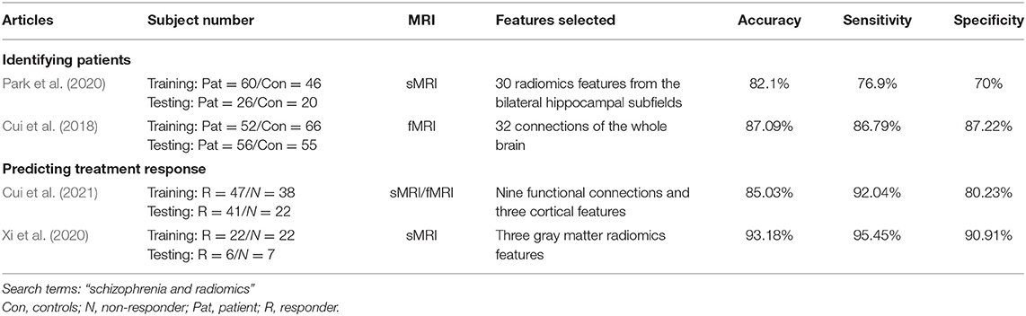
Table 1. Radiomics/machine learning and MRI in schizophrenia.
In addition to conventional features, another two studies considered abnormal functional connectivity as features (Cui et al., 2018, 2021). In a disease identification study, a total of 137 connections determined by functional MRI (fMRI) were detected between patients with schizophrenia and healthy controls (Cui et al., 2018). Then, they reduced to 32 using the LASSO binary LRM. The accuracy of detecting patients was 87.09%. One of the strengths of this study is the two types of cross-validation (CV), i.e., intra- and inter-data set CV. In an early treatment response prediction study, both functional connectivity and cortical measures with group difference were used to obtain baseline features (Cui et al., 2021). They used CV-LASSO to conduct feature selection and dimension reduction and constructed an SVM model to predict response to treatment. The combined features obtained an outstanding accuracy with 85.03%. Likewise, biologically meaningful group differences of MRI reflect the pathophysiology of schizophrenia in relation to diagnosis and treatment. Although these two studies included non-conventional radiomics features, MRI analyses produce tens of thousands of functional connections and cortical measurements. In line with radiomics, high-throughput mining of quantitative features from medical imaging, we can call it the radiomics strategy in schizophrenia research.
Nevertheless, dozens of MRI studies using machine learning are emerging in schizophrenia. Many of them are characterized by large international multicenter samples (Chen et al., 2020), multimodal MRI fusions (Lei et al., 2020), elegant machine learning models (Rozycki et al., 2018), considerable accuracy with high generalizability (Koutsouleris et al., 2018), or enhanced understanding of brain circuits that can serve as potential biomarkers (Zhao et al., 2020). For this reason, MRI-based machine learning approaches may offer better individual-level diagnostic and predictive value in mental disorders (Keshavan et al., 2020).
MRI-based radiomics/machine learning studies hold several strengths with regard to schizophrenia, e.g., having biological underpinnings (structure/function), extension of treatment prediction (APs/ECT), and validation methods (intra-/inter-data set CV). However, many critical challenges exist in this field from both clinical and research perspectives. First, most current classification studies treat a clinical diagnosis as the gold standard; however, with MRI or psychopathology, some recent unsupervised (Jauhar et al., 2018; Matsubara et al., 2019) or supervised (Jacobs et al., 2021) machine learning studies have tended to explore transdiagnostic characteristics of mental disorders and try to break the boundary of classic diagnosis and establish bioinformation-based disorder classification. Second, many novel machine learning models, such as generative adversarial networks (GANs), have been applied to large multicenter MRI data in this field (Zhong et al., 2020; Ren et al., 2021). GANs contribute a lot to improving reproducibility of radiomics features across manufacturers and increasing diagnostic accuracy (Marcadent et al., 2020). The use of “radiomics” combining novel machine learning models is considered an initiative and an important development over prior work in the precision medicine of mental disorders.
Driven by the need for better management of patients, as well as advances in neuroimaging-based machine learning approach, a quest for accurate detection of convention to illness, identification of patients, and prediction of treatment response and outcome is noted. MRI-based radiomics/machine learning researchers should promote the generalizability of findings across patients and pave the way to facilitate the guidance of clinical decision making by means of these findings.
L-BC conceptualized and wrote the first draft of the manuscript. All authors contributed to the article and approved the submitted version.
This work was supported by the grant support of Fourth Military Medical University (2019CYJH), Project funded by China Postdoctoral Science Foundation (2019TQ0130), and Scientific Research Project 2020 of The Second Medical Center (ZXBJ2005) and Innovative Project of Chinese PLA General Hospital (CX19028).
The authors declare that the research was conducted in the absence of any commercial or financial relationships that could be construed as a potential conflict of interest.
We thank Prof. Xiaopan Xu (Fourth Military Medical University) and Miss Jie Gong (Xidian University) for their help in revision.
Chaddad, A., Desrosiers, C., Hassan, L., and Tanougast, C. (2017). Hippocampus and amygdala radiomic biomarkers for the study of autism spectrum disorder. BMC Neurosci. 18:52. doi: 10.1186/s12868-017-0373-0
Chen, J., Patil, K. R., Weis, S., Sim, K., Nickl-Jockschat, T., Zhou, J., et al. (2020). Neurobiological divergence of the positive and negative schizophrenia subtypes identified on a new factor structure of psychopathology using non-negative factorization: an international machine learning study. Biol. Psychiatry 87, 282–293. doi: 10.1016/j.biopsych.2019.08.031
Cui, L. B., Fu, Y. F., Liu, L., Wu, X. S., Xi, Y. B., Wang, H. N., et al. (2021). Baseline structural and functional magnetic resonance imaging predicts early treatment response in schizophrenia with radiomics strategy. Eur. J. Neurosci. 53, 1961–1975. doi: 10.1111/ejn.15046
Cui, L. B., Liu, L., Wang, H. N., Wang, L. X., Guo, F., Xi, Y. B., et al. (2018). Disease definition for schizophrenia by functional connectivity using radiomics strategy. Schizophr. Bull. 44, 1053–1059. doi: 10.1093/schbul/sby007
Gong, J., Cui, L. B., Xi, Y. B., Zhao, Y. S., Yang, X. J., Xu, Z. L., et al. (2020). Predicting response to electroconvulsive therapy combined with antipsychotics in schizophrenia using multi-parametric magnetic resonance imaging. Schizophr. Res. 216, 262–271. doi: 10.1016/j.schres.2019.11.046
Huang, K., Lin, Y., Yang, L., Wang, Y., Cai, S., Pang, L., et al. (2020). A multipredictor model to predict the conversion of mild cognitive impairment to Alzheimer's disease by using a predictive nomogram. Neuropsychopharmacology 45, 358–366. doi: 10.1038/s41386-019-0551-0
Jacobs, G. R., Voineskos, A. N., Hawco, C., Stefanik, L., Forde, N. J., Dickie, E. W., et al. (2021). Integration of brain and behavior measures for identification of data-driven groups cutting across children with ASD, ADHD, or OCD. Neuropsychopharmacology 46, 643–653. doi: 10.1038/s41386-020-00902-6
Jauhar, S., Krishnadas, R., Nour, M. M., Cunningham-Owens, D., Johnstone, E. C., and Lawrie, S. M. (2018). Is there a symptomatic distinction between the affective psychoses and schizophrenia? A machine learning approach. Schizophr. Res. 202, 241–247. doi: 10.1016/j.schres.2018.06.070
Jiang, J.-B., Cao, Y., An, N.-Y., Yang, Q., and Cui, L.-B. (2020). Magnetic resonance imaging-based connectomics in first-episode schizophrenia: from preclinical study to clinical translation. Front. Psychiatry 11:948. doi: 10.3389/fpsyt.2020.565056
Kai, C., Uchiyama, Y., Shiraishi, J., Fujita, H., and Doi, K. (2018). Computer-aided diagnosis with radiogenomics: analysis of the relationship between genotype and morphological changes of the brain magnetic resonance images. Radiol. Phys. Technol. 11, 265–273. doi: 10.1007/s12194-018-0462-5
Keshavan, M. S., Collin, G., Guimond, S., Kelly, S., Prasad, K. M., and Lizano, P. (2020). Neuroimaging in schizophrenia. Neuroimaging Clin. N. Am. 30, 73–83. doi: 10.1016/j.nic.2019.09.007
Koutsouleris, N., Wobrock, T., Guse, B., Langguth, B., Landgrebe, M., Eichhammer, P., et al. (2018). Predicting response to repetitive transcranial magnetic stimulation in patients with schizophrenia using structural magnetic resonance imaging: a multisite machine learning analysis. Schizophr. Bull. 44, 1021–1034. doi: 10.1093/schbul/sbx114
Kraguljac, N. V., McDonald, W. M., Widge, A. S., Rodriguez, C. I., Tohen, M., and Nemeroff, C. B. (2021). Neuroimaging biomarkers in schizophrenia. Am. J. Psychiatry. doi: 10.1176/appi.ajp.2020.20030340. [Epub ahead of print].
Kupfer, D. J., Kuhl, E. A., and Regier, D. A. (2013). DSM-5–the future arrived. JAMA 309, 1691–1692. doi: 10.1001/jama.2013.2298
Lambin, P., Leijenaar, R. T. H., Deist, T. M., Peerlings, J., de Jong, E. E. C., van Timmeren, J., et al. (2017). Radiomics: the bridge between medical imaging and personalized medicine. Nat. Rev. Clin. Oncol. 14, 749–762. doi: 10.1038/nrclinonc.2017.141
Lei, D., Pinaya, W. H. L., Young, J., van Amelsvoort, T., Marcelis, M., Donohoe, G., et al. (2020). Integrating machining learning and multimodal neuroimaging to detect schizophrenia at the level of the individual. Hum. Brain Mapp. 41, 1119–1135. doi: 10.1002/hbm.24863
Li, Y., Jiang, J., Shen, T., Wu, P., and Zuo, C. (2018). Radiomics features as predictors to distinguish fast and slow progression of Mild Cognitive Impairment to Alzheimer's disease. Annu. Int. Conf. IEEE Eng. Med. Biol. Soc. 2018, 127–130. doi: 10.1109/EMBC.2018.8512273
Marcadent, S., Hofmeister, J., Preti, M. G., Martin, S. P., Van De Ville, D., and Montet, X. (2020). Generative adversarial networks improve the reproducibility and discriminative power of radiomic features. Radiol. Artif. Intell. 2:e190035. doi: 10.1148/ryai.2020190035
Matsubara, T., Tashiro, T., and Uehara, K. (2019). Deep neural generative model of functional MRI images for psychiatric disorder diagnosis. IEEE Trans. Biomed. Eng. 66, 2768–2779. doi: 10.1109/TBME.2019.2895663
Park, Y. W., Choi, D., Lee, J., Ahn, S. S., Lee, S. K., Lee, S. H., et al. (2020). Differentiating patients with schizophrenia from healthy controls by hippocampal subfields using radiomics. Schizophr. Res. 223, 337–344. doi: 10.1016/j.schres.2020.09.009
Ranjbar, S., Velgos, S. N., Dueck, A. C., Geda, Y. E., and Mitchell, J. R. (2019). Brain MR radiomics to differentiate cognitive disorders. J. Neuropsychiatry Clin. Neurosci. 31, 210–219. doi: 10.1176/appi.neuropsych.17120366
Ren, M., Dey, N., Fishbaugh, J., and Gerig, G. (2021). Segmentation-renormalized deep feature modulation for unpaired image harmonization. IEEE Trans. Med. Imaging doi: 10.1109/TMI.2021.3059726. [Epub ahead of print].
Rozycki, M., Satterthwaite, T. D., Koutsouleris, N., Erus, G., Doshi, J., Wolf, D. H., et al. (2018). Multisite machine learning analysis provides a robust structural imaging signature of schizophrenia detectable across diverse patient populations and within individuals. Schizophr. Bull. 44, 1035–1044. doi: 10.1093/schbul/sbx137
Samuel, A. L. (1959). Some studies in machine learning using the game of checkers. IBM J. Res. Dev. 3, 535–554. doi: 10.1147/rd.33.0210
Sun, H., Chen, Y., Huang, Q., Lui, S., Huang, X., Shi, Y., et al. (2018). Psychoradiologic utility of MR imaging for diagnosis of attention deficit hyperactivity disorder: a radiomics analysis. Radiology 287, 620–630. doi: 10.1148/radiol.2017170226
Tandon, N., and Tandon, R. (2018). Will machine learning enable us to finally cut the gordian knot of schizophrenia. Schizophr. Bull. 44, 939–941. doi: 10.1093/schbul/sby101
Wang, X. H., Yu, A., Zhu, X., Yin, H., and Cui, L. B. (2019). Cardiopulmonary comorbidity, radiomics and machine learning, and therapeutic regimens for a cerebral fMRI predictor study in psychotic disorders. Neurosci. Bull. 35, 955–957. doi: 10.1007/s12264-019-00409-1
Wang, Y., Sun, K., Liu, Z., Chen, G., Jia, Y., Zhong, S., et al. (2020). Classification of unmedicated bipolar disorder using whole-brain functional activity and connectivity: a radiomics analysis. Cereb. Cortex 30, 1117–1128. doi: 10.1093/cercor/bhz152
Xi, Y. B., Cui, L. B., Gong, J., Fu, Y. F., Wu, X. S., Guo, F., et al. (2020). Neuroanatomical features that predict response to electroconvulsive therapy combined with antipsychotics in schizophrenia: a magnetic resonance imaging study using radiomics strategy. Front. Psychiatry 11:456. doi: 10.3389/fpsyt.2020.00456
Zhao, W., Guo, S., Linli, Z., Yang, A. C., Lin, C. P., and Tsai, S. J. (2020). Functional, anatomical, and morphological networks highlight the role of basal ganglia-thalamus-cortex circuits in schizophrenia. Schizophr. Bull. 46, 422–431. doi: 10.1093/schbul/sbz062
Keywords: radiomics, machine learning, mental disorders, magnetic resonance imaging, schizophrenia
Citation: Cui L-B, Xu X and Cao F (2021) Building the Precision Medicine for Mental Disorders via Radiomics/Machine Learning and Neuroimaging. Front. Neurosci. 15:685005. doi: 10.3389/fnins.2021.685005
Received: 24 March 2021; Accepted: 10 May 2021;
Published: 15 June 2021.
Edited by:
Reza Lashgari, Institute for Research in Fundamental Sciences, IranReviewed by:
Mingrui Xia, Beijing Normal University, ChinaCopyright © 2021 Cui, Xu and Cao. This is an open-access article distributed under the terms of the Creative Commons Attribution License (CC BY). The use, distribution or reproduction in other forums is permitted, provided the original author(s) and the copyright owner(s) are credited and that the original publication in this journal is cited, in accordance with accepted academic practice. No use, distribution or reproduction is permitted which does not comply with these terms.
*Correspondence: Feng Cao, d2luZDg4MjhAZ21haWwuY29t; Long-Biao Cui, bGJjdWlAZm1tdS5lZHUuY24=; orcid.org/0000-0002-0784-181X
†These authors have contributed equally to this work
Disclaimer: All claims expressed in this article are solely those of the authors and do not necessarily represent those of their affiliated organizations, or those of the publisher, the editors and the reviewers. Any product that may be evaluated in this article or claim that may be made by its manufacturer is not guaranteed or endorsed by the publisher.
Research integrity at Frontiers
Learn more about the work of our research integrity team to safeguard the quality of each article we publish.