 Xuesong Wang
 Jia Wang2†
 Chaochao Yu
95% of researchers rate our articles as excellent or good
Learn more about the work of our research integrity team to safeguard the quality of each article we publish.
Find out more
REVIEW article
Front. Neurol. , 13 June 2024
Sec. Experimental Therapeutics
Volume 15 - 2024 | https://doi.org/10.3389/fneur.2024.1399925
Recently, there has been increasing attention on the impact of acupuncture on the dysregulated neural circuits in different disease. This has led to new understandings of how acupuncture works. This review presents a comprehensive analysis of research that have examined the impact of acupuncture on abnormal neural circuits associated with pain, anxiety, Parkinson’s disease, addiction disorders, cognitive problems, and gastrointestinal disorders. These studies have shown that acupuncture’s therapeutic effects are mediated by specific brain areas and neurons involved in neural circuit mechanisms, emphasising its wide-ranging influence. The positive impacts of acupuncture can be ascribed to its ability to modify the functioning of neurocircuits in various physiological conditions. Nevertheless, contemporary studies on acupuncture neural circuits frequently overlook the comprehensive circuit mechanism including the periphery, central nervous system, and target organ. Additionally, the scope of diseases studied is restricted. Future study should focus on broadening the range of diseases studied and exploring the neural circuit mechanisms of these diseases in depth in order to enhance our understanding of acupuncture’s neurobiological impacts.
Acupuncture has a rich historical background and holds significant cultural importance in China, it is an important part of traditional Chinese medicine (TCM). Acupuncture originated in ancient China and has been developed and inherited over thousands of years of history. In the ancient medical literature of China, such as the Miraculous Pivot (《灵枢》), there are numerous documented instances of acupuncture.
Currently, acupuncture remains extensively utilized in China and is progressively garnering global interest and acknowledgment. Acupoints, according to traditional meridian theory, are the convergence points of Qi (气, vital energy), blood, and meridians within the internal Zang-Fu (脏腑). They are also reflective areas on the body surface corresponding to the internal Zang-Fu (脏腑). Acupoints play a crucial role in the therapeutic effectiveness of acupuncture. Meridians have many functions, including connecting internal Zang-Fu (脏腑), communication between internal and external, responsible for the operation of Qi (气, vital energy) and blood, nutritional Zang-Fu (脏腑), defense against disease, balance of Yin (阴) and Yang (阳). The functioning of life activities in the human body is governed by Qi (气, vital energy), and acupuncture can be employed to regulate the functioning of Qi (气, vital energy) and blood by stimulating certain acupoints, so achieving the objective of healing diseases and maintaining health.
In recent years, the field of neuroscience has made significant progress in enhancing our comprehension of the neural circuits in organisation and operation of brain networks. “Neural Circuits” consist of neurons and glial cells (especially astrocytes and microglia), and are the basic units in the nervous system for transmitting and processing neural signals. Neural circuits play a crucial role in overseeing and managing a wide range of physiological functions in the body, including perception, movement, cognition, and emotion. Acupuncture has been discovered to have the ability to regulate the physiological activities of the organism and generate therapeutic effects by influencing various neural circuits and neurotransmitter systems. Hence, the study of neural circuits is essential for comprehending the mechanisms and impacts of acupuncture, while also offering novel concepts and directions for advancing the field of acupuncture. With the ongoing advancement of acupuncture standardization, more and more high-quality clinical and basic evidence of acupuncture is being reported, and the recognition of acupuncture is increasing internationally (1). This has led to a rising international acknowledgment of acupuncture. Modern medical research has shown that acupuncture exerts therapeutic effects through mechanisms such as anti-inflammatory effects (2), immunomodulation (3), regulation of synaptic plasticity (4), and anti-apoptotic processes (5). In recent years, acupuncture related neural circuit studies have become the focus of growing research interest, mainly in the areas of pain (6), pain-related anxiety (7), Parkinson’s disease (8), addiction (9), cognitive disorders (10), and gastrointestinal disorders (3). Here, we provide a review of current research with acupuncture-related neural circuit studies according to different diseases, and our objective is to provide new perspectives and a deeper understanding of how acupuncture works.
Pain is a physiological response of the body to potential injury or damage to tissues, manifested by way of nerve signaling. The condition can manifest as either acute, meaning it develops rapidly and subsides fast, or chronic, lasting for more than three months. Pain can arise from a range of factors, such as tissue damage, chronic diseases, and neurological abnormalities (11). Furthermore, pain is not solely a physiological occurrence, but is intricately connected to psychological and emotional reactions. Chronic pain is frequently accompanied by emotional disorders like anxiety, which can worsen the experience of pain and possibly impacts the effectiveness of treatment (12, 13). There have been numerous research conducted on the analgesic effects of acupuncture (14–17). Research in classical studies has demonstrated that acupuncture has the ability to stimulate the release of occurring opioid peptides, such as enkephalin and dynorphin. This stimulation also activates neural activity in the dorsal region of the grey matter surrounding the aqueduct, resulting in analgesic effects within the central nervous system (18, 19). Furthermore, acupuncture has the ability to regulate the secretion of pain-related molecules, including dopaminergic and serotonergic neurotransmitters (20), acetylcholine (21), ATP/adenosine (22), purinergic signaling (23). It also has the capacity to suppress the release of pain-inducing substances, hence alleviating pain.
Acupuncture is essential in regulating pain at the spinal level. During pathological conditions, acupuncture sites can have an impact on the circuits that transmit pain. Pain caused by harmful stimuli usually includes receptors located in the peripheral nerves. These receptors convey signals through the spinal nerve roots to the dorsal root ganglia (DRG), and eventually reach the thalamus and other brain structures (24). As primary afferent neurons, pain signals travel through C fibers or Aδ fibers in the DRG to the spinal cord. Studies have investigated the neurobiological processes of acupoints sensitization that are associated with the activation of primary afferent nerves (25). Studies indicate that C fibers in the DRG are implicated in acupoints sensitization (26). Further research, retrograde tracing and chemogenetics, revealed that the Zusanli (ST36) acupuncture point and the corresponding plantar area on the same side of the body had shared nociceptive dorsal root ganglia (DRGs). Alterations in the sensitivity of these commonly shared nociceptive dorsal root ganglia (DRGs) are associated with changes in the thresholds for mechanical and thermal pain at the Zusanli (ST36) acupoint in a mouse model treated with complete Freund’s adjuvant (CFA). This indicates that the overstimulation of the shared DRGs at the site of injury and the corresponding acupoint plays a role in the development of increased sensitivity to acupuncture. Furthermore, Zhu (27) found that acupoints not only generate sensitization signals but also communicate information to the central nervous system. This process activates the endogenous pain modulation system through descending inhibitory pathways, resulting in systemic analgesic effects. The endogenous pain modulation system encompasses modulation at multiple levels, including the spinal level, central nervous system, brainstem, and forebrain. The descending inhibitory system is essential for regulating pain and involves structures such as the periaqueductal grey (PAG), rostral ventromedial medulla (RVM), locus coeruleus (LC), and lateral reticular nucleus (LRN) (28). The interconnectedness of these tissues is facilitated by descending pathways that control the activation of primary afferents in the spinal dorsal horn. One of these components, the periaqueductal grey (PAG), serves as a central hub by engaging in the majority of higher-level central pain inhibitory pathways and triggering the descending inhibitory system. Acupuncture can alleviate pain by activating certain internal pain control structures or activating neuronal pathways in the inhibitory system that reduces pain sensation (29).
The thalamus functions as an intermediary in the transmission of pain signals, playing a crucial role in facilitating the passage of nociceptive information to the cortex. The thalamus is divided into three parts: the posterior, medial, and lateral thalamus. Each of these divisions receives signals related to pain from the spinal cord (30). The posterior thalamus primarily projects to the somatosensory cortex (SSC), contributing to the discrimination of pain sensation. Neurons in the medial thalamus not only project to the motor areas of the cingulate gyrus but also regulate the anterior cingulate cortex (ACC) and prefrontal cortex, thereby participating in the emotional and attentional modulation of pain perception. The lateral thalamus predominantly projects to the dorsolateral prefrontal cortex (DLPFC), engaging in the cognitive evaluation of pain (31). Acupuncture exerts regulatory effects on pain sensation, emotion, and cognition by modulating neural circuits within the thalamus and cortex (32). Acupuncture therapy for chronic pain may involve the regulation of GABAergic neurons surrounding the ventrolateral periaqueductal gray (vlPAG) via various pathways, thereby attenuating or relieving 5-HT-related descending inhibitory control and augmenting the pain-modulating function of the descending inhibitory system (33). Researchers have observed that enhanced expression of cannabinoid cannabinoid receptor 1 (CB1) on midbrain GABAergic neurons can alleviate 5-HT-related descending inhibitory control and diffuse noxious inhibitory control (DNIC) function, consequently alleviating chronic pain (34). In a mouse model of knee osteoarthritis (KOA), low-frequency, high-intensity electroacupuncture at the Zusanli and Sanyinjiao points enhances the expression of CB1 receptors on midbrain GABAergic neurons by activating CB1 receptors in the ventrolateral periaqueductal gray (vlPAG), thereby eliciting the aforementioned effects, indicating that acupuncture may alleviate chronic pain by regulating the expression of ventrolateral periaqueductal gray (vlPAG) CB1 receptors to enhance the descending inhibitory system (35). Similarly, researchers have noted the potential influence of the vlPAG’s activating effect on the descending inhibitory system on pain-related negative emotions in the rostral anterior cingulate cortex (rACC) (36). Researchers found that GABAergic neurons in the vlPAG receive glutamatergic projections from the rostral anterior cingulate cortex (rACC), and inhibiting the Glu-vlPAG circuit in the rostral anterior cingulate cortex (rACC) leads to feedforward inhibition of 5-HT neurons by GABAergic neurons in the vlPAG (37). Activating this circuit in spared nerve injury (SNI) model mice can counteract the analgesic effect of electroacupuncture but cannot reverse its anxiolytic effect, suggesting that electroacupuncture may alleviate neuropathic pain hypersensitivity through this neural circuit rather than its anxiolytic effect (38). Furthermore, Wu et al. (39) demonstrated that electroacupuncture alleviated chronic pain-induced anxiety-like behaviors via activation rACCCaMKII-DRN5-HT circuit. Shen et al. (38) found that electroacupuncture can treat anxiety-like behavior in Complete Freund’s Adjuvant (CFA) model rats by activating the rACC Glu-thalamus circuit without affecting pain. Xu et al. (40) found that electroacupuncture can exert analgesic and anxiolytic effects on spared nerve injury (SNI) model mice by activating the rACC Glu-dorsal raphe nucleus (DRN) circuit. Hsiao et al. (41) found that electroacupuncture to the Zusanli (ST36) could treat chronic inflammatory pain by inhibiting CaMKIIα signaling in the somatosensory cortex (SSC)-anterior cingulate cortex (ACC) circuit.
The limbic and reward systems are crucial brain regions linked to the control of emotions, motivation, and pain. The limbic system comprises the hypothalamus, amygdala, and hippocampus, primarily responsible for emotional regulation, memory formation, and stress reactions in response to pain. Concurrently, the reward system, comprising the nucleus accumbens (NAcc), ventral tegmental area (VTA), and substantia nigra, is believed to have a strong connection to several elements of pain perception, such as pleasure and addiction. Additionally, it plays a role in broader reward processing. The interplay between these systems impacts an individual’s perception and emotional reaction to pain, therefore influencing their capacity to manage and adjust to pain (42). Acupuncture can modulate the emotional and motivational aspects of pain by influencing neural circuits within the limbic and reward systems, while also impacting the rewarding effects of pain. Recent studies have shed light on the role of the lateral hypothalamus (LH) in regulating neuropathic pain (43). The study found that LH neurons projecting from the lateral habenula (LHb) are involved in modulating neuropathic pain (44). These discovered that GABAergic neurons in the lateral septum (LS) projecting to the lateral hypothalamus (LH) are implicated in the comorbidity of pain and anxiety. These findings suggest that lateral hypothalamus (LH) may serve as a crucial center for pain modulation, and acupuncture may exert therapeutic effects on pain and anxiety by influencing lateral hypothalamus (LH) neural circuits (45). The nucleus accumbens (NAcc) is a central component of the brain’s reward system, primarily involved in dopamine-mediated reward and pleasure behaviors, which also has implications for the rewarding effects of pain (46). The rewarding effects of pain refer to the pleasurable or satisfying sensation experienced when pain is relieved or alleviated, thus increasing pain tolerance and acceptance. Acupuncture can influence the rewarding effects of pain by activating neural circuits within the nucleus accumbens (NAcc). Wang et al. (47) found that signals from acupuncture points travel from the spinal cord to the hypothalamus, and electroacupuncture at bilateral Zusanli (ST36) in spared nerve injury (SNI) model rats can induce conditioned place preference (CCP) during the early stages of chronic pain. Activation of the orexinergic neural circuit from the lateral hypothalamus (LH) to the nucleus accumbens (NAcc) shell induced by electroacupuncture-induced pain relief elucidates acupuncture’s potential to exert rewarding effects in alleviating chronic pain through this neural pathway (47).
In conclusion, acupuncture, as a pain management technique, engages numerous neural circuits and offers promising therapeutic benefits for a range of pain and pain-related mental disorders.
Parkinson’s disease (PD) is a common neurodegenerative disease of the central nervous system (48), which is characterized by significant degeneration and loss of dopamine (DA) neurons in the substantia nigra (49). The primary symptoms of PD include bradykinesia, tremor in the static state, emotional abnormality (mainly depression, anxiety and apathy), and postural instability (50). Acupuncture is a therapeutic method that can improve symptoms of Parkinson’s disease. When used alongside medication, it helps reduce side effects and increase effectiveness (51). The mechanism behind acupuncture’s treatment for PD may involve reducing oxidative stress, alleviate immune-inflammatory reactions, and regulate the gut-brain axis (52, 53).
Early research and anatomical studies have revealed that glutamatergic neurons in the cortex and thalamus, as well as dopaminergic neurons in the midbrain, project to the basal ganglia’s primary input nucleus—the striatum (54). Loss of dopaminergic neurons in the substantia nigra results in insufficient dopamine projection to the thalamus and cerebral cortex, leading to impaired motor control function in the basal ganglia circuitry (55). The inability of the substantia nigra to coordinate the movements of agonist and antagonist muscles gives rise to motor symptoms such as resting tremors in PD. Hence, the loss and degeneration of dopaminergic neurons play a crucial role in PD’s motor symptoms (56). Additionally, in animal models of PD, dopaminergic pathways in the basal ganglia, thalamus, and limbic system are also compromised (57). Functional magnetic resonance imaging (fMRI) can reveal changes in brain neural activity signals induced by acupuncture, aiding in the identification of brain regions involved in the neural pathways of acupuncture treatment for PD in clinical research (58). Chae et al. (59) treated 10 PD patients with acupuncture at the left Yanglingquan (SP9) acupoint, resulting in improvements in hand motor function, as indicated by fMRI activation of the thalamus and primary motor cortex post-treatment. Similarly, Yeo et al. (60, 61) demonstrated increased signals in PD-affected brain regions such as the substantia nigra, striatum, prefrontal cortex, and anterior cingulate cortex following acupuncture at the right Yanglingquan acupoint. These findings suggest that acupuncture may alleviate PD motor symptoms by activating PD-affected brain regions and modulating the basal ganglia-thalamocortical and cortical circuits. Li et al. (62) showed that acupuncture at Baihui, Fengchi, and the tremor area improved tremor symptoms in PD patients, possibly by affecting the cerebello-thalamo-cortical (CTC) circuit. fMRI observed specific activation of the cerebellum, along with various changes in the thalamus and motor cortex. It is noteworthy that the inconsistency in the regional homogeneity of the motor cortex fMRI between the left and right sides may be due to handedness. In animal experiments, Jia et al. (63) found that electroacupuncture reduced the loss of dopaminergic neurons in the substantia nigra of unilateral medial forebrain bundle (MFB)-lesioned PD model rats, reversed the decrease in midbrain substance P levels and the increase in glutamic acid decarboxylase-67 (GAD 67) mRNA levels induced by the lesion, and significantly reduced abnormal movements in PD model rats. They also found that high-frequency electroacupuncture reversed the increase in midbrain GABA content and promoted motor coordination in MFB-injured rats (64). These studies suggest that electroacupuncture therapy may improve PD motor dysfunction by restoring the homeostasis of basal ganglia circuits and inhibiting excessive GABA output.
In summary, recent studies indicate that acupuncture predominantly regulates the activity of GABAergic neurons in the basal ganglia and is involved in specific neural pathways connecting the basal ganglia, thalamus, and brain. This modulation decreases the degeneration of dopaminergic neurons in the basal ganglia, enhances the activity of dopamine receptors, therefore leading to an improvement in aberrant motor symptoms in PD. Moreover, there is a correlation between thalamic dysfunction and non-motor symptoms of PD, such as rigidity and tremor. Acupuncture may enhance non-motor symptoms of PD by activating alternative pathways, such as the cerebello-thalamo-cortical (CTC) circuit. Subsequent investigations should delve further into the mechanisms by which these neural circuits operate in order to gain a more comprehensive understanding of the impact of acupuncture therapy on the treatment of PD.
Addiction disorder refers to an intense reliance on a substance or behavior, leading to an uncontrollable compulsion to use or engage in it. This reliance is typically accompanied by strong psychological urges and physical symptoms of withdrawal. Addiction disorders can be addictions to substances or drugs (e.g., drugs, alcohol, nicotine, etc.) or addictions to certain behaviors or activities (e.g., gambling, shopping, food intake, etc.) (65). Addiction disorders are considered to be complex disorders that are influenced by genetic, environmental, and psychological factors (66, 67). The primary emphasis of current research on addiction disorders is substance addiction. Acupuncture is regarded as a supplementary therapy for addiction problems, aiding in the reduction of withdrawal symptoms, alleviation of worry and dread, and promotion of bodily and mental equilibrium throughout the withdrawal process. It has the potential to control levels of neurotransmitters, mitigate withdrawal symptoms, and partially cure addictive behaviors (68–70).
The mesolimbic reward system plays a crucial role in the onset of addiction, exerting influence over dopamine neurons (71). The nucleus accumbens (NAcc), as the core of the reward system, is interconnected with the ventral tegmental area (VTA) (72). Alterations in the nucleus accumbens (NAcc) neural circuitry are pivotal for reward learning, receiving excitatory projections from brain regions such as the prefrontal cortex (PFC) and thalamus (73). Through acupuncture, modulation of endogenous opioid input and dopamine neuron activity within the nucleus accumbens (NAcc) and ventral tegmental area (VTA) upstream neural circuits can be achieved, thereby addressing the motor and emotional abnormalities induced by substance addiction (74). Research suggests that acupuncture at the Shenmen (HT7) can alleviate withdrawal tremors, anxiety, and compulsive drug-seeking behavior in ethanol-dependent rats by activating the arcuate nucleus of the hypothalamus (ARC) to input enkephalins into the nucleus accumbens (NAcc) (75). In studies on cocaine addiction, this team found that acupuncture at Shenmen (HT7) suppressed cocaine-induced motor symptoms via signals mediated by the ulnar nerve A fibers (76). Further investigations suggest that this acupuncture signal may activate the lateral habenula (LHb)-rostromedial tegmental nucleus (RMTg) circuitry via peripheral tactile pathways, ultimately inhibiting dopamine (DA) neurons (77).
During drug withdrawal, compulsive drug-seeking behavior is often accompanied by severe negative emotions such as anxiety and depression (78). The lateral habenula (LHb) plays a crucial role in negatively regulating reward behavior, aversive emotions, and behavioral inhibition (79). Primarily receiving input from the basal ganglia and ventral forebrain, LHb neurons possess the function of inhibiting dopamine neurons (80). Studies have elucidated multiple input pathways to the lateral habenula (LHb), including projections from glutamatergic neurons in the medial prefrontal cortex (mPFC), which elicit aversive responses (81). Addictive substances such as cocaine, nicotine, and ethanol can enhance lateral habenula (LHb) activity, leading to aberrant excitatory and inhibitory synaptic transmission and thus mediating the generation of negative emotions. Augmenting glutamatergic transmission from the medial prefrontal cortex (mPFC) to the lateral habenula (LHb) may be one of the mechanisms by which acupuncture suppresses psychomotor symptoms in cocaine-addicted rats (82). Furthermore, acupuncture can alleviate drug addiction by modulating gamma-aminobutyric acid (GABA) transmission and dopamine expression levels in the ventral tegmental area (VTA) (83). Studies using optogenetics and other techniques suggest that acupuncture may mediate the regulatory effect of GABAergic neurons in the ventral tegmental area (VTA) via activation of central amygdala (CeA), resulting in reduced DA release in the nucleus accumbens (NAcc) and thereby suppressing abnormal motor behavior and positive emotional states induced by methamphetamine (84).
In conclusion, recent research indicates that acupuncture improves symptoms of substance addiction primarily by modulating dopaminergic neural circuits between the prefrontal cortex, thalamus, and the reward systems nucleus accumbens (NAcc) and ventral tegmental area (VTA). Additionally, acupuncture may exert its effects by inhibiting LHb-mediated negative regulation of reward behavior, modulating relevant cortical or reward system inputs to the LHb, suppressing dopamine release in the nucleus accumbens (NAcc) and ventral tegmental area (VTA), and ameliorating substance addiction. Studies have also highlighted the dorsal column pathway as a potential route for acupuncture treatment of cocaine addiction, providing insight into the complete pathway of acupuncture signals from the periphery to the central nervous system.
Cognitive impairment refers to a state when an individual’s capacity to think, recall, and process information is affected due to various conditions that impact brain function. Mild cognitive impairment (MCI), dementia, Alzheimer’s disease (AD), vascular dementia (VaD), frontotemporal dementia (FD), and traumatic brain injury are frequent causes of cognitive impairment, with Alzheimer’s disease being particularly prevalent. This condition is a degenerative neurological disease characterized by the gradual demise of nerve cells and the deterioration of connections between them in the brain. The initial symptoms typically involve memory loss and cognitive decline. As the disease advances, the patient’s ability to carry out daily activities gradually deteriorates, finally resulting in severe cognitive impairment (85). While a treatment for the condition is not yet available, timely detection and management can effectively decelerate the advancement of the illness and enhance the overall well-being of patients (86). Several recent studies have documented the benefits of acupuncture in the management of Alzheimer’s disease. The mechanism of acupuncture in treating Alzheimer’s disease involves several factors, such as regulating Aβ metabolism and tau protein phosphorylation in a beneficial way, modulating neurotransmitters, reducing damage to synaptic and neuronal function, suppressing neuroinflammatory responses, and enhancing brain energy metabolism (87–89). Thus, acupuncture intervention in Alzheimer’s disease has a multi-target, multi-pathway network modulation impact, which aligns with the overall modulation properties of acupuncture.
The hippocampus stores each experience as a unique memory representation through a mechanism called pattern separation (PS), then retrieves this representation from partial cues via pattern completion (PC) to support memory (90). Impaired pattern separation is often observed in neurocognitive and neurodegenerative disorders. Adult hippocampal neurogenesis and synaptic plasticity in CA1 and CA3 regions are closely linked to pattern separation (91). Furthermore, the medial septum (MS) is involved in central cholinergic (ACh) neuron projections to the hippocampus and entorhinal cortex via the fimbria-fornix, exciting hippocampal pyramidal cells to facilitate learning and memory tasks (91). The M1 receptor plays a crucial role in long-term potentiation (LTP) in the hippocampal CA1 region. Researchers have found that the medial septum (MS)-vertical limb of the diagonal band (MS/VDB)-dentate gyrus (DG) cholinergic neural circuitry may participate in promoting hippocampal neurogenesis and reducing impaired pattern separation by enhancing cholinergic neuron survival, promoting acetylcholine transport, among other mechanisms. Dysfunction in the medial septum (MS)-hippocampal cholinergic system may lead to decreased learning and memory abilities. While, electroacupuncture can improve memory impairment in dementia model mice by activating this circuitry. Researchers have suggested that electroacupuncture may treat early learning and memory impairment caused by diabetes by modulating the medial septum (MS)-hippocampal cholinergic circuitry, inhibiting excessive acetylcholine output from the medial septum (MS) to the hippocampus, enhancing M1 receptor activity, and rectifying nerve growth factor (NGF) and pro-nerve growth factor (proNGF) level imbalances in the medial septum (MS) and hippocampus (92).
Diabetes may also induce this dysfunction, concomitant with impaired hippocampal plasticity and dysregulated levels of pro-nerve growth factor (proNGF) (93). In addition to the hippocampus, research has shown that the dorsal raphe nucleus serotonergic neurons and other upstream brain regions also project to the hippocampus, which is related to cognitive function. This suggests that the hippocampus and upstream brain regions may be key areas for research on cognitive disorders. The neurons in the hippocampus are mainly glutamatergic neurons, which are closely related to the activity of glutamate AMPA receptors, NMDA receptors, and CaMKII, all of which are crucial for the formation of long-term potentiation (LTP). Acupuncture therapy has been shown to improve cognitive function by affecting the hippocampal glutamatergic neuron circuit (94). Although the role of GABAergic neurons in the acupuncture treatment of cognitive disorders has not been fully studied, there is evidence to suggest that inhibition of the activity and function of GABAergic interneurons may lead to structural abnormalities in excitatory synaptic transmission pathways, reducing synaptic plasticity and resulting in cognitive disorders (95). Therefore, GABAergic interneuron-related neural circuits may serve as a new target for research on cognitive disorders.
As a whole, researchers conducted comprehensive assessments of changes in learning abilities in animal experiments before and after electroacupuncture treatment. They used various behavioral tests, including novel object recognition, novel location recognition, Morris water maze, and Y-maze experiments, thereby enhancing the accuracy of cognitive learning ability evaluations. These tests improved the accuracy of evaluating cognitive learning abilities. However, current studies mainly focus on animal spatial memory and short-term memory capabilities, further research is needed to elucidate long-term memory and the hippocampal cholinergic or glutamatergic pathways.
Gastrointestinal diseases are prevalent globally, and acupuncture has a lengthy historical background and extensive clinical expertise in managing such conditions (96, 97). Previous literature has shown that acupuncture is effective in the treatment of functional dyspepsia, functional diarrhea, irritable bowel syndrome with diarrhea, functional constipation, and ulcerative colitis (98–101). The mechanism may be related to acupuncture can regulate the function of gastrointestinal tract, relieve pain, regulate immune function, improve mood, and maintain intestinal microecological balance (96, 102).
Studies have indicated that higher central nuclei associated with the treatment of gastrointestinal diseases include the dorsal vagal complex (DVC), paraventricular nucleus (PVN), and central amygdala (CeA). Within the brainstem, the dorsal vagal complex (DVC) is considered a critical region for regulating gastrointestinal smooth muscle movement and glandular secretion (103). It connects bidirectionally the nucleus tractus solitarius (NTS) and the dorsal motor nucleus of the vagus nerve (DMV) (104). It is speculated that gastrointestinal sensory instructions may reach the dorsal motor nucleus of the vagus nerve (DMV) or nucleus tractus solitarius (NTS) directly or indirectly via the vagus nerve. After signal integration in these regions, the dorsal motor nucleus of the vagus nerve (DMV) communicates the information through the vagus nerve to higher central nuclei, thereby regulating gastrointestinal activity and glandular secretion (105). Studies have found that electroacupuncture at the Zusanli (ST36) point may regulate gastrointestinal hormone levels and affect gastrointestinal motility via the DVC-vagus nerve-stomach pathway (106). Another study has indicated that acupuncture is involved in the PVN-DVC-vagus nerve pathway, influencing gastric motility (107). The hypothalamus influences gastrointestinal movement through the autonomic nervous system, parasympathetic nervous system, and endocrine regulation. The cerebellum, besides participating in motor coordination, also plays a role in regulating visceral activity, thus affecting gastrointestinal motility in synergy. Through fMRI, it has been observed that acupuncture at the Zusanli (ST36) and Tianshu (ST25) points enhances neural signals in the right cerebellar cortex and bilateral hypothalamus, suggesting involvement of the cerebellum-hypothalamus circuit in regulating gastrointestinal function along the stomach meridian (108). Another study found that acupuncture at Zusanli (ST36) may improve gastrointestinal function via the lateral hypothalamic area (LHA)-dentate nucleus (DN) cerebellar circuit (109). These studies suggest that acupuncture may exert its effects on gastrointestinal motility through different neurons within the hypothalamus-cerebellum circuit. Further research indicates that the paraventricular nucleus (PVN) receives projections from GABAergic neurons in the central amygdala (CeA), and electroacupuncture at the Zusanli (ST36) point may regulate appetite, promote gastric emptying, and thus improve gastrointestinal function through the CeAGABA-PVN circuit (110).
From the above studies, the neurological pathways of acupuncture treatment for gastrointestinal diseases mostly concentrates on the brainstem, hypothalamus, and cerebellum. These regions are responsible for regulating visceral activity. Acupuncture can enhance gastrointestinal function by influencing the action of digestive smooth muscles through the regulation of several gastrointestinal hormones within neural circuits. Researchers can employ advanced methods like fMRI, neural tracing, and chemogenetics to detect the activation of specific brain areas and nuclei, such as the hypothalamus, cerebellum, and central amygdala (CeA), along with their projections, during acupuncture treatment for gastrointestinal illnesses. However, in order to conduct a more accurate examination of the exact activities of neurons within the neural circuitry that is affected by acupuncture, and to gain a more complete understanding of its central mechanisms, additional study using advanced techniques like optogenetics may be required.
Furthermore, acupuncture has been shown to affect the circuitry mechanisms of post-stroke functional impairments, chronic itch, comorbid anxiety and depression, and sleep disorders. Recent studies suggest that electroacupuncture at Baihui (GV20) and Dazhui (GV14) points may improve blood circulation in the contralateral primary somatosensory cortex (S1)—motor cortex (M1) circuit, enhancing the activity of neurons in this circuit and thereby promoting the recovery of limb motor function after unilateral cerebral infarction (111). Additionally, the paraventricular nucleus of the hypothalamus (PVN) may be involved in electroacupuncture treatment for post-stroke dysphagia (PSD) and is associated with fiber connections between the parabrachial nucleus (PBN) and nucleus tractus solitarius (NTS) (112). Further research indicates that electroacupuncture at Lianquan (CV23) point may treat PSD through the M1-PBN-NTS neural circuit by reducing the activation of contralateral M1/L5 on parabrachial nucleus (PBN) and nucleus tractus solitarius (NTS) (113). Moreover, electroacupuncture treatment for chronic itch may be associated with the expression of CB1 receptors in the descending inhibitory system. High-frequency and high-intensity electroacupuncture reduce the expression of CB1 receptors in GABAergic neurons of the ventrolateral periaqueductal gray (vlPAG), inhibit serotonin (5-HT) levels in the spinal cord, and thereby alleviate chronic itch (114). Additionally, the mesolimbic reward system circuitry not only participates in acupuncture analgesia and addiction treatment but has also been found to play a role in acupuncture treatment for comorbid anxiety and depression and sleep disorders. For example, acupuncture can treat comorbid anxiety and depression by modulating neural adaptation in the brain reward circuitry of atopic dermatitis mice (115). Moreover, Xi et al. (116) found that electroacupuncture intervention at the Yintang (GV29) and Shenting (GV24) points resulted in decreased dopamine (DA) levels in the ventral tegmental area (VTA) and nucleus accumbens (NAcc), along with reduced expression of dopamine receptor 1 (D1R) and increased expression of dopamine receptor 2 (D2R) in the nucleus accumbens (NAcc) in a chronic sleep deprivation model in rats. These findings suggest that electroacupuncture at the Yintang and Shenting points may treat chronic insomnia through the VTA-NAc dopamine (DA) circuit. Additionally, Zhu et al. (117) proved that electroacupuncture at Zusanli (ST 36), Fenglong (ST 40), Guanyuan (CV 4), and Zhongwan (CV 2) effectively ameliorated insulin resistance via activating NTSGLP-1-VTADA circuit.
Recent research on acupuncture pathways has mostly focused on pain and its related problems, including anxiety, Parkinson’s disease (PD), addiction disorders, cognitive dysfunction, gastrointestinal diseases, and other afflictions. Out of them, research on pain modulation pathways has been more common. We have conducted research that covers a wide range of topics, including the sensitization of nociceptive pathways, the mechanisms of endogenous pain modulation systems, and the modulation pathways involving the thalamus, cortex, and mesolimbic reward systems at various levels. Various brain regions and nuclei have different impacts on the perception of pain. For example, the periaqueductal grey (PAG) and rostral ventromedial medulla (RVM) can modify the transmission of pain signals in the spinal cord to suppress pain. On the other hand, the thalamus, anterior cingulate cortex (ACC), and prefrontal cortex (PFC) regulate the emotional and attentional aspects of pain. These structures interact through projections of many types of neurons, including GABAergic, glutamatergic, and serotonergic neurons, to either enhance or suppress the feeling of pain and emotion. Acupuncture can influence these pathways, indicating that acupuncture signals can alleviate pain by targeting several objectives at different phases of pain perception, affecting diverse locations and nuclei in the brain. The relative positions of all the mentioned acupoints on the human body are shown in Figure 1.
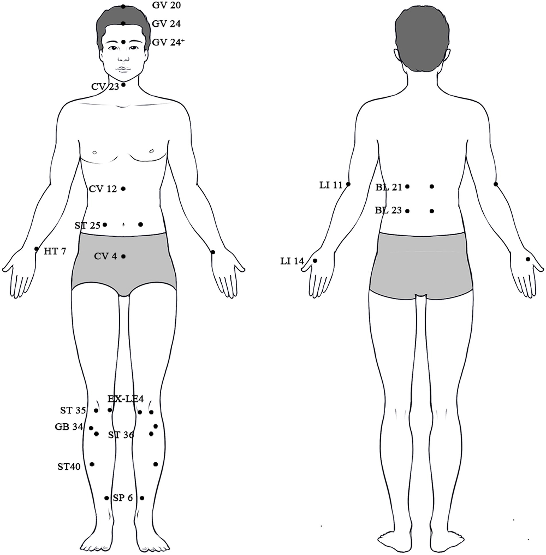
Figure 1. The locations of all the mentioned acupoints on the human body.
Research indicates that acupuncture is largely effective in treating motor symptoms of Parkinson’s disease (PD) by improving balance in the basal ganglia and thalamocortical circuits. Furthermore, this suggests the importance of identifying alternative pathways, such as the corticothalamic circuit (CTC), that can compensate for the decreased basal ganglia pathways in order to improve the effectiveness of acupuncture in treating Parkinson’s disease. Acupuncture has the ability to regulate the activity of dopaminergic neural circuits in specific areas of the brain, such as the prefrontal cortex, thalamus, and reward systems. This can be used as a treatment for substance addiction. Acupuncture in cognitive impairment therapy primarily affects the cholinergic neuronal pathways connecting the medial septum (MS) and diagonal band (DB) to the hippocampus. It also influences the glutamatergic neuronal circuitry within the hippocampus. The neurological pathways primarily engaged in acupuncture treatment for gastrointestinal illnesses include the brainstem, hypothalamus, and cerebellar circuitries, as well as the dorsal vagal complex route.
Overall, the aforementioned studies have utilized modern techniques such as neural tracing, chemogenetics/optogenetics, and functional brain imaging (fMRI), combined with behavioral testing, to demonstrate specific neural projections, regulate or precisely regulate specific cells within circuits, and depict neural signal responses before and after acupuncture in brain areas or nuclei, enhancing the comprehensiveness and objectivity of acupuncture pathway research. They have mainly explored the regulatory effects of acupuncture on specific neuronal projections within the central nervous system in the aforementioned diseases, with some studies also involving research on the peripheral sensory level transmission pathways. This aids in elucidating the central nervous system effects of acupuncture therapy, suggesting that acupuncture may exert its therapeutic effects by modulating neural circuit mechanisms that exhibit functional differences in various physiological and pathological states.
Neural pathways are crucial focal points for studying acupuncture effects; however, there are still some issues to address. Firstly, due to limitations in tracing technology and techniques like chemical genetics/optogenetics, research subjects are predominantly animal models, with varied standards for constructing these models. Clinical studies are relatively limited, with most having small sample sizes. It is hoped that future advancements in technology will facilitate large-sample, multicenter clinical studies to enhance the reliability of conclusions. Secondly, in experimental animals, electroacupuncture is usually the preferred method, and male mice are predominantly used as subjects. Does gender affect circuitry mechanism research? In clinical research, are there differences between needling on the left and right sides due to handedness? It is hoped that future research will strengthen the standardization of acupuncture, promote standardized acupoint selection and operation procedures, and further investigate these differences to enhance the objectivity of experimental results. Thirdly, central nervous system circuitry research currently predominates, with a limited number of diseases being studied. Therefore, it is hoped that future research will gradually expand the scope of disease research on acupuncture pathways, improve the comprehensive study of acupuncture effects on complete pathways from the periphery to the central nervous system, and then to target organs.
As the aforementioned concerns are gradually resolved, future research on the neural circuit mechanics of acupuncture is expected to become more thorough and comprehensive. Specifically, it is necessary to broaden the range of disease research and thoroughly investigate the acupuncture pathway mechanisms of beneficial diseases. This will enhance our comprehension of the neurobiological impacts of acupuncture therapy and direct clinical practice towards more efficient treatments.
XW: Writing – review & editing, Writing – original draft, Resources, Investigation, Conceptualization. JW: Writing – review & editing, Writing – original draft, Resources, Investigation, Conceptualization. RH: Writing – original draft, Resources, Investigation. CY: Writing – review & editing, Writing – original draft, Supervision, Resources, Funding acquisition, Conceptualization. FS: Writing – review & editing, Supervision, Resources, Funding acquisition, Conceptualization.
The author(s) declare that financial support was received for the research, authorship, and/or publication of this article. This work was funded by Shenzhen Science and Technology Program (No. JCYJ20210324111609024); National Natural Science Foundation of China (No. 82205271); Key Chinese Medicine Project supported by Hubei Provincial Natural Science Foundation (2023AFD113); Chinese Medicine Research Project supported by Hubei Administration of Traditional Chinese Medicine, China (No. ZY2023F138).
The authors declare that the research was conducted in the absence of any commercial or financial relationships that could be construed as a potential conflict of interest.
All claims expressed in this article are solely those of the authors and do not necessarily represent those of their affiliated organizations, or those of the publisher, the editors and the reviewers. Any product that may be evaluated in this article, or claim that may be made by its manufacturer, is not guaranteed or endorsed by the publisher.
1. Liu, B. Elaborating and revealing the effectiveness of acupuncture in TCM: with the curative effect as the guidance and focusing on outcome management. Zhongguo Zhen Jiu. (2022) 42:1.
2. Liu, S, Wang, Z-F, Su, Y-S, Ray, RS, Jing, X-H, Wang, Y-Q, et al. Somatotopic organization and intensity dependence in driving distinct NPY-expressing sympathetic pathways by electroacupuncture. Neuron. (2020) 108:436–450.e7. e7. doi: 10.1016/j.neuron.2020.07.015
3. Liu, S, Wang, Z, Su, Y, Qi, L, Yang, W, Fu, M, et al. A neuroanatomical basis for electroacupuncture to drive the vagal-adrenal axis. Nature. (2021) 598:641–5. doi: 10.1038/s41586-021-04001-4
4. Lyu, Z, Guo, Y, Gong, Y, Fan, W, Dou, B, Li, N, et al. The role of neuroglial crosstalk and synaptic plasticity-mediated central sensitization in acupuncture analgesia. Neural Plast. (2021) 2021:1–18. doi: 10.1155/2021/8881557
5. Cai, W, and Shen, W-D. Anti-apoptotic mechanisms of acupuncture in neurological diseases: a review. Am J Chin Med. (2018) 46:515–35. doi: 10.1142/S0192415X1850026X
6. Mercer Lindsay, N, Chen, C, Gilam, G, Mackey, S, and Scherrer, G. Brain circuits for pain and its treatment. Sci Transl Med. (2021) 13:7360. doi: 10.1126/scitranslmed.abj7360
7. Williams, LM. Precision psychiatry: a neural circuit taxonomy for depression and anxiety. Lancet Psychiatry. (2016) 3:472–80. doi: 10.1016/S2215-0366(15)00579-9
8. Cavallo, A, and Neumann, WJ. Dopaminergic reinforcement in the motor system: implications for Parkinson’s disease and deep brain stimulation. Eur J Neurosci. (2024) 59:457–72. doi: 10.1111/ejn.16222
9. Ersche, KD, Jones, PS, Williams, GB, Turton, AJ, Robbins, TW, and Bullmore, ET. Abnormal brain structure implicated in stimulant drug addiction. Science. (2012) 335:601–4. doi: 10.1126/science.1214463
10. Grieco, SF, Holmes, TC, and Xu, X. Probing neural circuit mechanisms in Alzheimer’s disease using novel technologies. Mol Psychiatry. (2023) 28:4407–20. doi: 10.1038/s41380-023-02018-x
11. Bouard, G, Bazille, C, Rochcongar, G, Audoual, R, Michon, J, and Bonhomme, J. Chronic painful lesion. Clin Infect Dis. (2020) 70:169–72. doi: 10.1093/cid/ciz279
12. Zhuo, M. Neural mechanisms underlying anxiety-chronic pain interactions. Trends Neurosci. (2016) 39:136–45. doi: 10.1016/j.tins.2016.01.006
13. Koechlin, H, Coakley, R, Schechter, N, Werner, C, and Kossowsky, J. The role of emotion regulation in chronic pain: a systematic literature review. J Psychosom Res. (2018) 107:38–45. doi: 10.1016/j.jpsychores.2018.02.002
14. Chen, T, Zhang, WW, Chu, Y-X, and Wang, Y-Q. Acupuncture for pain management: molecular mechanisms of action. Am J Chin Med. (2020) 48:793–811. doi: 10.1142/S0192415X20500408
15. Molsberger, AF, Schneider, T, Gotthardt, H, and Drabik, A. German randomized acupuncture trial for chronic shoulder pain (GRASP)—a pragmatic, controlled, patient-blinded, multi-centre trial in an outpatient care environment. Pain. (2010) 151:146–54. doi: 10.1016/j.pain.2010.06.036
16. Song, X, Zhu, Q, Su, L, Shi, L, Chi, H, Yan, Y, et al. New perspectives on migraine treatment: a review of the mechanisms and effects of complementary and alternative therapies. Front Neurol. (2024) 15:1372509. doi: 10.3389/fneur.2024.1372509
17. Zhou, X, Zhang, J, Jiang, L, Zhang, S, Gu, Y, Tang, J, et al. Therapeutic efficacy of acupuncture point stimulation for stomach cancer pain: a systematic review and meta-analysis. Front Neurol. (2024) 15:1334657. doi: 10.3389/fneur.2024.1334657
18. Wang, S-M, Kain, ZN, and White, P. Acupuncture analgesia: I. The scientific basis. Anesth Analg. (2008) 106:602–10. doi: 10.1213/01.ane.0000277493.42335.7b
19. Han, J-S. Acupuncture and endorphins. Neurosci Lett. (2004) 361:258–61. doi: 10.1016/j.neulet.2003.12.019
20. Martikainen, IK, Hagelberg, N, Jääskeläinen, SK, Hietala, J, and Pertovaara, A. Dopaminergic and serotonergic mechanisms in the modulation of pain: in vivo studies in human brain. Eur J Pharmacol. (2018) 834:337–45. doi: 10.1016/j.ejphar.2018.07.038
21. Yao, D, Chen, Y, and Chen, G. The role of pain modulation pathway and related brain regions in pain. Rev Neurosci. (2023) 34:899–914. doi: 10.1515/revneuro-2023-0037
22. Illes, P, Burnstock, G, and Tang, Y. Astroglia-derived ATP modulates CNS neuronal circuits. Trends Neurosci. (2019) 42:885–98. doi: 10.1016/j.tins.2019.09.006
23. Tang, Y, Yin, H-Y, Rubini, P, and Illes, P. Acupuncture-induced analgesia: a neurobiological basis in purinergic signaling. Neuroscientist. (2016) 22:563–78. doi: 10.1177/1073858416654453
24. Cui, X, Liu, K, Gao, X, and Zhu, B. Advancing the understanding of acupoint sensitization and plasticity through cutaneous C-nociceptors. Front Neurosci. (2022) 16:822436. doi: 10.3389/fnins.2022.822436
25. Li, W, Liu, J, Chen, A, Dai, D, Zhao, T, Liu, Q, et al. Shared nociceptive dorsal root ganglion neurons participating in acupoint sensitization. Front Mol Neurosci. (2022) 15:974007. doi: 10.3389/fnmol.2022.974007
26. Zhang, M, Guo, H, Ma, Y, Xu, F, Bai, F, Liang, S, et al. Acupoint sensitization is associated with increased excitability and hyperpolarization-activated current (Ih) in C-but not Aδ-type neurons. Neuroscience. (2019) 404:499–509. doi: 10.1016/j.neuroscience.2019.02.028
27. Zhu, B. The sensitization phenomenon of acupoint and biological significances. Zhongguo Zhen Jiu. (2019) 39:115–21. doi: 10.13703/j.0255-2930.2019.02.001
28. Neyama, H, Wu, Y, Nakaya, Y, Kato, S, Shimizu, T, Tahara, T, et al. (2023). Opioidergic activation of descending pain inhibitory system underlies placebo analgesia. bioRxiv. Available at: https://doi.org/10.1101/2023.06.26.546410. [Epub ahead of preprint]
29. Su, N, Cai, P, Dou, Z, Yin, X, Xu, H, He, J, et al. Brain nuclei and neural circuits in neuropathic pain and brain modulation mechanisms of acupuncture: a review on animal-based experimental research. Front Neurosci. (2023) 17:1243231. doi: 10.3389/fnins.2023.1243231
30. Zhang, M, Yang, Z, Zhong, J, Zhang, Y, Lin, X, Cai, H, et al. Thalamocortical mechanisms for nostalgia-induced analgesia. J Neurosci. (2022) 42:2963–72. doi: 10.1523/JNEUROSCI.2123-21.2022
31. Nakahama, H. Pain mechanisms in the central nervous system. Int Anesthesiol Clin. (1975) 13:109–48. doi: 10.1097/00004311-197513010-00006
32. Pan, S, Wang, S, Xue, X, Yuan, H, Li, J, Liu, Y, et al. Multidimensional pain modulation by acupuncture analgesia: the reward effect of acupuncture on pain relief. Evid Based Complement Alternat Med. (2022) 2022:1–9. doi: 10.1155/2022/3759181
33. Lai, H-C, Lin, Y-W, and Hsieh, C-L. Acupuncture-analgesia-mediated alleviation of central sensitization. Evid Based Complement Alternat Med. (2019) 2019:6173412. doi: 10.1155/2019/6173412
34. Ma, Y, Zhan, Y, Pei, J, Ye, G, Chen, Y, Zhu, W, et al. Involvement of 5-HT1A receptors of the thalamic descending pathway in the analgesic effect of intramuscular heating-needle stimulation in a rat model of lumbar disc herniation. Front Neurosci. (2023) 17:1222286. doi: 10.3389/fnins.2023.1222286
35. Yuan, X-C, Zhu, B, Jing, X-H, Xiong, L-Z, Wu, C-H, Gao, F, et al. Electroacupuncture potentiates cannabinoid receptor-mediated descending inhibitory control in a mouse model of knee osteoarthritis. Front Mol Neurosci. (2018) 11:112. doi: 10.3389/fnmol.2018.00112
36. Zhu, X, Xu, Y, Shen, Z, Zhang, H, Xiao, S, Zhu, Y, et al. Rostral anterior cingulate cortex-ventrolateral periaqueductal gray circuit underlies electroacupuncture to alleviate hyperalgesia but not anxiety-like behaviors in mice with spared nerve injury. Front Neurosci. (2022) 15:757628. doi: 10.3389/fnins.2021.757628
37. Journée, SH, Mathis, VP, Fillinger, C, Veinante, P, and Yalcin, I. Janus effect of the anterior cingulate cortex: pain and emotion. Neurosci Biobehav Rev. (2023) 153:105362. doi: 10.1016/j.neubiorev.2023.105362
38. Shen, Z, Zhang, H, Wu, Z, He, Q, Liu, J, He, X, et al. Electroacupuncture alleviates chronic pain-induced anxiety disorders by regulating the rACC-thalamus circuitry. Front Neurosci. (2021) 14:615395. doi: 10.3389/fnins.2020.615395
39. Wu, Z, Shen, Z, Xu, Y, Chen, S, Xiao, S, Ye, J, et al. A neural circuit associated with anxiety-like behaviors induced by chronic inflammatory pain and the anxiolytic effects of electroacupuncture. CNS Neurosci Ther. (2023) 30:e14520. doi: 10.1111/cns.14520
40. Xu, Y, Zhu, X, Chen, Y, Chen, Y, Zhu, Y, Xiao, S, et al. Electroacupuncture alleviates mechanical allodynia and anxiety-like behaviors induced by chronic neuropathic pain via regulating rostral anterior cingulate cortex-dorsal raphe nucleus neural circuit. CNS Neurosci Ther. (2023) 29:4043–58. doi: 10.1111/cns.14328
41. Hsiao, I-H, Yen, C-M, Hsu, H-C, Liao, H-Y, and Lin, Y-W. Chemogenetics modulation of electroacupuncture analgesia in mice spared nerve injury-induced neuropathic pain through TRPV1 signaling pathway. Int J Mol Sci. (2024) 25:1771. doi: 10.3390/ijms25031771
42. Vachon-Presseau, E. Effects of stress on the corticolimbic system: implications for chronic pain. Prog Neuro-Psychopharmacol Biol Psychiatry. (2018) 87:216–23. doi: 10.1016/j.pnpbp.2017.10.014
43. Ma, C, Zou, Y, Ye, Y, Cao, M, and Yan, X. Progress in the mechanism of acupuncture intervention on pain emotion and pain cognition mediated by limbic system. J Acupunct Tuina Sci. (2022) 20:499–504. doi: 10.1007/s11726-022-1351-3
44. Gu, H-W, Zhang, G-F, Liu, P-M, Pan, W-T, Tao, Y-X, Zhou, Z-Q, et al. Contribution of activating lateral hypothalamus-lateral habenula circuit to nerve trauma-induced neuropathic pain in mice. Neurobiol Dis. (2023) 182:106155. doi: 10.1016/j.nbd.2023.106155
45. Wang, D, Pan, X, Zhou, Y, Wu, Z, Ren, K, Liu, H, et al. Lateral septum-lateral hypothalamus circuit dysfunction in comorbid pain and anxiety. Mol Psychiatry. (2023) 28:1090–100. doi: 10.1038/s41380-022-01922-y
46. Benarroch, EE. Involvement of the nucleus accumbens and dopamine system in chronic pain. Neurology. (2016) 87:1720–6. doi: 10.1212/WNL.0000000000003243
47. Wang, C, Chen, M, Qin, C, Qu, X, Shen, X, and Liu, S. Lateral hypothalamic orexin neurons mediate the reward effects of pain relief induced by electroacupuncture. Front Mol Neurosci. (2022) 15:812035. doi: 10.3389/fnmol.2022.812035
48. Sung, YH, and Kang, SY. Pain in atypical parkinsonism, vascular parkinsonism, and Parkinson’s disease. Neurol Sci. (2022) 43:4797–802. doi: 10.1007/s10072-022-06035-6
49. Simon, DK, Tanner, CM, and Brundin, P. Parkinson disease epidemiology, pathology, genetics, and pathophysiology. Clin Geriatr Med. (2020) 36:1–12. doi: 10.1016/j.cger.2019.08.002
50. Yu, S-W, Lin, S-H, Tsai, C-C, Chaudhuri, KR, Huang, Y-C, Chen, Y-S, et al. Acupuncture effect and mechanism for treating pain in patients with Parkinson’s disease. Front Neurol. (2019) 10:1114. doi: 10.3389/fneur.2019.01114
51. Fan, J-Q, Lu, W-J, Tan, W-Q, Feng, W-C, and Zhuang, L-X. Acupuncture for Parkinson’s disease: from theory to practice. Biomed Pharmacother. (2022) 149:112907. doi: 10.1016/j.biopha.2022.112907
52. Zhao, Y, Zhang, Z, Qin, S, Fan, W, Li, W, Liu, J, et al. Acupuncture for Parkinson’s disease: efficacy evaluation and mechanisms in the dopaminergic neural circuit. Neural Plast. (2021) 2021:1–23. doi: 10.1155/2021/9926445
53. Tamtaji, OR, Naderi Taheri, M, Notghi, F, Alipoor, R, Bouzari, R, and Asemi, Z. The effects of acupuncture and electroacupuncture on Parkinson’s disease: current status and future perspectives for molecular mechanisms. J Cell Biochem. (2019) 120:12156–66. doi: 10.1002/jcb.28654
54. Moore, R, and Bloom, F. Central catecholamine neuron systems: anatomy and physiology of the dopamine systems. Annu Rev Neurosci. (1978) 1:129–69. doi: 10.1146/annurev.ne.01.030178.001021
55. Deuschl, G, Raethjen, J, Baron, R, Lindemann, M, Wilms, H, and Krack, P. The pathophysiology of parkinsonian tremor: a review. J Neurol. (2000) 247:V33–48. doi: 10.1007/PL00007781
56. McGregor, MM, and Nelson, AB. Circuit mechanisms of Parkinson’s disease. Neuron. (2019) 101:1042–56. doi: 10.1016/j.neuron.2019.03.004
57. Singh, A. Oscillatory activity in the cortico-basal ganglia-thalamic neural circuits in Parkinson’s disease. Eur J Neurosci. (2018) 48:2869–78. doi: 10.1111/ejn.13853
58. Dhond, RP, Kettner, N, and Napadow, V. Neuroimaging acupuncture effects in the human brain. J Altern Complement Med. (2007) 13:603–16. doi: 10.1089/acm.2007.7040
59. Chae, Y, Lee, H, Kim, H, Kim, CH, Chang, DI, Kim, KM, et al. Parsing brain activity associated with acupuncture treatment in Parkinson’s diseases. Mov Disord. (2009) 24:1794–802. doi: 10.1002/mds.22673
60. Yeo, S, Lim, S, Choe, IH, Choi, YG, Chung, KC, Jahng, GH, et al. Acupuncture stimulation on GB 34 activates neural responses associated with Parkinson’s disease. CNS Neurosci Ther. (2012) 18:781–90. doi: 10.1111/j.1755-5949.2012.00363.x
61. Yeo, S, Choe, I-H, van den Noort, M, Bosch, P, Jahng, G-H, Rosen, B, et al. Acupuncture on GB34 activates the precentral gyrus and prefrontal cortex in Parkinson’s disease. BMC Complement Altern Med. (2014) 14:1–9. doi: 10.1186/1472-6882-14-336
62. Li, Z, Chen, J, Cheng, J, Huang, S, Hu, Y, Wu, Y, et al. Acupuncture modulates the cerebello-thalamo-cortical circuit and cognitive brain regions in patients of Parkinson’s disease with tremor. Front Aging Neurosci. (2018) 10:206. doi: 10.3389/fnagi.2018.00206
63. Jia, J, Sun, Z, Li, B, Pan, Y, Wang, H, Wang, X, et al. Electro-acupuncture stimulation improves motor disorders in parkinsonian rats. Behav Brain Res. (2009) 205:214–8. doi: 10.1016/j.bbr.2009.06.024
64. Jia, J, Li, B, Sun, Z-L, Yu, F, Wang, X, and Wang, X-M. Electro-acupuncture stimulation acts on the basal ganglia output pathway to ameliorate motor impairment in parkinsonian model rats. Behav Neurosci. (2010) 124:305–10. doi: 10.1037/a0018931
65. Rehm, J, and Shield, KD. Global burden of disease and the impact of mental and addictive disorders. Curr Psychiatry Rep. (2019) 21:10. doi: 10.1007/s11920-019-0997-0
66. Kalin, NH. Substance use disorders and addiction: mechanisms, trends, and treatment implications. Am Psychiatric Assoc. (2020) 177:1015–8. doi: 10.1176/appi.ajp.2020.20091382
67. Goudriaan, A. Gambling disorder and other behavioral addictions: mechanisms, recognition and treatment. Eur Psychiatry. (2023) 66:S2–2. doi: 10.1192/j.eurpsy.2023.28
68. Lee, MY, Lee, BH, Kim, HY, and Yang, CH. Bidirectional role of acupuncture in the treatment of drug addiction. Neurosci Biobehav Rev. (2021) 126:382–97. doi: 10.1016/j.neubiorev.2021.04.004
69. Sun, L, and Wang, H. Acupuncture in the treatment of cocaine addiction: how does it work? Acupunct Med. (2024) 9645284241248473. doi: 10.1177/09645284241248473
70. Theodoratou, K. The effects of acupuncture on addictive behaviours. Eur J Integr Med. (2016) 8:35. doi: 10.1016/j.eujim.2016.08.084
71. Zheng, H, Zhai, T, Lin, X, Dong, G, Yang, Y, and Yuan, T-F. The resting-state brain activity signatures for addictive disorders. Med. (2024) 5:201–223.e6. doi: 10.1016/j.medj.2024.01.008
72. Watabe-Uchida, M, Zhu, L, Ogawa, SK, Vamanrao, A, and Uchida, N. Whole-brain mapping of direct inputs to midbrain dopamine neurons. Neuron. (2012) 74:858–73. doi: 10.1016/j.neuron.2012.03.017
73. LeGates, TA, Kvarta, MD, Tooley, JR, Francis, TC, Lobo, MK, Creed, MC, et al. Reward behaviour is regulated by the strength of hippocampus-nucleus accumbens synapses. Nature. (2018) 564:258–62. doi: 10.1038/s41586-018-0740-8
74. Chang, S, Ryu, Y, Fan, Y, Kim, NJ, and Lee, BH. Involvement of the cuneate nucleus in the acupuncture inhibition of drug-seeking behaviors. Front Neurosci. (2019) 13:436052. doi: 10.3389/fnins.2019.00928
75. Chang, S, Kim, DH, Jang, EY, Yoon, SS, Gwak, YS, Yi, YJ, et al. Acupuncture attenuates alcohol dependence through activation of endorphinergic input to the nucleus accumbens from the arcuate nucleus. Sci Adv. (2019) 5:1342. doi: 10.1126/sciadv.aax1342
76. Zhao, RJ, Yoon, SS, Lee, BH, Kwon, YK, Kim, KJ, Shim, I, et al. Acupuncture normalizes the release of accumbal dopamine during the withdrawal period and after the ethanol challenge in chronic ethanol-treated rats. Neurosci Lett. (2006) 395:28–32. doi: 10.1016/j.neulet.2005.10.043
77. Chang, S, Ryu, Y, Gwak, YS, Kim, NJ, Kim, JM, Lee, JY, et al. Spinal pathways involved in somatosensory inhibition of the psychomotor actions of cocaine. Sci Rep. (2017) 7:5359. doi: 10.1038/s41598-017-05681-7
78. West, R, and Gossop, M. Overview: a comparison of withdrawal symptoms from different drug classes. Addiction. (1994) 89:1483–9. doi: 10.1111/j.1360-0443.1994.tb03747.x
79. Benekareddy, M, Stachniak, TJ, Bruns, A, Knoflach, F, von Kienlin, M, Künnecke, B, et al. Identification of a corticohabenular circuit regulating socially directed behavior. Biol Psychiatry. (2018) 83:607–17. doi: 10.1016/j.biopsych.2017.10.032
80. Hu, H, Cui, Y, and Yang, Y. Circuits and functions of the lateral habenula in health and in disease. Nat Rev Neurosci. (2020) 21:277–95. doi: 10.1038/s41583-020-0292-4
81. Chang, S, Kim, HK, Ryu, Y, Jang, HB, Ahn, D, Lee, BH, et al. Mediation of mPFC-LHb pathway in acupuncture inhibition of cocaine psychomotor activity. Addict Biol. (2023) 28:e13321. doi: 10.1111/adb.13321
82. Wang, X, Zhang, B, Zhang, L, and Liu, S. Electroacupuncture suppresses morphine reward-seeking behavior: lateral hypothalamic orexin neurons implicated. Neurosci Lett. (2017) 661:84–9. doi: 10.1016/j.neulet.2017.09.057
83. Yoon, SS, Kwon, YK, Kim, MR, Shim, I, Kim, KJ, Lee, MH, et al. Acupuncture-mediated inhibition of ethanol-induced dopamine release in the rat nucleus accumbens through the GABAB receptor. Neurosci Lett. (2004) 369:234–8. doi: 10.1016/j.neulet.2004.07.095
84. Kim, MS, Fan, Y, Lee, SM, Chang, SC, Kim, HK, Ryu, Y, et al. Role of the central amygdala in acupuncture inhibition of methamphetamine-induced behaviors in rats. Addict Biol. (2021) 26:e12862. doi: 10.1111/adb.12862
85. Knopman, DS, Amieva, H, Petersen, RC, Chételat, G, Holtzman, DM, Hyman, BT, et al. Alzheimer disease. Nat Rev Dis Primers. (2021) 7:577–159033. doi: 10.1038/s41572-021-00269-y
86. Alzheimer’s Association. 2023 Alzheimer’s disease facts and figures. Alzheimers Dement. (2023) 19:1598–695. doi: 10.1002/alz.13016
87. Petersen, RC, and Negash, S. Mild cognitive impairment: an overview. CNS Spectr. (2008) 13:45–53. doi: 10.1017/S1092852900016151
88. Wu, L, Dong, Y, Zhu, C, and Chen, Y. Effect and mechanism of acupuncture on Alzheimer’s disease: a review. Front Aging Neurosci. (2023) 15:1035376. doi: 10.3389/fnagi.2023.1035376
89. Yu, C-C, Du, Y-J, Wang, S-Q, Liu, L-B, Shen, F, Wang, L, et al. Experimental evidence of the benefits of acupuncture for Alzheimer’s disease: an updated review. Front Neurosci. (2020) 14:549772. doi: 10.3389/fnins.2020.549772
90. Jeffery, KJ. The hippocampus: from memory, to map, to memory map. Trends Neurosci. (2018) 41:64–6. doi: 10.1016/j.tins.2017.12.004
91. Reichelt, AC, Kramar, CP, Ghosh-Swaby, OR, Sheppard, PA, Kent, BA, Bekinschtein, P, et al. The spontaneous location recognition task for assessing spatial pattern separation and memory across a delay in rats and mice. Nat Protoc. (2021) 16:5616–33. doi: 10.1038/s41596-021-00627-w
92. Protto, V, Soligo, M, De Stefano, ME, Farioli-Vecchioli, S, Marlier, LN, Nisticò, R, et al. Electroacupuncture in rats normalizes the diabetes-induced alterations in the septo-hippocampal cholinergic system. Hippocampus. (2019) 29:891–904. doi: 10.1002/hipo.23088
93. Zilliox, LA, Chadrasekaran, K, Kwan, JY, and Russell, JW. Diabetes and cognitive impairment. Curr Diab Rep. (2016) 16:87. doi: 10.1007/s11892-016-0775-x
94. Li, L, Li, J, Dai, Y, Yang, M, Liang, S, Wang, Z, et al. Electro-acupuncture improve the early pattern separation in Alzheimer’s disease mice via basal forebrain-hippocampus cholinergic neural circuit. Front Aging Neurosci. (2022) 13:770948. doi: 10.3389/fnagi.2021.770948
95. Xu, Y, Zhao, M, Han, Y, and Zhang, H. GABAergic inhibitory interneuron deficits in Alzheimer’s disease: implications for treatment. Front Neurosci. (2020) 14:536784. doi: 10.3389/fnins.2020.00660
96. Kim, M-J, Lee, S, and Kim, S-N. Effects of acupuncture on gastrointestinal diseases and its underlying mechanism: a literature review of animal studies. Front Med. (2023) 10:1167356. doi: 10.3389/fmed.2023.1167356
97. Rabitti, S, Giovanardi, CM, and Colussi, D. Acupuncture and related therapies for the treatment of gastrointestinal diseases. J Clin Gastroenterol. (2021) 55:207–17. doi: 10.1097/MCG.0000000000001455
98. Yang, J-W, Wang, L-Q, Zou, X, Yan, S-Y, Wang, Y, Zhao, J-J, et al. Effect of acupuncture for postprandial distress syndrome: a randomized clinical trial. Ann Intern Med. (2020) 172:777–85. doi: 10.7326/M19-2880
99. Wang, X, Shi, X, Lv, J, Zhang, J, Huo, Y, Zuo, G, et al. Acupuncture and related therapies for the anxiety and depression in irritable bowel syndrome with diarrhea (IBS-D): a network meta-analysis of randomized controlled trials. Front Psychiatry. (2022) 13:1067329. doi: 10.3389/fpsyt.2022.1067329
100. Wang, Y, Liu, Y, Zhou, K, Bauer, BA, Liu, B, Su, T, et al. The duration of acupuncture effects and its associated factors in chronic severe functional constipation: secondary analysis of a randomized controlled trial. Ther Adv Gastroenterol. (2019) 12:175628481988185. doi: 10.1177/1756284819881859
101. Ji, J, Huang, Y, Wang, X-F, Ma, Z, Wu, H-G, Im, H, et al. Review of clinical studies of the treatment of ulcerative colitis using acupuncture and moxibustion. Gastroenterol Res Pract. (2016) 2016:9248589–10. doi: 10.1155/2016/9248589
102. Takahashi, T. Mechanism of acupuncture on neuromodulation in the gut—a review. Neuromodulation. (2011) 14:8–12. doi: 10.1111/j.1525-1403.2010.00295.x
103. Filpa, V, Moro, E, Protasoni, M, Crema, F, Frigo, G, and Giaroni, C. Role of glutamatergic neurotransmission in the enteric nervous system and brain-gut axis in health and disease. Neuropharmacology. (2016) 111:14–33. doi: 10.1016/j.neuropharm.2016.08.024
104. McMenamin, CA, Travagli, RA, and Browning, KN. Inhibitory neurotransmission regulates vagal efferent activity and gastric motility. Exp Biol Med. (2016) 241:1343–50. doi: 10.1177/1535370216654228
105. Chen, S, Yong, C-y, Chen, H, Chu, X, Zhang, C, Tan, C, et al. Gastric distention-related neurons in dorsal nucleus of the vagus nerve of rats in response to different acupuncture acupoints. Chin J Tissue Eng Res. (2014) 18:5842. doi: 10.3969/j.issn.2095-4344.2014.36.018
106. Wang, H, Shen, GM, Liu, WJ, Huang, S, and Zhang, MT. The neural mechanism by which the dorsal vagal complex mediates the regulation of the gastric motility by Weishu (RN12) and Zhongwan (BL21) stimulation. Evid Based Complement Alternat Med. (2013) 2013:291764:1–7. doi: 10.1155/2013/291764
107. Wang, L, Shen, G, Wang, H, Hu, M, Yao, Y, and Ye, S. The effects of electroacupuncture at shu and mu points of stomach on gastric motility, the NMDA of vagus nerve dorsal nucleus and serum NO expression in functional dyspepsia rats. Chin Acupunct Moxibustion. (2018) 38:285–90. doi: 10.13703/j.0255-2930.2018.03.016
108. Junping, Z, Cheng, L, Zhuyuan, H, Jianan, LI, Hua, MA, Bo, J, et al. A preliminary study on the cerebellar function of acupuncturing the stomach Meridian foot-Yang Ming by fMRI. Chin Imaging J Integr Tradit West Med. (2018) 16:331–4. doi: 10.3969/j.issn.1672-0512.2018.04.001
109. Yu, Z, Wang, Y, Liang, C, and Xu, B. Effects of acupuncture at" Zusanli"(ST 36) on sensitive neurons of gastric distention in LHA-FN circuit in rats. Zhongguo Zhen Jiu. (2016) 36:851–6. doi: 10.13703/j.0255-2930.2016.08.018
110. Wang, H, Shen, G-M, Wang, X-Y, and Zhang, M-T. Involvement of GABAergic projection from central amygdaloid nucleus to paraventricular nucleus of hypothalamus in electroacupuncture induced regulation of gastric function in GAD2-Cre mice. Zhen Ci Yan Jiu. (2020) 45:351–6. doi: 10.13702/j.1000-0607.190898
111. Yao, L-L, Yuan, S, Wu, Z-N, Luo, J-Y, Tang, X-R, Tang, C-Z, et al. Contralateral S1 function is involved in electroacupuncture treatment-mediated recovery after focal unilateral M1 infarction. Neural Regen Res. (2022) 17:1310–7. doi: 10.4103/1673-5374.327355
112. Yuan, S, Deng, B, Ye, Q, Wu, Z, Wu, J, Wang, L, et al. Excitatory neurons in paraventricular hypothalamus contributed to the mechanism underlying acupuncture regulating the swallowing function. Sci Rep. (2022) 12:5797. doi: 10.1038/s41598-022-09470-9
113. Yao, L, Ye, Q, Liu, Y, Yao, S, Yuan, S, Xu, Q, et al. Electroacupuncture improves swallowing function in a post-stroke dysphagia mouse model by activating the motor cortex inputs to the nucleus tractus solitarii through the parabrachial nuclei. Nat Commun. (2023) 14:810. doi: 10.1038/s41467-023-36448-6
114. Ge, W-Q, Zhan-Mu, O-Y, Chen, C, Zhang, H, Wang, X-Y, Liu, X, et al. Electroacupuncture reduces chronic itch via cannabinoid CB1 receptors in the ventrolateral periaqueductal gray. Front Pharmacol. (2022) 13:931600. doi: 10.3389/fphar.2022.931600
115. Yeom, M, Ahn, S, Jang, SY, Jang, JH, Lee, Y, Hahm, DH, et al. Acupuncture attenuates comorbid anxiety-and depressive-like behaviors of atopic dermatitis through modulating neuroadaptation in the brain reward circuit in mice. Biol Res. (2022) 55:28. doi: 10.1186/s40659-022-00396-0
116. Xi, H, Wu, W, Qin, S, Wang, X, and Liu, C. Effects of electroacupuncture on the ventral tegmental area-nucleus accumbens dopamine pathway in rats with chronic sleep deprivation. Acupunct Med. (2023) 41:336–44. doi: 10.1177/09645284221146197
Keywords: acupuncture, neural circuit mechanisms, pain, Parkinson’s disease, addictive disorders, cognitive disorders, gastrointestinal disorders, review
Citation: Wang X, Wang J, Han R, Yu C and Shen F (2024) Neural circuit mechanisms of acupuncture effect: where are we now? Front. Neurol. 15:1399925. doi: 10.3389/fneur.2024.1399925
Edited by:
Shuai Liu, Hunan Normal University, ChinaReviewed by:
Peter Illes, Leipzig University, GermanyCopyright © 2024 Wang, Wang, Han, Yu and Shen. This is an open-access article distributed under the terms of the Creative Commons Attribution License (CC BY). The use, distribution or reproduction in other forums is permitted, provided the original author(s) and the copyright owner(s) are credited and that the original publication in this journal is cited, in accordance with accepted academic practice. No use, distribution or reproduction is permitted which does not comply with these terms.
*Correspondence: Chaochao Yu, a2l0ZXJ1bm5lcjJAMTYzLmNvbQ==; Feng Shen, NDAwNTI5NThAcXEuY29t
†These authors have contributed equally to this work
Disclaimer: All claims expressed in this article are solely those of the authors and do not necessarily represent those of their affiliated organizations, or those of the publisher, the editors and the reviewers. Any product that may be evaluated in this article or claim that may be made by its manufacturer is not guaranteed or endorsed by the publisher.
Research integrity at Frontiers
Learn more about the work of our research integrity team to safeguard the quality of each article we publish.