 Charles L. Francoeur
 François Lauzier1,2,3
 Patrice Brassard
- 1Population Health and Optimal Health Practices Research Unit (Trauma—Emergency—Critical Care Medicine), Centre Hospitalier Universitaire (CHU) de Québec—Université Laval Research Centre, Université Laval, Québec City, QC, Canada
- 2Department of Anesthesiology and Critical Care, CHU de Québec—Université Laval, Critical Care Division, Québec City, QC, Canada
- 3Critical Care Medicine Service, CHU de Québec—Université Laval, Québec City, QC, Canada
- 4Department of Kinesiology, Faculty of Medicine, Université Laval, Québec City, QC, Canada
- 5Research Center of the Institut Universitaire de Cardiologie et de Pneumologie de Québec, Québec City, QC, Canada
Delayed cerebral ischemia (DCI) disproportionately affects poor grade aneurysmal subarachnoid hemorrhage (aSAH) patients. An unreliable neurological exam and the lack of appropriate monitoring leads to unrecognized DCI, which in turn is associated with severe long-term deficits and higher mortality. Near Infrared Spectroscopy (NIRS) offers simple, continuous, real time, non-invasive cerebral monitoring. It provides regional cerebral oxygen saturation (c-rSO2), which reflects the balance between cerebral oxygen consumption and supply. Reports have demonstrated a good correlation with other cerebral oxygen and blood flow monitoring, and credible cerebrovascular reactivity indices were also derived from NIRS signals. Multiple critical c-rSO2 values have been reported in aSAH patients, based on various thresholds, duration, variation from baseline or cerebrovascular reactivity indices. Some were associated with vasospasm, some with DCI and others with clinical outcomes. However, the poor grade aSAH population has not been specifically studied and no randomized clinical trial has been published. The available literature does not support a specific NIRS-based intervention threshold to guide diagnostic or treatment in aSAH patients. We review herein the fundamental basic concepts behind NIRS technology, relationship of c-rSO2 to other brain monitoring values and their potential clinical interpretation. We follow with a critical evaluation of the use of NIRS in the aSAH population, more specifically its ability to diagnose vasospasm, to predict DCI and its association to outcome. In summary, NIRS might offer significant potential for poor grade aSAH in the future. However, current evidence does not support its use in clinical decision-making, and proper technology evaluation is required.
Introduction
For patients surviving the initial injury of an aneurysmal subarachnoid hemorrhage (aSAH), delayed cerebral ischemia (DCI) deteriorating into cerebral infarction represents the main threat to a favorable outcome (1). Patients with poor grade aSAH [IV and V on the World Federation of Neurosurgeons (WFNS) scale (2)] are the most at-risk (3). They often present an altered level of consciousness, diminishing the reliability of the neurological exam and jeopardizing the application of current consensus DCI definition (4). Compared with clinically apparent DCI, these unrecognized ischemic episodes (5) are associated with worst long-term deficits and higher mortality (6). Transcranial Doppler ultrasonography (TCD), serial vascular or perfusion imaging, electroencephalographic (EEG) monitoring and invasive multimodality monitoring have all been called upon to mitigate these limitations in our ability to detect ischemia. Unfortunately, the evidence supporting their use has been disappointing so far and none has established itself as a reliable modality to circumvent the loss of clinical exam in poor grade aSAH (7).
Near Infrared Spectroscopy (NIRS) technology has been advocated as a solution to this conundrum. It is a simple, portable, continuous, real time, non-invasive monitoring of cerebral oximetry. It should therefore be ideal for timely detection of ischemia and to improve management in poor grade aSAH. The technology has been around for more than 40 years, after it was introduced in 1977 by Jöbsis (8), and subsequently popularized 20 years later by Kirkpatrick (9). Since then, multiple NIRS systems came along, algorithms have improved, a substantial corpus of literature—albeit contradictory and of variable quality—has emerged, and NIRS technology has gained sufficient popularity to be incorporated as a standard of care in many operating theaters and critical care units around the world. However, the quality of evidence supporting it use remains low, and parameters that should influence management, if any, are still unknown.
In this concise review, we lay out the basic concepts required for non-experts to comprehend the technology, we provide a critical appraisal of the possible interpretations and use of the data and its potential impact on poor grade aSAH patients. Closing remarks will review ongoing studies and give some future directions.
Basic Concepts
The first basic concepts concern optical physics and light attenuation. A NIRS device emits light from a light emitting diode (LED) or a laser in the near infrared range, usually between 700 and 850 nm, and collects what reaches the photodetectors. The loss of light between emitter and detectors represent optical attenuation and is a consequence of two phenomena affecting photon trajectory: absorption and scattering. The absorption is proportional to the concentration of chromophores, which are substances that absorb the light of a specific frequency. In our context, the chromophores of interest are oxyhemoglobin (HbO2) and deoxyhemoglobin (HHb). The differential absorption profile should therefore allow the determination of HbO2 and HHb content in the region of interest as described by the Beer-Lambert law. There are however competing chromophores and manufacturers must select wavelengths where the absorption spectra of HbO2 and HHb are maximally separated and where the overlap with water and melatonin (10) absorption is minimal. The phenomenon of scattering is the other critical variable and the major contributor to attenuation. As light travels through biological tissue, the initial trajectory of the photon is lost, and it is deviated to another direction. The consequences are that some light never reaches the detector, while some reaches it only after being scattered multiple times, traveling a greater distance than the one separating the source and the detector. Deflection is so significant that the path of tissue-reflected photons in the adult head is parabolic rather than in a straight line, explaining why the light source and detectors are placed on adjacent areas of the head. Disentangling the effect of absorption and scattering is one of the crucial roles of the various proprietary algorithms embedded in NIRS devices. They rely on many assumptions, including a fixed arterial: venous ratio (usually 30:70 or 25:75) and a constant scattering, both of which do not reflect what is happening in vivo. For example, brain edema creates large shifts in intracranial photon scattering that may profoundly alter readings in an unpredictable manner.
Commercially available oximeters rely on continuous wave data acquisition and spatially resolved spectroscopy (11). The latter is based on the use of multiple detectors, the number of which varies amongst models. As the depth of tissue interrogated is proportional to the distance between emitter and detector, it is assumed that the closest detector receives light that has passed through the scalp, whereas that arriving at the farthest detector has passed through brain tissue. Those distances vary between manufacturers. Multiple detectors therefore allow a gain in spatial resolution and help to mitigate extracranial contamination. This is a considerable weakness of NIRS technology and estimates for the degree of contamination from extracranial tissues range from 7 to 35% (12–14) and vary amongst manufacturers. The biggest impact of spatially resolved spectroscopy, however, is to derive a scaled absolute hemoglobin concentration by combining measures of those closely spaced detectors. That is, the absolute HbO2 and HHb contents are unknown, but their relative proportion can be calculated. Percentage of HbO2 over total hemoglobin thus provides the cerebral regional oxygen saturation (c-rSO2), which is variably coined and abbreviated by manufacturers (rSO2, TOI, TCCO, StcO2).
Other concepts are about understanding what is being monitored. The probes are normally placed on each side of the forehead, 3 cm above the superciliary line to avoid the frontal sinuses. Consistent positioning is required to minimize variations and obtain reproducible results. As the hemoglobin in larger vessels traps all incident infrared light, surface-detected infrared reflections arise from blood vessels that are <1 mm in diameter (15) and ~1.5 cm under the skin. The presence of intracranial extravascular blood may influence readings because of this infrared photon sequestration. Contrary to pulse oximetry (SpO2), NIRS does not require actively flowing and pulsatile flow. It measures a weighted average of arterial, capillary, and venous compartments. C-rSO2 thus reflects primarily the small venous compartment of 1.5 cm3 of gray matter brain tissue in the territory between the anterior and middle cerebral arteries (16), with some degree of extracranial contamination. True reference values do not exist. Some widely available systems, such as INVOS™, benefit from a high volume of published data allowing the conclusion that for healthy and preoperative adult patients, normal baseline probably ranges between 60 and 75% (17–19). Reports comparing technologies (20, 21) show us that although different NIRS devices provide similar trends, they give different baseline estimates, rendering comparisons difficult. Potential baseline values for aSAH patients are reported in Table 1 (27).
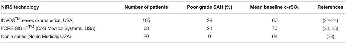
Table 1. Baseline regional cerebral oxygen saturation (c-rSO2) in aSAH patients.
Physiological Data—Oxygenation, Blood Flow, Cerebrovascular Reactivity
C-rSO2 is assumed to reflect the balance between cerebral oxygen consumption and supply in the region of interest and therefore to essentially be determined by cerebral metabolic rate of oxygen (CMRO2) and oxygen delivery. A pathological decline in c-rSO2 might therefore signal arterial hypotension, low cardiac output, systemic hypoxia, hyperventilation, stroke or impending DCI. Conversely, high c-rSO2 might be secondary to hyperemia, acidosis, hypoglycemia, high levels of sedation or hypothermia. Convulsions might present as low, high or oscillating c-rSO2 (28). How does NIRS-determined c-rSO2 compared to other brain oxygen monitoring technologies to detect meaningful events? A few reports evaluated its performance against invasive brain tissue oxygen monitoring (PtiO2) and jugular bulb venous oxygenation (SjvO2). The comparisons are obviously limited, as PtiO2 reflects the partial pressure of dissolved oxygen in interstitial white matter, while SjvO2 is the global venous saturation of drained hemispheric blood. Overall, the correlation between NIRS and PtiO2 is good. They reflect similar dynamic changes of cerebral oxygen metabolism (29–32), but they vary in their degree and speed of response (33). The sensitivity of NIRS to detect cerebral ischemia, here defined as a PtiO2 of <15 mmHg, seems problematic (30), including in the SAH population (34). The same conclusions apply when comparing SjvO2 with NIRS. There is a good correlation between modalities (33, 35) and a somewhat lower sensitivity of NIRS to detect significant desaturation (36). Data suggest that NIRS, PtiO2 and SjvO2 assess different processes that are intimately related, but that crude substitution is not warranted (37).
Several investigators also observed an association between c-rSO2 and cerebral blood flow (CBF). Fluctuations in c-rSO2 correlate with CBF measurements made by xenon enhanced computed tomography (38, 39) and computed tomography perfusion imaging (40). Ventilation-based CBF manipulations were also used to show the correlation between c-rSO2 and CBF as measured with an invasive thermodilution probe (22). Finally, cardiac output augmentation with either dobutamine (41) or milrinone (25) in DCI patients also resulted in a better c-rSO2.
More recently, various teams used the information obtained from NIRS monitoring to evaluate cerebrovascular reactivity (42, 43). The reactivity index, variously named tissue oxygenation index (TOx) or cerebral oxygenation index (COx), is calculated as a rolling correlation coefficient between averaged CPP (or MAP) values and the corresponding NIRS signals. It has been validated against pressure reactivity index (PRx) (44) and TCD-derived reactivity index (Mx) (45). It does, however, rely on assumptions that have been regularly challenged (46). Other approaches to evaluate cerebrovascular reactivity have been described, such as those based on frequency-domain analysis, but they have not been applied to our population of interest (47).
If we accept that abnormal c-rSO2 reflects a pathological alteration in brain oxygenation, how should it be managed? Most clinicians would suggest investigating plausible and reversible causes as a first step. Correcting obvious systemic physiological derangements such as significant hypoxemia, arterial hypotension, or iatrogenic hyperventilation is also sensible. However, the appropriate management of abnormal c-rSO2 in aSAH patients, beyond what was just mentioned, is unclear. The impact of vasopressors, for example, is controversial. Studies have reported a decrease in c-rSO2 with vasopressor infusion both in aSAH (48) and healthy patients (49), whereas it improved c-rSO2 values in the first few hours post-cardiac arrest (50). Studies reporting the effect of red blood cell transfusions in neurocritical care patients, a minority of which were aSAH, have also yielded conflicting results (51–53). Controlled hypercapnia, in the range of 50–60 mmHg, had some success to improve c-rSO2 in two studies involving aSAH patients (22, 54). This approach, still investigational, is deemed temporary to avoid rebound vasoconstriction. Hyperoxia, targeting supraphysiological levels of arterial partial pressure of oxygen, seems to improve c-rSO2 (55), but is also potentially associated with a higher incidence of vasospasm (56), DCI and poor outcome in aSAH (57). It would therefore be inappropriate, based on currently available data, to recommend any specific intervention to attempt correction of abnormal c-rSO2 in aSAH patients.
Clinical Data—Vasospasm, DCI, Prognosis
The ultimate objective behind adequate monitoring of cerebral oxygenation, perfusion and cerebrovascular reactivity is early detection of secondary brain injury and management guidance to improve patient outcomes. Vasospasm detection, DCI prediction, and prognostication have been specifically evaluated in the aSAH population. Comparisons between TCD and commercially available NIRS seem to support a moderate degree of correlation between the two modalities (58). A decrease of more than 12% in c-rSO2 from the baseline was associated with a better predictive value than same-side TCD using the traditional threshold value of 200 cm/s to detect severe vasospasm on CT angiography (59). More interestingly, the same 12% cut-off yielded a sensitivity of 94% (95% CI: 73–99%) and a specificity of 71% (95% CI: 53–85%) to detect DCI (59). Another study on 24 patients presenting with poor grade aSAH reported a sensitivity of 86% (95% CI: 67–98%) and specificity of 86% (95% CI: 67–96%) for DCI detection using a greater than 15% decrease in c-rSO2 (60).
Others took a different approach and evaluated the association between impaired cerebrovascular reactivity and DCI. Using an index of |R| ≥ 0.5 on either side as a definition for impaired cerebrovascular reactivity, and the consensus definition for DCI, one group reported odds of DCI of 36 (95% CI: 6–211%) when impaired cerebrovascular reactivity was present (23). Another investigator used TOx and found that impaired cerebrovascular reactivity on the side contralateral to the aneurysm was significantly more frequent in the DCI group than in the non-DCI group (58 vs. 16%, p = 0.014) and was associated with an OR of DCI of 19 (95% CI: 1.2–320) (24). Using a threshold of 0.07 conferred TOx a sensitivity of 58% and a specificity of 91% to predict DCI.
Only three studies have examined the relationship between c-rSO2 and outcome in aSAH patients. In one study, 163 aSAH patients were monitored between day 5 and 10 after aneurysm rupture (26). Using the definition of cerebral desaturation of <50% for 30 min on either side, and poor outcomes as a mRS of 4–6, it was observed that cerebral desaturation was independently associated with poor functional outcomes at 3 months (OR 2.72, 95%CI 1.02–7.20) but not at 12 months. In a small study of 38 patients using a definition of cerebral desaturation of <60% for at least 30 min on either side, patients with an unfavorable outcome spent more time with a cerebral desaturation than those with a good outcome (5 h 43 vs. 1 h 47, p = 0.02) (61). Patients with episodes of cerebral desaturation lasting more than 2 h were at much higher risk of poor short-term outcomes than those without [OR 15.4 (95% CI: 1.1–214.2%)]. Another small study of 31 patients evaluated the association between NIRS-based cerebrovascular reactivity indices and optimal blood pressure with functional outcomes at 3 months and defined unfavorable outcome as ≥ 3 on the mRS scale (44). In this study, preserved cerebrovascular reactivity, as defined by a negative or near-zero TOx was associated with good functional outcomes at 90 days (OR, 2.5; 95% CI, 1.3–4.8), including after adjustment for age, WFNS and DCI. Using NIRS-derived optimal blood pressures, %time outside the limits of autoregulation was significantly associated with poor 90-day outcomes (OR, 1.9; 95% CI, 1.3–2.9) and deviation from NIRS-derived autoregulatory limits predicted poor 90-day outcomes with high sensitivity (0.82; 95% CI, 0.67–0.98) and specificity (0.88; 95% CI, 0.76–1.00) (34). The studies before mentioned are observational. No NIRS-based intervention study has been conducted in SAH patients with long term functional outcome as a primary outcome. There is no data to support specific NIRS-based management recommendations to improve outcome in this population.
Discussion and Perspective
Despite widespread availability for more than two decades and obvious user-friendly characteristics, the role of NIRS in aSAH management is still ill-defined. The technology possesses obvious advantages: simple, safe, non-invasive, continuous. Unfortunately, limitations are significant. One is the important differences amongst devices: types and amounts of light sources, wavelengths used, the distance between the emitters and detectors, the number of detectors, arterial:venous ratio assumptions, underlying proprietary algorithms, reported normal baseline values, degree of extracranial contamination. All these affect data accuracy and interpretation, complexifies the adoption of conclusions obtained from one device to another and is akin to work with uncalibrated instruments. Technical problems, although rarely reported, should also be considered. In some studies, up to 20% of the readings were non-valid (34, 62). Competition for space on the forehead for electroencephalography and lack of long-term adherence of probes because of sweat are also potentially common (62). Frontal hematoma, wounds on the forehead and deficient NIRS signal are sometimes used as exclusion criteria (30) and bifrontal decompressive craniectomies prevents usual NIRS placement and interpretation (53). This review only covered commonly used NIRS technologies at the bedside, but other devices rely on time domain (63) and frequency domain spectroscopy (64) rather than continuous wave spectroscopy. Some also employ hybrid technologies such as ultrasound tagged near-infrared spectroscopy (65) or diffuse correlation spectroscopy and diffuse optical spectroscopy (66) to provide relative estimates of CBF changes. However, promising these technological advances might sound, none was proven superior for clinical applications in aSAH.
The lack of a normal range of c-rSO2 for neurocritical care patients, especially aSAH, is troublesome. The lack of consensus regarding a lower limit of NIRS-derived c-rSO2 values, serving as an intervention threshold, is even more problematic. Various thresholds, duration, variations from the baseline, asymmetries, cerebrovascular reactivity indices and ischemic burdens have been described. None has established itself as a significant physiological or clinical marker that should modify management and the authors would be hard-pressed to suggest a specific one. The overall low quality of the clinical research is important to highlight. Published data on the use of NIRS in aSAH since 1998 are all but one unblinded, uncontrolled, single center observational studies, a third of which included fewer than 15 patients. Consensus definition of DCI and cerebral infarction are used in less than a third of the studies, and 90 days outcome are reported in <25%. Poor-grade aSAH, the population most susceptible to benefit from such a monitoring, is vastly underrepresented.
Conducting research on DCI in poor grade aSAH is difficult. DCI is a complex, elusive and evolving entity lacking a gold standard in patients without a proper neurological exam. No evidence-based treatment exists. Our understanding of brain physiology, including oxygenation, perfusion and autoregulation, is incomplete and the impact of a monitoring strategy on clinical decision-making is more complex than simply assessing accuracy. Nonetheless, NIRS technology should be submitted to rigorous evaluation and assessment. We suggest that clinical investigators focus on pragmatic, bedside-applicable hypotheses rather than exploratory ones. The heterogeneity of “ischemic” indices already hinders clinical research tremendously. The population of poor grade aSAH should be targeted. Observational studies are to be prospective and blinded, and intervention studies should be properly conducted, pragmatic and multicenter randomized clinical trials. Outcomes should include cerebral infarction on MRI as a surrogate for DCI and 90 days or more functional and quality-of-life outcomes using a validated scale, aligned with published common data elements (67). As no gold standard exists for monitoring poor grade aSAH, comparison with other monitoring tools seems futile. At least two upcoming trials might help to shade some light on the use of NIRS in aSAH patients. One is the NeurO2 study, a prospective, blinded, multicenter observational study that will recruit close to 300 TBI and aSAH patients, monitoring them with NIRS and evaluating the outcome at 6 months using the Glasgow Outcome Scale extended and EQ-5D-5L (68). The other one is an interventional, multicenter, single-blinded, randomized clinical trial aiming to enroll 150 aSAH patients to evaluate NIRS-directed optimal cerebral perfusion pressure on Glasgow outcome scale at 6 months (69).
Improving outcomes in aSAH patients is intrinsically associated with earlier detection and treatment of DCI, preventing evolution toward cerebral infarction and the associated sequelae. Poor grade aSAH are at high risk of DCI, their clinical examination is suboptimal, and monitoring alternatives are limited. NIRS technology profile is promising, but current evidence does not support its use to guide management in this population. High-quality research is urgently needed.
Author Contributions
CF contributed by reviewing the current literature and writing the manuscript. FL, PB, and AT contributed by reviewing the manuscript and making substantive intellectual contributions. All authors read and approved the manuscript.
Conflict of Interest
The authors declare that the research was conducted in the absence of any commercial or financial relationships that could be construed as a potential conflict of interest.
Publisher's Note
All claims expressed in this article are solely those of the authors and do not necessarily represent those of their affiliated organizations, or those of the publisher, the editors and the reviewers. Any product that may be evaluated in this article, or claim that may be made by its manufacturer, is not guaranteed or endorsed by the publisher.
References
1. Rosengart AJ, Schultheiss KE, Tolentino J, Macdonald RL. Prognostic factors for outcome in patients with aneurysmal subarachnoid hemorrhage. Stroke. (2007) 38:2315–21. doi: 10.1161/STROKEAHA.107.484360
2. Teasdale GM, Drake CG, Hunt W, Kassell N, Sano K, Perat B, et al. A universal subarachnoid hemorrhage scale: report of a committee of the World Federation of Neurosurgical Societies. J Neurol Neurosurg Psychiatry. (1988) 51:1457. doi: 10.1136/jnnp.51.11.1457
3. Crobeddu E, Mittal MK, Dupont S, Wijdicks EFM, Lanzino G, Rabinstein AA. Predicting the lack of development of delayed cerebral ischemia after aneurysmal subarachnoid hemorrhage. Stroke. (2012) 43:697–701. doi: 10.1161/STROKEAHA.111.638403
4. Vergouwen MDI, Vermeulen M, van Gijn J, Rinkel GJE, Wijdicks EF, Muizelaar JP, et al. Definition of delayed cerebral ischemia after aneurysmal subarachnoid hemorrhage as an outcome event in clinical trials and observational studies: proposal of a multidisciplinary research group. Stroke. (2010) 41:2391–5. doi: 10.1161/STROKEAHA.110.589275
5. Shimoda M, Takeuchi M, Tominaga J, Oda S, Kumasaka A, Tsugane R, et al. Asymptomatic versus symptomatic infarcts from vasospasm in patients with subarachnoid hemorrhage: serial magnetic resonance imaging. Neurosurgery. (2001) 49:1341–50. doi: 10.1097/00006123-200112000-00010
6. Schmidt JM, Wartenberg K, Fernandez A, Claassen J, Rincon F, Ostapkovich NO, et al. Frequency and clinical impact of asymptomatic cerebral infarction due to vasospasm after subarachnoid hemorrhage. J Neurosurg. (2008) 109:1052–9. doi: 10.3171/JNS.2008.109.12.1052
7. le Roux P, Menon DK, Citerio G, Vespa P, Bader MK, Brophy GM, et al. Consensus summary statement of the International Multidisciplinary Consensus Conference on Multimodality Monitoring in Neurocritical Care: a statement for healthcare professionals from the Neurocritical Care Society and the European Society of Intensive. Neurocritical Care. (2014) 21:S1–S26. doi: 10.1007/s12028-014-0041-5
8. Jöbsis FF. Noninvasive, infrared monitoring of cerebral and myocardial oxygen sufficiency and circulatory parameters. Science. (1977) 198:1264–7. doi: 10.1126/science.929199
9. Kirkpatrick PJ. Use of near-infrared spectroscopy in the adult. Philos Trans R Soc B Biol Sci. (1997) 352:701–5. doi: 10.1098/rstb.1997.0052
10. Sun X, Ellis J, Corso PJ, Hill PC, Chen F, Lindsay J. Skin pigmentation interferes with the clinical measurement of regional cerebral oxygen saturation. Br J Anaesth. (2015) 114:276–80. doi: 10.1093/bja/aeu335
11. Lange F, Tachtsidis I. Clinical brain monitoring with time domain NIRS: a review and future perspectives. Appl Sci. (2019) 9:1612. doi: 10.3390/app9081612
12. Davie SN, Grocott HP. Impact of extracranial contamination on regional cerebral oxygen saturation. Anesthesiology. (2012) 116:834–40. doi: 10.1097/ALN.0b013e31824c00d7
13. Sørensen H, Rasmussen P, Siebenmann C, Zaar M, Hvidtfeldt M, Ogoh S, et al. Extra-cerebral oxygenation influence on near-infrared-spectroscopy-determined frontal lobe oxygenation in healthy volunteers: a comparison between INVOS-4100 and NIRO-200NX. Clin Physiol Funct Imaging. (2015) 35:177–84. doi: 10.1111/cpf.12142
14. Kato S, Yoshitani K, Kubota Y, Inatomi Y, Ohnishi Y. Effect of posture and extracranial contamination on results of cerebral oximetry by near-infrared spectroscopy. J Anesth. (2017) 31:103–10. doi: 10.1007/s00540-016-2275-1
15. Ferrari M, Mottola L, Quaresima V. Principles, techniques, and limitations of near infrared spectroscopy. Can J Appl Physiol. (2004) 29:463–87. doi: 10.1139/h04-031
16. Edmonds H. Detection and Correction of Brain Oxygen Imbalance. Surgical and Critical Care Applications of the INVOS Cerebral/Somatic Oximeter. Boulder, CO: Medtronics (2018).
17. Baikoussis NG, Karanikolas M, Siminelakis S, Matsagas M, Papadopoulos G. Baseline cerebral oximetry values in cardiac and vascular surgery patients: a prospective observational study. J Cardiothorac Surg. (2010) 5:1–7. doi: 10.1186/1749-8090-5-41
18. Heringlake M, Garbers C, Med C, Kä J-H, Anderson I, Heinze H, et al. Preoperative cerebral oxygen saturation and clinical outcomes in cardiac surgery. Anesthesiology. (2010) 114:58–68. doi: 10.1097/ALN.0b013e3181fef34e
19. Chan MJ, Chung T, Glassford NJ, Bellomo R. Near-infrared spectroscopy in adult cardiac surgery patients: a systematic review and meta-analysis. J Cardiothorac Vasc Anesth. (2017) 31:1155–65. doi: 10.1053/j.jvca.2017.02.187
20. Owojuyigbe AM, Adenekan AT, Kawamae K, Aaron O. Cerebral oximetry in healthy adults: a comparison of three commercial Near-Infrared Spectrophotometers. Middle East J Anesthesiol. (2021) 28:37–44.
21. Gagnon RE, Macnab AJ, Gagnon FA, Blackstock D, Leblanc JG, Re G, et al. Comparison of two spatially resolved NIRS oxygenation indices. J Clin Monit Comput. (2002) 17:385–91. doi: 10.1023/A:1026274124837
22. Westermaier T, Stetter C, Kunze E, Willner N, Holzmeier J, Kilgenstein C, et al. Controlled transient hypercapnia: a novel approach for the treatment of delayed cerebral ischemia after subarachnoid hemorrhage? J Neurosurg. (2014) 121:1056–62. doi: 10.3171/2014.7.JNS132611
23. Liu G, Guo Z, Sun X, Chai WN Qi L, Li H, et al. Monitoring of the effect of cerebral autoregulation on delayed cerebral ischemia in patients with aneurysmal subarachnoid hemorrhage. World Neurosurg. (2018) 118:e269–75. doi: 10.1016/j.wneu.2018.06.170
24. Uryga A, Czyz M, Adamik B, Tabakow P, Kasprowicz M, Burzyńska M. Serum biomarkers and cerebral autoregulation as early warnings of delayed cerebral ischemia risk in patients after aneurysmal subarachnoid haemorrhage. J Clin Neurosci. (2021) 87:35–43. doi: 10.1016/j.jocn.2021.02.009
25. Ghanem MA, Shabana AM. Effects of Milrinone continuous intravenous infusion on global cerebral oxygenation and cerebral vasospasm after cerebral aneurysm surgical clipping. Egypt J Anaesthesia. (2014) 30:73–82. doi: 10.1016/j.egja.2013.07.006
26. Yousef KM, Balzer JR, Crago EA, Poloyac SM, Sherwood PR, Jo KYE. Transcranial regional cerebral oxygen desaturation predicts delayed cerebral ischaemia and poor outcomes after subarachnoid haemorrhage: a correlational study. Intensive Crit Care Nurs. (2014) 30:346–52. doi: 10.1016/j.iccn.2014.05.001
27. Bensaidane MR, Turgeon AF, Lauzier F, English SW, Leblanc G, Francoeur CL. Neuromonitoring with near-infrared spectroscopy (NIRS) in aneurysmal subarachnoid haemorrhage: a systematic review protocol. BMJ Open. (2020) 10:e043300. doi: 10.1136/bmjopen-2020-043300
28. Sokol DK, Markand ON, Daly EC, Luerssen TG, Malkoff MD. Near infrared spectroscopy (NIRS) distinguishes seizure types. Seizure. (2000) 9:323–7. doi: 10.1053/seiz.2000.0406
29. Brawanski A, Faltermeier R, Rothoerl RD, Woertgen C. Comparison of near-infrared spectroscopy and tissue PO2 time series in patients after severe head injury and aneurysmal subarachnoid hemorrhage. J Cereb Blood Flow Metab. (2002) 22:605–11. doi: 10.1097/00004647-200205000-00012
30. Leal-Noval SR, Cayuela A. Invasive and noninvasive assessment of cerebral oxygenation in patients with severe traumatic brain injury. Intensive Care Med. (2010) 36:1283–5. doi: 10.1007/s00134-010-1921-6
31. Rothoerl RD, Faltermeier R, Burger R, Woertgen C, Brawanski A. Dynamic correlation between tissue PO2 and near infrared spectroscopy. Acta Neurochirurg Suppl. (2002) 81:311–3. doi: 10.1007/978-3-7091-6738-0_79
32. Holzschuh M, Woertgen C, Metz C, Brawanski A. Dynamic changes of cerebral oxygenation measured by brain tissue oxygen pressure and near infrared spectroscopy. Neurol Res. (1997) 19:246–8. doi: 10.1080/01616412.1997.11740807
33. McLeod AD, Igielman F, Elwell C, Cope M, Smith M. Measuring cerebral oxygenation during normobaric hyperoxia: a comparison of tissue microprobes, near-infrared spectroscopy, and jugular venous oximetry in head injury. Anesth Analg. (2003) 97:851–6. doi: 10.1213/01.ANE.0000072541.57132.BA
34. Kerz T, Beyer C, Huthmann A, Kalasauskas D, Amr AN, Boor S, et al. Continuous-wave near-infrared spectroscopy is not related to brain tissue oxygen tension. J Clin Monit Comput. (2016) 30:641–7. doi: 10.1007/s10877-015-9755-y
35. Rosenthal G, Furmanov A, Itshayek E, Shoshan Y, Singh V. Assessment of a noninvasive cerebral oxygenation monitor in patients with severe traumatic brain injury: clinical article. J Neurosurg JNS. (2014) 120:901–7. doi: 10.3171/2013.12.JNS131089
36. Jeong H, Jeong S, Lim HJ, Lee J, Yoo KY. Cerebral oxygen saturation measured by near-infrared spectroscopy and jugular venous bulb oxygen saturation during arthroscopic shoulder surgery in beach chair position under sevoflurane-nitrous oxide or propofol-remifentanil anesthesia. Anesthesiology. (2012) 116:1047–56. doi: 10.1097/ALN.0b013e31825154d2
37. Forcione M, Ganau M, Prisco L, Chiarelli AM, Bellelli A, Belli A, et al. Mismatch between tissue partial oxygen pressure and near-infrared spectroscopy neuromonitoring of tissue respiration in acute brain trauma: the rationale for implementing a multimodal monitoring strategy. Int J Mol Sci. (2021) 22:112. doi: 10.3390/ijms22031122
38. Kerr M, Nemoto EM, Edwin M, Yonas H, Kassam A. Cerebral oximetry by near-infrared spectroscopy (NIRS) as an early indicator of delayed cerebral ischemia (DCI) following subarachnoid hemorrhage (SAH). Crit Care Med. (1999) 27:78–80. doi: 10.1097/00003246-199912001-00132
39. Kim MN, Durduran T, Frangos S, Edlow BL, Buckley EM, Moss HE, et al. Noninvasive measurement of cerebral blood flow and blood oxygenation using near-infrared and diffuse correlation spectroscopies in critically brain-injured adults. Neurocrit Care. (2010) 12:173–80. doi: 10.1007/s12028-009-9305-x
40. Taussky P, O'Neal B, Daugherty WP, Luke S, Thorpe D, Pooley RA, et al. Validation of frontal near-infrared spectroscopy as noninvasive bedside monitoring for regional cerebral blood flow in brain-injured patients. Neurosurg Focus. (2012) 32:1–6. doi: 10.3171/2011.12.FOCUS11280
41. Mutoh T, Ishikawa T, Suzuki A, Yasui N. Continuous cardiac output and near-infrared spectroscopy monitoring to assist in management of symptomatic cerebral vasospasm after subarachnoid hemorrhage. Neurocrit Care. (2010) 13:331–8. doi: 10.1007/s12028-010-9383-9
42. Steiner LA, Pfister D, Strebel SP, Radolovich D, Smielewski P, Czosnyka M. Near-infrared spectroscopy can monitor dynamic cerebral autoregulation in adults. Neurocrit Care. (2009) 10:122–8. doi: 10.1007/s12028-008-9140-5
43. de Hert S, Moerman A. Recent advances in cerebral oximetry. Assessment of cerebral autoregulation with near-infrared spectroscopy: myth or reality? F1000Research. (2017) 6:1615. doi: 10.12688/f1000research.11351.1
44. Silverman A, Kodali S, Strander S, Gilmore EJ, Kimmel A, Wang A, et al. Deviation from personalized blood pressure targets is associated with worse outcome after subarachnoid hemorrhage. Stroke. (2019) 50:2729–37. doi: 10.1161/STROKEAHA.119.026282
45. Zweifel C, Castellani G, Czosnyka M, Carrera E, Brady KM, Kirkpatrick PJ, et al. Continuous assessment of cerebral autoregulation with near-infrared spectroscopy in adults after subarachnoid hemorrhage. Stroke. (2010) 41:1963–8. doi: 10.1161/STROKEAHA.109.577320
46. Brassard P, Labrecque L, Smirl JD, Tymko MM, Caldwell HG, Hoiland RL, et al. Losing the dogmatic view of cerebral autoregulation. Physiol Rep. (2021) 9:e14982. doi: 10.14814/phy2.14982
47. Andersen AV, Simonsen SA, Schytz HW, Iversen HK. Assessing low-frequency oscillations in cerebrovascular diseases and related conditions with near-infrared spectroscopy: a plausible method for evaluating cerebral autoregulation? Neurophotonics. (2018) 5:1. doi: 10.1117/1.NPh.5.3.030901
48. Yousef KM, Crago E, Chang Y, Lagattuta TF, Mahmoud K, Shutter L, et al. Vasopressor infusion after subarachnoid hemorrhage does not increase regional cerebral tissue oxygenation. J Neurosci Nurs. (2018) 50:225–30. doi: 10.1097/JNN.0000000000000382
49. Brassard P, Seifert T, Secher NH. Is cerebral oxygenation negatively affected by infusion of norepinephrine in healthy subjects? Br J Anaesth. (2009) 102:800–5. doi: 10.1093/bja/aep065
50. Ameloot K, de Deyne C, Eertmans W, Ferdinande B, Dupont M, Palmers P-J, et al. Early goal-directed haemodynamic optimization of cerebral oxygenation in comatose survivors after cardiac arrest: the Neuroprotect post-cardiac arrest trial. Eur Heart J. (2019) 40:1804–14. doi: 10.1093/eurheartj/ehz120
51. Muthuchellappan R, Shaikh NA, Surve RM, Ganne URS, Philip M. Regional cerebral tissue oxygen saturation changes following blood transfusion in neuro-intensive care unit patients – a pilot observational study. Trans Med. (2018) 28:304–9. doi: 10.1111/tme.12504
52. Leal-Noval SR, Muñoz-Gómez M, Casado-Méndez M, Murillo-Cabezas F, Rincón-Ferrari MD, Amaya-Villar R, et al. Red blood cell transfusion guided by near infrared spectroscopy in neurocritically ill patients with moderate or severe anemia: a randomized, controlled trial. J Neurotrauma. (2017) 34:2553–9. doi: 10.1089/neu.2016.4794
53. McCredie VA, Piva S, Santos M, Xiong W, de Oliveira Manoel AL, Rigamonti A, et al. The impact of red blood cell transfusion on cerebral tissue oxygen saturation in severe traumatic brain injury. Neurocrit Care. (2017) 26:247–55. doi: 10.1007/s12028-016-0310-6
54. Stetter C, Weidner F, Lilla N, Weiland J, Kunze E, Ernestus RI, et al. Therapeutic hypercapnia for prevention of secondary ischemia after severe subarachnoid hemorrhage: physiological responses to continuous hypercapnia. Sci Rep. (2021) 11:11715. doi: 10.1038/s41598-021-91007-7
55. Jakkula P, Reinikainen M, Hästbacka J, Loisa P, Tiainen M, Pettilä V, et al. Targeting two different levels of both arterial carbon dioxide and arterial oxygen after cardiac arrest and resuscitation: a randomised pilot trial. Intensive Care Med. (2018) 44:2112–21. doi: 10.1007/s00134-018-5453-9
56. Reynolds R, Amin SS, Jonathan V, Tang AR, Lan M, Wang C, et al. Hyperoxia and delayed cerebral vasospasm in aneurysmal subarachnoid hemorrhage. Neurosurgery. (2020) 67:98. doi: 10.1007/s12028-020-01136-6
57. Jeon S-B, Choi HA, Badjatia N. Hyperoxia may be related to delayed cerebral ischemia and poor outcome after subarachnoid haemorrhage. J Neurol Neurosurg Psychiatry. (2014) 85:1301–7. doi: 10.1136/jnnp-2013-307314
58. Ekelund A, Kongstad P, Säveland H, Romner B, Reinstrup P, Kristiansson KA, et al. Transcranial cerebral oximetry related to transcranial Doppler after aneurysmal subarachnoid haemorrhage. Acta Neurochir. (1998) 140:1029–36. doi: 10.1007/s007010050211
59. Park JJ, Kim C, Jeon JP. Monitoring of delayed cerebral ischemia in patients with subarachnoid hemorrhage via near-infrared spectroscopy. J Clin Med. (2020) 9:1595. doi: 10.3390/jcm9051595
60. Park JJ, Kim Y, Chai CL, Jeon JP. Application of near-infrared spectroscopy for the detection of delayed cerebral ischemia in poor-grade subarachnoid hemorrhage. Neurocrit Care. (2021) 35:767–74. doi: 10.1007/s12028-021-01223-2
61. Burzyńska M, Uryga A, Kasprowicz M, Czosnyka M, Dragan B, Kübler A. The relationship between the time of cerebral desaturation episodes and outcome in aneurysmal subarachnoid haemorrhage: a preliminary study. J Clin Monit Comput. (2020) 34:705–714. doi: 10.1007/s10877-019-00377-x
62. Maslehaty H, Krause-Titz U, Petridis AK, Barth H, Mehdorn HM. Continuous measurement of cerebral oxygenation with near-infrared spectroscopy after spontaneous subarachnoid hemorrhage. ISRN Neurol. (2012) 2012:1–7. doi: 10.5402/2012/907187
63. Yokose N, Sakatani K, Murata Y, Awano T, Igarashi T, Nakamura S, et al. Bedside monitoring of cerebral blood oxygenation and hemodynamics after aneurysmal subarachnoid hemorrhage by quantitative time-resolved near-infrared spectroscopy. World Neurosurg. (2010) 73:508–13. doi: 10.1016/j.wneu.2010.02.061
64. Calderon-Arnulphi M, Alaraj A, Amin-Hanjani S, Mantulin WW, Polzonetti CM, Gratton E, et al. Detection of cerebral ischemia in neurovascular surgery using quantitative frequency-domain near-infrared spectroscopy. J Neurosurg. (2007) 106:283–90. doi: 10.3171/jns.2007.106.2.283
65. Schytz HW, Guo S, Jensen LT, Kamar M, Nini A, Gress DR, et al. A new technology for detecting cerebral blood flow: a comparative study of ultrasound tagged NIRS and 133Xe-SPECT. Neurocrit Care. (2012) 17:139–45. doi: 10.1007/s12028-012-9720-2
66. Busch DR, Balu R, Baker WB, Guo W, He L, Diop M, et al. Detection of brain hypoxia based on noninvasive optical monitoring of cerebral blood flow with diffuse correlation spectroscopy. Neurocritical Care. (2019) 30:72–80. doi: 10.1007/s12028-018-0573-1
67. Suarez JI, Sheikh MK, Macdonald RL, Amin-Hanjani S, Brown RD, Leonardo De Oliveira Manoel A, et al. Common data elements for unruptured intracranial aneurysms and subarachnoid hemorrhage clinical research: a national institute for neurological disorders and stroke and national library of medicine project on behalf of the unruptured intracranial aneurysms and SAH CDE Project Investigators. Neurocrit Care. (2028) 30:4–19. doi: 10.1007/s12028-019-00723-6
68. ClinicalTrials.gov [Internet]. Optimal Brain Oxygenation in Neurologic Intensive Care Unit: The NeurO2 Study. Identifier NCT04935866. Bethesda, MD: National Library of Medicine (US) (2021). Available online at: https://clinicaltrials.gov/ct2/show/NCT04935866?term=04935866&draw=2&rank=1 (accessed March 23, 2022).
69. ClinicalTrials.gov [Internet]. NIRS Directed Optimal Cerebral Perfusion Pressure on the Outcome of Aneurysmal Subarachnoid Hemorrhage Patients. Identifier NCT05003232. Bethesda, MD: National Library of Medicine (US) (2021). Available online at: https://clinicaltrials.gov/ct2/show/NCT05003232?term=05003232&draw=2&rank=1 (accessed March 23, 2022).
Keywords: near infrared spectroscopy, delayed cerebral ischemia, subarachnoid hemorrhage, vasospasm, poor grade aneurysmal SAH, neuromonitoring
Citation: Francoeur CL, Lauzier F, Brassard P and Turgeon AF (2022) Near Infrared Spectroscopy for Poor Grade Aneurysmal Subarachnoid Hemorrhage—A Concise Review. Front. Neurol. 13:874393. doi: 10.3389/fneur.2022.874393
Received: 12 February 2022; Accepted: 14 March 2022;
Published: 18 April 2022.
Edited by:
Miriam Weiss, University Hospital RWTH Aachen, GermanyReviewed by:
Markus Skrifvars, University of Helsinki, FinlandGiulio Bicciato, University Hospital Zürich, Switzerland
Copyright © 2022 Francoeur, Lauzier, Brassard and Turgeon. This is an open-access article distributed under the terms of the Creative Commons Attribution License (CC BY). The use, distribution or reproduction in other forums is permitted, provided the original author(s) and the copyright owner(s) are credited and that the original publication in this journal is cited, in accordance with accepted academic practice. No use, distribution or reproduction is permitted which does not comply with these terms.
*Correspondence: Charles L. Francoeur, Y2hhcmxlcy1sYW5naXMuZnJhbmNvZXVyLm1lZEBzc3NzLmdvdXYucWMuY2E=