95% of researchers rate our articles as excellent or good
Learn more about the work of our research integrity team to safeguard the quality of each article we publish.
Find out more
CORRECTION article
Front. Neuroanat. , 05 January 2022
Volume 15 - 2021 | https://doi.org/10.3389/fnana.2021.835799
This article is a correction to:
Perforating Arteries of the Lemniscal Trigone: A Microsurgical Neuroanatomic Description
 Santino Ottavio Tomasi1,2,3*
 Giuseppe Emmanuele Umana4
 Gianluca Scalia5
 Roberto Luis Rubio-Rodriguez6,7,8
 Giuseppe Raudino9
 Julian Rechberger2
 Philipp Geiger2
 Bipin Chaurasia10
 Kaan Yagmurlu11
 Michael T. Lawton12†
 Peter A. Winkler1,2,3†
A Corrigendum on
Perforating Arteries of the Lemniscal Trigone: A Microsurgical Neuroanatomic Description
by Tomasi, S. O., Umana, G. E., Scalia, G., Rubio-Rodriguez, R. L., Raudino, G., Rechberger, J., Geiger, P., Chaurasia, B., Yagmurlu, K., Lawton, M. T., and Winkler, P. A. (2021). Front. Neuroanat. 15:675313. doi: 10.3389/fnana.2021.675313
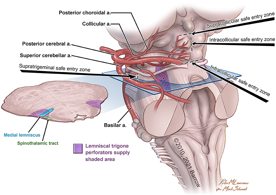
In the original article, there was a mistake in Figure 1 as published. The word trigone (in purple type) was misspelled (“trigon”). The corrected Figure 1 appears below.

Figure 1. Anatomical illustration of the lemniscal trigone zone. a., artery. Used with permission from Barrow Neurological Institute, Phoenix, Arizona.
The authors apologize for this error and state that this does not change the scientific conclusions of the article in any way. The original article has been updated.
All claims expressed in this article are solely those of the authors and do not necessarily represent those of their affiliated organizations, or those of the publisher, the editors and the reviewers. Any product that may be evaluated in this article, or claim that may be made by its manufacturer, is not guaranteed or endorsed by the publisher.
Keywords: lemniscal trigone, dorsolateral midbrain perforating zone, microsurgical anatomy, arterial capillary network, perforating arteries, anatomical variability
Citation: Tomasi SO, Umana GE, Scalia G, Rubio-Rodriguez RL, Raudino G, Rechberger J, Geiger P, Chaurasia B, Yagmurlu K, Lawton MT and Winkler PA (2022) Corrigendum: Perforating Arteries of the Lemniscal Trigone: A Microsurgical Neuroanatomic Description. Front. Neuroanat. 15:835799. doi: 10.3389/fnana.2021.835799
Received: 14 December 2021; Accepted: 15 December 2021;
Published: 05 January 2022.
Approved by:
Frontiers Editorial Office, Frontiers Media SA, SwitzerlandCopyright © 2022 Tomasi, Umana, Scalia, Rubio-Rodriguez, Raudino, Rechberger, Geiger, Chaurasia, Yagmurlu, Lawton and Winkler. This is an open-access article distributed under the terms of the Creative Commons Attribution License (CC BY). The use, distribution or reproduction in other forums is permitted, provided the original author(s) and the copyright owner(s) are credited and that the original publication in this journal is cited, in accordance with accepted academic practice. No use, distribution or reproduction is permitted which does not comply with these terms.
*Correspondence: Santino Ottavio Tomasi, dG9tYXNpLmJyYWluYW5kc3BpbmVzdXJnZXJ5QGdtYWlsLmNvbQ==; cy50b21hc2lAc2Fsay5hdA==
†These authors have contributed equally to this work and share senior authorship
Disclaimer: All claims expressed in this article are solely those of the authors and do not necessarily represent those of their affiliated organizations, or those of the publisher, the editors and the reviewers. Any product that may be evaluated in this article or claim that may be made by its manufacturer is not guaranteed or endorsed by the publisher.
Research integrity at Frontiers

Learn more about the work of our research integrity team to safeguard the quality of each article we publish.