 Roberta Haddad-Tóvolli1
 Fabian A. Paul2
 Yuanfeng Zhang1
 Xunlei Zhou1
 Thomas Theil3
 Luis Puelles4*
 Sandra Blaess2*
 Gonzalo Alvarez-Bolado1*
- 1Department of Neuroanatomy, University of Heidelberg, Heidelberg, Germany
- 2Laboratory of Neurodevelopmental Genetics, Life and Brain Center, Institute of Reconstructive Neurobiology, University of Bonn, Bonn, Germany
- 3Centre for Integrative Physiology, University of Edinburgh, Edinburgh, UK
- 4Department of Morphology, University of Murcia and Instituto Murciano de Investigación Biosanitaria, Murcia, Murcia, Spain
A commentary on
Differential requirements for Gli2 and Gli3 in the regional specification of the mouse hypothalamus
by Haddad-Tóvolli, R., Paul, F. A., Zhang, Y., Zhou, X., Theil, T., Puelles, L., et al. (2015). Front. Neuroanat. 9:34. doi: 10.3389/fnana.2015.00034
By mistake, Figure 2 of the article by Haddad-Tóvolli et al. (2015) showed in panels (A) and (B) the same image of Gli1 expression in E8.5 wildtype mouse embryos. It should have shown Gli1 expression in (A) and Gli2 expression in (B). Therefore, we provide a corrected Figure 2, now with panel (B) showing Gli2 expression, as we originally intended and as the Figure legend indicates. This is a minor change not affecting the scientific content of the article.
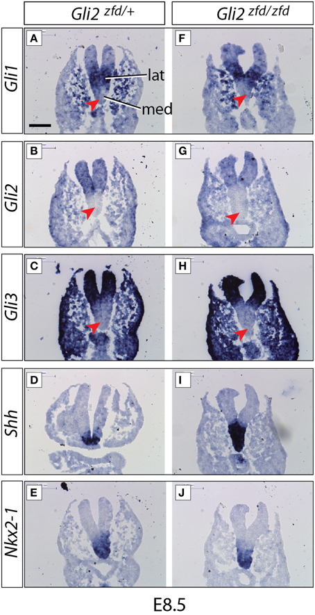
Figure 2. Expression of Gli genes in the presumptive hypothalamus at E8.5. In situ detection of marker gene expression in Gli2zfd/+ and Gli2zfd/zfd mutant E8.5 embryos as indicated. “lat” and “med” in (A) indicate progenitor domains. Red arrowheads in (A–C) and (F–H) indicate lack of expression in the medial progenitor domain. Nkx2-1 expression (E,J) identifies the presumptive hypothalamus. Scale bar (in A) 100 μm.
Conflict of Interest Statement
The authors declare that the research was conducted in the absence of any commercial or financial relationships that could be construed as a potential conflict of interest.
Keywords: embryo, Gli1, Gli2, Gli3, hypothalamus, mouse, mutant, Shh
Citation: Haddad-Tóvolli R, Paul FA, Zhang Y, Zhou X, Theil T, Puelles L, Blaess S and Alvarez-Bolado G (2015) Corrigendum: Differential requirements for Gli2 and Gli3 in the regional specification of the mouse hypothalamus. Front. Neuroanat. 9:58. doi: 10.3389/fnana.2015.00058
Received: 17 April 2015; Accepted: 24 April 2015;
Published: 13 May 2015.
Edited and reviewed by: Agustín González, Universidad Complutense de Madrid, Spain
Copyright © 2015 Haddad-Tóvolli, Paul, Zhang, Zhou, Theil, Puelles, Blaess and Alvarez-Bolado. This is an open-access article distributed under the terms of the Creative Commons Attribution License (CC BY). The use, distribution or reproduction in other forums is permitted, provided the original author(s) or licensor are credited and that the original publication in this journal is cited, in accordance with accepted academic practice. No use, distribution or reproduction is permitted which does not comply with these terms.
*Correspondence: Sandra Blaess,c2FuZHJhLmJsYWVzc0B1bmktYm9ubi5kZQ==;
Luis Puelles,cHVlbGxlc0B1bS5lcw==;
Gonzalo Alvarez-Bolado,YWx2YXJlekBhbmEudW5pLWhlaWRlbGJlcmcuZGU=