 Amanpreet K. Sidhu
 Sanskruthi B. Agrawal
 Naveen Verma
 Priya Kaushal
 Madhvi Sharma
94% of researchers rate our articles as excellent or good
Learn more about the work of our research integrity team to safeguard the quality of each article we publish.
Find out more
REVIEW article
Front. Nanotechnol., 14 April 2025
Sec. Nanomaterials
Volume 7 - 2025 | https://doi.org/10.3389/fnano.2025.1549713
This article is part of the Research TopicMyconanotechnology for SustainabilityView all articles
Multimetallic nanoparticles (MMNPs) produced by fungus-mediated synthesis have attracted a lot of interest as an environmentally friendly and sustainable nanotechnology method. Fungi are effective bio-factories that create complex nanoparticles with special qualities by using their metabolic and enzymatic capabilities. When compared to their monometallic counterparts, MMNPs—which are composed of combinations of two or more metals—offer synergistic benefits such increased catalytic activity, higher stability, and superior biocompatibility. In addition to highlighting the structural diversity of MMNPs, such as core-shell, alloy, and Janus configurations, this review investigates the mechanisms underpinning fungal-mediated synthesis, including enzymatic reduction and stabilisation pathways. Additionally covered are characterisation methods for examining functionality, morphology, and composition. The potential applications of MMNPs synthesized by fungi in biomedicine, environmental remediation, biosensing, and catalysis are highlighted in the article. This green synthesis method, which makes use of the natural benefits of fungus and multimetallic systems, responds to the increasing need for sustainable nanomaterials and opens the door to novel uses in both the scientific and industrial fields.
Nanotechnology has emerged as a revolutionary force, poised to transform industries and tackle some of the most pressing global challenges. As an interdisciplinary field, it harnesses the unique properties of materials at the nanoscale, offering unprecedented potential across various sectors, including medicine, environmental remediation, biosensors, and agriculture (Yang et al., 2019; Tao et al., 2023; Sidhu et al., 2022; Verma et al., 2024; Gaber et al., 2024). Researchers worldwide are dedicating significant time and resources to exploring the vast applications of nanotechnology, recognizing its potential to drive innovation and create sustainable solutions. From enhancing drug delivery systems to enabling eco-friendly materials and revolutionizing diagnostics, nanotechnology is rapidly becoming a cornerstone of modern science. As human civilization faces an array of complex challenges—ranging from climate change and resource scarcity to emerging diseases and technological gaps—the application of nanotechnology presents an innovative and transformative path forward. This dynamic field holds the promise of addressing critical issues, paving the way for a future where science and technology work in harmony to improve quality of life and sustainability at a global scale (Verma et al., 2023).
While most studies focus on the delivery and application of single metallic nanoparticles (MNPs), recent advancements have explored the combinatorial potential of multi-metallic nanoparticles (MMNPs), nanocomposites (NCs), and synergistic blends of mono-metallic nanoparticles. Multi-metallic nanoparticles are emerging as highly functional materials that integrate two or more metals, forming alloys with enhanced properties, such as superior catalytic activity, optical tunability, and high stability. The ability to modify their chemical composition, morphology, and structure enables MMNPs to achieve maximum synergistic performance (Zhang et al., 2017).
The synthesis of MMNPs can be achieved through various methods, including physical, chemical, and biological routes. Among these, biological synthesis especially using fungi, presents a highly promising and underexplored avenue. Fungi offer several advantages for nanoparticle synthesis due to their ability to secrete a wide range of bioactive metabolites, including enzymes, proteins, and secondary metabolites. These biomolecules act as natural reducing, capping, and stabilizing agents, facilitating the environmentally friendly and cost-effective synthesis of nanoparticles (Madigan et al., 1997; Zhao et al., 2024). Furthermore, fungal synthesis aligns with the principles of green chemistry by eliminating the need for toxic chemicals, high energy inputs, or harsh physical conditions. This energy-efficient and sustainable approach minimizes environmental impact while maximizing scalability. Fungi are also easy to cultivate on inexpensive substrates, enabling large-scale biomass production and, consequently, the synthesis of MMNPs in an economical manner (Honary et al., 2013). Their inherent ability to act as biological factories for nanoparticle synthesis eliminates the need for external reducing agents, making the process even more eco-friendly (Yadav et al., 2015; Sidhu and Kaushal, 2023; Hashem et al., 2023). Despite these advantages, the potential of fungi in synthesizing MMNPs remains relatively underexplored compared to other natural systems, such as plants and bacteria.
This review provides a comprehensive discussion on the fungal synthesis of MMNPs, emphasizing the mechanisms of synthesis, their characterization, and their diverse applications across medicine, antimicrobial agents, drug delivery systems, and bioimaging agents, biosensors, agriculture, and environmental sustainability. By delving into the unique capabilities of fungi in nanoparticle synthesis, this work aims to highlight the untapped potential of fungi-mediated approaches for the production of MMNPs and their transformative impact on multiple sectors.
Fungi have emerged as highly effective agents for the biogenic synthesis of nanoparticles due to their distinct biological features and considerable benefits over alternative systems. Their ability to resist high metal concentrations, combined with the development of numerous extracellular proteins, ensures that nanoparticles remain stable during synthesis. Many fungi are resistant to metal toxicity by active and incidental mechanisms, making them ideal candidates for metal immobilisation and mineral dissolution (Dhillon et al., 2012; Sidhu and Kaushal, 2023). Fungi also create a variety of reducing agents, including enzymes and proteins, which aid in nanoparticle production. These characteristics, together with their quick growth and simple biomass processing, make fungi an efficient and feasible option for large-scale nanoparticle manufacturing (Fouda et al., 2020; Rai et al., 2021). Furthermore, fungi produce more biomass and reduce the need for sophisticated extraction techniques, giving them a considerable advantage over bacterial systems (Hashem et al., 2023). The structural features of fungi make them ideal for nanoparticle production. Their filamentous mycelial network, which has a high surface area-to-mass ratio, is rich in metal-binding functional groups in the cell wall and extracellular polymeric substances (EPS), making it an effective template for nanoparticle nucleation. Moreover, the hydrated mucilaginous coating promotes geochemical processes, while branching hyphae provide several locations for nanoparticle synthesis, highlighting fungi’s versatility and effectiveness as green nanotechnology agents (Gadd, 2010; Gadd and Raven, 2010; Li et al., 2022). The environmentally benign methodologies demonstrate fungi’s enormous potential for large-scale nanoparticle manufacturing. Their distinct biological and structural properties make them critical for furthering green nanotechnology.
Fungi use complex metabolic processes to synthesise metallic nanoparticles, taking advantage of their special features. Fungi can synthesize nanoparticles via both intracellular and extracellular methods, using their metabolic pathways to convert metal ions into nanoparticles (Figure 1).
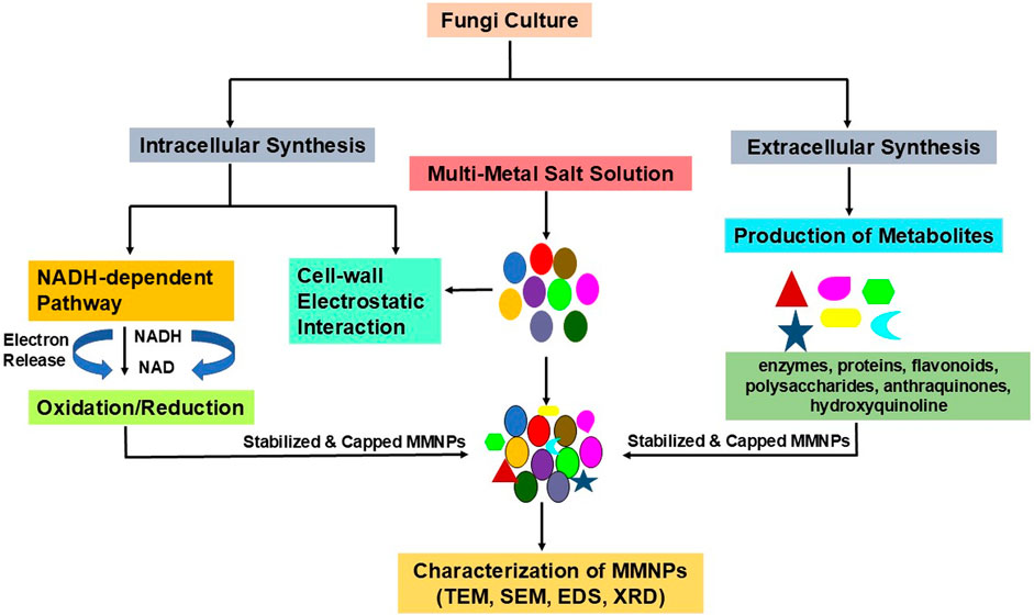
Figure 1. A schematic illustration of fungal-mediated synthesis of multimetallic nanoparticles (MMNPs) via external and intracellular pathways. Secreted fungal enzymes, proteins, and metabolites act as reducing, stabilising, and capping agents in the extracellular environment, allowing MMNP production to occur more easily. The intracellular approach involves the bioaccumulation of metal ions within fungal cells, where enzymatic and metabolic processes decrease the ions to produce MMNPs, which are then recovered from the fungal biomass.
Fungi release a wide variety of extracellular metabolites, including enzymes, proteins, polysaccharides, flavonoids, and phenolic compounds, which act as both reducing and stabilising agents during nanoparticle (NP) formation (Zhao et al., 2024). These metabolites reduce metal ions like Ag+ or Au+ in the extracellular environment, resulting in the synthesis of nanoparticles (Xu et al., 2024). The NADH-dependent nitrate reductase process delivers electrons to metal ions, resulting in their neutral metallic state (M0). Furthermore, secondary metabolites such as anthraquinones and hydroxyquinoline act as electron donors, helping to reduce and stabilise the nanoparticles (Rai et al., 2021). Given that the particles are formed outside of fungal cells, the extracellular synthesis route has considerable advantages in terms of efficiency, scalability, and nanoparticle recovery simplicity.
In the intracellular approach, the metallic precursor is added to the fungal mycelia culture. Fungal cells synthesise nanoparticles by binding metal ions (M+) to their surfaces. This adsorption occurs due to electrostatic interactions between positively charged metal ions and negatively charged lysine residues on the fungal cell membrane (Yadav et al., 2015). Once attached, metal ions are reduced by enzymes and metabolites found within the fungal cell membrane. Biochemical agents transform metal ions into neutral metal atoms (M0), resulting in the formation and aggregation of nanoparticles beneath the cell surface (Rajput et al., 2016). This reduction process is dependent on enzymes such as nitrate reductase and cofactors such as NADH. Fusarium oxysporum, for example, uses NADH-dependent nitrate reductase to drive the intracellular production of silver nanoparticles (AgNPs), demonstrating the importance of specific enzymatic pathways in nanoparticle formation (Rai et al., 2021). In a study demonstrating intracellular synthesis of NPs, Au3+ ions were reported to penetrate the cell membrane via ion channels and then reduced by cytosolic redox enzymes (Xu et al., 2024).
Fungi serve as a stabilising and reducing agent in the production of metal nanoparticles (Sidhu et al., 2022). Fungi produce biomolecules that attach to nanoparticles, increasing their stability and preventing agglomeration, which helps to improve the stability and biological activity of the nanoparticles (Priya et al., 2021). Proteins and amino acid residues cover the surfaces of the nanoparticles to give stability and prevent agglomeration, and these biomolecules have a high tendency to adhere to the surfaces of the nanoparticles (Basavaraja et al., 2008). It is believed that the free amino groups—often referred to as cysteine residues—interact with and bind the surfaces of the nanoparticles. Additionally, it is believed that the negative carboxyl groups provided by cell wall enzymes help to create the electrostatic attraction between biomolecules and nanoparticles (Durán et al., 2011; Husseiny et al., 2015; Gole et al., 2001). Several investigations have shown that biomolecules from fungal extracts work as a capping agent. Studies have employed FTIR, XRD, EDS analysis, and UV-visible spectroscopy to show that nanoparticles have a capping agent on their surface. In the majority of research, proteins are discovered to be the primary capping biomolecule. Researchers observed signals for biomolecules (C, N, and O) as well as a strong signal for the metal used to synthesise nanoparticles (Das et al., 2009; Elgorban et al., 2016). The presence of capping protein on the surface of the nanoparticles may also be determined by recording fluorescence spectra at an excitation wavelength of 280 nm, which represents the transitions arising from aromatic amino acids tyrosine and tryptophan of proteins present on the particle surface (Kadam et al., 2019).
The production of nanoparticles by biological methods results in great variation in form, size, and polydispersity, which is regulated by a variety of interacting factors. For instance, biologically synthesised AgNPs typically range in size from 20 to 40 nm, but they are frequently larger than those made chemically (Spagnoletti et al., 2021). These nanoparticles can have spherical forms, however they tend to form agglomerates, resulting in less uniform dispersion. Important variables that affect nanoparticle properties include pH, temperature, and reaction time (Cruz et al., 2024) (Table 1). For example, biogenic particles are typically 42.4 nm in size at pH 6, but they are smaller and monodisperse at pH 11 (21.4 nm) (Amaladhas et al., 2012). While neutral to slightly basic pH (5–7) primarily creates spherical nanoparticles, acidic circumstances (pH 3) produce a variety of morphologies, such as rods, triangles, and spheres (Rajput et al., 2016). The synthesis of nanoparticles is also greatly influenced by temperature and reaction time. Spherical nanoparticles between 2 and 5 nm in size form in 24 h at 30°C and pH 7, whereas bigger particles (up to 80 nm) are produced after 72 h of incubation. Extending the reaction time causes the nanoparticle shape to change from spherical to a combination of spherical, triangular, and rectangular forms at higher pH 9, according to Kumari et al. (2017). These results demonstrate that the characteristics of biogenic nanoparticles are not determined by a single component, but rather by a special equilibrium of environmental and physical factors, which makes biological synthesis a highly flexible but intricate process (Rozhin et al., 2021).
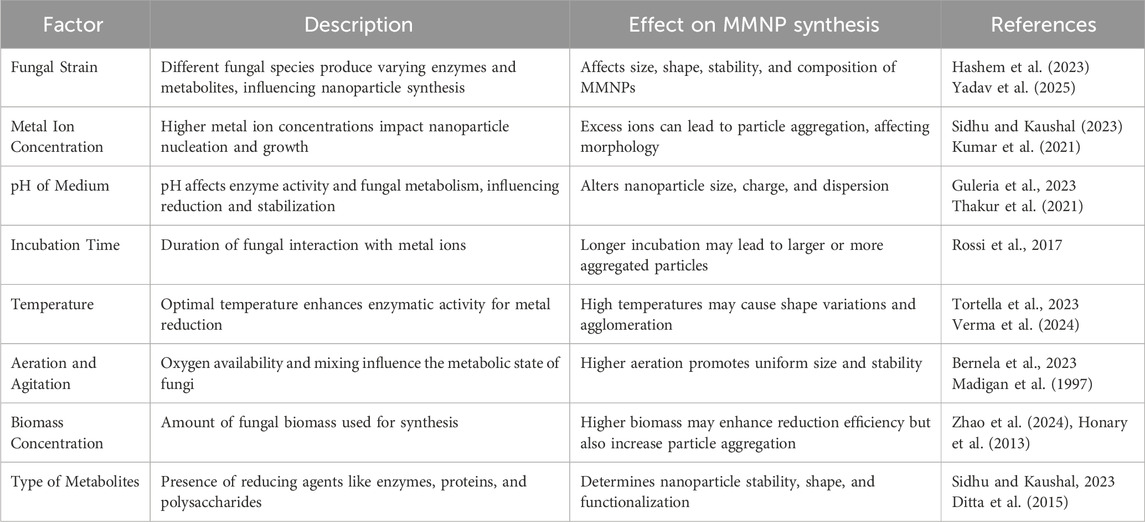
Table 1. Factors influencing fungal-mediated synthesis of multimetallic nanoparticles (MMNPs).
To synthesise multimetallic nanoparticles (MMNPs) for instance bimetallic nanoparticles (BMNPs), two aqueous metal precursor salt solutions are mixed with the fungal extracts. The existence of two metal ions with distinct reduction potentials leads to competitive reduction in the process. Metal ions with higher reduction potential are quickly reduced, followed by those with lower reduction potential (Sasireka and Lalitha, 2021). During the synthesis of bimetallic Cd/Hg nanoparticles, Romero-Núñez et al. (2019), proposed that NPs may develop inside the cell as follows: initially toxic metals are delivered into the cytoplasm via components that interact with these cations. Once in the cytoplasm, Cd2+ and Hg2+ bind to anions, which inhibits transport and forms coordinated complexes with biomolecules and sulfhydryl groups that keep them there and reduce them to form Bimetallic Nanoparticles.
Metallic nanoparticles are classified into monometallic, bimetallic, and trimetallic categories based on the number of metals involved in their synthesis. Various metals have been utilized for their production, often with biological extracts serving as reducing and stabilizing agents (Gebru et al., 2013). Multimetallic nanoparticles are typically synthesized using co-reduction or successive reduction techniques. The integration of two or three metals or metal oxides into a single structure results in materials with unique and multifaceted properties that arise from the synergistic interaction of their components. These properties enable multimetallic nanoparticles to exhibit enhanced functionality compared to their monometallic counterparts (Sumbal et al., 2019).
To broaden their applications, researchers have focused on optimizing the size and morphology of multimetallic nanoparticles, creating structures such as heterodimers, core-shells, and alloys. These structural modifications further enhance their utility across various fields, making them a promising class of nanomaterials for advanced applications (Hashem et al., 2023). However, it could be hypothesized that, nanostructures could exhibit one or more of the following topographical features, random, cluster-in-cluster, core-shell and nano-alloy structures. In random structure, atoms are oriented randomly. While in cluster-in-cluster structure, one metal organized as nano-cluster and the others acted as binders. In core-shell structure, one metal is nucleated firstly in reaction liquor to form the core, while the other metals were clustered later as the outer shells. In nano-alloy structure, the metal nanoparticles were assumed to be synthesized in reaction medium nearly with the same rate. In case of nano-alloy, when the as-nucleated nanoparticles exhibited similar particle sizes it could result in production of a random nano-alloy, while that with different atomic sizes were resulted in inter-metallic nano-alloy (Yang et al., 2008; Ahmed and Emam, 2020).
Chemical co-reduction technique is a type of simultaneous methodologies that generatemultimetallic nanostructures from the reduction of metal precursors to zero-valent metallic forms. This method is mainly advantageous by its versatility and simplicity.Simultaneous reduction of metal precursors is a straightforward route to prepare random alloy nanoparticles, especially when the alloyed metal elements have similar reduction potentials. In this process, supersaturation of monomers is induced by the introduction of an appropriate reducing agent, such as hydrazine, NaBH4, or polyol, into the precursor solution, followed by the nucleation and growth processes (Kim et al., 2022).
The successive or seed mediated growth method is processed via the reduction of metal ions over the surface of other pre-nucleated nanoparticles followed by the cluster growth of the other particles by time. The seed-mediated growthproceeds by synthesizing a seed nanoparticle from metal precursor under the action of reducing agent and subsequently started for growing in reaction liquor. Such synthetic technique was typically applied for preparation of core-shell and intermetallic alloyed nanostructures owing to its controllability under the effect of the reducing agent for production of highly composition, size and shape regulated multimetallic nanostructure (Xia et al., 2017). Seed-mediated synthesis involves a two-step process comprising the preparation of seed nanoparticles and incorporation of secondary (or more) metal species on seeds forming random alloy nanoparticles. The prerequisites to achieving homogeneous random mixing of metals are similar physicochemical properties of the constituent metals and sufficiently high temperatures (Kim et al., 2022).
Multimetallic nanoparticles (MNPs), composed of two or more different metals, have gained significant attention for their unique functionalities and advanced material properties. These nanoparticles, often forming alloys or core–shell nanocomposites, exhibit enhanced chemical, optical, and catalytic properties compared to monometallic and bimetallic NPs. The synergistic interactions between different metals or metal oxides contribute to their superior performance in various applications, particularly in catalysis, where combined metal action significantly improves efficiency and selectivity (Buchwalter et al., 2015).
Multimetallic NPs are classified into categories such as bimetallic, trimetallic, and quadrometallic, based on the number of metals incorporated into their structure. Among these, binary, ternary, and quaternary combinations often display specialized characteristics, including increased chemical reactivity and improved optical and catalytic behavior, making them highly versatile materials. These advanced properties underline the growing interest in multimetallic NPs for diverse scientific and industrial applications (Zhang et al., 2017; Basavegowda and Baek, 2021). The majority of the research on multimetallic nanoparticle synthesis utilising fungi focus on the production of bimetallic and timetallic nanoparticles (Table 2).
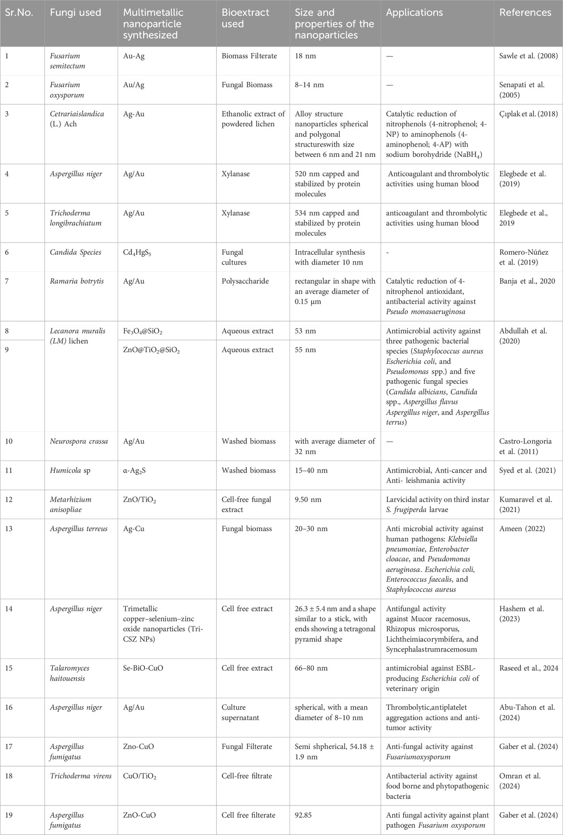
Table 2. Table summarising multimetallic nanoparticles (MMNPs) synthesised by fungi, including their structural configurations, essential features, and applications in biomedicine, agriculture and environmental remediation, and catalysis.
Bi-metallic nanoparticles (BMNs) are nanostructures composed of two different metals, either in alloyed or core–shell configurations, exhibiting unique physical, chemical, and biological properties. Their exceptional biological potential arises from the synergistic activities of their constituent metals (Sharma et al., 2019). These synergistic effects influence variations in configuration, strength, binding, and interaction, contributing to their remarkable reactivity.
The spatial arrangement of metal atoms, driven by physical and chemical interactions, determines whether BMNs form core–shell structures or alloys (Godfrey et al., 2017). Incorporating a second metal enables precise tuning of geometric and electronic structures, enhancing catalytic activity and selectivity. This structural and functional versatility creates novel opportunities for diverse biomedical and industrial applications (Elemike et al., 2019).
BMNs, such as Ag/Au, Ag/Cu, Au/Pt, Au/Pd, Ag/Fe, Fe/Pt, Cu/Zn, Cu/Ni, Au/CuS, and Fe3S4/Ag, exhibit distinct surface activities, while bimetallic oxides like MgO/ZnO, CuO/ZnO, and Fe3O4/ZnO show unique antibacterial properties. These enhanced antibacterial effects are often attributed to tensile strain and the synergism between their metallic components. By leveraging their size, shape, and morphology, BMNs offer unparalleled potential in catalysis, biomedical applications, and environmental solutions (Das and Karankar, 2019).
The biosynthesis of nanoparticles employing fungal and microbial species has shown tremendous promise in a variety of applications due to its environmental friendliness, efficiency, and cost-effectiveness. Romero-Núñez et al. (2019) found that Candida species may synthesise bimetallic (Cd/Hg) nanoparticles with a circular morphology and diameter of 10 nm. These Cd4HgS5 nanoparticles show great promise for a variety of applications. Singh et al. (2020) used Trichoderma reesei (NCIM992) microbial biomass to rapidly bioreduce Ag and Au, resulting in the creation of Au-Ag nanoparticles. These nanoparticles were discovered to be extremely efficient against nosocomial pathogen infections, demonstrating their potential for medical applications. Similarly, Castro-Longoria et al. (2011) used Neurospora crassa to synthesise Ag-Au bimetallic nanoparticles (BNPs) and monometallic nanoparticles. The wild-type strain N150 was grown in Vogel’s Minimal Medium containing sucrose, then subjected to aqueous solutions of AgNO3 and HAuCl4. The bio-reduction process was tracked with UV-Vis spectroscopy, and the nanoparticles were characterised with confocal and transmission electron microscopy (TEM). This study highlighted the versatility of fungal-mediated synthesis by demonstrating that a single fungus species may successfully generate a wide range of nanomaterials. Abdullah et al. (2020) found that Lecanora muralis (LM) lichen aqueous extract can biosynthesise Fe3O4/SiO2 and ZnO/TiO2/SiO2 nanocomposites in a simple, cost-effective, and quick manner. The lichen extract was analysed using GC-Mass, which confirmed the presence of bioactive phytochemicals that played an important part in the synthesis process. These green-synthesized nanocomposites demonstrated significant bioactivity against common harmful bacteria and fungus, indicating their potential for antibacterial applications.
Trimetallic nanoparticles (TNPs) are composed of three distinct metals, designed to reduce metal consumption, optimize atomic ordering, and fine-tune their size and morphology. These nanoparticles exhibit superior catalytic selectivity, activity, and efficiency compared to monometallic and bimetallic counterparts, making them valuable in applications such as biomedical, antimicrobial, catalytic, active food packaging, and sensing technologies. The incorporation of three metals allows for diverse structural possibilities, including core–shell, mixed structures, subcluster-segregated, and multishell configurations (Ali et al., 2020; Basavegowda and Baek, 2021).
By modifying atomic distribution and surface composition, TNPs can also be engineered as alloys or intermetallic nanoparticles to enhance catalytic performance. Their innovative physicochemical properties stem from synergistic and multifunctional effects, enabling their potential in diverse applications (Zaleska-Medynska et al., 2016). Despite these advantages, studies on antimicrobial effects of TNPs remain limited compared to those on mono- and bimetallic nanoparticles. However, TNPs demonstrate efficient antibacterial activity, often outperforming bi- and monometallic nanoparticles at lower concentrations (Yadav et al., 2015; Das and Karankar, 2019).
An example is the synthesis of Tri-CSZ nanoparticles using AspergillusnigerAH1 biomass filtrate, reported by Hashem et al. (2023). The fungus-produced cell-free filtrate was combined with copper acetate, sodium selenite, and zinc acetate. The resulting nanoparticles, confirmed by a color change to dark green, showed significant antimicrobial activity against Mucorales fungi.
Characterization of NPs is important for unravelling their physical and chemical properties. This process helps in determining the average size, shape, and unique features of synthesized NPs. Xray based methods like X ray diffraction (XRD) and Xray Photoelectron Spectroscopy (XPS) are used to determine crystal nature and elemental composition of particles. Microscopic analyses, such as scanning electron microscopy (SEM), transmission electron microscopy (TEM), high-resolution TEM (HRTEM), atomic-force microscopy (AFM), have been used to examine the size, morphology, and distribution of nanomaterials. Energy dispersive spectrometry (EDS) is an accessory of electron microscopy instruments (TEM and SEM)that is used to determine the chemical nature of the core and shell. EDS displays the distribution of elements in the samples. Characterization techniques are discussed below which givesthe widespread overview of approaches used to characterize fungal synthesized monomeric and multimetallic nanoparticles.
According to Khatami et al. (2018), core-shell nanoparticles (NPs) are made up of two or more nanomaterials, including a variety of organic and inorganic nanomaterials (such as metals or polymers). One of these nanomaterial serves as the core, and the second nanomaterial is positioned around the core, known as the shell. They are made so that the shell material can improve the core material’s oxidative state, thermal stability, or reactivity. Type of shell material used in NPs depends on their application. Based on research interests and unique properties different shapes of core -shell nanoparticlesare made (Shanmuganathan et al., 2020). Alloy nanoparticles with different compounds can be synthesized by altering molar proportion of the metal ions in the synthetic solutions (Boroumand Moghaddam et al., 2015). Core-shell structures have different spectral behaviours as compared to alloy structures. The core–shell type structures show two plasmon bands while alloy NPs shows only one plasmon band (Chen et al., 2006; Sawle et al., 2008; Tripathi et al., 2015 showed that the fungal biomass of Trichoderma harzianum acts as a reducing and capping agent for the synthesis and stabilisation of Au–Ag alloy.
Multimetallic nanoparticle analysis using UV-visible spectroscopy is a potent method that makes it possible to quantify surface-conjugated molecules and characterise the characteristics of nanoparticles. A study by Çıplak et al. (2018) used the UV-Vis spectra of Ag, Au monometallic, and Ag-Au bimetallic NPs to perform a preliminary examination of the Ag-Au bimetallic nanoparticles. Metal NPs were successfully generated using C. islandica (L.) Ach, as evidenced by the presence of a surface plasmon resonance (SPR) band of monometallic and bimetallic NPs. The SPR band maxima of monometallic Ag (Ag100Au0) and Au (Ag0Au100) nanoparticles are 410 nm and 534 nm, respectively. In contrast, the absorption bands of Ag and Au monometallic NPs are between the SPR bands of bimetallic NPs. Although UV-visible spectroscopy is a reliable technique for examining multimetallic nanoparticles, complementary methods like transmission electron microscopy and dynamic light scattering must be taken into account in order to validate results and guarantee thorough characterisation (Potts et al., 2023). X-ray Diffraction (XRD) is a non-destructive technique used to examine the crystal structure and phase purity of the synthesised nanoparticles (Khatami et al., 2018). X-rays of a fixed wavelength and the intensity are passed over a crystalline or powdered sample placed over a sample holder and the intensity of the reflected radiation is recorded using a goniometer. Data are then analysed by Bragg’s equation and Debye–Scherrer to calculate inter atomic spacing for the reflection angle and crystalline nature and size, respectively (Khandel and Shahi, 2018). Xray Photoelectron Spectroscopy (XPS) analysis is conducted for further identification of the oxidation states, elemental compositions, and electronic states of the nanoparticles (Omran et al., 2024). XPS analysis irradiates the surface of material with X-ray’s and measures the kinetic energy of ejected photoelectrons. Researchers have used either or both techniques for characterization of monomeric and multimetallic nanoparticles where XRD confirmed crystalline structure (Table 3). XPS analysis showed peaks for the individual elements like Cu, Ti, O and Ag, Au present in fungus mediated CuO/TiO bimetallic and coreshellAg@Au nanoparticle, respectively (Omran et al., 2024; Bhanja et al., 2020).
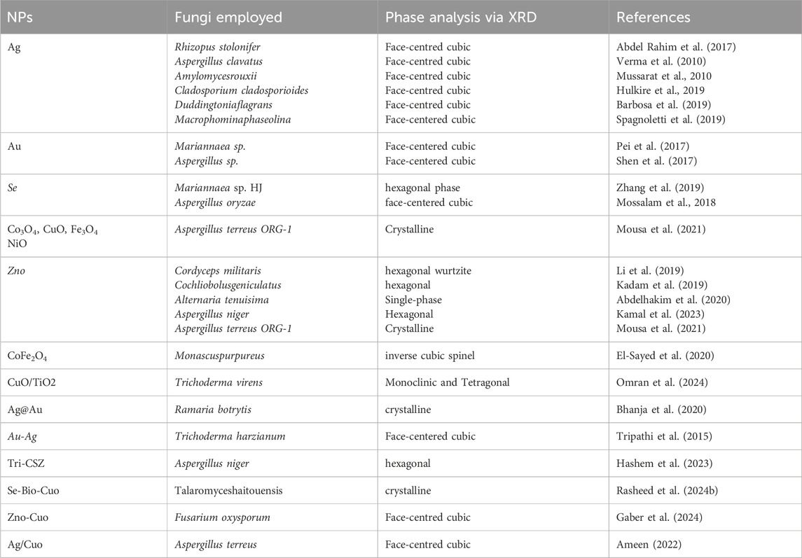
Table 3. Phase analysis via XRD of fungal mediated multimetallic nanoparticles.
Inductively Coupled Plasma Mass Spectrometry (ICP-MS) is a ultrasensitive method used to identify and quantify the elemental composition and concentrations of samples. ICP-MS provides rapid multielement analysis with low detection limits. Usually Single particle ICP-MS (SP-ICPMS) which is an emerging technique used for analysis. Thistechnique relies on two factors: i) their number concentration (ii) the size or the element mass per NP. The numerical concentration of NPs has a direct correlation with the pulse frequency. The intensity of each pulse is proportional to the mass of element, in fact to the number of atoms, in each detected NP (Laborda et al., 2014). Earlier ICP-MS have been used for determination of NPs in complex matrices (Peters et al., 2015).
The morphological characterization of NPs is important to know about their properties. Transmission electron microscopy (TEM) and scanning electron microscopy (SEM) are essential methods for describing multimetallic nanoparticles (MMNPs), offering vital information about their surface characteristics, size, and shape. These methods are commonly used in nearly all MMNP studies because, when paired with energy-dispersive X-ray spectroscopy (EDS), they allow for a thorough understanding of particle distribution, shape uniformity, and elemental composition (Romero-Núñez et al., 2019; Anjum et al., 2022; Saqib et al., 2022; Nyabadza et al., 2023). Their combined use is essential for verifying the successful synthesis of nanoparticles and customising their characteristics for a certain use. Different methods exist to characterize NPs for studying their morphology, but techniques that use microscopy such as SEM, TEM is widely used. In SEM which is a surface imaging method, surface of specimen is scanned using electron beam of accelerated voltage. Backscattered electrons are collected by the detector and analysed to obtain an image. This method is capable of resolving different particle sizes and their distributions, nanomaterial shapes and the surface topology of the particles. The limitation of SEM is that, it is not able to resolve the internal structure (Khandel and Shahi, 2018). SEM coupled withenergy-dispersive X-ray spectrometry (EDS) allow the analysis ofsample surface morphology, the inner structure and the elemental composition. TEM depends on electron transmissionto image a nanoparticle sample, providing much higher resolution. It is preferred method to directly measure nanoparticle size and their distribution. SEM, TEM, EDS have been widely used for determination of shape, size and elemental analysis of mono-, bi- and multi-metallic nanoparticles (Table 4). SEM images are not applicable for studying the structure of core–shell NPs since they characterise the surfaces while TEM images are useful for studying the structure of core–shell NPs considering its ability in measuring the thickness and spacing between core and shells.
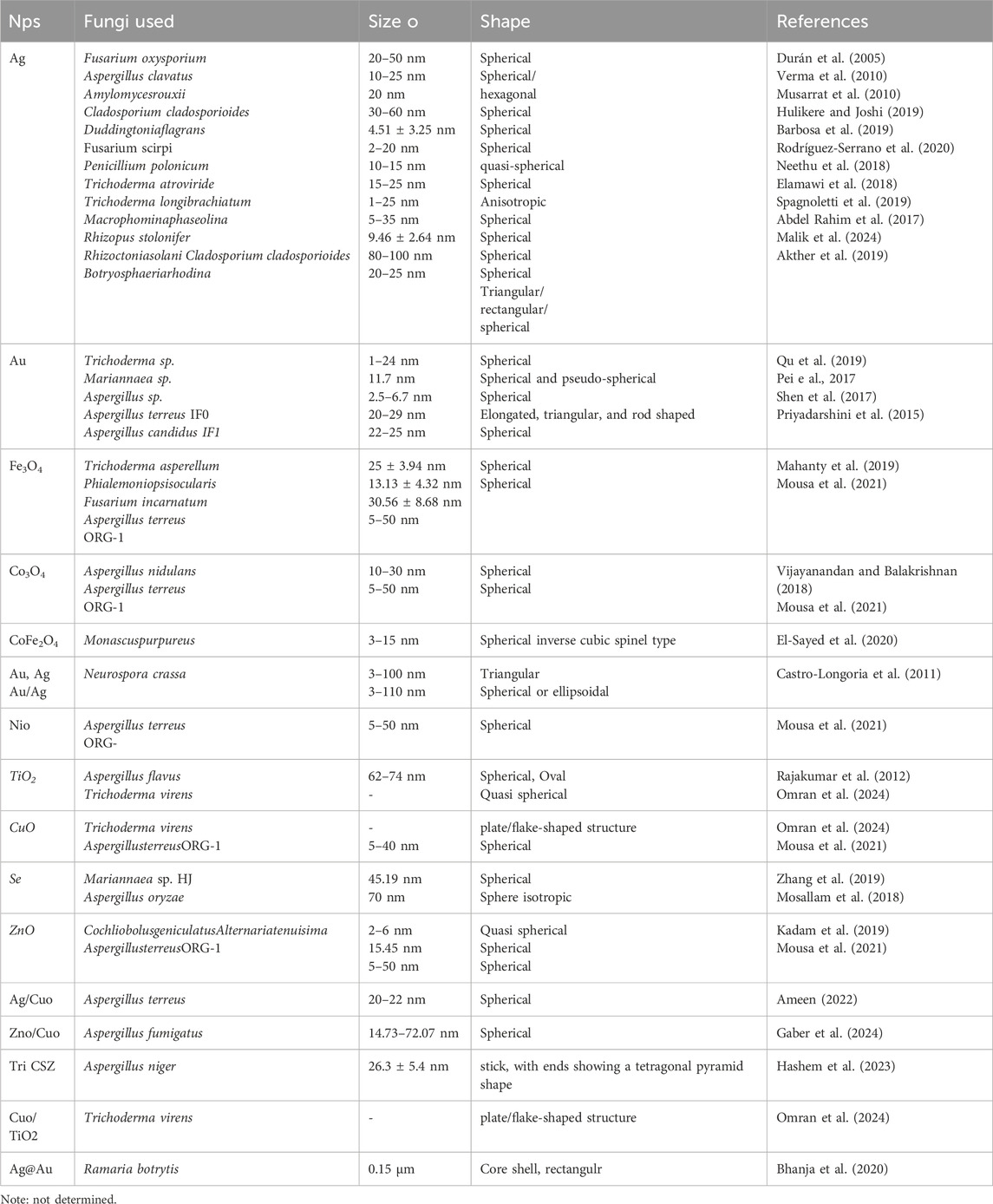
Table 4. SEM/TEM analysis of mono and multimetallic nanoparticles.
The synergistic interactions of two different metals in a nanoparticle system produce novel nanostructural topologies that are very different from those of monometallic nanoparticles (Sasireka and Lalitha, 2021). Multimetallic nanoparticles (MMNPs) generated from fungi often have smaller diameters, greater surface-to-volume ratios, and improved surface reactivity, which results in more active sites for catalysis and adsorption (Zhao et al., 2024). This lowers the energy barrier for electron transport and reaction kinetics, which improves catalytic efficiency.
The charge transfer and electronic characteristics of metals give rise to their catalytic potential, and metallisation in multimetallic systems can greatly increase these characteristics—a result that is not commonly seen in monometallic nanoparticles (Sasireka and Lalitha, 2021; Li et al., 2022). Superior selectivity and stability in a range of applications are made possible by the fine adjustment of geometrical designs and functionalities made possible by the inclusion of several metal components (Rodriguez and Goodman, 1992; Wang et al., 2017). The remarkable catalytic, electrical, and optical capabilities of these bimetallic and multimetallic systems are a result of their unique bi-functional and synergistic effects (Habas et al., 2007). The multifunctional properties of MMNPs are further enhanced by band structure alterations, lattice strain effects, and electronic interactions among the metal components (Chen et al., 2005; Senapati et al., 2005).
Despite these advances, research on fungal-mediated MMNPs is still limited, and more studies are needed to completely understand their structural evolution, stability, and functional features in various application domains.
The synergistic interactions between different metals in MMNPs allow for customisable features, making them applicable in a variety of sectors. Furthermore, fungal-mediated synthesis is consistent with green chemistry principles, providing a sustainable alternative to traditional chemical and physical synthesis processes. These features make fungal-synthesized MMNPs interesting candidates for a variety of applications, including biomedicine, environmental remediation, catalysis, and more (Figure 2).
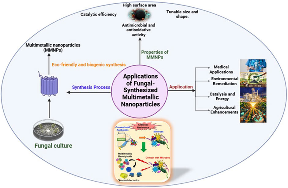
Figure 2. Illustration demonstrating the various characteristics and broad applications of multimetallic nanoparticles (MMNPs) synthesised using fungal-mediated methods. These nanoparticles’ key properties include increased catalytic activity, higher thermal and chemical durability, improved biocompatibility, optical tunability, and novel structural configurations. The image also shows the adaptability of fungal-synthesized MMNPs in a variety of domains, including biomedicine, environmental cleanup, catalysis, and agricultural development.
In recent years, biologically produced metal nanoparticles have developed as a lively field of research with substantial implications for biomedical applications. The various uses of biologically synthesised particles in biomedicine are drawing more and more attention. These particles are frequently produced using environmentally friendly or biologically friendly synthesis techniques (Soni et al., 2021; Osman et al., 2024). They are especially well-suited for delicate medical applications due to their improved biocompatibility and environmentally friendly manufacturing process (Das et al., 2017). The capacity to overcome biological barriers, surface functionalisation, and customised size of biologically synthesised nanoparticles make them effective drug carriers (Ahmad et al., 2022; Deshmukh et al., 2024). They increase treatment efficacy and minimise negative effects by improving targeted medication delivery. Biologically produced nanoparticles are perfect for bioimaging because of their optical characteristics. Fluorescence imaging, magnetic resonance imaging (MRI), and other imaging modalities use biologically synthesised metal-based nanoparticles, magnetic nanoparticles, and quantum dots (Mirabello et al., 2015; Das et al., 2019; Truong, et al., 2024). Their sensitivity and accuracy in illness diagnosis are improved by their capacity to be functionalised for particular biomarkers (Shnoudeh et al., 2019; Han et al., 2019; Thakare, 2023). In photothermal therapy (PTT), cancer cells are specifically killed by use of nanoparticles that have the ability to absorb light and transform it into heat (Alamdari, et al., 2022). In PTT, biologically produced particles—especially those made of gold or graphene—are beneficial because of their adjustable photothermal characteristics and lower toxicity (Chen, et al., 2019). The biocompatibility of these particles, in contrast to their chemically synthesised counterparts, minimises toxicity problems and immunological reactions in all of these applications. They are therefore a viable path towards safer and more efficient biological treatments (Abbasi et al., 2023; Oladipo et al., 2023). Like, Gold nanoparticles, including those containing gold cores, provide a versatile platform with desirable properties for application in biomedicine, particularly in tissue imaging, photothermal therapy, and theragnostic (Yang et al., 2019; Ehrhardt Jr and Güleç, 2020). Moreover, by adding two or more contrast agents, such as fluorophores, MRI agents, photoacoustic agents, or surface-enhanced Raman spectroscopy reporters, gold nanoparticles can be used to give multimodal imaging. This method minimises the effect on patients’ immune systems while improving the characterisation of the disease (Pillai, 2019). Gold nanoparticle-based targeted photothermal therapy in conjunction with a chemotherapeutic agent has proven to be a successful treatment for multidrug-resistant cancers (Kesharwani et al., 2023).
Entomopathogenic fungi (EPF) are a significant source of secondary metabolites, particularly low-molecular-weight organic compounds released in response to environmental stimuli. These metabolites are crucial for maintaining host functionality and successfully infecting pathogens by damaging their nervous system or lowering insect resistance (Donzelli and Krasnoff, 2016; Vivekanandhan et al., 2018; Karthi et al., 2018).
Due to their high enzymatic activity, ability to produce bioactive compounds, and adaptability to culture media, EPF is also being explored in nanoparticle biosynthesis and bioconversion of flavonoids and steroids, which hold significant economic potential (Różalska et al., 2018). One widely studied EPF species, Metarhizium anisopliae, is commonly used as a biocontrol agent against agricultural pests (Faria and Wraight, 2007). These fungi produce various metabolites, including destruxins (DTXs)—cyclic depsipeptides composed of L-proline, L-isoleucine, N-methyl-L-valine, N-methyl-L-alanine, β-alanine, and a D-α-hydroxy acid, which varies among destruxin subtypes (A-E), primarily isolated from Metarhizium species (Arroyo-Manzanares et al., 2017).
The biocompatibility of biologically synthesized nanoparticles, in contrast to their chemically synthesized counterparts, minimizes toxicity issues and immunological reactions in various applications. As a result, they offer a safer and more efficient approach to biological treatments. Their production through biological processes—using fungi, microorganisms, or plants—reduces environmental impact during synthesis and contributes to large-scale environmental cleanup initiatives due to their scalability and versatility (Abdellatif et al., 2021; Oladipo et al., 2023).
Biologically produced nanoparticles, such as those composed of iron, silver, and zinc, exhibit remarkable adsorption and reduction capacities for removing heavy metals like lead, cadmium, and arsenic from contaminated water (Kumar et al., 2021; Zaimee et al., 2021; Razzak et al., 2022).
These nanoparticles also demonstrate potential in degrading organic contaminants, including dyes, insecticides, and pharmaceutical residues. For example, silver nanoparticles have been used under mild conditions to catalyze the breakdown of industrial dyes (Abbas et al., 2016; Feisal et al., 2024), while biologically synthesized iron nanoparticles efficiently degrade chlorinated organic compounds (Ismail et al., 2019; Chaudhari et al., 2024; Venkateshaiah et al., 2022).
Additionally, these nanoparticles have been employed in soil remediation and air purification. Microbially synthesized iron oxide nanoparticles have shown effectiveness in reducing soil contamination caused by hydrocarbons and other hazardous pollutants (Bolade et al., 2021; Tao et al., 2023). Compared to conventional methods, biologically synthesized nanoparticles offer superior efficiency with lower environmental toxicity, making them an eco-friendly solution for pollution control. Their scalability and versatility are increasingly driving their adoption in global environmental restoration programs.
Biologically synthesized nanoparticles have gained attention for their potential role in bioenergy production. Certain fungi and microorganisms used in the biosynthesis of nanoparticles also contribute to bioconversion processes, where they facilitate the breakdown of organic matter into biofuels and other energy-rich compounds.
For instance, some entomopathogenic fungi (EPF) with high enzymatic activity are being investigated for their role in converting biological waste into energy sources. These fungi, by producing secondary metabolites and enzymes, can aid in the degradation of lignocellulosic biomass—a crucial step in bioethanol and biogas production. Their efficiency in breaking down complex organic materials highlights their potential in sustainable bioenergy initiatives.
Certain soil-borne plant pathogens, particularly those affecting economically important crops, cause severe yield losses by impairing plant growth at various stages (Granzow et al., 2017; Ali et al., 2021). Wilting diseases and root rot are often attributed to latent soil fungi (Stoddard et al., 2010; Levenfors, 2003). Among them, Fusarium wilt disease is particularly damaging, as it compromises plant immunity, significantly reducing both yield and quality (Elamawi and Al-Harbi, 2014; Mahmoud, 2016; Maity et al., 2022).
During the planting season, Fusarium wilt begins in the root system, gradually spreading throughout the plant. The pathogen obstructs vascular tissues, disrupting water and nutrient transport, ultimately leading to wilting, leaf necrosis, and plant death (Elamawi and Al-Harbi, 2014). Chemical control methods have proven challenging and environmentally harmful, making biological control strategies a more suitable alternative. Biocontrol approaches are cost-effective, eco-friendly, and efficient in managing Fusarium wilt (Yu et al., 2021; Abdelaziz et al., 2022; Hashem et al., 2022; Attia et al., 2022; Abdelaziz et al., 2023).
Entomopathogenic fungi, particularly species like Metarhizium anisopliae, have demonstrated effectiveness in controlling soil-borne pathogens through their ability to produce antifungal metabolites and induce plant resistance mechanisms. The integration of biological nanoparticles and fungal biocontrol agents presents an innovative strategy for soil health management and sustainable agriculture.
Sensors and biosensors are essential to contemporary diagnostics because they provide quick, precise, and sensitive detection techniques, especially for medical and environmental monitoring applications (Haleem, et al., 2021; Bhatia et al., 2024). To identify chemical, biological, or physical changes in a system, these instruments take advantage of special optical and electrical characteristics (Singh et al., 2020; Hemdan et al., 2024). They are made up of a reader device for data collection, a signal transducer that transforms the biological reaction into an electrical signal, and a recognition component that recognises the analyte and produces a corresponding signal. In industries ranging from environmental monitoring to healthcare, these instruments are essential for identifying and measuring a variety of chemicals (Jain, 2012). Additionally, biosensors such as the DNA and microbial biosensors are essential for monitoring contaminants in environmental applications, enabling prompt actions that greatly aid in environmental conservation.Research in the Journal of Medical Internet Research suggests that the use of wearable biosensors for remote patient monitoring has significantly reduced hospital readmission rates, saving healthcare systems throughout the world a significant amount of money (Turner, 2013). Moreover, biosensors make it possible to conduct high-throughput screening in drug discovery, which significantly cuts down on the amount of time needed to find possible therapeutic options. Adoption of biosensors has significantly increased research productivity worldwide. The extensive use of biosensors in research labs across continents is expected to drive the global biosensors market to reach USD 35.5 billion by 2026, according to a report by Research and Markets.Moreover, analytes are detected and measured by optical sensors using light. Methods including Raman spectroscopy, surface plasmon resonance (SPR), and fluorescence are frequently employed (Wang et al., 2017; Bousiakou et al., 2019; Philip and Kumar, 2022). SPR-based biosensors, for example, are perfect for medical diagnostics such as identifying biomarkers in blood or saliva because they can identify molecular interactions in real-time without the need for labels (Das et al., 2023; Swami et al., 2024). High-sensitivity detection in environmental monitoring, such as the detection of pollutants or toxins, is made possible by fluorescence-based sensors, which are frequently augmented with nanomaterials like quantum dots (Thakur and Kumar, 2022; Mohkam et al., 2023; Udhayakumari, 2024). When interacting with the target analyte, electrical sensors rely on changes in electrical signals, such as resistance, capacitance, or current. In medical diagnostics, electrochemical biosensors—like glucose meters—are well-known examples. Ion-selective electrodes (ISEs) are employed in environmental applications to identify ions such as heavy metals or nitrates in water. These sensors’ sensitivity and selectivity are improved by the incorporation of nanostructured materials like graphene or carbon nanotubes (Sharma et al., 2021; Polidori et al., 2024). The integration of electrical and optical sensing modalities, cost reduction, and portability are the main goals of recent developments (Heikenfeld et al., 2018). Wearable biosensors, smartphone-integrated platforms, and point-of-care (POC) devices are all growing in popularity. These developments address issues in both resource-rich and resource-limited environments by guaranteeing that sensitive and accurate diagnostics are available in a variety of contexts (Colombo, 2022). Finding pollutants in water, soil health indicators, and air quality measures is known as environmental diagnostics. Monitoring chronic diseases, identifying infectious disorders, and identifying cancer biomarkers are all examples of medical diagnostics. Next-generation sensors are being developed as a result of the intersection of nanotechnology, material science, and data analytics, which has the potential to revolutionize environmental sustainability and global health. For instance, among the most destructive plant diseases are Fusarium species. In order to inhibit the growth of F. oxysporum, Aspergillus fumigatus was used to myco-synthesize bimetallic zinc oxide-copper oxide nanoparticles (ZnO-CuO NPs). After being separated from soil, A. fumigatus was detected both physically and genetically (Gaber et al., 2024).
For example, biosensors such as the enzyme-linked immunosorbent assay (ELISA) and glucose biosensor have revolutionised diabetes treatment by facilitating precise and real-time glucose level monitoring, which is essential for efficient disease management. Biosensors such as the fiber-optic and piezoelectric biosensors have made it easier to quickly identify pollutants in food products, hence improving food safety. By quickly detecting infections like Salmonella and E. coli in food samples, these biosensors protect the public’s health. Additionally, biosensors such as the DNA and microbial biosensors are essential for monitoring contaminants in environmental applications, enabling prompt actions that greatly aid in environmental conservation (Bhatia et al., 2024). For instance, the potential of gold nanoparticles as multifunctional sensors has been thoroughly studied. Gold nanomaterials can be tailored for certain uses by varying their shape; moreover, gold nanoparticles, nanorods, nano-shells, and nanocages all have unique thermal and optical properties that make them promising theragnostic agents (Prakash, 2023).
Plant infections caused by Fusarium species are among the most damaging. Using A. fumigatus, bimetallic zinc oxide-copper oxide nanoparticles (ZnO-CuO NPs) were myco-synthesised to prevent the growth of F. oxysporum. Physically and genetically, A. fumigatus was identified after being isolated from soil. Omran et al., 2024 studied, the cell-free filtrate of the endophytic fungus T. virens was used to create CuO/TiO2 NPs. Mycosynthesis of CuO/TiO2 NPs was the outcome of the fungal filtrate’s reaction with the two metal salts, CuSO4.5H2O and TiO2. Utilizing XRD, FTIR spectroscopy, EDX, DLS, zeta potential analysis, XPS, Raman scattering, FESEM, and HRTEM, the produced bimetal oxide nanoparticles were studied. The monoclinic CuO and tetragonal rutile TiO2 crystal planes were represented by the diffractionPeaks of the mycosynthesizedCuo/TiO2 nanoparticles. This study evaluated an environmentally acceptable approach for the mycogenic synthesis of the bimetallic oxide, CuO/TiO2 NPs, using the cell-free filtrate of Trichoderma virens. In order to address the severity of plant diseases and foodborne illnesses and contribute to meeting the global food and agricultural needs, the effectiveness of the mycogenicCuO/TiO2 NPs against foodborne and phytopathogenic bacteria was then assessed.
MMNPs have great interest as compared to single metals-based nanoparticles because of their good stability, numerous morphologies, synergistic effect of multiple metals, improved optical, catalytic, magnetic, and electronic properties (Basavegowda and Baek, 2021). Biogenic synthesis of MMNPs is considered as simple, economically feasible and scale-up approach due to the presence of fast growth of fungal cultures, different reducing agents (enzymes, proetins), non-toxicity of fungal cell extract (Rasheed R. et al., 2024). Despite its advantageous usage, there are some limitations of MMNPs including selection, type of nanoparticle, structure, size, stability, dosage, mode of application, batch to batch variability, toxicity, environmental, and health hazards. Firstly, selection of multiple metals specific precursors, compatibility with different precursors, ratio of multiple metals, reaction time, and conditions (pH, temperature) for uniform mixing of all the metals is the biggest challenge in the synthesis process of MNPs. Secondly, requirement of advanced characterization approach of MMNPs is also the major problem (Dey et al., 2024). Moreover, different types of metals have their own physical and chemical properties that may interfere with the crystallization, aggregation, and penetration of MMNPs to the target site (Yapa et al., 2024). Besides this, optimization of fungal culture media, different growth conditions is also important for the production of bioactive compounds that acts as stabilizing, reducing agents and also controls the uniformity, shape and size of the synthesized MMNPs (Adeleke et al., 2024). In Addition to above, batch-to-batch variability remains a key challenge due to fluctuations in environmental factors such as pH, temperature, and nutrient availability, which can influence fungal metabolism and nanoparticle characteristics. To address this, advancements in bioreactor-based production, genetic engineering for metabolically stable fungal strains, and process optimization strategies are being explored to enhance reproducibility. Therefore, proper research is required for the selection of metallic nanoparticles, their physical and chemical nature, and more focus should be on studying the toxicity of MMNPs. Regulatory approvals, safety and handling rules, and economic viability should be imposed for the efficient development of MMNPs. Moreover, the fungal metabolism should also be carefully examined for determining the mode of action of reducing agents that may controlled the aggregation, dispersion, shape and size of nanoparticles.
The study and production of metallic NPs using fungi are increasing and have received great attention from scientific and technological communities due to their ease of handling and high metal tolerance. The biogenic method provides natural agents for reduction, capping and stabilization of NPs (Sidhu et al., 2022). Metal nanoparticles, due to their quantum effects and high surface-to-volume ratio, have outstanding ultraviolet-visible sensitivity, electrical, catalytic, thermal, and antibacterial properties (Mekuye and Abera, 2023). Fungal-mediated production of multimetallic nanoparticles (MMNPs) has various intriguing future directions. Optimising fungal strains through genetic engineering and metabolic pathway alteration can improve the efficiency, yield, and specificity of MMNP production. Addressing scalability and reproducibility issues is critical, necessitating the creation of standardised processes to assure uniform particle composition, size, and morphology in large-scale production. Integrating MMNPs with other nanomaterials to form hybrid systems opens up possibilities for multifunctional applications in sectors such as medication delivery, biosensing, and environmental remediation. Furthermore, getting deeper mechanistic insights into the enzymatic and metabolic pathways involved in fungal-mediated synthesis can lead to improved process control and nanoparticle property customisation.
In multi-metallic nanoparticles metals form layer around each other exhibiting good optical properties (Srinoi et al., 2018). Although studies about the formation of multi-metallic NPs using fungus are few. Employing fungus these kinds of nanoparticles can be rapidly synthesized, giving NPs of different morphologies. (Aigbe and Osibote, 2024). To ensure safe and sustainable use, thorough research on the toxicity, environmental effect, and biocompatibility of fungal-derived MMNPs are required. Tailoring MMNP features to specific applications in agriculture, healthcare, catalysis, and environmental research can increase their practical utility. Finally, fostering multidisciplinary collaborations among microbiologists, chemists, materials scientists, and engineers will be pivotal in accelerating research advancements and industrial applications of this green synthesis approach. Together, these activities will realise the full potential of fungal-mediated MMNPs, resulting in long-term solutions across a wide range of industries.
The production of multimetallic nanoparticles (MMNPs) using fungus-mediated synthesis offers an environmentally responsible and sustainable substitute for traditional chemical and physical processes. Fungi’s natural capacity for enzymatic reduction, biostabilization, and the release of bioactive metabolites makes it possible to produce MMNPs with special structural and functional characteristics, like increased catalytic activity, improved stability, and biocompatibility. Because of their synergistic qualities, MMNPs are very adaptable for use in biomedicine, environmental remediation, biosensing, and catalysis.
Despite tremendous advancements, issues such as reproducibility, scalability, and precise control over particle composition continue to be important impediments to the widespread use of fungal-mediated synthesis. Addressing these problems would necessitate a multidisciplinary strategy that includes sophisticated strain optimisation, genetic engineering, and the integration of fungal systems with hybrid technology. Furthermore, the study of novel fungus species and their metabolic processes may open up new avenues for adapting MMNP features to specific applications.
This study highlights the potential of fungi as bio-factories for green MMNP synthesis, offering an attractive path to meeting the growing need for sustainable nanomaterials. Researchers and industry can expedite the creation of creative solutions to global difficulties in agriculture, medicine, and environmental sustainability by capitalising on the benefits of fungus and multimetallic systems. Future research should focus on bridging the gap between laboratory-scale synthesis and industrial-scale production, paving the door for practical fungal-mediated MMNP applications in a variety of domains.
AS: Conceptualization, Data curation, Formal Analysis, Supervision, Validation, Writing – original draft, Writing – review and editing. SA: Conceptualization, Data curation, Writing – original draft. NV: Conceptualization, Visualization, Writing – original draft. PK: Conceptualization, Visualization, Writing – original draft. MS: Conceptualization, Data curation, Validation, Visualization, Writing – original draft.
The author(s) declare that no financial support was received for the research and/or publication of this article.
The authors declare that the research was conducted without commercial or financial relationships that could create a conflict of interest.
The author(s) declare that no Generative AI was used in the creation of this manuscript.
All claims expressed in this article are solely those of the authors and do not necessarily represent those of their affiliated organizations, or those of the publisher, the editors and the reviewers. Any product that may be evaluated in this article, or claim that may be made by its manufacturer, is not guaranteed or endorsed by the publisher.
Abbas, A., Al-Amer, A. M., Laoui, T., Al-Marri, M. J., Nasser, M. S., Khraisheh, M., et al. (2016). Heavy metal removal from aqueous solution by advanced carbon nanotubes: critical review of adsorption applications. Sep. Purif. Technol. 157, 141–161. doi:10.1016/j.seppur.2015.11.039
Abbasi, R., Shineh, G., Mobaraki, M., Doughty, S., and Tayebi, L. (2023). Structural parameters of nanoparticles affecting their toxicity for biomedical applications: a review. J. Nanoparticle Res. 25 (3), 43. doi:10.1007/s11051-023-05690-w
Abdelaziz, A. M., Hashem, A. H., El-Sayyad, G. S., El-Wakil, D. A., Selim, S., Alkhalifah, D. H., et al. (2023). Biocontrol of soil borne diseases by plant growth promoting rhizobacteria. Trop. Plant Pathol. 48 (2), 105–127. doi:10.1007/s40858-022-00544-7
Abdelaziz, A. M., Kalaba, M. H., Hashem, A. H., Sharaf, M. H., and Attia, M. S. (2022). Biostimulation of tomato growth and biocontrol of Fusarium wilt disease using certain endophytic fungi. Bot. Stud. 63 (1), 34. doi:10.1186/s40529-022-00364-7
Abdelhakim, H. K., El-Sayed, E. R., and Rashidi, F. B. (2020). Biosynthesis of zinc oxide nanoparticles with antimicrobial, anticancer, antioxidant and photocatalytic activities by the endophytic Alternaria tenuissima. J. Appl. Microbiol. 128 (6), 1634–1646. doi:10.1111/jam.14581
Abdellatif, A. A., Mohammed, H. A., Khan, R. A., Singh, V., Bouazzaoui, A., Yusuf, M., et al. (2021). Nano-scale delivery: a comprehensive review of nano-structured devices, preparative techniques, site-specificity designs, biomedical applications, commercial products, and references to safety, cellular uptake, and organ toxicity. Nanotechnol. Rev. 10 (1), 1493–1559. doi:10.1515/ntrev-2021-0096
Abdel Rahim, K., Mahmoud, S. Y., Ali, A. M., Almaary, K. S., Mustafa, A. E. Z. M., and Husseiny, S. M. (2017). Extracellular biosynthesis of silver nanoparticles using Rhizopus stolonifer. Saudi J. Biol. Sci. 24 (1), 208–216. doi:10.1016/j.sjbs.2016.02.025
Abdullah, S. M., Kolo, K., and Sajadi, S. M. (2020). Greener pathway toward the synthesis of lichen-based ZnO@ TiO2@ SiO2 and Fe3O4@ SiO2 nanocomposites and investigation of their biological activities. Food Sci. and Nutr. 8 (8), 4044–4054. doi:10.1002/fsn3.1661
Abu-Tahon, M. A., Alshammari, F. A., Shahhat, I. M., Ghareib, M., and Abdallah, W. E. (2024). Eco-friendly synthesis, characterization, and biomedical applications of biosynthesized bimetallic silver-gold nanoparticles by culture supernatant of Aspergillus Niger. Appl. Biochem. Biotechnol. 197, 137–158. doi:10.1007/s12010-024-05035-w
Adeleke, B. S., Olowe, O. M., Ayilara, M. S., Fasusi, O. A., Omotayo, O. P., Fadiji, A. E., et al. (2024). Biosynthesis of nanoparticles using microorganisms: a focus on endophytic fungi. Heliyon 10 (21), e39636. doi:10.1016/j.heliyon.2024.e39636
Ahmad, F., Salem-Bekhit, M. M., Khan, F., Alshehri, S., Khan, A., Ghoneim, M. M., et al. (2022). Unique properties of surface-functionalized nanoparticles for bio-application: functionalization mechanisms and importance in application. Nanomaterials 12 (8), 1333. doi:10.3390/nano12081333
Ahmed, H. B., and Emam, H. E. (2020). Overview for multimetallic nanostructures with biomedical, environmental and industrial applications. J. Mol. Liq. 321, 114669. doi:10.1016/j.molliq.2020.114669
Aigbe, U. O., and Osibote, A. O. (2024). Green synthesis of metal oxide nanoparticles, and their various applications. J. Hazard. Mater. Adv. 13, 100401. doi:10.1016/j.hazadv.2024.100401
Akther, T., Mathipi, V., Kumar, N. S., Davoodbasha, M., and Srinivasan, H. (2019). Fungal-mediated synthesis of pharmaceutically active silver nanoparticles and anticancer property against A549 cells through apoptosis. Environ. Sci. Pollut. Res. 26 (13), 13649–13657. doi:10.1007/s11356-019-04718-w
Alamdari, S. G., Amini, M., Jalilzadeh, N., Baradaran, B., Mohammadzadeh, R., Mokhtarzadeh, A., et al. (2022). Recent advances in nanoparticle-based photothermal therapy for breast cancer. J. Control. Release 349, 269–303. doi:10.1016/j.jconrel.2022.06.050
Ali, O., Ramsubhag, A., and Jayaraman, J. (2021). Biostimulant properties of seaweed extracts in plants: implications towards sustainable crop production. Plants 10 (3), 531. doi:10.3390/plants10030531
Ali, S., Sharma, A. S., Ahmad, W., Zareef, M., Hassan, M. M., Viswadevarayalu, A., et al. (2020). Noble metals based bimetallic and trimetallic nanoparticles: controlled synthesis, antimicrobial and anticancer applications. Crit. Rev. Anal. Chem. 51, 454–481. doi:10.1080/10408347.2020.1743964
Amaladhas, T. P., Sivagami, S., Devi, T. A., Ananthi, N., and Velammal, S. P. (2012). Biogenic synthesis of silver nanoparticles by leaf extract of Cassia angustifolia. Adv. Nat. Sci. Nanosci. Nanotechnol. 3, 045006. doi:10.1088/2043-6262/3/4/045006
Ameen, F. (2022). Optimization of the synthesis of fungus-mediated bi-metallic Ag-Cu nanoparticles. Appl. Sci. 12 (3), 1384. doi:10.3390/app12031384
Anjum, S., Nawaz, K., Ahmad, B., Hano, C., and Abbasi, B. H. (2022). Green synthesis of biocompatible core–shell (Au–Ag) and hybrid (Au–ZnO and Ag–ZnO) bimetallic nanoparticles and evaluation of their potential antibacterial, antidiabetic, antiglycation and anticancer activities. RSC Adv. 12 (37), 23845–23859. doi:10.1039/d2ra03196e
Arroyo-Manzanares, N., Diana Di Mavungu, J., Garrido-Jurado, I., Arce, L., Vanhaecke, L., Quesada-Moraga, E., et al. (2017). Analytical strategy for determination of known and unknown destruxins using hybrid quadrupole-Orbitrap high-resolution mass spectrometry. Anal. Bioanal. Chem. 409, 3347–3357. doi:10.1007/s00216-017-0276-z
Attia, M. S., El-Wakil, D. A., Hashem, A. H., and Abdelaziz, A. M. (2022). Antagonistic effect of plant growth-promoting fungi against fusarium wilt disease in tomato: in vitro and in vivo study. Appl. Biochem. Biotechnol. 194 (11), 5100–5118. doi:10.1007/s12010-022-03975-9
Barbosa, A. C. M. S., Silva, L. P. C., Ferraz, C. M., Tobias, F. L., de Araújo, J. V., Loureiro, B., et al. (2019). Nematicidal activity of silver nanoparticles from the fungus Duddingtoniaflagrans. Int. J. Nanomedicine 14, 2341–2348. doi:10.2147/IJN.S193679
Basavaraja, S., Balaji, S. D., Lagashetty, A., Rajasab, A. H., and Venkataraman, A. (2008). Extracellular biosynthesis of silver nanoparticles using the fungus Fusarium semitectum. Mater. Res. Bull. 43 (5), 1164–1170. doi:10.1016/j.materresbull.2007.06.020
Basavegowda, N., and Baek, K. H. (2021). Multimetallic nanoparticles as alternative antimicrobial agents: challenges and perspectives. Molecules 26 (4), 912. doi:10.3390/molecules26040912
Bernela, M., Kaushal, P., Verma, N., Thakur, R., Ahuja, M., and Kaur, P. (2023). Anti-inflammatory therapeutics: conventional concepts and future with nanotechnology. Recent Adv. Inflamm. and Allergy Drug Discov. 17 (1), 7–19. doi:10.2174/2772270817666221027154402
Bhanja, S. K., Samanta, S. K., Mondal, B., Jana, S., Ray, J., Pandey, A., et al. (2020). Green synthesis of Ag@ Au bimetallic composite nanoparticles using a polysaccharide extracted from Ramaria botrytis mushroom and performance in catalytic reduction of 4-nitrophenol and antioxidant, antibacterial activity. Environ. Nanotechnol. Monit. and Manag. 14, 100341. doi:10.1016/j.enmm.2020.100341
Bhatia, D., Paul, S., Acharjee, T., and Ramachairy, S. S. (2024). Biosensors and their widespread impact on human health. Sensors Int. 5, 100257. doi:10.1016/j.sintl.2023.100257
Bolade, O. P., Akinsiku, A. A., Oluwafemi, O. S., Williams, A. B., and Benson, N. U. (2021). Biogenic iron oxide nanoparticles and activated sodium persulphate for hydrocarbon remediation in contaminated soil. Environ. Technol. and Innovation 23, 101719. doi:10.1016/j.eti.2021.101719
Boroumand Moghaddam, A., Namvar, F., Moniri, M., Md. Tahir, P., Azizi, S., and Mohamad, R. (2015). Nanoparticles biosynthesized by fungi and yeast: a review of their preparation, properties, and medical applications. Molecules 20 (9), 16540–16565. doi:10.3390/molecules200916540
Bousiakou, L. G., Gebavi, H., Mikac, L., Karapetis, S., and Ivanda, M. (2019). Surface enhanced Raman spectroscopy for molecular identification-A review on surface plasmon resonance (SPR) and localised surface plasmon resonance (LSPR) in optical nanobiosensing. Croat. Chem. Acta 92 (4), 479–494. doi:10.5562/cca3558
Buchwalter, P., Rosé, J., and Braunstein, P. (2015). Multimetallic catalysis based on heterometallic complexes and clusters. Chem. Rev. 115, 28–126. doi:10.1021/cr500208k
Castro-Longoria, E., Vilchis-Nestor, A. R., and Avalos-Borja, M. (2011). Biosynthesis of silver, gold and bimetallic nanoparticles using the filamentous fungus Neurospora crassa. Colloids surfaces B Biointerfaces 83 (1), 42–48. doi:10.1016/j.colsurfb.2010.10.035
Chaudhari, D. S., Upadhyay, R. P., Shinde, G. Y., Gawande, M. B., Filip, J., Varma, R. S., et al. (2024). A review on sustainable iron oxide nanoparticles: syntheses and applications in organic catalysis and environmental remediation. Green Chem. 26, 7579–7655. doi:10.1039/d4gc01870b
Chen, H. M., Liu, R. S., Jang, L. Y., Lee, J. F., and Hu, S. F. (2006). Characterization of core–shell type and alloy Ag/Au bimetallic clusters by using extended X-ray absorption fine structure spectroscopy. Chem. Phys. Lett. 421 (1-3), 118–123. doi:10.1016/j.cplett.2006.01.043
Chen, J., Ning, C., Zhou, Z., Yu, P., Zhu, Y., Tan, G., et al. (2019). Nanomaterials as photothermal therapeutic agents. Prog. Mater. Sci. 99, 1–26. doi:10.1016/j.pmatsci.2018.07.005
Chen, M., Kumar, D., Yi, C. W., and Goodman, D. W. (2005). The promotional effect of gold in catalysis by palladium-gold. Science 310, 291–293. doi:10.1126/science.1115800
Çıplak, Z., Gökalp, C., Getiren, B., Yıldız, A., and Yıldız, N. (2018). Catalytic performance of Ag, Au and Ag-Au nanoparticles synthesized by lichen extract. Green Process. Synthesis 7 (5), 433–440. doi:10.1515/gps-2017-0074
Colombo, R. N. (2022). “Biosensors in point-of-care: molecular analysis, strategies and perspectives to health care,” in Advances in bioelectrochemistry volume 3: biosensors, wearable devices and biomedical applications (Cham: Springer International Publishing), 169–198.
Cruz, J. N., Muzammil, S., Ashraf, A., Ijaz, M. U., Siddique, M. H., Abbas, R., et al. (2024). A review on mycogenic metallic nanoparticles and their potential role as antioxidant, antibiofilm and quorum quenching agents. Heliyon 10, e29500. doi:10.1016/j.heliyon.2024.e29500
Das, P., and Karankar, V. S. (2019). New avenues of controlling microbial infections through anti-microbial and anti-biofilm potentials of green mono-and multi-metallic nanoparticles: a review. A Rev. J. Microbiol. Methods 167, 105766. doi:10.1016/j.mimet.2019.105766
Das, R. K., Pachapur, V. L., Lonappan, L., Naghdi, M., Pulicharla, R., Maiti, S., et al. (2017). Biological synthesis of metallic nanoparticles: plants, animals and microbial aspects. Nanotechnol. Environ. Eng. 2, 18–21. doi:10.1007/s41204-017-0029-4
Das, S., Devireddy, R., and Gartia, M. R. (2023). Surface plasmon resonance (SPR) sensor for cancer biomarker detection. Biosensors 13 (3), 396. doi:10.3390/bios13030396
Das, S., Kotcherlakota, R., and Patra, C. R. (2019). Noninvasive imaging techniques of metal nanoparticles and their future diagnostic applications. Med. Imaging Methods Recent Trends, 119–141. doi:10.1007/978-981-13-9121-7_5
Das, S. K., Das, A. R., and Guha, A. K. (2009). Gold nanoparticles: microbial synthesis and application in water hygiene management. Langmuir 25 (14), 8192–8199. doi:10.1021/la900585p
Deshmukh, R., Sethi, P., Singh, B., Shiekmydeen, J., Salave, S., Patel, R. J., et al. (2024). Recent review on biological barriers and host–material interfaces in precision drug delivery: advancement in biomaterial engineering for better treatment therapies. Pharmaceutics 16 (8), 1076. doi:10.3390/pharmaceutics16081076
Dey, S., Ghosh, N., Nath, S., Gopal, G., Paul, S., Mukherjee, A., et al. (2024). Application of multi-metallic nanoparticles in agriculture: the more, the better? Biocatal. Agric. Biotechnol. 58, 103238. doi:10.1016/j.bcab.2024.103238
Dhillon, G. S., Brar, S. K., Kaur, S., and Verma, M. (2012). Green approach for nanoparticle biosynthesis by fungi: current trends and applications. Crit. Rev. Biotechnol. 32 (1), 49–73. doi:10.3109/07388551.2010.550568
Ditta, A., Arshad, M., and Ibrahim, M. (2015). Nanoparticles in sustainable agricultural crop production: applications and perspectives. Nanotechnol. plant Sci. nanoparticles their impact plants, 55–75. doi:10.1007/978-3-319-14502-0_4
Durán, N., Marcato, P. D., Alves, O. L., De Souza, G. I., and Esposito, E. (2005). Mechanistic aspects of biosynthesis of silver nanoparticles by several Fusarium oxysporum strains. J. nanobiotechnology 3, 8–7. doi:10.1186/1477-3155-3-8
Durán, N., Marcato, P. D., Durán, M., Yadav, A., Gade, A., and Rai, M. (2011). Mechanistic aspects in the biogenic synthesis of extracellular metal nanoparticles by peptides, bacteria, fungi, and plants. Appl. Microbiol. Biotechnol. 90 (5), 1609–1624. doi:10.1007/s00253-011-3249-8
Donzelli, B. G. G., and Krasnoff, S. B. (2016). Molecular genetics of secondary chemistry in Metarhizium fungi. Adva. Genetics. 94, 365–436. doi:10.1016/bs.adgen.2016.01.005
Ehrhardt Jr, J. D., and Güleç, S. (2020). A review of the history of radioactive iodine theranostics: the origin of nuclear ontology. Mol. imaging Radionucl. Ther. 29 (3), 88–97. doi:10.4274/mirt.galenos.2020.83703
Elamawi, R. M., and Al-Harbi, R. E. (2014). Effect of biosynthesized silver nanoparticles on Fusarium oxysporum fungus the cause of seed rot disease of faba bean, tomato and barley. J. Plant Prot. Pathology 5 (2), 225–237. doi:10.21608/jppp.2014.87901
Elamawi, R. M., Al-Harbi, R. E., and Hendi, A. A. (2018). Biosynthesis and characterization of silver nanoparticles using Trichoderma longibrachiatum and their effect on phytopathogenic fungi. Egypt. J. Biol. pest control 28 (1), 28–31. doi:10.1186/s41938-018-0028-1
Elegbede, J. A., Lateef, A., Azeez, M. A., Asafa, T. B., Yekeen, T. A., Oladipo, I. C., et al. (2019). Silver-gold alloy nanoparticles biofabricated by fungal xylanases exhibited potent biomedical and catalytic activities. Biotechnol. Prog. 35 (5), e2829. doi:10.1002/btpr.2829
Elemike, E. E., Onwudiwe, D. C., Fayemi, O. E., and Botha, T. L. (2019). Green synthesis and electrochemistry of Ag, Au, and Ag–Au bimetallic nanoparticles using golden rod (Solidago canadensis) leaf extract. Appl. Phys. A Mater. Sci. Process. 125, 42. doi:10.1007/s00339-018-2348-0
Elgorban, A. M., Aref, S. M., Seham, S. M., Elhindi, K. M., Bahkali, A. H., Sayed, S. R., et al. (2016). Extracellular synthesis of silver nanoparticles using Aspergillus versicolor and evaluation of their activity on plant pathogenic fungi. Mycosphere 7 (6), 844–852. doi:10.5943/mycosphere/7/6/15
El-Sayed, E. S. R., Abdelhakim, H. K., and Zakaria, Z. (2020). Extracellular biosynthesis of cobalt ferrite nanoparticles by Monascus purpureus and their antioxidant, anticancer and antimicrobial activities: yield enhancement by gamma irradiation. Mater. Sci. Eng. C 107, 110318. doi:10.1016/j.msec.2019.110318
Faria, M., Hajek, A. E., and Wraight, S. P. (2009). Imbibitional damage in conidia of the entomopathogenic fungi Beauveria bassiana, Metarhizium acridum, and Metarhizium anisopliae. Biolog. Cont. 51 (3), 346–354. doi:10.1016/j.biocontrol.2009.06.012
Feisal, N. A. S., Kamaludin, N. H., Ahmad, M. A., and Ibrahim, T. N. B. T. (2024). A comprehensive review of nanomaterials for efficient heavy metal ions removal in water treatment. J. Water Process Eng. 64, 105566. doi:10.1016/j.jwpe.2024.105566
Fouda, A., Hassan, S. E.-D., Abdo, A. M., and El-Gamal, M. S. (2020). Antimicrobial, antioxidant and larvicidal activities of spherical silver nanoparticles synthesized by endophytic Streptomyces spp. Biol. Trace Elem. Res. 195, 707–724. doi:10.1007/s12011-019-01883-4
Gaber, S. E., Hashem, A. H., El-Sayyad, G. S., and Attia, M. S. (2024). Antifungal activity of myco-synthesized bimetallic ZnO-CuO nanoparticles against fungal plant pathogen Fusarium oxysporum. Biomass Convers. Biorefinery 14 (20), 25395–25409. doi:10.1007/s13399-023-04550-w
Gadd, G. M. (2010). Metals, minerals and microbes: geomicrobiology and bioremediation. Microbiology 156 (3), 609–643. doi:10.1099/mic.0.037143-0
Gadd, G. M., and Raven, J. A. (2010). Geomicrobiology of eukaryotic microorganisms. Geomicrobiol. J. 27 (6-7), 491–519. doi:10.1080/01490451003703006
Gebru, H., Taddesse, A., Kaushal, J., and Yadav, O. P. J. (2013). Green synthesis of silver nanoparticles and their antibacterial activity. Surf. Sci. Technol. 29, 47–66.
Godfrey, I. J., Dent, A. J., Parkin, I. P., Maenosono, S., and Sankar, G. (2017). Structure of gold-silver nanoparticles. J. Phys. Chem. C 121, 1957–1963. doi:10.1021/acs.jpcc.6b11186
Gole, A., Dash, C., Ramakrishnan, V., Sainkar, S. R., Mandale, A. B., Rao, M., et al. (2001). Pepsin− gold colloid conjugates: preparation, characterization, and enzymatic activity. Langmuir 17 (5), 1674–1679. doi:10.1021/la001164w
Granzow, S., Kaiser, K., Wemheuer, B., Pfeiffer, B., Daniel, R., Vidal, S., et al. (2017). The effects of cropping regimes on fungal and bacterial communities of wheat and faba bean in a greenhouse pot experiment differ between plant species and compartment. Front. Microbiol. 8, 902. doi:10.3389/fmicb.2017.00902
Guleria, G., Thakur, S., Shandilya, M., Sharma, S., Thakur, S., and Kalia, S. (2023). Nanotechnology for sustainable agro-food systems: the need and role of nanoparticles in protecting plants and improving crop productivity. Plant Physiology Biochem. 194, 533–549. doi:10.1016/j.plaphy.2022.12.004
Habas, S. E., Lee, H., Radmilovic, V., Somorjai, G. A., and Yang, P. (2007). Shaping binary metal nanocrystals through epitaxial seeded growth. Nat. Mater. 6, 692–697. doi:10.1038/nmat1957
Haleem, A., Javaid, M., Singh, R. P., Suman, R., and Rab, S. (2021). Biosensors applications in medical field: a brief review. Sensors Int. 2, 100100. doi:10.1016/j.sintl.2021.100100
Han, X., Xu, K., Taratula, O., and Farsad, K. (2019). Applications of nanoparticles in biomedical imaging. Nanoscale 11 (3), 799–819. doi:10.1039/c8nr07769j
Hashem, A., Abdelaziz, A. M., and Attia, M. S. (2022). Impact of plant growth promoting fungi on biochemical defense performance of tomato under fusarial infection. Egypt. J. Chem. 65 (132), 291–301. doi:10.21608/ejchem.2022.124008.5532
Hashem, A. H., Al-Askar, A. A., Haponiuk, J., Abd-Elsalam, K. A., and Hasanin, M. S. (2023). Biosynthesis, characterization, and antifungal activity of novel trimetallic copper oxide–selenium–zinc oxide nanoparticles against some Mucorales fungi. Microorganisms 11, 1380. doi:10.3390/microorganisms11061380
Heikenfeld, J., Jajack, A., Rogers, J., Gutruf, P., Tian, L., Pan, T., et al. (2018). Wearable sensors: modalities, challenges, and prospects. Lab a Chip 18 (2), 217–248. doi:10.1039/c7lc00914c
Hemdan, M., Ali, M. A., Doghish, A. S., Mageed, S. S. A., Elazab, I. M., Khalil, M. M., et al. (2024). Innovations in biosensor technologies for healthcare diagnostics and therapeutic drug monitoring: applications, recent progress, and future research challenges. Sensors Basel, Switz. 24 (16), 5143. doi:10.3390/s24165143
Honary, S., Barabadi, H., Gharaei-Fathabad, E., and Naghibi, F. (2013). Green synthesis of silver nanoparticles induced by the fungus Penicillium citrinum. Trop. J. Pharm. Res. 12 (1), 7–11. doi:10.4314/tjpr.v12i1.2Available online at: https://openknowledge.fao.org/server/api/core/bitstreams/3a8baf8c-960d-4105-9538-e4bd9b1d4503/content/index.html.
Hulikere, M. M., and Joshi, C. G. (2019). Characterization, antioxidant and antimicrobial activity of silver nanoparticles synthesized using marine endophytic fungus-Cladosporium cladosporioides. Process Biochem. 82, 199–204. doi:10.1016/j.procbio.2019.04.011
Husseiny, S. M., Salah, T. A., and Anter, H. A. (2015). Biosynthesis of size controlled silver nanoparticles by Fusarium oxysporum, their antibacterial and antitumor activities. Beni-Seuf Univ. J. Appl. Sci. 4 (3), 225–231. doi:10.1016/j.bjbas.2015.07.004
Ismail, M., Akhtar, K., Khan, M. I., Kamal, T., Khan, M. A., M Asiri, A., et al. (2019). Pollution, toxicity and carcinogenicity of organic dyes and their catalytic bio-remediation. Curr. Pharm. Des. 25 (34), 3645–3663. doi:10.2174/1381612825666191021142026
Jain, R. (2012). Biosensors for environmental monitoring: a review. J. Environ. Monit. 14 (2), 302–317.
Karthi, S., Vaideki, K., Shivakumar, M. S., Ponsankar, A., Thanigaivel, A., Chellappandian, M., et al. (2018). Effect of Aspergillus flavus on the mortality and activity of antioxidant enzymes of Spodoptera litura Fab.(Lepidoptera: Noctuidae) larvae. Pestici. Biochem. Physiol. 149, 54–60. doi:10.1016/j.pestbp.2018.05.009
Kadam, V. V., Ettiyappan, J. P., and Balakrishnan, R. M. (2019). Mechanistic insight into the endophytic fungus mediated synthesis of protein capped ZnO nanoparticles. Mater. Sci. Eng. B 243, 214–221. doi:10.1016/j.mseb.2019.04.017
Kamal, A., Saba, M., Kamal, A., Batool, M., Asif, M., Al-Mohaimeed, A. M., et al. (2023). Bioinspired green synthesis of bimetallic iron and zinc oxide nanoparticles using mushroom extract and use against Aspergillus niger; the most devastating fungi of the green world. Catalysts 13 (2), 400. doi:10.3390/catal13020400
Kesharwani, P., Ma, R., Sang, L., Fatima, M., Sheikh, A., Abourehab, M. A., et al. (2023). Gold nanoparticles and gold nanorods in the landscape of cancer therapy. Mol. cancer 22 (1), 98. doi:10.1186/s12943-023-01798-8
Khandel, P., and Shahi, S. K. (2018). Mycogenic nanoparticles and their bio-prospective applications: current status and future challenges. J. Nanostructure Chem. 8, 369–391. doi:10.1007/s40097-018-0285-2
Khatami, M., Alijani, H. Q., and Sharifi, I. (2018). Biosynthesis of bimetallic and core–shell nanoparticles: their biomedical applications–a review. IET nanobiotechnology 12 (7), 879–887. doi:10.1049/iet-nbt.2017.0308
Kim, H., Yoo, T. Y., Bootharaju, M. S., Kim, J. H., Chung, D. Y., and Hyeon, T. (2022). Noble metal-based multimetallic nanoparticles for electrocatalytic applications. Adv. Sci. 9 (1), 2104054. doi:10.1002/advs.202104054
Kumar, M., Seth, A., Singh, A. K., Rajput, M. S., and Sikandar, M. (2021). Remediation strategies for heavy metals contaminated ecosystem: a review. Environ. Sustain. Indic. 12, 100155. doi:10.1016/j.indic.2021.100155
Kumaravel, J., Lalitha, K., Arunthirumeni, M., and Shivakumar, M. S. (2021). Mycosynthesis of bimetallic zinc oxide and titanium dioxide nanoparticles for control of Spodoptera frugiperda. Pesticide Biochem. Physiology 178, 104910. doi:10.1016/j.pestbp.2021.104910
Kumari, M., Pandey, S., Giri, V. P., Bhattacharya, A., Shukla, R., Mishra, A., et al. (2017). Tailoring shape and size of biogenic silver nano-particles to enhance antimicrobial efficacy against MDR bacteria. Microb. Pathog. 105, 346–355. doi:10.1016/j.micpath.2016.11.012
Laborda, F., Bolea, E., and Jiménez-Lamana, J. (2014). Single particle inductively coupled plasma mass spectrometry: a powerful tool for nanoanalysis, 2270–2278.
Levenfors, J. (2003). Soil-borne pathogens in intensive legume cropping-aphanomyces spp. and root rots. Dr. thesis.
Li, J. F., Rupa, E. J., Hurh, J., Huo, Y., Chen, L., Han, Y., et al. (2019). Cordyceps militaris fungus mediated Zinc Oxide nanoparticles for the photocatalytic degradation of Methylene blue dye. Optik 183, 691–697. doi:10.1016/j.ijleo.2019.02.081
Li, Q., Liu, F., Li, M., Chen, C., and Gadd, G. M. (2022). Nanoparticle and nanomineral production by fungi. Fungal Biol. Rev. 41, 31–44. doi:10.1016/j.fbr.2021.07.003
Madigan, M. T., Martinko, J. M., and Parker, J. (1997). Brock biology of microorganisms, 11. Upper Saddle River, NJ: Prentice-Hall.
Mahanty, S., Bakshi, M., Ghosh, S., Chatterjee, S., Bhattacharyya, S., Das, P., et al. (2019). Green synthesis of iron oxide nanoparticles mediated by filamentous fungi isolated from Sundarban mangrove ecosystem, India. BioNanoScience 9, 637–651. doi:10.1007/s12668-019-00644-w
Mahmoud, A. (2016). Evaluation of certain antagonistic fungal species for biological control of faba bean wilt disease incited by Fusarium oxysporum. J. Phytopathology Pest Manag. 3 (2), 1–14.
Maity, D., Gupta, U., and Saha, S. (2022). Biosynthesized metal oxide nanoparticles for sustainable agriculture: next-generation nanotechnology for crop production, protection and management. Nanoscale 14 (38), 13950–13989. doi:10.1039/d2nr03944c
Malik, M. A., Wani, A. H., Bhat, M. Y., Siddiqui, S., Alamri, S. A., and Alrumman, S. A. (2024). Fungal-mediated synthesis of silver nanoparticles: a novel strategy for plant disease management. Front. Microbiol. 15, 1399331. doi:10.3389/fmicb.2024.1399331
Mekuye, B., and Abera, B. (2023). Nanomaterials: an overview of synthesis, classification, characterization, and applications. Nano Sel. 4 (8), 486–501. doi:10.1002/nano.202300038
Mirabello, V., Calatayud, D. G., Arrowsmith, R. L., Ge, H., and Pascu, S. I. (2015). Metallic nanoparticles as synthetic building blocks for cancer diagnostics: from materials design to molecular imaging applications. J. Mater. Chem. B 3 (28), 5657–5672. doi:10.1039/c5tb00841g
Mohkam, M., Sadraeian, M., Lauto, A., Gholami, A., Nabavizadeh, S. H., Esmaeilzadeh, H., et al. (2023). Exploring the potential and safety of quantum dots in allergy diagnostics. Microsystems and Nanoeng. 9 (1), 145. doi:10.1038/s41378-023-00608-x
Mosallam, F. M., El-Sayyad, G. S., Fathy, R. M., and El-Batal, A. I. (2018). Biomolecules-mediated synthesis of selenium nanoparticles using Aspergillus oryzae fermented Lupin extract and gamma radiation for hindering the growth of some multidrug-resistant bacteria and pathogenic fungi. Microb. Pathog. 122, 108–116. doi:10.1016/j.micpath.2018.06.013
Mousa, S. A., El-Sayed, E. S. R., Mohamed, S. S., Abo El-Seoud, M. A., Elmehlawy, A. A., and Abdou, D. A. (2021). Novel mycosynthesis of Co3O4, CuO, Fe3O4, NiO, and ZnO nanoparticles by the endophytic Aspergillus terreus and evaluation of their antioxidant and antimicrobial activities. Appl. Microbiol. Biotechnol. 105, 741–753. doi:10.1007/s00253-020-11046-4
Musarrat, J., Dwivedi, S., Singh, B. R., Al-Khedhairy, A. A., Azam, A., and Naqvi, A. (2010). Production of antimicrobial silver nanoparticles in water extracts of the fungus Amylomyces rouxii strain KSU-09. Bioresour. Technol. 101 (22), 8772–8776. doi:10.1016/j.biortech.2010.06.065
Neethu, S., Midhun, S. J., Radhakrishnan, E. K., and Jyothis, M. (2018). Green synthesized silver nanoparticles by marine endophytic fungus Penicillium polonicum and its antibacterial efficacy against biofilm forming, multidrug-resistant Acinetobacter baumanii. Microb. Pathog. 116, 263–272. doi:10.1016/j.micpath.2018.01.033
Nyabadza, A., McCarthy, É., Makhesana, M., Heidarinassab, S., Plouze, A., Vazquez, M., et al. (2023). A review of physical, chemical and biological synthesis methods of bimetallic nanoparticles and applications in sensing, water treatment, biomedicine, catalysis and hydrogen storage. Adv. Colloid Interface Sci. 321, 103010. doi:10.1016/j.cis.2023.103010
Oladipo, A. O., Lebelo, S. L., and Msagati, T. A. (2023). Nanocarrier design–function relationship: the prodigious role of properties in regulating biocompatibility for drug delivery applications. Chemico-Biological Interact. 377, 110466. doi:10.1016/j.cbi.2023.110466
Omran, B. A., Rabbee, M. F., Abdel-Salam, M. O., and Baek, K. H. (2024). Mycofabrication of bimetal oxide nanoparticles (CuO/TiO2) using the endophytic fungus Trichoderma virens: material properties and microbiocidal effects against bacterial pathogens. Ceram. Int. 50, 30843–30857. doi:10.1016/j.ceramint.2024.05.389
Osman, A. I., Zhang, Y., Farghali, M., Rashwan, A. K., Eltaweil, A. S., Abd El-Monaem, E. M., et al. (2024). Synthesis of green nanoparticles for energy, biomedical, environmental, agricultural, and food applications: a review. Environ. Chem. Lett. 22 (2), 841–887. doi:10.1007/s10311-023-01682-3
Pei, X., Qu, Y., Shen, W., Li, H., Zhang, X., Li, S., et al. (2017). Green synthesis of gold nanoparticles using fungus Mariannaea sp. HJ and their catalysis in reduction of 4-nitrophenol. Environ. Sci. Pollut. Res. 24, 21649–21659. doi:10.1007/s11356-017-9684-z
Peters, R., Herrera-Rivera, Z., Undas, A., van der Lee, M., Marvin, H., Bouwmeester, H., et al. (2015). Single particle ICP-MS combined with a data evaluation tool as a routine technique for the analysis of nanoparticles in complex matrices. J. Anal. Atomic Spectrom. 30 (6), 1274–1285. doi:10.1039/c4ja00357h
Philip, A., and Kumar, A. R. (2022). The performance enhancement of surface plasmon resonance optical sensors using nanomaterials: a review. Coord. Chem. Rev. 458, 214424. doi:10.1016/j.ccr.2022.214424
Pillai, G. (2019). Nanotechnology toward treating cancer: a comprehensive review. Appl. Target. nano drugs Deliv. Syst., 221–256. doi:10.1016/B978-0-12-814029-1.00009-0
Polidori, G., Tonello, S., and Serpelloni, M. (2024). Ion-selective all-solid-state printed sensors: a systematic review. IEEE Sensors J. 24, 7375–7394. doi:10.1109/jsen.2024.3354321
Potts, J. C., Jain, A., Amabilino, D. B., Rawson, F. J., and Pérez-García, L. (2023). Molecular surface quantification of multifunctionalized gold nanoparticles using UV–visible absorption spectroscopy deconvolution. Anal. Chem. 95 (35), 12998–13002. doi:10.1021/acs.analchem.3c01649
Prakash, S. (2023). Nano-based drug delivery system for therapeutics: a comprehensive review. Biomed. Phys. and Eng. Express 9 (5), 052002. doi:10.1088/2057-1976/acedb2
Priyadarshini, E., Pradhan, N., Panda, P. K., and Mishra, B. K. (2015). Biogenic unmodified gold nanoparticles for selective and quantitative detection of cerium using UV–vis spectroscopy and photon correlation spectroscopy (DLS). Biosens. Bioelectron. 68, 598–603. doi:10.1016/j.bios.2015.01.048
Priya, N., Kaur, K., and Sidhu, A. K. (2021). Green synthesis: An eco-friendly route for the synthesis of iron oxide nanoparticles. Front. Nanotechnol. 3, 655062. doi:10.3389/fnano.2021.655062
Qu, Y., Li, X., Lian, S., Dai, C., Jv, Z., Zhao, B., et al. (2019). Biosynthesis of gold nanoparticles using fungus Trichoderma sp. WL-Go and their catalysis in degradation of aromatic pollutants. IET nanobiotechnology 13 (1), 12–17. doi:10.1049/iet-nbt.2018.5177
Rai, M., Bonde, S., Golinska, P., Trzcińska-Wencel, J., Gade, A., Abd-Elsalam, K. A., et al. (2021). Fusarium as a novel fungus for the synthesis of nanoparticles: mechanism and applications. J. Fungi 7, 139. doi:10.3390/jof7020139
Różalska, B., Sadowska, B., Budzyńska, A., Bernat, P., and Różalska, S. (2018). Biogenic nanosilver synthesized in Metarhizium robertsii waste mycelium extract–As a modulator of Candida albicans morphogenesis, membrane lipidome and biofilm. PLoS One. 13 (3), e0194254. doi:10.1371/journal.pone.0194254
Rajakumar, G., Rahuman, A. A., Roopan, S. M., Khanna, V. G., Elango, G., Kamaraj, C., et al. (2012). Fungus-mediated biosynthesis and characterization of TiO2 nanoparticles and their activity against pathogenic bacteria. Spectrochimica Acta Part A Mol. Biomol. Spectrosc. 91, 23–29. doi:10.1016/j.saa.2012.01.011
Rajput, S., Werezuk, R., Lange, R. M., and McDermott, M. T. (2016). Fungal isolate optimized for biogenesis of silver nanoparticles with enhanced colloidal stability. Langmuir 32, 8688–8697. doi:10.1021/acs.langmuir.6b01813
Rasheed, R., Uzair, B., Raza, A., Binsuwaidan, R., and Alshammari, N. (2024a). Fungus-mediated synthesis of Se-BiO-CuOmultimetallic nanoparticles as a potential alternative antimicrobial against ESBL-producing Escherichia coli of veterinary origin. Front. Cell. Infect. Microbiol. 14, 1301351. doi:10.3389/fcimb.2024.1301351
Rasheed, S., Kanwal, T., Ahmad, N., Fatima, B., Najam-ul-Haq, M., and Hussain, D. (2024b). Advances and challenges in portable optical biosensors for onsite detection and point-of-care diagnostics. TrAC Trends Anal. Chem. 173, 117640. doi:10.1016/j.trac.2024.117640
Razzak, S. A., Faruque, M. O., Alsheikh, Z., Alsheikhmohamad, L., Alkuroud, D., Alfayez, A., et al. (2022). A comprehensive review on conventional and biological-driven heavy metals removal from industrial wastewater. Environ. Adv. 7, 100168. doi:10.1016/j.envadv.2022.100168
Rodriguez, J. A., and Goodman, D. W. (1992). The nature of the metal-metal bond in bimetallic surfaces. Science 257, 897–903. doi:10.1126/science.257.5072.897
Rodríguez-Serrano, C., Guzmán-Moreno, J., Ángeles-Chávez, C., Rodríguez-González, V., Ortega-Sigala, J. J., Ramírez-Santoyo, R. M., et al. (2020). Biosynthesis of silver nanoparticles by Fusarium scirpi and its potential as antimicrobial agent against uropathogenicEscherichia coli biofilms. Plos one 15 (3), e0230275. doi:10.1371/journal.pone.0230275
Romero-Núñez, A., González, G., Romero-Ibarra, J. E., Vega-González, A., Cruz-Jiménez, G., Hernández-Cristóbal, O., et al. (2019). Synthesis of bimetallic nanoparticles of Cd4HgS5 by Candida species. Crystals 9 (2), 61. doi:10.3390/cryst9020061
Rossi, L., Zhang, W., and Ma, X. (2017). Cerium oxide nanoparticles alter the salt stress tolerance of Brassica napus L. by modifying the formation of root apoplastic barriers. Environ. Pollut. 229, 132–138. doi:10.1016/j.envpol.2017.05.083
Rozhin, A., Batasheva, S., Kruychkova, M., Cherednichenko, Y., Rozhina, E., and Fakhrullin, R. (2021). Biogenic silver nanoparticles: synthesis and application as antibacterial and antifungal agents. Micromachines 12 (12), 1480. doi:10.3390/mi12121480
Saqib, S., Faryad, S., Afridi, M. I., Arshad, B., Younas, M., Naeem, M., et al. (2022). Bimetallic assembled silver nanoparticles impregnated in Aspergillus fumigatus extract damage the bacterial membrane surface and release cellular contents. Coatings 12 (10), 1505. doi:10.3390/coatings12101505
Sasireka, K. S., and Lalitha, P. (2021). Biogenic synthesis of bimetallic nanoparticles and their applications. Rev. Inorg. Chem. 41 (4), 223–244. doi:10.1515/revic-2020-0024
Sawle, D. B., Salimath, B., Deshpande, R., Dhondojirao Bedre, M., Krishnamurthy Prabhakar, B., and Venkataraman, A. (2008). Biosynthesis and stabilization of Au and Au-Ag alloy nanoparticles by fungus, Fusarium semitectum. Sci. Technol. Adv. Mater. 9, 035012. doi:10.1088/1468-6996/9/3/035012
Senapati, S., Ahmad, A., Khan, M. I., Sastry, M., and Kumar, R. (2005). Extracellular biosynthesis of bimetallic Au–Ag alloy nanoparticles. Small 1 (5), 517–520. doi:10.1002/smll.200400053
Shanmuganathan, R., LewisOscar, F., Shanmugam, S., Thajuddin, N., Alharbi, S. A., Alharbi, N. S., et al. (2020). Core/shell nanoparticles: synthesis, investigation of antimicrobial potential and photocatalytic degradation of Rhodamine B. J. Photochem. Photobiol. B Biol. 202, 111729. doi:10.1016/j.jphotobiol.2019.111729
Sharma, G., Kumar, A., Sharma, S., Naushad, M., Dwivedi, R. P., Alothman, Z. A., et al. (2019). Novel development of nanoparticles to bimetallic nanoparticles and their composites: a review. J. King Saud. Univ. -Sc. 31, 257–269. doi:10.1016/j.jksus.2017.06.012
Sharma, R., Geranpayehvaghei, M., Ejeian, F., Razmjou, A., and Asadnia, M. (2021). Recent advances in polymeric nanostructured ion selective membranes for biomedical applications. Talanta 235, 122815. doi:10.1016/j.talanta.2021.122815
Shen, W., Qu, Y., Pei, X., Li, S., You, S., Wang, J., et al. (2017). Catalytic reduction of 4-nitrophenol using gold nanoparticles biosynthesized by cell-free extracts of Aspergillus sp. WL-Au. J. Hazard. Mater. 321, 299–306. doi:10.1016/j.jhazmat.2016.07.051
Shnoudeh, A. J., Hamad, I., Abdo, R. W., Qadumii, L., Jaber, A. Y., Surchi, H. S., et al. (2019). “Synthesis, characterization, and applications of metal nanoparticles,” in Biomaterials and bionanotechnology (Academic Press), 527–612.
Sidhu, A. K., and Kaushal, P. (2023). “Biosynthesis of iron oxide nanoparticles using fungi,” in Mycosynthesis of nanomaterials (Taylor & Francis, CRC Press), 81–98.
Sidhu, A. K., Verma, N., and Kaushal, P. (2022). Role of biogenic capping agents in the synthesis of metallic nanoparticles and evaluation of their therapeutic potential. Front. Nanotechnol. 3, 801620. doi:10.3389/fnano.2021.801620
Singh, S., Kumar, V., Dhanjal, D. S., Datta, S., Prasad, R., and Singh, J. (2020). Biological biosensors for monitoring and diagnosis. Microb. Biotechnol., 317–335. doi:10.1007/978-981-15-2817-0_14
Soni, V., Raizada, P., Singh, P., Cuong, H. N., Rangabhashiyam, S., Saini, A., et al. (2021). Sustainable and green trends in using plant extracts for the synthesis of biogenic metal nanoparticles toward environmental and pharmaceutical advances: a review. Environ. Res. 202, 111622. doi:10.1016/j.envres.2021.111622
Spagnoletti, F. N., Kronberg, F., Spedalieri, C., Munarriz, E., and Giacometti, R. (2021). Protein corona on biogenic silver nanoparti-cles provides higher stability and protects cells from toxicity in comparison to chemical nanoparticles. J. Environ. Manag. 297, 113434. doi:10.1016/j.jenvman.2021.113434
Spagnoletti, F. N., Spedalieri, C., Kronberg, F., and Giacometti, R. (2019). Extracellular biosynthesis of bactericidal Ag/AgCl nanoparticles for crop protection using the fungus Macrophominaphaseolina. J. Environ. Manag. 231, 457–466. doi:10.1016/j.jenvman.2018.10.081
Srinoi, P., Chen, Y. T., Vittur, V., Marquez, M. D., and Lee, T. R. (2018). Bimetallic nanoparticles: enhanced magnetic and optical properties for emerging biological applications. Appl. Sci. 8 (7), 1106. doi:10.3390/app8071106
Stoddard, F. L., Nicholas, A. H., Rubiales, D., Thomas, J., and Villegas-Fernández, A. M. (2010). Integrated pest management in faba bean. Field crops Res. 115 (3), 308–318. doi:10.1016/j.fcr.2009.07.002
Sumbal, , Nadeem, A., Naz, S., Ali, J. S., Mannan, A., and Zia, M. (2019). Synthesis, characterization and biological activities of monometallic and bimetallic nanoparticles using Mirabilis jalapa leaf extract. Biotechnol. Rep. . 22, e00338. doi:10.1016/j.btre.2019.e00338
Swami, S., Kayenat, F., and Wajid, S. (2024). SPR biosensing: cancer diagnosis and biomarkers quantification. Microchem. J. 197, 109792. doi:10.1016/j.microc.2023.109792
Syed, A., Al Saedi, M. H., Bahkali, A. H., Elgorban, A. M., Kharat, M., Pai, K., et al. (2021). Biological synthesis of α-Ag2S composite nanoparticles using the fungus Humicola sp. and its biomedical applications. J. Drug Deliv. Sci. Technol. 66, 102770. doi:10.1016/j.jddst.2021.102770
Tao, Z., Zhou, Q., Zheng, T., Mo, F., and Ouyang, S. (2023). Iron oxide nanoparticles in the soil environment: adsorption, transformation, and environmental risk. J. Hazard. Mater. 459, 132107. doi:10.1016/j.jhazmat.2023.132107
Thakare, V. G. (2023). “Metal oxides: bioimaging and biomedical applications,” in Luminescent metal oxides (Taylor & Francis, CRC Press), 263–282.
Thakur, A., and Kumar, A. (2022). Recent advances on rapid detection and remediation of environmental pollutants utilizing nanomaterials-based (bio) sensors. Sci. Total Environ. 834, 155219. doi:10.1016/j.scitotenv.2022.155219
Thakur, S., Asthir, B., Kaur, G., Kalia, A., and Sharma, A. (2021). Zinc oxide and titanium dioxide nanoparticles influence heat stress tolerance mediated by antioxidant defense system in wheat. Cereal Res. Commun. 50, 385–396. doi:10.1007/s42976-021-00190-w
Tortella, G., Rubilar, O., Pieretti, J. C., Fincheira, P., de Melo Santana, B., Fernández-Baldo, M. A., et al. (2023). Nanoparticles as a promising strategy to mitigate biotic stress in agriculture. Antibiotics 12 (2), 338. doi:10.3390/antibiotics12020338
Tripathi, R. M., Gupta, R. K., Bhadwal, A. S., Singh, P., Shrivastav, A., and Shrivastav, B. R. (2015). Fungal biomolecules assisted biosynthesis of Au–Ag alloy nanoparticles and evaluation of their catalytic property. IET nanobiotechnology 9 (4), 178–183. doi:10.1049/iet-nbt.2014.0043
Truong, T. T., Mondal, S., Doan, V. H. M., Tak, S., Choi, J., Oh, H., et al. (2024). Precision-engineered metal and metal-oxide nanoparticles for biomedical imaging and healthcare applications. Adv. Colloid Interface Sci. 332, 103263. doi:10.1016/j.cis.2024.103263
Turner, A. P. (2013). Biosensors: sense and sensibility. Chem. Soc. Rev. 42 (8), 3184–3196. doi:10.1039/c3cs35528d
Udhayakumari, D. (2024). A review of nanotechnology-enabled fluorescent chemosensors for environmental toxic ion detection. J. Fluoresc., 1–19. doi:10.1007/s10895-024-03793-8
Venkateshaiah, A., Silvestri, D., Wacławek, S., Ramakrishnan, R. K., Krawczyk, K., Saravanan, P., et al. (2022). A comparative study of the degradation efficiency of chlorinated organic compounds by bimetallic zero-valent iron nanoparticles. Environ. Sci. Water Res. and Technol. 8 (1), 162–172. doi:10.1039/d1ew00791b
Verma, N., Kaushal, P., Gahalot, D., Sidhu, A. K., and Kaur, K. (2023). Mechanistic aspect of zinc oxide nanoparticles in alleviating abiotic stress in plants—a sustainable agriculture approach. BioNanoScience 13 (4), 1645–1661. doi:10.1007/s12668-023-01192-0
Verma, N., Kaushal, P., and Sidhu, A. K. (2024). Harnessing biological synthesis: zinc oxide nanoparticles for plant biotic stress management. Front. Chem. 12, 1432469. doi:10.3389/fchem.2024.1432469
Verma, V. C., Kharwar, R. N., and Gange, A. C. (2010). Biosynthesis of antimicrobial silver nanoparticles by the endophytic fungus Aspergillus clavatus. Nanomedicine, 5(1), 33–40. doi:10.2217/nnm.09.77
Vijayanandan, A. S., and Balakrishnan, R. M. (2018). Biosynthesis of cobalt oxide nanoparticles using endophytic fungus Aspergillus nidulans. J. Environ. Manag. 218, 442–450. doi:10.1016/j.jenvman.2018.04.032
Vivekanandhan, P., Karthi, S., Shivakumar, M. S., and Benelli, G. (2018). Synergistic effect of entomopathogenic fungus Fusarium oxysporum extract in combination with temephos against three major mosquito vectors. Pathog. Global. Health. 112 (1), 37–46. doi:10.1080/20477724.2018.1438228
Wang, J., Lin, W., Cao, E., Xu, X., Liang, W., and Zhang, X. (2017). Surface plasmon resonance sensors on Raman and fluorescence spectroscopy. Sensors 17 (12), 2719. doi:10.3390/s17122719
Xia, Y., Gilroy, K. D., Peng, H. C., and Xia, X. (2017). Seed-mediated growth of colloidal metal nanocrystals. Angew. Int. Ed. 56, 60–95. doi:10.1002/anie.201604731
Xu, F., Li, Y., Zhao, X., Liu, G., Pang, B., Liao, N., et al. (2024). Diversity of fungus-mediated synthesis of gold nanoparticles: properties, mechanisms, challenges, and solving methods. Crit. Rev. Biotechnol. 44 (5), 924–940. doi:10.1080/07388551.2023.2225131
Yadav, A., Kon, K., Kratosova, G., Duran, N., Ingle, A. P., and Rai, M. (2015). Fungi as an efficient mycosystem for the synthesis of metal nanoparticles: progress and key aspects of research. Biotechnol. Lett. 37, 2099–2120. doi:10.1007/s10529-015-1901-6
Yang, W., Liang, H., Ma, S., Wang, D., and Huang, J. (2019). Gold nanoparticle based photothermal therapy: development and application for effective cancer treatment. Sustain. Mater. Technol. 22, e00109. doi:10.1016/j.susmat.2019.e00109
Yang, Y., Shi, J., Kawamura, G., and Nogami, M. (2008). Preparation of Au–Ag, Ag–Au core–shell bimetallic nanoparticles for surface-enhanced Raman scattering. Scr. Mater. 58, 862–865. doi:10.1016/j.scriptamat.2008.01.017
Yapa, P. N., Munaweera, I., Weerasekera, M. M., and Weerasinghe, L. (2024). Nanoarchitectonics for synergistic activity of multimetallic nanohybrids as a possible approach for antimicrobial resistance (AMR). JBIC J. Biol. Inorg. Chem. 29 (5), 477–498. doi:10.1007/s00775-024-02066-w
Yu, J., Wang, D., Geetha, N., Khawar, K. M., Jogaiah, S., and Mujtaba, M. (2021). Current trends and challenges in the synthesis and applications of chitosan-based nanocomposites for plants: a review. Carbohydr. Polym. 261, 117904. doi:10.1016/j.carbpol.2021.117904
Zaimee, M. Z. A., Sarjadi, M. S., and Rahman, M. L. (2021). Heavy metals removal from water by efficient adsorbents. Water 13 (19), 2659. doi:10.3390/w13192659
Zaleska-Medynska, A., Marchelek, M., Diak, M., and Grabowska, E. (2016). Noble metal-based bimetallic nanoparticles: the effect of the structure on the optical, catalytic and photocatalytic properties. Adv. Colloid Interface Sci. 229, 80–107. doi:10.1016/j.cis.2015.12.008
Zhang, H., Zhou, H., Bai, J., Li, Y., Yang, J., Ma, Q., et al. (2019). Biosynthesis of selenium nanoparticles mediated by fungus Mariannaea sp. HJ and their characterization. Colloids Surfaces A Physicochem. Eng. Aspects 571, 9–16. doi:10.1016/j.colsurfa.2019.02.070
Zhang, J., Ma, J., Fan, X., Peng, W., Zhang, G., Zhang, F., et al. (2017). Graphene supported Au-Pd-Fe3O4 alloy trimetallic nanoparticles with peroxidase-like activities as mimic enzyme. Catal. Commun. 89, 148–151. doi:10.1016/j.catcom.2016.08.027
Keywords: Biological nanoparticle synthesis, catalysis and biomedicine, eco-friendly nanotechnology, fungal-mediated synthesis, green nanomaterials, multimetallic nanoparticles
Citation: Sidhu AK, Agrawal SB, Verma N, Kaushal P and Sharma M (2025) Fungal-mediated synthesis of multimetallic nanoparticles: mechanisms, unique properties, and potential applications. Front. Nanotechnol. 7:1549713. doi: 10.3389/fnano.2025.1549713
Received: 21 December 2024; Accepted: 24 March 2025;
Published: 14 April 2025.
Edited by:
Ramesh Namdeo Pudake, Amity University, IndiaReviewed by:
Vadanasundari Vedarethinam, Cornell University, United StatesCopyright © 2025 Sidhu, Agrawal, Verma, Kaushal and Sharma. This is an open-access article distributed under the terms of the Creative Commons Attribution License (CC BY). The use, distribution or reproduction in other forums is permitted, provided the original author(s) and the copyright owner(s) are credited and that the original publication in this journal is cited, in accordance with accepted academic practice. No use, distribution or reproduction is permitted which does not comply with these terms.
*Correspondence: Madhvi Sharma, bWFkaHZpc2hhcm1hNDEzQGdtYWlsLmNvbQ==
Disclaimer: All claims expressed in this article are solely those of the authors and do not necessarily represent those of their affiliated organizations, or those of the publisher, the editors and the reviewers. Any product that may be evaluated in this article or claim that may be made by its manufacturer is not guaranteed or endorsed by the publisher.
Research integrity at Frontiers
Learn more about the work of our research integrity team to safeguard the quality of each article we publish.