 Frederico B. Sousa1,2†
 Lucas Lafeta
 Gladystone R. Fonseca
 Ana M. de Paula
- 1Departamento de Física, Instituto de Ciências Exatas, Universidade Federal de Minas Gerais, Belo Horizonte, Minas Gerais, Brazil
- 2Departamento de Física, Universidade Federal de São Carlos, São Paulo, Brazil
- 3Department of Chemistry and Center of NanoScience (CeNS), Ludwig-Maximilians-Universität, Munich, Germany
Since the obtaining of graphene, two-dimensional materials have emerged as a new class of nanomaterials with a plethora of new basic properties leading to a wide range of possible applications. In particular, 2D transition metal dichalcogenides (TMDs) and hexagonal boron nitride (h-BN) have been extensively studied due to their high nonlinear optical properties. In this review, we focused on the nonlinear properties of 2D nanomaterials covering the researches that explored their nonlinearities through optical imaging of the crystal structures.
1 Introduction
Soon after the advent of the laser, nonlinear optical effects were experimentally demonstrated, in which the second harmonic generation (SHG) was the first phenomenon to be observed (Franken et al., 1961). Since then, many applications have been accomplished in a diversity of research and technology areas. For instance, nonlinear optical phenomena are the major source of new wavelength range pulses obtained from changing the frequency of pulsed femtosecond laser by interactions with nonlinear materials (Manzoni and Cerullo, 2016). In the last two decades, the development of user-friendly tabletop femtosecond Ti:Sapphire laser systems has led to the implementation of nonlinear optical microscopy by point scanning the pulsed laser beam over a material area and measuring the intensity map of the generated nonlinear signal. Nonlinear microscopy by harmonic generation, especially the second and the third harmonic generation (SHG and THG), four-wave mixing (FWM) and fluorescence by two-photon excitation (2PEF) have allowed to optically obtain the structural characterization of 2D materials (Malard et al., 2013; Oliveira et al., 2015) and biological materials (Zipfel et al., 2003; Ouellette et al., 2021; Cunha et al., 2021; Gomes et al., 2023). The overwhelming amount of publications on nonlinear properties of 2D nanomaterials make it hard to provide a comprehensive review of the literature. Thus, in this mini-review, we present and discuss how different nonlinear imaging techniques was employed to investigate and characterize 2D materials. The discussion is separated into sections of second and third-order nonlinear optical imaging.
2 Second-order nonlinear imaging
The continuous unveiling of novel properties in 2D materials with great appeal for future technologies is followed by a significant effort to improve growth techniques to provide large-area flakes in a scalable production (Zhang et al., 2019; Aras et al., 2022). Hence, methods to rapidly and easily characterize the crystal quality of these grown samples can highly contribute to developing the synthesis of 2D materials. In particular, synthesized large-area flakes ordinarily exhibit polycrystalline domains (Lin et al., 2016), highlighting the necessity of mapping the crystallographic orientation over the sample. For instance, transmission electron microscopy (TEM) measurements precisely determine the sample’s crystallographic orientation (Huang et al., 2011; Yu et al., 2011; Ly et al., 2014). However, the mandatory use of a TEM grid and the limited measured area make such an experiment time-consuming, hampering its broad use. The SHG, which is a second-order nonlinear optical effect in which two incident fields with the same frequency
where
where
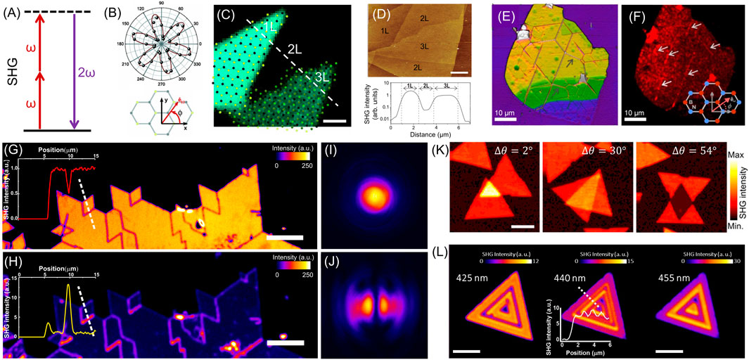
Figure 1. SHG Imaging: (A) SHG energy level diagram representing two photons of frequency
Due to the sensitivity of SHG to the material’s crystalline properties, one-dimensional defects such as edges, grain boundaries, and wrinkles are examples of common features presented by 2D materials that can be probed by SHG imaging (Oliveira et al., 2015; Yin et al., 2014; Karvonen et al., 2017; Carvalho et al., 2019). For instance, polarization-dependent SHG results have shown the formation of crystallographically-oriented origami-type wrinkles in annealed hexagonal boron nitride (h-BN) layers (Oliveira et al., 2015), Figures 1E, F. Additionally, Cunha et al. (2020) revealed the important role of defects in the increased efficiency of the SHG in h-BN flakes. SHG has also been used to reveal the crystalline details of grain boundaries. While the single crystalline grains exhibit uniform SHG intensities, edges and grain boundaries present a suppressed SH emission due to their translational symmetry breaking, which results in destructive interference of the SH fields and allows rapid visualization of these defective regions (Yin et al., 2014). Dark-field SHG imaging can also probe edges and grain boundaries in TMD monolayers (Carvalho et al., 2019). This method consists of blocking the central spot of the SH signal to collect only the emission at high angles, leading to an enhanced SH intensity at edges and grain boundaries, as displayed in Figures 1G–J. While there is destructive interference of the SH fields at small angles for these defective regions, large angles compensate for the fields’ phase difference and result in constructive interference of the SHG at grain boundaries and edges (Carvalho et al., 2019). These angle-dependent constructive and destructive interferences are confirmed by the spatial mode of the SH emission of a
In addition to the rich information directly given by the SHG imaging, it is also possible to employ data processing of the measured images to map distinct properties of the sample. For instance, a map of the crystallographic orientations of a TMD monolayer can be generated from polarization-resolved SHG imaging by fitting the angle-dependent SH expression (Equation 2) to the SH data of each measured pixel (David et al., 2015; Psilodimitrakopoulos et al., 2018). Besides, it was also reported a strain mapping of a TMD monolayer from polarization-resolved SHG imaging (Mennel et al., 2018; Li et al., 2019). As strain breaks the crystal symmetry of the material, it induces asymmetric intensities in the polarized SH six-fold pattern due to modifications in the second-order susceptibility tensor (Liang et al., 2017; Mennel et al., 2018; Mennel et al., 2019); Li et al., 2019). Hence, a photoelastic tensor was introduced to account for the role of the strain tensor on the nonlinear susceptibility tensor, allowing the evaluation of the strain field on the sample from polarization-resolved SHG measurements (Mennel et al., 2018; Mennel et al., 2019). Therefore, strain fields over a TMD monolayer can be mapped using this approach for each measured pixel of the polarized SHG images (Mennel et al., 2018).
Exploring the 2D materials field, we find that enormous efforts are also being placed in producing and investigating 2D heterostructures (Geim and Grigorieva, 2013; Novoselov et al., 2016; Castellanos-Gomez et al., 2022). For vertically stacked 2D materials, for example, novel and promising physical phenomena were revealed to be highly dependent on the twist angle between the forming layers (Castellanos-Gomez et al., 2022). Hence, SHG imaging emerges as a powerful non-invasive technique to determine the relative orientation between layers in 2D TMD heterostructures as well. This crystallographic orientation monitoring is simpler when the fabricated vertical heterostructure presents individual monolayers with non-overlapping areas (Hsu et al., 2014; Kim et al., 2021; Yuan et al., 2023). In such cases, it is possible to determine the crystallographic orientation of each monolayer from their isolated regions. However, as mentioned before, samples with
Twist angle analysis through SHG imaging experiments is not straightforward when the heterostructure contains monolayers without isolated regions. For a twisted TMD homobilayer in which one monolayer presents an isolated region and the other is entirely overlapped, it is elementary to obtain
Also regarding 2D heterostructures, it has been recently demonstrated that the stacking of TMD monolayers can be used to boost the nonlinear optical gain and reach strong SHG signals (Trovatello et al., 2021), achieving optical parametric amplification in 2D TMDs. Furthermore, high nonlinear conversion efficiencies were obtained by controlling the phase-matching in multilayer 3R-
TMD monolayers also exhibit singular valley phenomena (Xiao et al., 2012; Mak et al., 2012; Cao et al., 2012; Sousa et al., 2024a) due to their strong spin-orbit coupling and broken inversion symmetry, leading to a spin-valley locking effect at the inequivalent K and K′ valleys. This coupling between spin and valley degrees of freedom, which can be selectively accessed by circularly polarized light (Mak et al., 2012; Cao et al., 2012), sheds light on a novel field with promising potential for data storage, manipulation, and readout named valleytronics (Schaibley et al., 2016). Hence, control over valley polarization is essential in view of technological applications, resulting in a demand for methods to probe valley asymmetries in 2D materials. In this sense, a SHG imaging technique was recently used to map the valley imbalance in TMD monolayers (Mouchliadis et al., 2021). A valley polarization out of equilibrium impacts the crystal symmetry, thus inducing new terms in the material’s second-order susceptibility tensor and modifying the SH emission (Hipolito and Pereira, 2017). Particularly, the intrinsic
Beyond the largely investigated 2D materials like graphene, h-BN, and TMDs, SHG has also been employed to probe magnetic symmetries in emergent layered magnets such as
Despite the standard polarized SHG techniques presented above as consolidated and powerful tools to determine the orientation and symmetry properties of 2D materials, other innovative approaches using SHG improved and extended this second-order nonlinear technique to determine other properties and explore different materials. An example of this recent development is the Fourier space SH imaging, which provides a straightforward method to determine the crystallographic orientation and symmetry based on the images generated by the SH signal in Fourier space using an azimuthal laser mode to excite the sample (Lafeta et al., 2025). In addition, there is also a great interest in developing the potential of SHG techniques to improve their spatial resolution. Although considerable efforts are employed to achieve increased spatial resolutions for SHG imaging (Psilodimitrakopoulos et al., 2018), there is the intrinsic confocal optics resolution limitation of hundreds of nm due to the diffraction limit of light. Therefore, nonlinear near-field techniques emerge as a powerful alternative for imaging the nanoscale SH responses (Yao et al., 2022; Luo et al., 2023). For example, localized variations of the stacking order in a TMD homobilayer as well as excitonic resonances were recently probed by nano-SHG imaging with a spatial resolution down to 20 nm (Yao et al., 2022). Moreover, nano-SHG imaging could also detect localized variations in the symmetry of a TMD monolayer (Luo et al., 2023).
3 Third-order nonlinear imaging
Similar to second-order optical effects, third-order nonlinear processes play an important role and significantly contribute to the study of 2D materials. The general third-order nonlinear optical phenomenon is known as four-wave mixing (FWM), which depends on the third-order electrical susceptibility
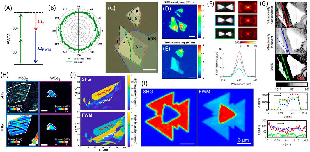
Figure 2. FWM Imaging: (A) Energy diagram of degenerate four-wave mixing process. (B) Polar plot of the third harmonic intensity from
Previous studies have explored the dependence of polarization in exceptional cases of FWM. In particular, Wang et al. (2014); Woodward et al. (2016) investigated the polarization dependence of THG, showing that third-harmonic signals from
In general, TMDs present a strong FWM signal that increases with the number of layers (Li et al., 2016; Säynätjoki et al., 2017; Rosa et al., 2018; Autere et al., 2018; Balla et al., 2018; Lange et al., 2024), as shown in Figures 2C–E, H–J. This FWM response complements the second-order techniques that cannot probe centrosymmetric materials such as even-layered TMDs due to their
Moreover, FWM techniques using broadband lasers were also used to investigate TMDs. This method employs broadband incident laser pulses to generate a combination of third-order nonlinear responses over a large range of frequencies, which enables the investigation of several effects such as resonances with excitons in one single measurement (Ko et al., 2019) and the increase in the nonlinear signal produced by the control of the phase of the incident fields (Lange et al., 2024).
FWM imaging has also been employed to study other 2D materials such as graphene and h-BN, with a special interest in monolayer graphene because of its inversion symmetry and consequent absent SHG signal (Shan et al., 2018). Such investigations in these materials exploit a particular case of DFWM called coherent anti-Stokes Raman scattering (CARS), which occurs when the energy difference of the incident fields (
4 Conclusion and future perspectives
In this mini-review, we explored the progress of nonlinear optical imaging techniques for the study of nonlinear responses in 2D materials, focusing on second-order, specifically SHG, and third-order, FWM and its special cases. In particular, we discussed the potential of SHG experiments to probe symmetry information of h-BN and odd-layered TMD samples. This capability of SHG measurements to image the crystallographic orientations of these materials enables, for example, the study of one-dimensional defects such as wrinkles, edges, and grain boundaries, the determination of twist angles in 2D heterostructures, and the mapping of strain fields over the samples. Additionally, we discussed the importance of FWM experiments to investigate nonlinearities of centrosymmetric materials as graphene and even-layered TMDs, which present a negligible second-order nonlinear emission. Furthermore, we also addressed the potential of resonant SHG and FWM experiments to probe excitonic effects in TMDs. In summary, we highlighted that these non-invasive nonlinear techniques are powerful tools for the rapid imaging of 2D material properties, paving the way for the development of sample fabrication and the research of fundamental aspects of nonlinear optics. Moreover, we shed light on different possibilities to increase the potential of nonlinear imaging. For instance, the recent use of near-field SHG and FWM experiments allows the imaging of 2D materials with nanometric resolution, while the constant developments in nonlinear optics theory and data analysis enable to unveil notable properties as valley population imbalance in these samples.
Author contributions
FS: Investigation, Writing–original draft, Writing–review and editing. LL: Investigation, Writing–original draft, Writing–review and editing. GF: Investigation, Writing–original draft, Writing–review and editing. AdP: Conceptualization, Funding acquisition, Investigation, Writing–original draft, Writing–review and editing.
Funding
The author(s) declare that financial support was received for the research, authorship, and/or publication of this article. This work was financially supported by the Brazilian agencies Conselho Nacional de Desenvolvimento Científico e Tecnológico (CNPq), Fundação de Amparo à Pesquisa do Estado de Minas Gerais (Fapemig), Coordenação de Aperfeiçoamento de Pessoal de Nível Superior (Capes) and Institute of Science and Technology (INCT) in Carbon Nanomaterials. LL gratefully acknowledges the Alexander von Humboldt Foundation for its financial support.
Conflict of interest
The authors declare that the research was conducted in the absence of any commercial or financial relationships that could be construed as a potential conflict of interest.
Generative AI statement
The author(s) declare that no Generative AI was used in the creation of this manuscript.
Publisher’s note
All claims expressed in this article are solely those of the authors and do not necessarily represent those of their affiliated organizations, or those of the publisher, the editors and the reviewers. Any product that may be evaluated in this article, or claim that may be made by its manufacturer, is not guaranteed or endorsed by the publisher.
References
Aras, F. G., Yilmaz, A., Tasdelen, H. G., Ozden, A., Ay, F., Perkgoz, N. K., et al. (2022). A review on recent advances of chemical vapor deposition technique for monolayer transition metal dichalcogenides (MX2: Mo, W; S, Se, Te). Mater. Sci. Semicond. Process. 148, 106829. doi:10.1016/j.mssp.2022.106829
Autere, A., Jussila, H., Marini, A., Saavedra, J. R. M., Dai, Y., Säynätjoki, A., et al. (2018). Optical harmonic generation in monolayer group-VI transition metal dichalcogenides. Phys. Rev. B 98, 115426. doi:10.1103/PhysRevB.98.115426
Balla, N. K., O’Brien, M., McEvoy, N., Duesberg, G. S., Rigneault, H., Brasselet, S., et al. (2018). Effects of excitonic resonance on second and third order nonlinear scattering from few-layer mos2. ACS Photonics 5, 1235–1240. doi:10.1021/acsphotonics.7b00912
Bauer, J. M., Chen, L., Wilhelm, P., Watanabe, K., Taniguchi, T., Bange, S., et al. (2022). Excitonic resonances control the temporal dynamics of nonlinear optical wave mixing in monolayer semiconductors. Nat. Photonics 16, 777–783. doi:10.1038/s41566-022-01080-1
Cao, T., Wang, G., Han, W., Ye, H., Zhu, C., Shi, J., et al. (2012). Valley-selective circular dichroism of monolayer molybdenum disulphide. Nat. Commun. 3, 887. doi:10.1038/ncomms1882
Carvalho, B. R., Wang, Y., Fujisawa, K., Zhang, T., Kahn, E., Bilgin, I., et al. (2019). Nonlinear dark-field imaging of one-dimensional defects in monolayer dichalcogenides. Nano Lett. 20, 284–291. doi:10.1021/acs.nanolett.9b03795
Castellanos-Gomez, A., Duan, X., Fei, Z., Gutierrez, H. R., Huang, Y., Huang, X., et al. (2022). Van der Waals heterostructures. Nat. Rev. Methods Prim. 2, 58. doi:10.1038/s43586-022-00139-1
Cheng, J., Jiang, T., Ji, Q., Zhang, Y., Li, Z., Shan, Y., et al. (2015). Kinetic nature of grain boundary formation in as-grown MoS2 monolayers. Adv. Mater. Deerf. Beach, Fla 27, 4069–4074. doi:10.1002/adma.201501354
Cheng, J.-X., and Xie, X. S. (2004). Coherent anti-Stokes Raman scattering Microscopy: instrumentation, theory, and applications. J. Phys. Chem. B 108, 827–840. doi:10.1021/jp035693v
Chu, H., Roh, C. J., Island, J. O., Li, C., Lee, S., Chen, J., et al. (2020). Linear magnetoelectric phase in ultrathin mnps 3 probed by optical second harmonic generation. Phys. Rev. Lett. 124, 027601. doi:10.1103/physrevlett.124.027601
Cunha, R., Cadore, A., Ramos, S. L., Watanabe, K., Taniguchi, T., Kim, S., et al. (2020). Second harmonic generation in defective hexagonal boron nitride. J. Phys. Condens. Matter 32, 19LT01. doi:10.1088/1361-648x/ab6cbf
Cunha, R., Lafeta, L., Fonseca, E. A., Barbosa, A., Romano-Silva, M. A., Vieira, R., et al. (2021). Nonlinear and vibrational microscopy for label-free characterization of amyloid-β plaques in Alzheimer's disease model. Analyst 146, 2945–2954. doi:10.1039/D1AN00074H
Dai, Y., Wang, Y., Das, S., Li, S., Xue, H., Mohsen, A., et al. (2021). Broadband plasmon-enhanced four-wave mixing in monolayer MoS2. Nano Lett. 21, 6321–6327. doi:10.1021/acs.nanolett.1c02381
David, S. N., Zhai, Y., Van Der Zande, A. M., O’Brien, K., Huang, P. Y., Chenet, D. A., et al. (2015). Rapid, all-optical crystal orientation imaging of two-dimensional transition metal dichalcogenide monolayers. Appl. Phys. Lett. 107. doi:10.1063/1.4930232
Duncan, M. D., Reintjes, J., and Manuccia, T. J. (1982). Scanning coherent anti-Stokes Raman microscope. Opt. Lett. 7, 350–352. doi:10.1364/OL.7.000350
Fano, U. (1961). Effects of configuration interaction on intensities and phase shifts. Phys. Rev. 124, 1866–1878. doi:10.1103/PhysRev.124.1866
Franken, P. A., Hill, A. E., Peters, C. W., and Weinreich, G. (1961). Generation of optical harmonics. Phys. Rev. Lett. 7, 118–119. doi:10.1103/PhysRevLett.7.118
Geim, A. K., and Grigorieva, I. V. (2013). Van der Waals heterostructures. Nature 499, 419–425. doi:10.1038/nature12385
Golub, L., and Tarasenko, S. (2014). Valley polarization induced second harmonic generation in graphene. Phys. Rev. B 90, 201402. doi:10.1103/physrevb.90.201402
Gomes, E. F. A., Paulino Junior, E., de Lima, M. F. R., Reis, L. A., Paranhos, G., Mamede, M., et al. (2023). Prostate cancer tissue classification by multiphoton imaging, automated image analysis and machine learning. J. Biophot. 16, e202200382. doi:10.1002/jbio.202200382
Gordeev, G., Lafeta, L., Flavel, B. S., Jorio, A., and Malard, L. M. (2023). Excitonic resonances in coherent anti-Stokes Raman scattering from single-walled carbon nanotubes. J. Phys. Chem. C 127, 20438–20444. doi:10.1021/acs.jpcc.3c05696
Herrmann, P., Klimmer, S., Lettau, T., Monfared, M., Staude, I., Paradisanos, I., et al. (2023). Nonlinear all-optical coherent generation and read-out of valleys in atomically thin semiconductors. Small 19, 2301126. doi:10.1002/smll.202301126
Hipolito, F., and Pereira, V. M. (2017). Second harmonic spectroscopy to optically detect valley polarization in 2D materials. 2D Mater. 4, 021027. doi:10.1088/2053-1583/aa6f4d
Ho, Y. W., Rosa, H. G., Verzhbitskiy, I., Rodrigues, M. J., Taniguchi, T., Watanabe, K., et al. (2020). Measuring valley polarization in two-dimensional materials with second-harmonic spectroscopy. ACS Photonics 7, 925–931. doi:10.1021/acsphotonics.0c00174
Hsu, W.-T., Zhao, Z.-A., Li, L.-J., Chen, C.-H., Chiu, M.-H., Chang, P.-S., et al. (2014). Second harmonic generation from artificially stacked transition metal dichalcogenide twisted bilayers. ACS Nano 8, 2951–2958. doi:10.1021/nn500228r
Huang, P. Y., Ruiz-Vargas, C. S., Van Der Zande, A. M., Whitney, W. S., Levendorf, M. P., Kevek, J. W., et al. (2011). Grains and grain boundaries in single-layer graphene atomic patchwork quilts. Nature 469, 389–392. doi:10.1038/nature09718
Karvonen, L., Säynätjoki, A., Huttunen, M. J., Autere, A., Amirsolaimani, B., Li, S., et al. (2017). Rapid visualization of grain boundaries in monolayer MoS2 by multiphoton microscopy. Nat. Commun. 8, 15714. doi:10.1038/ncomms15714
Kim, W., Ahn, J. Y., Oh, J., Shim, J. H., and Ryu, S. (2020). Second-harmonic young’s interference in atom-thin heterocrystals. Nano Lett. 20, 8825–8831. doi:10.1021/acs.nanolett.0c03763
Kim, Y. C., Yoo, H., Nguyen, V. T., Lee, S., Park, J.-Y., and Ahn, Y. H. (2021). High-speed imaging of second-harmonic generation in MoS2 bilayer under femtosecond laser ablation. Nanomaterials 11, 1786. doi:10.3390/nano11071786
Ko, B. A., Sokolov, A. V., Scully, M. O., Zhang, Z., and Lee, H. W. H. (2019). Enhanced four-wave mixing process near the excitonic resonances of bulk MoS2. Phot. Res. 7, 251–259. doi:10.1364/PRJ.7.000251
Lafeta, L., Cadore, A. R., Mendes-de Sa, T. G., Watanabe, K., Taniguchi, T., Campos, L. C., et al. (2017). Anomalous nonlinear optical response of graphene near phonon resonances. Nano Lett. 17, 3447–3451. doi:10.1021/acs.nanolett.7b00329
Lafeta, L., Corradi, A., Zhang, T., Kahn, E., Bilgin, I., Carvalho, B. R., et al. (2021). Second- and third-order optical susceptibilities across excitons states in 2D monolayer transition metal dichalcogenides. 2D Mater. 8, 035010. doi:10.1088/2053-1583/abeed4
Lafeta, L., Hartmann, S., Rosa, B., Reitzenstein, S., Malard, L. M., and Hartschuh, A. (2025). Probing noncentrosymmetric 2D materials by Fourier space second harmonic imaging. ACS Photonics 12, 357–363. doi:10.1021/acsphotonics.4c01724
Lange, L., Wang, K., Bange, S., Lafeta, L., Rosa, B., Reitzenstein, S., et al. (2024). Ultrafast phase-control of the nonlinear optical response of 2D semiconductors. ACS Photonics 11, 3112–3122. doi:10.1021/acsphotonics.4c00388
Lee, K., Dismukes, A. H., Telford, E. J., Wiscons, R. A., Wang, J., Xu, X., et al. (2021). Magnetic order and symmetry in the 2d semiconductor crsbr. Nano Lett. 21, 3511–3517. doi:10.1021/acs.nanolett.1c00219
Li, D., Wei, C., Song, J., Huang, X., Wang, F., Liu, K., et al. (2019). Anisotropic enhancement of second-harmonic generation in monolayer and bilayer MoS2 by integrating with TiO2 nanowires. Nano Lett. 19, 4195–4204. doi:10.1021/acs.nanolett.9b01933
Li, D., Xiong, W., Jiang, L., Xiao, Z., Rabiee Golgir, H., Wang, M., et al. (2016). Multimodal nonlinear optical imaging of mos2 and mos2-based van der waals heterostructures. ACS Nano 10, 3766–3775. doi:10.1021/acsnano.6b00371
Li, S., Li, Y., Yi, R., Liu, L., and Qu, J. (2020). Coherent anti-Stokes Raman scattering microscopy and its applications. Front. Phys. 8. doi:10.3389/fphy.2020.598420
Li, Y., Rao, Y., Mak, K. F., You, Y., Wang, S., Dean, C. R., et al. (2013). Probing symmetry properties of few-layer MoS2 and h-BN by optical second-harmonic generation. Nano Lett. 13, 3329–3333. doi:10.1021/nl401561r
Liang, J., Zhang, J., Li, Z., Hong, H., Wang, J., Zhang, Z., et al. (2017). Monitoring local strain vector in atomic-layered MoSe2 by second-harmonic generation. Nano Lett. 17, 7539–7543. doi:10.1021/acs.nanolett.7b03476
Lin, Z., Carvalho, B. R., Kahn, E., Lv, R., Rao, R., Terrones, H., et al. (2016). Defect engineering of two-dimensional transition metal dichalcogenides. 2D Mater. 3, 022002. doi:10.1088/2053-1583/3/2/022002
Ling, J., Miao, X., Sun, Y., Feng, Y., Zhang, L., Sun, Z., et al. (2019). Vibrational imaging and quantification of two-dimensional hexagonal Boron Nitride with stimulated Raman scattering. ACS Nano 13, 14033–14040. doi:10.1021/acsnano.9b06337
Luo, W., Whetten, B. G., Kravtsov, V., Singh, A., Yang, Y., Huang, D., et al. (2023). Ultrafast nanoimaging of electronic coherence of monolayer WSe2. Nano Lett. 23, 1767–1773. doi:10.1021/acs.nanolett.2c04536
Ly, T. H., Chiu, M.-H., Li, M.-Y., Zhao, J., Perello, D. J., Cichocka, M. O., et al. (2014). Observing grain boundaries in CVD-grown monolayer transition metal dichalcogenides. ACS Nano 8, 11401–11408. doi:10.1021/nn504470q
Mak, K. F., He, K., Shan, J., and Heinz, T. F. (2012). Control of valley polarization in monolayer MoS2 by optical helicity. Nat. Nanotechnol. 7, 494–498. doi:10.1038/nnano.2012.96
Malard, L. M., Alencar, T. V., Barboza, A. P. M., Mak, K. F., and de Paula, A. M. (2013). Observation of intense second harmonic generation from atomic crystals. Phys. Rev. B 87, 201401. doi:10.1103/physrevb.87.201401
Malard, L. M., Lafeta, L., Cunha, R. S., Nadas, R., Gadelha, A., Cançado, L. G., et al. (2021). Studying 2D materials with advanced Raman spectroscopy: CARS, SRS and TERS. Phys. Chem. Chem. Phys. 23, 23428–23444. doi:10.1039/D1CP03240B
Manzoni, C., and Cerullo, G. (2016). Design criteria for ultrafast optical parametric amplifiers. J. Opt. 18, 103501. doi:10.1088/2040-8978/18/10/103501
Mennel, L., Furchi, M. M., Wachter, S., Paur, M., Polyushkin, D. K., and Mueller, T. (2018). Optical imaging of strain in two-dimensional crystals. Nat. Commun. 9, 516. doi:10.1038/s41467-018-02830-y
Mennel, L., Paur, M., and Mueller, T. (2019). Second harmonic generation in strained transition metal dichalcogenide monolayers: MoS2, MoSe2, WS2, and WSe2. Apl. Photonics 4, 034404. doi:10.1063/1.5051965
Mouchliadis, L., Psilodimitrakopoulos, S., Maragkakis, G. M., Demeridou, I., Kourmoulakis, G., Lemonis, A., et al. (2021). Probing valley population imbalance in transition metal dichalcogenides via temperature-dependent second harmonic generation imaging. npj 2D Mater. Appl. 5 (6), 6. doi:10.1038/s41699-020-00183-z
Novoselov, K. S., Mishchenko, A., Carvalho, A., and Castro Neto, A. (2016). 2d materials and van der Waals heterostructures. Science 353, aac9439. doi:10.1126/science.aac9439
Oliveira, C. K., Gomes, E. F. A., Prado, M. C., Alencar, T. V., Nascimento, R., Malard, L. M., et al. (2015). Crystal-oriented wrinkles with origami-type junctions in few-layer hexagonal boron nitride. Nano Res. 8, 1680–1688. doi:10.1007/s12274-014-0665-y
Ouellette, J. N., Drifka, C. R., Pointer, K. B., Liu, Y., Lieberthal, T. J., Kao, W. J., et al. (2021). Navigating the collagen jungle: the biomedical potential of fiber organization in cancer. Bioengineering 8, 17. doi:10.3390/bioengineering8020017
Paddubskaya, A., Rutkauskas, D., Karpicz, R., Dovbeshko, G., Nebogatikova, N., Antonova, I., et al. (2020). Recognition of spatial distribution of CNT and graphene in hybrid structure by mapping with coherent anti-Stokes Raman microscopy. Nanoscale Res. Lett. 15, 37. doi:10.1186/s11671-020-3264-8
Palekar, C. C., Faria Junior, P. E., Rosa, B., Sousa, F. B., Malard, L. M., Fabian, J., et al. (2024). Amplification of interlayer exciton emission in twisted WSe2/WSe2/MoSe2 heterotrilayers. npj 2D Mater. Appl. 8, 49. doi:10.1038/s41699-024-00483-8
Paradisanos, I., Raven, A. M. S., Amand, T., Robert, C., Renucci, P., Watanabe, K., et al. (2022). Second harmonic generation control in twisted bilayers of transition metal dichalcogenides. Phys. Rev. B 105, 115420. doi:10.1103/physrevb.105.115420
Polli, D., Kumar, V., Valensise, C. M., Marangoni, M., and Cerullo, G. (2018). Broadband coherent Raman scattering microscopy. Laser and Photonics Rev. 12, 1800020. doi:10.1002/lpor.201800020
Potma, E. O., and Xie, X. S. (2004). Cars microscopy for biology and medicine. Opt. Phot. News 15, 40–45. doi:10.1364/OPN.15.11.000040
Psilodimitrakopoulos, S., Mouchliadis, L., Maragkakis, G. M., Kourmoulakis, G., Lemonis, A., Kioseoglou, G., et al. (2020). Real-time spatially resolved determination of twist angle in transition metal dichalcogenide heterobilayers. 2D Mater. 8, 015015. doi:10.1088/2053-1583/abbf88
Psilodimitrakopoulos, S., Mouchliadis, L., Paradisanos, I., Kourmoulakis, G., Lemonis, A., Kioseoglou, G., et al. (2019). Twist angle mapping in layered WS2 by polarization-resolved second harmonic generation. Sci. Rep. 9, 14285. doi:10.1038/s41598-019-50534-0
Psilodimitrakopoulos, S., Mouchliadis, L., Paradisanos, I., Lemonis, A., Kioseoglou, G., and Stratakis, E. (2018). Ultrahigh-resolution nonlinear optical imaging of the armchair orientation in 2D transition metal dichalcogenides. Light Sci. and Appl. 7, 18005. doi:10.1038/lsa.2018.5
Rosa, B. L. T., Fujisawa, K., Santos, J. C. C., Zhang, T., Matos, M. J. S., Sousa, F. B., et al. (2022). Investigation of spatially localized defects in synthetic monolayers. Phys. Rev. B 106, 115301. doi:10.1103/PhysRevB.106.115301
Rosa, H. G., Ho, Y. W., Verzhbitskiy, I., Rodrigues, M. J. F. L., Taniguchi, T., Watanabe, K., et al. (2018). Characterization of the second- and third-harmonic optical susceptibilities of atomically thin tungsten diselenide. Sci. Rep. 8, 10035. doi:10.1038/s41598-018-28374-1
Rycerz, A., Tworzydło, J., and Beenakker, C. (2007). Valley filter and valley valve in graphene. Nat. Phys. 3, 172–175. doi:10.1038/nphys547
Säynätjoki, A., Karvonen, L., Rostami, H., Autere, A., Mehravar, S., Lombardo, A., et al. (2017). Ultra-strong nonlinear optical processes and trigonal warping in mos2 layers. Nat. Commun. 8, 893. doi:10.1038/s41467-017-00749-4
Schaibley, J. R., Yu, H., Clark, G., Rivera, P., Ross, J. S., Seyler, K. L., et al. (2016). Valleytronics in 2D materials. Nat. Rev. Mater. 1, 16055. doi:10.1038/natrevmats.2016.55
Seyler, K. L., Schaibley, J. R., Gong, P., Rivera, P., Jones, A. M., Wu, S., et al. (2015). Electrical control of second-harmonic generation in a WSe2 monolayer transistor. Nat. Nanotechnol. 10, 407–411. doi:10.1038/nnano.2015.73
Shan, Y., Li, Y., Huang, D., Tong, Q., Yao, W., Liu, W.-T., et al. (2018). Stacking symmetry governed second harmonic generation in graphene trilayers. Sci. Adv. 4, eaat0074. doi:10.1126/sciadv.aat0074
Shree, S., Lagarde, D., Lombez, L., Robert, C., Balocchi, A., Watanabe, K., et al. (2021). Interlayer exciton mediated second harmonic generation in bilayer MoS2. Nat. Commun. 12, 6894. doi:10.1038/s41467-021-27213-8
Sie, E. J., Lui, C. H., Lee, Y.-H., Fu, L., Kong, J., and Gedik, N. (2017). Large, valley-exclusive Bloch-Siegert shift in monolayer WS2. Science 355, 1066–1069. doi:10.1126/science.aal2241
Sousa, F. B., Lafeta, L., Cadore, A. R., Sahoo, P. K., and Malard, L. M. (2021). Revealing atomically sharp interfaces of two-dimensional lateral heterostructures by second harmonic generation. 2D Mater. 8, 035051. doi:10.1088/2053-1583/ac0731
Sousa, F. B., Matos, M. J. S., Carvalho, B. R., Liu, M., Ames, A., Zhou, D., et al. (2024a). Giant Valley zeeman splitting in vanadium-doped WSe2 monolayers. Small 20, 2405434. doi:10.1002/smll.202405434
Sousa, F. B., Nadas, R., Martins, R., Barboza, A. P. M., Soares, J. S., Neves, B. R. A., et al. (2024b). Disentangling doping and strain effects at defects of grown MoS2 monolayers with nano-optical spectroscopy. Nanoscale 16, 12923–12933. doi:10.1039/D4NR00837E
Sousa, F. B., Zheng, B., Liu, M., Resende, G. C., Zhou, D., Pimenta, M. A., et al. (2024c). Effects of vanadium doping on the optical response and electronic structure of WS2 monolayers. Adv. Opt. Mater. 12, 2400235. doi:10.1002/adom.202400235
Stepanov, E., Semin, S., Woods, C., Vandelli, M., Kimel, A., Novoselov, K., et al. (2020). Direct observation of incommensurate–commensurate transition in graphene-hbn heterostructures via optical second harmonic generation. ACS Appl. Mater. and interfaces 12, 27758–27764. doi:10.1021/acsami.0c05965
Sun, Z., Yi, Y., Song, T., Clark, G., Huang, B., Shan, Y., et al. (2019). Giant nonreciprocal second-harmonic generation from antiferromagnetic bilayer cri3. Nature 572, 497–501. doi:10.1038/s41586-019-1445-3
Trovatello, C., Marini, A., Xu, X., Lee, C., Liu, F., Curreli, N., et al. (2021). Optical parametric amplification by monolayer transition metal dichalcogenides. Nat. Photonics 15, 6–10. doi:10.1038/s41566-020-00728-0
Van Der Zande, A. M., Huang, P. Y., Chenet, D. A., Berkelbach, T. C., You, Y., Lee, G.-H., et al. (2013). Grains and grain boundaries in highly crystalline monolayer molybdenum disulphide. Nat. Mater. 12, 554–561. doi:10.1038/nmat3633
Virga, A., Ferrante, C., Batignani, G., De Fazio, D., Nunn, A. D. G., Ferrari, A. C., et al. (2019). Coherent anti-Stokes Raman spectroscopy of single and multi-layer graphene. Nat. Commun. 10, 3658. doi:10.1038/s41467-019-11165-1
Wang, G., Gerber, I. C., Bouet, L., Lagarde, D., Balocchi, A., Vidal, M., et al. (2015a). Exciton states in monolayer MoSe2: impact on interband transitions. 2D Mater. 2, 045005. doi:10.1088/2053-1583/2/4/045005
Wang, G., Marie, X., Gerber, I., Amand, T., Lagarde, D., Bouet, L., et al. (2015b). Giant enhancement of the optical second-harmonic emission of monolayers by laser excitation at exciton resonances. Phys. Rev. Lett. 114, 097403. doi:10.1103/physrevlett.114.097403
Wang, R., Chien, H.-C., Kumar, J., Kumar, N., Chiu, H.-Y., and Zhao, H. (2014). Third-harmonic generation in ultrathin films of mos2. ACS Appl. Mater. and Interfaces 6, 314–318. doi:10.1021/am4042542
Wen, X., Gong, Z., and Li, D. (2019). Nonlinear optics of two-dimensional transition metal dichalcogenides. InfoMat 1, 317–337. doi:10.1002/inf2.12024
Woodward, R. I., Murray, R. T., Phelan, C. F., de Oliveira, R. E. P., Runcorn, T. H., Kelleher, E. J. R., et al. (2016). Characterization of the second- and third-order nonlinear optical susceptibilities of monolayer MoS2 using multiphoton microscopy. 2D Mater. 4, 011006. doi:10.1088/2053-1583/4/1/011006
Wu, J.-B., Wu, H., and Tan, P.-H. (2024). Magneto-optical interactions in layered magnets. Adv. Funct. Mater. 34, 2312214. doi:10.1002/adfm.202312214
Xiao, D., Liu, G.-B., Feng, W., Xu, X., and Yao, W. (2012). Coupled spin and valley physics in monolayers of and other group-VI dichalcogenides. Phys. Rev. Lett. 108, 196802. doi:10.1103/PhysRevLett.108.196802
Xiao, D., Yao, W., and Niu, Q. (2007). Valley-contrasting physics in graphene: magnetic moment and topological transport. Phys. Rev. Lett. 99, 236809. doi:10.1103/physrevlett.99.236809
Xu, M., Ji, H., Zhang, M., Zheng, L., Li, W., Luo, L., et al. (2024). CVD synthesis of twisted bilayer WS2 with tunable second harmonic generation. Adv. Mater. 36, 2313638. doi:10.1002/adma.202313638
Xu, X., Trovatello, C., Mooshammer, F., Shao, Y., Zhang, S., Yao, K., et al. (2022). Towards compact phase-matched and waveguided nonlinear optics in atomically layered semiconductors. Nat. Photonics 16, 698–706. doi:10.1038/s41566-022-01053-4
Yao, K., Zhang, S., Yanev, E., McCreary, K., Chuang, H.-J., Rosenberger, M. R., et al. (2022). Nanoscale optical imaging of 2D semiconductor stacking orders by exciton-enhanced second harmonic generation. Adv. Opt. Mater. 10, 2200085. doi:10.1002/adom.202200085
Yao, W., Xiao, D., and Niu, Q. (2008). Valley-dependent optoelectronics from inversion symmetry breaking. Phys. Rev. B—Condensed Matter Mater. Phys. 77, 235406. doi:10.1103/physrevb.77.235406
Yin, X., Ye, Z., Chenet, D. A., Ye, Y., O’Brien, K., Hone, J. C., et al. (2014). Edge nonlinear optics on a MoS 2 atomic monolayer. Science 344, 488–490. doi:10.1126/science.1250564
Yu, Q., Jauregui, L. A., Wu, W., Colby, R., Tian, J., Su, Z., et al. (2011). Control and characterization of individual grains and grain boundaries in graphene grown by chemical vapour deposition. Nat. Mater. 10, 443–449. doi:10.1038/nmat3010
Yuan, Y., Liu, P., Wu, H., Chen, H., Zheng, W., Peng, G., et al. (2023). Probing the twist-controlled interlayer coupling in artificially stacked transition metal dichalcogenide bilayers by second-harmonic generation. ACS Nano 17, 17897–17907. doi:10.1021/acsnano.3c03795
Zhang, Y., Yao, Y., Sendeku, M. G., Yin, L., Zhan, X., Wang, F., et al. (2019). Recent progress in cvd growth of 2d transition metal dichalcogenides and related heterostructures. Adv. Mater. 31, 1901694. doi:10.1002/adma.201901694
Zhao, M., Ye, Z., Suzuki, R., Ye, Y., Zhu, H., Xiao, J., et al. (2016). Atomically phase-matched second-harmonic generation in a 2D crystal. Light Sci. and Appl. 5, e16131. doi:10.1038/lsa.2016.131
Keywords: nonlinear imaging, second-harmonic generation, four-wave mixing, transition metal dichalcogenides, hexagonal boron nitride, heterostructures
Citation: Sousa FB, Lafeta L, Fonseca GR and de Paula AM (2025) Nonlinear optical imaging of two-dimensional nanomaterials. Front. Nanotechnol. 7:1537299. doi: 10.3389/fnano.2025.1537299
Received: 30 November 2024; Accepted: 10 January 2025;
Published: 29 January 2025.
Edited by:
Chiara Trovatello, Columbia University, United StatesReviewed by:
Yinan Dong, Columbia University, United StatesSebastian Klimmer, Friedrich Schiller University Jena, Germany
Copyright © 2025 Sousa, Lafeta, Fonseca and de Paula. This is an open-access article distributed under the terms of the Creative Commons Attribution License (CC BY). The use, distribution or reproduction in other forums is permitted, provided the original author(s) and the copyright owner(s) are credited and that the original publication in this journal is cited, in accordance with accepted academic practice. No use, distribution or reproduction is permitted which does not comply with these terms.
*Correspondence: Ana M. de Paula, YW5hQGZpc2ljYS51Zm1nLmJy
†These authors have contributed equally to this work