 Yan-ping Tang
 Yi-xin Yin1†
 Ji-lin Li
 Ke-zhi Li
 Bang-li Hu
- 1Department of Research, Guangxi Medical University Cancer Hospital, Nanning, China
- 2Department of Chemotherapy, Guangxi Medical University Cancer Hospital, Nanning, China
Background: The role of hyaluronan-mediated motility receptor (HMMR) in colorectal cancer (CRC) remains unclear. The present study aimed to explore the association of HMMR with the development and prognosis of CRC using sequence datasets, clinical tissues, blood samples, and cell lines.
Methods: CRC datasets were downloaded from TCGA and GEO databases. Forty CRC tissue samples, 120 CRC blood samples, and 100 healthy controls were collected. Four CRC cell lines (HCT116, HT-29, LoVo, and SW480) and one normal human colon mucosal epithelial cell line (NCM460) were cultured. RT-qPCR was used to determine the expression of HMMR in the tissues and cell lines. ELISA was used to measure HMMR levels in the blood samples.
Results: The expression of HMMR was significantly increased in CRC tissues than in corresponding adjacent tissues based on TCGA and GEO datasets, and clinical CRC tissues. No associations were found between the expression of HMMR and the TNM stage or other clinical parameters. The expression of HMMR varied in different CRC cell lines. The blood levels of HMMR tended to be higher in patients with CRC than in healthy controls. TCGA and GEO datasets showed inconsistent results regarding the association of HMMR expression with the survival of patients with CRC.
Conclusion: The expression of HMMR is increased in CRC tissues but not in the blood. The expression of HMMR is independent of CRC development and has no prognostic significance in patients with CRC.
Introduction
Colorectal cancer (CRC) is one of the most common malignancies worldwide, according to the GLOBOCAN 2020 reports (Sung et al., 2021). Despite advances in surgical techniques, and adjuvant and neoadjuvant therapies, the prognosis of patients with CRC at a later stage remains poor. The pathogenesis of CRC is a multifactorial process, and molecular changes are one of the crucial factors that lead to the development of CRC. Several molecules are associated with the development and progression of CRC, and some of them can serve as indicators of CRC diagnosis, treatment monitoring, and prognosis (Di Nicolantonio et al., 2021). However, other molecules still need to be explored.
The hyaluronan-mediated motility receptor (HMMR, also known as RHAMM), a hyaluronan receptor, is localized in the cytoplasm, nucleus, and cell surface and regulates cell motility and cell cycle (Evanko and Wight, 1999; Jiang et al., 2013; Carvalho et al., 2021). Evidence suggests that the HMMR is involved in the occurrence and development of tumors that are dependent on hyaluronan-mediated signaling (Tolg et al., 2003; He et al., 2020). Previous studies indicate that the HMMR is overexpressed in and is a potential prognostic factor in various cancer types, including breast cancer (Liu et al., 2016), head and neck squamous cell carcinoma (Lu et al., 2021), hepatocellular carcinoma (Lu et al., 2020), and other cancers.
Several studies have reported the role of HMMR in CRC. Zlobec et al. (Zlobec et al., 2008c) reported that high expression of HMMR was significantly associated with the five-year survival rate in patients with CRC. Sun et al. (Sun et al., 2020) analyzed data from The Cancer Genome Atlas (TCGA) and Gene Expression Omnibus (GEO) databases and found that the HMMR was significantly associated with overall survival in patients with CRC. The HMMR was also found to involve in the metastasis and treatment response in CRC (Lugli et al., 2006; Mele et al., 2017; Burren et al., 2021). These results indicate that the HMMR is a predictor of CRC in patients. However, due to limited data, the robustness of the results is undermined. Thus, the role of HMMR in CRC needs to be further elucidated. In the present study, we explored the role of HMMR in CRC by comprehensively analyzing its expression in CRC sequencing databases, tissue and serum samples, and cell lines; this study may provide more reliable results to validate the role of HMMR in CRC.
Materials and Methods
HMMR Data in CRC From TCGA and GEO Databases
TCGA data of HMMR in colon adenocarcinoma (COAD) and rectal adenocarcinoma (READ) were downloaded from the UCSC Xena database (http://xena.ucsc.edu/) that includes 440 colon cancer and 158 rectal cancer samples. The HMMR values and phenotypes of patients were extracted together. Twelve CRC microarray datasets were downloaded from the GEO database with accession numbers GSE21510 (num. 148), GSE21815 (num. 141), GSE31279 (num. 110), GSE44076 (num. 246), GSE32323 (num. 44), GSE113513 (num. 28), GSE164191 (num. 121), GSE47756 (num. 93), GSE17537 (num. 55), GSE12945 (num. 62), GSE17536 (num. 177), and GSE14333 (num. 290). Of these, GSE164191 and GSE47756 provided data from the blood of patients with CRC, and the remaining samples were from CRC tissues.
Clinical CRC Sample Collection
We collected 40 CRC tissues and corresponding adjacent normal tissues from the BioBank of the Guangxi Medical University Cancer Hospital between January 2017 and December 2019. Fresh tissue samples were frozen immediately after surgery and stored in liquid nitrogen until further use. All tissues were histologically confirmed to be CRC. In addition, the blood samples of 120 CRC patients and 100 healthy controls were collected. The patients with CRC did not receive chemotherapy or other treatments prior to surgery, or suffer from diseases, such as immune diseases and severe major organ dysfunction. This study was approved by the Ethics Committee of the Guangxi Medical University Cancer Hospital (2021-KY-043), and written informed consent was obtained from all participants.
Cell Lines and Culture
Four CRC cell lines, HCT116, LoVo, SW480, and HT-29, and one human colon mucosal epithelial cell line, NCM460, were obtained from the Chinese Academy of Sciences (Shanghai, China). HCT116, LoVo, SW480, and HT-29 cells were cultured in DMEM with GlutaMAX (Gibco) supplemented with 10% FBS and 1% streptomycin/penicillin. NCM460 cells were cultured in phenol red–free RPMI 1640 (Gibco) supplemented with 10% FBS and 1% streptomycin/penicillin. The identity of the cell lines was confirmed by STR profiling.
RNA Isolation From Colon Cancer Tissues and Cells
Total RNA from CRC tissues and cell lines was isolated using TRIzol reagent (Invitrogen; Thermo Fisher Scientific, Inc., Waltham, MA, United States), according to the manufacturer’s instructions. The RNA concentration was tested using NanoDrop ND-1000, and the quality was assessed using electrophoresis with 1.5% denaturing agarose gels.
RT-qPCR Procedure
First-strand cDNA was synthesized by reverse transcription from 1 µg RNA using the PrimeScript RT reagent Kit with gDNA Eraser (Takara, Dalian, China). qPCR was performed using the standard protocol from the SYBR® Premix Ex Taq kit (Takara, Dalian, China), according to the manufacturer’s instructions. GAPDH was used as an internal control. The following primers were used: HMMR: forward, 5′-GCA GAA CCA ACT CAA GCA ACA G-3′, and reverse, 5′-TCT TCA TAG AGG AGA CGC CAC T-3'; GAPDH: forward, 5′-GCA CCG TCA AGG CTG AGA AC-3′, and reverse, 5′-TGG TGA AGA CGC CAG TGG A-3'. The expression of HMMR was quantified using the 2−ΔΔCT method.
Enzyme-Linked Immunosorbent Assay (ELISA) for HMMR
A 5 ml fasting peripheral blood sample was collected from each subject. Serum samples were obtained by centrifuging at 1,500 ×g for 10 min at 4°C and then stored at −80°C for further use. Serum samples from healthy controls were collected in the morning. The ELISA kit (Signalway Antibody, California, United States, No. EK4982) was used to measure serum HMMR levels. The experiments were performed according to the manufacturer’s instructions.
Statistical Analysis
Data are presented as mean ± standard deviation. The χ2 test was used to compare differences in categorical variables, and Student’s t-test was used to compare differences between two groups for continuous variables. The one-way ANOVA test was employed to compare the continuous variables for over three groups. Kaplan–Meier survival curves were applied to depict the survival of patients, and the log-rank test was used to compare the survival rates. Statistical analyses were performed using R (version 3.4.1). A p-value < 0.05 was considered to be statistically significant.
Results
Expression of HMMR in Cancers in TCGA and GEO Datasets
We first analyzed the expression of HMMR mRNA in gastrointestinal cancers in TCGA, including colon cancer (COAD), rectal cancer (READ), liver cancer (LIHC), gastric cancer (STAD), and esophageal cancer (ESCA) datasets, and found that HMMR mRNA was significantly overexpressed in gastrointestinal cancer tissues than in normal tissues (Figure 1A, p < 0.05). Furthermore, we analyzed the expression of HMMR mRNA in GSE21815, GSE31279, GSE44076, GSE32323, and GSE113513 datasets and found that the expression of HMMR was significantly increased in the CRC tissues than in normal tissues (Figures 1B–F, p < 0.05).
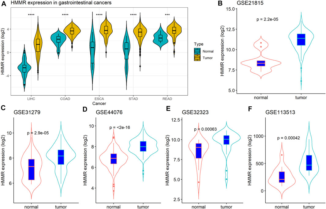
FIGURE 1. (A) Expression of HMMR in colorectal cancer (CRC) based on The Cancer Genome Atlas (TCGA) and Gene Expression Omnibus (GEO) datasets. (B-F) Comparison of HMMR in CRC and normal tissues in five GEO datasets.
Validation of HMMR in Clinical Colon Cancer Tissues
Next, we checked the expression of HMMR in 40 colon cancer tissues and corresponding adjacent colon tissues by RT-qPCR. The analysis suggested that HMMR mRNA was significantly overexpressed in colon cancer tissues than in adjacent colon tissues (Figure 2A, p < 0.05), which verified the results from the online datasets.
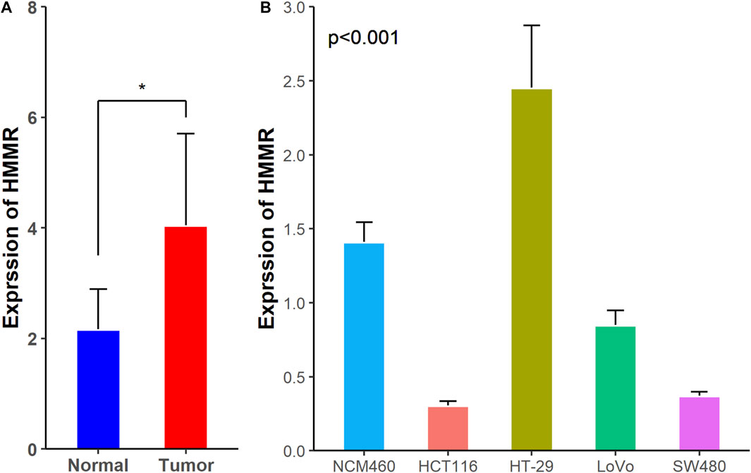
FIGURE 2. Expression of HMMR mRNA in (A) clinical tissues and (B) CRC cell lines and colon mucosal epithelial cell line. *p < 0.05.
Expression of HMMR in Colon Cancer Cells
We checked the expression of HMMR mRNA in HCT116, HT-29, LoVo, SW480, and NCM460 using the RT-qPCR assay. The expression of HMMR mRNA was significantly increased in HT-29 cells, but decreased in HCT116, LoVo, and SW480 cells, especially in the HCT116 cells, than in NCM460 cells (Figure 2B).
Clinical Significance of HMMR in CRC
We analyzed the clinical significance of HMMR in CRC using TCGA dataset, GEO dataset (GSE21815), and clinical datasets. The median HMMR level in CRC tissues was calculated and used as the cutoff value. As listed in Table 1, the expression of HMMR in CRC tissues was not associated with the patients’ age, sex, TNM stage, clinical stage, tumor location, and KRAS mutation status (p > 0.05), indicating that the expression of HMMR in CRC tissues was independent of these clinical parameters.
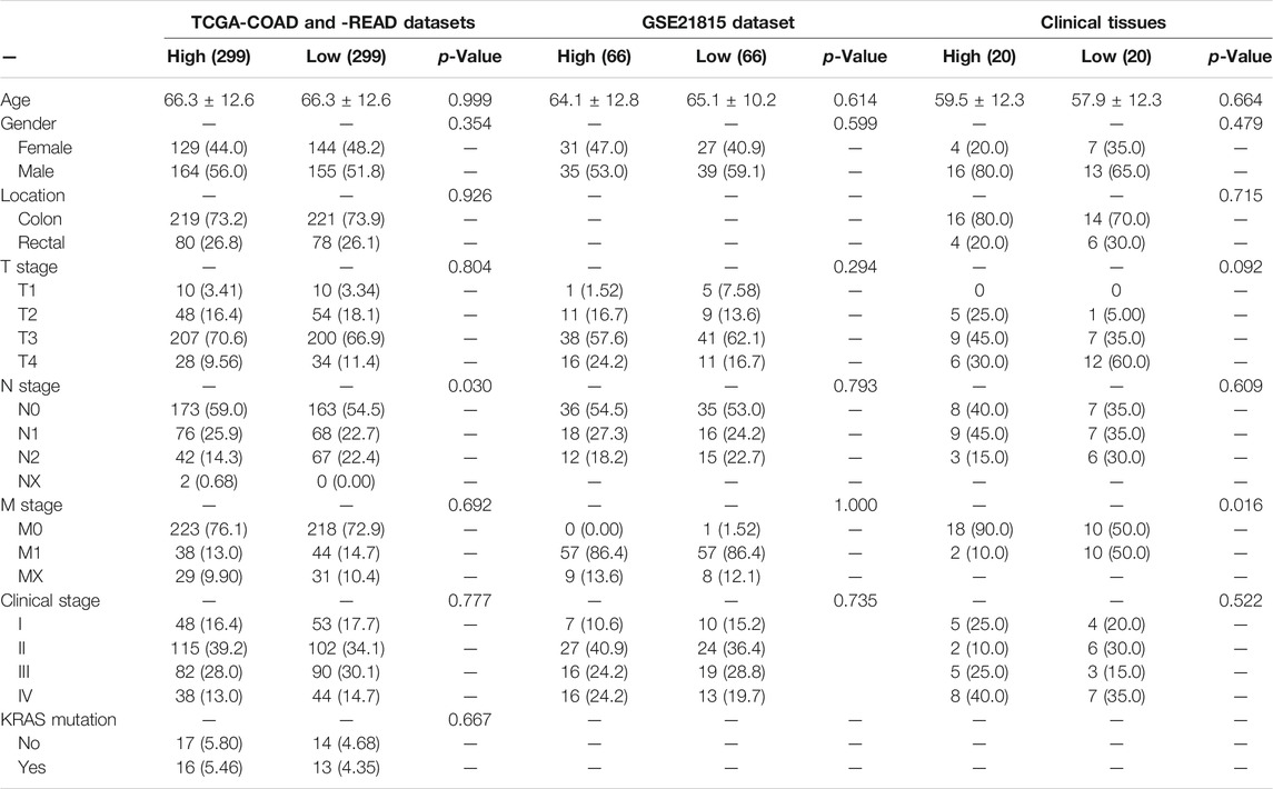
TABLE 1. Results of clinical significance of HMMR in CRC using three datasets.
HMMR Levels in Blood Samples
Furthermore, to explore the expression of HMMR in peripheral blood, two GEO datasets (GSE164191 and GSE47756) analyzing the expression of HMMR in peripheral blood were included in the analysis, and the blood samples of 120 patients with CRC and 100 healthy controls were collected and tested by ELISA. As shown in Figures 3A, B, HMMR levels in blood samples were not significantly different between patients with CRC and healthy controls in the GSE164191 and GSE47756 datasets; but HMMR levels, analyzed by ELISA, were significantly elevated in blood samples from patients with CRC than in those from healthy controls (Figure 3C, p < 0.01).
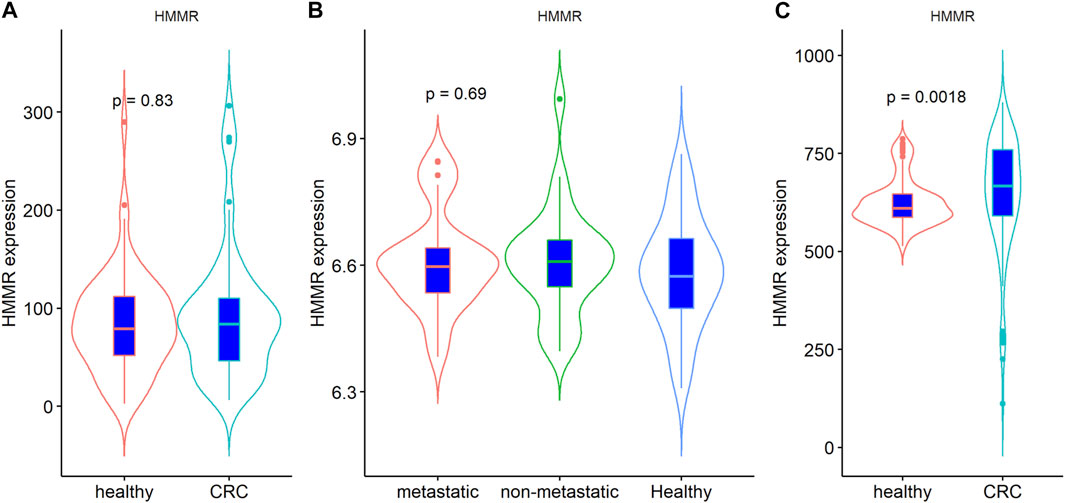
FIGURE 3. Comparison of blood levels of HMMR in CRC and healthy controls: (A) results of GSE164191; (B) results of GSE47756, including patients with metastatic and non-metastatic cancer; (C) results of clinical blood samples.
Next, we examined the association between HMMR levels and clinical parameters in CRC patients, as listed in Table 2, using median HMMR levels as the cutoff value. No significant differences in patients’ age, sex, TNM stage, clinical stage, tumor location, and the four tumor biomarkers (CEA, CA125, CA153, and CA199) (p < 0.05) were observed between samples with high and low HMMR levels; this suggested that HMMR levels in the peripheral blood of patients with CRC were independent of the clinical parameters and tumor biomarkers.
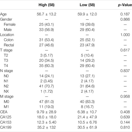
TABLE 2. Association of blood levels of HMMR with the clinical parameters.
Survival Analysis of HMMR in Patients With CRC
To determine the prognostic value of HMMR in patients with CRC, we analyzed the overall survival (OS) and disease-free survival (DFS) using data from TCGA and four GEO datasets (GSE17537, GSE12945, GSE17536, and GSE14333). As shown in Figures 4A–D, low expression of HMMR indicated poor survival rate in TCGA; but there was no significant association between HMMR expression and OS in patients with CRC in three GEO datasets. Furthermore, analyses of two GEO datasets (GSE17536 and GSE14333) indicated a significant association between HMMR expression and DFS in patients with CRC, although the other two datasets (GSE17537 and GSE12945) were inconsistent with these results (Figures 4E–H). Taken together, these results fail to confirm the association between HMMR expression and survival in patients with CRC.
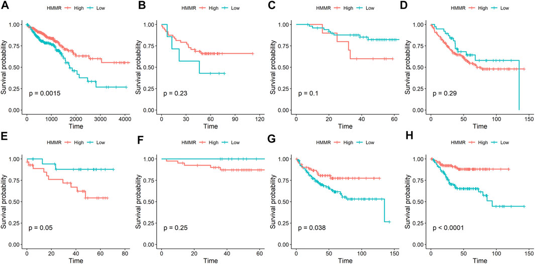
FIGURE 4. Association of the expression of HMMR with survival in patients with CRC. (A) OS results in TCGA-COAD and -READ datasets; (B) OS results in the GSE17537 dataset; (C) OS results in the GSE12945 dataset; (D) OS results in the GSE17536 dataset; (E) DFS results in the GSE17537 dataset; (F) DFS results in the GSE12945 dataset; (G) DFS results in the GSE17536 dataset; (H) DFS results in the GSE14333 dataset. OS: overall survival; DFS: disease-free survival.
Discussion
In the present study, we comprehensively analyzed the expression of HMMR in multiple types of CRC sample tissues, blood, and cell lines and in sequence datasets. The results indicated that the expression of HMMR mRNA was significantly increased in CRC tissues than in corresponding control tissues. However, the blood levels of HMMR were not significantly different between patients with CRC and healthy controls. Moreover, the clinical analysis failed to reveal the association of HMMR expression in tissues and peripheral blood with the TNM stage and clinical stage, suggesting that HMMR expression was independent of the progression of CRC. Furthermore, although the HMMR mRNA level was elevated in HT-29 cells than in normal colon mucosal epithelial cells, there was considerably decreased expression in the other three CRC cell lines, highlighting the variable expression of HMMR mRNA in CRC. Finally, analyses of datasets from TCGA and GEO failed to confirm the prognostic value of HMMR in OS and DFS in patients with CRC, suggesting that the predictive role of HMMR in CRC prognosis remains to be verified.
The molecular structure and biological function of HMMR have been documented in previous studies. Evidence suggests that the HMMR is a hydrophilic protein that lacks hydrophobic signal peptides or potential hydrophobic transmembrane domains (Choi et al., 2019); it also lacks the structural properties required for canonical extracellular export (Kouvidi et al., 2014). The expression of HMMR is critical for orientation of the mitotic spindle in human mitotic cells and is regulated by proliferation-associated transcription factors or signaling pathway members, such as FOXM1, E2F4, MYC (Lachmann et al., 2018), and Hippo (Wang et al., 2014). A study also revealed that the expression of HMMR is transcriptionally downregulated by the tumor suppressor p53 (Sohr and Engeland, 2008). The presence of HMMR-positive CRC cells is significantly associated with poor survival outcomes, independent of the TNM stage and adjuvant therapy (Koelzer et al., 2015). These findings suggest that the HMMR plays a critical role in the pathogenesis of CRC.
The expression of HMMR was reported to be upregulated in several cancer types (Shigeishi et al., 2014; Singleton, 2014; Buttermore et al., 2017), but it was also downregulated in other cancer types than in normal controls based on the results from the LAML and TGCT projects. In the present study, we found that the HMMR was significantly upregulated in gastrointestinal tract cancers, and results from TCGA and GEO datasets and clinical samples further validated the elevated expression of HMMR in CRC tissues. To determine the role of HMMR in the development of CRC, we analyzed the association between the expression of HMMR and clinical parameters. However, the results from the two large cohorts failed to reveal such associations. Additionally, the experimental analyses also confirmed only small associations, which indicated that the expression of HMMR in CRC tissues was independent of the tumor stage. Moreover, the expression of HMMR varied significantly between different CRC cell lines. These results demonstrated that the expression of HMMR was different in the tissues and cell lines.
As compared to the tissue samples, the role of HMMR levels in the blood of patients is more interesting to clinicians. Therefore, we examined HMMR levels in the blood of patients with CRC. However, the expression of HMMR in the blood was not significantly different between patients with CRC and healthy controls. The ELISA indicated significant differences in the expression of HMMR between patients with CRC and healthy controls; but considering the relatively small sample size, these results still need validation. Additionally, similar to the results from tissues, HMMR levels in the blood were not significantly associated with the clinical parameters, including the TNM stage.
A previous study suggested that high expression of HMMR was associated with poor survival in patients with CRC (Zlobec et al., 2008a). Another study showed similar results using immunohistochemistry (Zlobec et al., 2008b). However, in the present study, the OS and DFS in four GEO datasets were inconsistent, and those from TCGA were even opposite to the results of a previous study. Moreover, the association between HMMR and DFS was not significant in the three datasets. These discrepancies may be due to differences in treatment after diagnosis of CRC or the different detection methods of HMMR. Therefore, the prognostic value of HMMR in CRC remains to be validated in a larger cohort using a consistent detection method.
Previous studies have reported the expression of HMMR in CRC (Karamitopoulou et al., 2011; Mele et al., 2017); however, due to the limited number or types of samples, the clinical and prognostic significance of HMMR in CRC remains to be elucidated. The present study, as opposed to previous studies, included many types of samples with larger sample sizes and, thus, achieved more reliable results. However, some limitations of this study should be noted. First, several confounders, such as gene mutation and microsatellite instability status, may have affected the gene expression and prognosis of patients; but we could not analyze the effect of these factors on the expression of HMMR due to the limited data. Second, the number of clinical samples was relatively small, and the results still need to be verified in a larger cohort. Third, the mechanism of action of HMMR in CRC pathogenesis needs to be explored in vivo and in vitro.
Conclusion
By comprehensively analyzing the sequencing and microarray data, clinical samples, blood samples, and cell lines, the present study demonstrated that the expression of HMMR was increased in CRC tissues but not in the blood. Moreover, the expression of HMMR was independent of CRC development and had no prognostic significance in patients with CRC.
Data Availability Statement
The raw data supporting the conclusions of this article will be made available by the authors, without undue reservation.
Ethics Statement
The studies involving human participants were reviewed and approved by the Ethics Committee of Guangxi Medical University Cancer Hospital. The patients/participants provided their written informed consent to participate in this study.
Author Contributions
TY and HB conceptualized and designed the study. YY and XM collected and assembled the data. YX, XM, and LK performed the experiment. HB, TY, LK, LX, LJ, and XM were involved in data analysis and interpretation. All authors wrote, reviewed, read, and approved the manuscript in its current state.
Funding
This study was partially supported by research funding from the National Natural Science Foundation (No. 81860417), Natural Science Foundation of Guangxi (No. 2018JJA140136), and Basic Competence Promotion Project for Young and Middle-aged Teachers in Guangxi (No. 2020KY0348).
Conflict of Interest
The authors declare that the research was conducted in the absence of any commercial or financial relationships that could be construed as a potential conflict of interest.
Publisher’s Note
All claims expressed in this article are solely those of the authors and do not necessarily represent those of their affiliated organizations, or those of the publisher, the editors, and the reviewers. Any product that may be evaluated in this article, or claim that may be made by its manufacturer, is not guaranteed or endorsed by the publisher.
References
Burren, S., Reche, K., Blank, A., Galvàn, J. A., Dawson, H., Berger, M. D., et al. (2021). RHAMM in Liver Metastases of Stage IV Colorectal Cancer with Mismatch-Repair Proficient Status Correlates with Tumor Budding, Cytotoxic T-Cells and PD-1/pd-L1. Pathol. - Res. Pract. 223, 153486. doi:10.1016/j.prp.2021.153486
Buttermore, S. T., Hoffman, M. S., Kumar, A., Champeaux, A., Nicosia, S. V., and Kruk, P. A. (2017). Increased RHAMM Expression Relates to Ovarian Cancer Progression. J. Ovarian Res. 10 (1), 66. doi:10.1186/s13048-017-0360-1
Carvalho, A. M., Soares da Costa, D., Paulo, P. M. R., Reis, R. L., and Pashkuleva, I. (2021). Co-localization and Crosstalk between CD44 and RHAMM Depend on Hyaluronan Presentation. Acta Biomater. 119, 114–124. doi:10.1016/j.actbio.2020.10.024
Choi, S., Wang, D., Chen, X., Tang, L. H., Verma, A., Chen, Z., et al. (2019). Function and Clinical Relevance of RHAMM Isoforms in Pancreatic Tumor Progression. Mol. Cancer 18 (1), 92. doi:10.1186/s12943-019-1018-y
Di Nicolantonio, F., Vitiello, P. P., Marsoni, S., Siena, S., Tabernero, J., Trusolino, L., et al. (2021). Precision Oncology in Metastatic Colorectal Cancer - from Biology to Medicine. Nat. Rev. Clin. Oncol. 18, 506–525. doi:10.1038/s41571-021-00495-z
Evanko, S. P., and Wight, T. N. (1999). Intracellular Localization of Hyaluronan in Proliferating Cells. J. Histochem. Cytochem. 47 (10), 1331–1341. doi:10.1177/002215549904701013
He, Z., Mei, L., Connell, M., and Maxwell, C. A. (2020). Hyaluronan Mediated Motility Receptor (HMMR) Encodes an Evolutionarily Conserved Homeostasis, Mitosis, and Meiosis Regulator rather Than a Hyaluronan Receptor. Cells 9 (4), 819. doi:10.3390/cells9040819
Jiang, J., Mohan, P., and Maxwell, C. A. (2013). The Cytoskeletal Protein RHAMM and ERK1/2 Activity Maintain the Pluripotency of Murine Embryonic Stem Cells. PLoS One 8 (9), e73548. doi:10.1371/journal.pone.0073548
Karamitopoulou, E., Zlobec, I., Panayiotides, I., Patsouris, E. S., Peros, G., Rallis, G., et al. (2011). Systematic Analysis of Proteins from Different Signaling Pathways in the Tumor center and the Invasive Front of Colorectal Cancer. Hum. Pathol. 42 (12), 1888–1896. doi:10.1016/j.humpath.2010.06.020
Koelzer, V. H., Huber, B., Mele, V., Iezzi, G., Trippel, M., Karamitopoulou, E., et al. (2015). Expression of the Hyaluronan-Mediated Motility Receptor RHAMM in Tumor Budding Cells Identifies Aggressive Colorectal Cancers. Hum. Pathol. 46 (11), 1573–1581. doi:10.1016/j.humpath.2015.07.010
Kouvidi, K., Nikitovic, D., Berdiaki, A., and Tzanakakis, G. N. (2014). Hyaluronan/RHAMM Interactions in Mesenchymal Tumor Pathogenesis. Adv. Cancer Res. 123, 319–349. doi:10.1016/B978-0-12-800092-2.00012-5
Lachmann, A., Torre, D., Keenan, A. B., Jagodnik, K. M., Lee, H. J., Wang, L., et al. (2018). Massive Mining of Publicly Available RNA-Seq Data from Human and Mouse. Nat. Commun. 9 (1), 1366. doi:10.1038/s41467-018-03751-6
Liu, W., Ma, J., Cheng, Y., Zhang, H., Luo, W., and Zhang, H. (2016). HMMR Antisense RNA 1, a Novel Long Noncoding RNA, Regulates the Progression of Basal-like Breast Cancer Cells. Breast Cancer (London: Dove Med Press), 8, 223–229. doi:10.2147/BCTT.S119997
Lu, D., Bai, X., Zou, Q., Gan, Z., and Lv, Y. (2020). Identification of the Association between HMMR Expression and Progression of Hepatocellular Carcinoma via Construction of a Co-expression N-etwork. Oncol. Lett. 20 (3), 2645–2654. doi:10.3892/ol.2020.11844
Lu, T., Zheng, Y., Gong, X., Lv, Q., Chen, J., Tu, Z., et al. (2021). High Expression of Hyaluronan-Mediated Motility Receptor Predicts Adverse Outcomes: A Potential Therapeutic Target for Head and Neck Squamous Cell Carcinoma. Front. Oncol. 11, 608842. doi:10.3389/fonc.2021.608842
Lugli, A., Zlobec, I., Günthert, U., Minoo, P., Baker, K., Tornillo, L., et al. (2006). Overexpression of the Receptor for Hyaluronic Acid Mediated Motility Is an Independent Adverse Prognostic Factor in Colorectal Cancer. Mod. Pathol. 19 (10), 1302–1309. doi:10.1038/modpathol.3800648
Mele, V., Sokol, L., Kölzer, V. H., Pfaff, D., Muraro, M. G., Keller, I., et al. (2017). The Hyaluronan-Mediated Motility Receptor RHAMM Promotes Growth, Invasiveness and Dissemination of Colorectal Cancer. Oncotarget 8 (41), 70617–70629. doi:10.18632/oncotarget.19904
Shigeishi, H., Higashikawa, K., and Takechi, M. (2014). Role of Receptor for Hyaluronan-Mediated Motility (RHAMM) in Human Head and Neck Cancers. J. Cancer Res. Clin. Oncol. 140 (10), 1629–1640. doi:10.1007/s00432-014-1653-z
Singleton, P. A. (2014). Hyaluronan Regulation of Endothelial Barrier Function in Cancer. Adv. Cancer Res. 123, 191–209. doi:10.1016/B978-0-12-800092-2.00007-1
Sohr, S., and Engeland, K. (2008). RHAMM Is Differentially Expressed in the Cell Cycle and Downregulated by the Tumor Suppressor P53. Cell Cycle 7 (21), 3448–3460. doi:10.4161/cc.7.21.7014
Sun, Z., Liu, C., and Cheng, S. Y. (2021). Identification of Four Novel Prognosis Biomarkers and Potential Therapeutic Drugs for Human Colorectal Cancer by Bioinformatics Analysis. J. Biomed. Res. 35 (1), 21–35. doi:10.7555/JBR.34.20200021
Sung, H., Ferlay, J., Siegel, R. L., Laversanne, M., Soerjomataram, I., Jemal, A., et al. (2021). Global Cancer Statistics 2020: GLOBOCAN Estimates of Incidence and Mortality Worldwide for 36 Cancers in 185 Countries. CA A. Cancer J. Clin. 71 (3), 209–249. doi:10.3322/caac.21660
Tolg, C., Poon, R., Fodde, R., Turley, E. A., and Alman, B. A. (2003). Genetic Deletion of Receptor for Hyaluronan-Mediated Motility (Rhamm) Attenuates the Formation of Aggressive Fibromatosis (Desmoid Tumor). Oncogene 22 (44), 6873–6882. doi:10.1038/sj.onc.1206811
Wang, Z., Wu, Y., Wang, H., Zhang, Y., Mei, L., Fang, X., et al. (2014). Interplay of Mevalonate and Hippo Pathways Regulates RHAMM Transcription via YAP to Modulate Breast Cancer Cell Motility. Proc. Natl. Acad. Sci. USA 111 (1), E89–E98. doi:10.1073/pnas.1319190110
Zlobec, I., Baker, K., Terracciano, L. M., and Lugli, A. (2008b). RHAMM, P21 Combined Phenotype Identifies Microsatellite Instability-High Colorectal Cancers with a Highly Adverse Prognosis. Clin. Cancer Res. 14 (12), 3798–3806. doi:10.1158/1078-0432.CCR-07-5103
Zlobec, I., Baker, K., Terracciano, L., Peter, S., Degen, L., Beglinger, C., et al. (2008a). Two-marker Protein Profile Predicts Poor Prognosis in Patients with Early Rectal Cancer. Br. J. Cancer 99 (10), 1712–1717. doi:10.1038/sj.bjc.6604729
Keywords: colorectal cancer, hyaluronan-mediated motility receptor, tissues, blood, cell lines
Citation: Tang Y-p, Yin Y-x, Xie M-z, Liang X-q, Li J-l, Li K-z and Hu B-l (2021) Systematic Analysis of the Clinical Significance of Hyaluronan-Mediated Motility Receptor in Colorectal Cancer. Front. Mol. Biosci. 8:733271. doi: 10.3389/fmolb.2021.733271
Received: 30 June 2021; Accepted: 23 September 2021;
Published: 26 October 2021.
Edited by:
Hossain Shekhar, University of Dhaka, BangladeshReviewed by:
Dragana Nikitovic, University of Crete, GreecePrihantono Prihantono, Hasanuddin University, Indonesia
Copyright © 2021 Tang, Yin, Xie, Liang, Li, Li and Hu. This is an open-access article distributed under the terms of the Creative Commons Attribution License (CC BY). The use, distribution or reproduction in other forums is permitted, provided the original author(s) and the copyright owner(s) are credited and that the original publication in this journal is cited, in accordance with accepted academic practice. No use, distribution or reproduction is permitted which does not comply with these terms.
*Correspondence: Ke-zhi Li, bGlrZXpoaUBneG11LmVkdS5jbg==; Bang-li Hu, aHViYW5nbGlAZ3htdS5lZHUuY24=
†These authors have contributed equally to this work