
94% of researchers rate our articles as excellent or good
Learn more about the work of our research integrity team to safeguard the quality of each article we publish.
Find out more
ORIGINAL RESEARCH article
Front. Microbiol. , 27 February 2025
Sec. Food Microbiology
Volume 16 - 2025 | https://doi.org/10.3389/fmicb.2025.1521202
 Kandhan Srinivas1,2
 Sandeep Ghatak2*
 Arockiasamy Arun Prince Milton2*
 Samir Das2*
 Kekungu-u Puro2
 Daniel Aibor Pyngrope2
 Madesh Angappan1,2Mosuri Chendu Bharat Prasad2Dadimi Bhargavi1,2Nur Abdul Kader2Vanita Lyngdoh2Heiborkie Shilla2John Pynhun Lamare2
Introduction: Escherichia albertii is an emerging food-borne pathogen with zoonotic potential which is often under-reported due to misidentifications.
Materials and methods: The current study identified E. albertii from retail fish sold in market which was confirmed by phenotypic (colorless colonies on Xylose-Rhamnose-Melibiose MacConkey Agar), genotypic (dual target uniplex PCR-based detection) and genomic methods (CheckM analysis). In this paper we report the phenotypic characters of the isolate and genomic features such as resistome, virulome and mobilome followed by in silico O and H antigen based typing and comparative phylogenomics using various tools (RAST, RGI v6.0.0, ABRicate v1.0.1, PathogenFinder v1.1, PlasmidFinder v2.0, BacAnt v3.3.1, Phigaro v2.4.0, MAFFT v7.490, FigTree v1.4.4).
Results and discussion: Multidrug resistance was identified with reduced susceptibility to gentamicin, azithromycin, ceftazidime and cefotaxime with a Multiple Antibiotic Resistance (MAR) index of 0.33. Clinically important virulence genes such as eae, cdt, east1 formed a part of the virulome and the probability of being pathogenic to humans was found to be 0.883. The genome was found to harbor mobile genetic elements such as plasmids [IncFIA, IncFIB(pB171), IncFII(pSE11)], transposons (Tn3411, Tn6292) and prophages (Siphoviridae, Myoviridae, Podoviridae). Various typing methods such as biotyping, multilocus sequence typing and in silico O and H antigen typing classified the isolate into biotype 3, multi locus sequence type 4596, O-genotype 4 and H-genotype 1. Phylogenomically, the isolate was placed close to isolate from neighboring country of China. Identification of virulent multidrug-resistant E. albertii from new food source such as fishes increases the risk for fish eating population and necessitates the requirement of further elucidation and development of appropriate control strategies.
Escherichia albertii has emerged as a potentially important food-borne pathogen over the last few years (Masuda et al., 2020; Hirose et al., 2022; Muchaamba et al., 2022). Since its initial identification from diarrhoeagenic infant patients from Bangladesh, the hazard level has steadily ascended over the years (Huys et al., 2003; Muchaamba et al., 2022). Recent reports have hinted that the virulence potential of E. albertii could be on par with Escherichia coli due to shared repertoire of genes and subsequentially it has been implicated in clinical manifestations in humans as well (Bours et al., 2010; Luo et al., 2021). Frequent association of the pathogen with various clinical conditions affecting humans such as diarrhea and urinary tract infections over a wide geographical area has gradually raised global concerns associated with this pathogen (Bours et al., 2010; Brandal et al., 2015; Zaki et al., 2022; Iguchi et al., 2023). The recent reports have showcased the ability of E. albertii to harbor diverse clinically important virulence genes such as intimin, cytolethal distending toxin, Shiga toxin and Enteroaggregative E. coli heat-stable enterotoxin 1 (EAST1) which often cause gastro-intestinal disorders in humans (Luo et al., 2021; Muchaamba et al., 2022).
The resistances profile of E. albertii have leaped from reporting susceptible patterns to a multidrug-resistant strains with inclusion of resistance against clinically important drugs such as carbapenem (Wang et al., 2022) and colistin (Sonnevend et al., 2022). Genotypically, they were also found to carry Extended Spectrum Beta Lactamase (ESBL) genes such as blaTEM-141 and blaCTX-M-55 (Guo et al., 2024) as well as mcr genes responsible for colistin resistance (Li et al., 2018; Sonnevend et al., 2022). Mobile genetic elements such as transposons, plasmids, phages and integrons play an important role in the horizontal transfer and further dissemination of antimicrobial resistance genes and E. albertii is no exception (Partridge et al., 2018). Various typing methods have been used to study the epidemiology of E. albertii which include the conventional biotyping (Murakami et al., 2019), Multilocus sequence typing (MLST) (Luo et al., 2021) and pulsotyping (Leszczyńska et al., 2023). In addition, PCR based methods harnessing the variations in repetitive elements interspersed between the genes have also been used (Grillová et al., 2018).
Food and water as a vehicle for the transmission of E. albertii was proven in a series of outbreaks in Japan (Masuda et al., 2020). Additionally, E. albertii has also been isolated from foods of plant as well as animal origin. Plant based sources include lettuce, Japanese parsley, Mitsuba, Mizu, watercress and cucumber; whereas, chicken, pork, milk, cheese, mutton, duck meat, Japanese rock oyster and Pacific oyster (Fiedler et al., 2018; Arai et al., 2022; Hirose et al., 2022; Muchaamba et al., 2022). Additionally, the organism has also been spotted in river water and drinking water systems (Fiedoruk et al., 2014; Maheux et al., 2014).
With this background, the current study aims to document the first report of the isolation of E. albertii from fish sold in the retail markets. Further we attempted to characterize the genome of the isolate in terms of resistance genes, virulence genes, mobile genetic elements, in silico typing and phylogenomic positioning with respect to global dataset of genomes to obtain cues on the transmission and genetic diversity.
Single ichthyic isolate of E. albertii (Ea_FM1) was recovered from fresh fish meat sample collected from Laitumkhrah market of Meghalaya as part of routine surveillance work of food samples in the laboratory (Institutional Animal Ethics Committee clearance no. V-11011(13)/12/2023-CPCSEA-DADF dated December 5, 2023). The sample (25 g) was enriched in novobiocin (HiMedia, Mumbai, India) supplemented modified EC broth (HiMedia, Mumbai, India) incubated at 42°C for 24 h (Arai et al., 2021) followed by plating onto modified Xylose-Rhamnose-Melibiose MacConkey (XRM-MacConkey) agar (MacConkey agar base; D-xylose [HiMedia, Mumbai, India]; L-rhamnose [Sisco Research Laboratories, Mumbai, India]; D-(+)-Melibiose [Sigma-Aldrich, MA, USA]) incubated at 37°C for 48 h (Hinenoya et al., 2020). Colorless colonies were later confirmed with the help of combination of conventional PCRs targeting a 393 bp region of DNA binding transcriptional activator of cysteine biosynthesis gene (Lindsey et al., 2017) and a 449 bp region of E. albertii specific cytolethal distending toxin (Eacdt) gene (Hinenoya et al., 2019).
Phenotypic characterization involved the assessment of antimicrobial susceptibility profile using CLSI guidelines (Clinical and Laboratory Standards Institute, 2023). The isolate was pitted against 12 clinically important antibiotics namely ampicillin (10 mcg), cefotaxime (30 mcg), ceftriaxone (30 mcg), cefoxitin (30 mcg), ceftazidime (30 mcg), imipenem (10 mcg), gentamicin (10 mcg), azithromycin (15 mcg), tetracycline (30 mcg), ciprofloxacin (5 mcg), co-trimoxazole (25 mcg) and chloramphenicol (30 mcg) belonging to various antimicrobial classes. Interpretative criteria for Enterobacteriaceae as stipulated by Clinical and Laboratory Standards Institute were followed (Clinical and Laboratory Standards Institute, 2023). Resistance to 3 or more different classes of antibiotics was taken as the criteria to confer multidrug resistance (Magiorakos et al., 2012). MAR index of the isolate was calculated as described earlier (Krumperman, 1983). Biofilm forming ability of the isolate was also tested by microtiter plate assay using previously established methods with Acinetobacter baumannii (ATCC 19606) and E. coli DH5α (New England Biolabs) as positive and negative control, respectively (Srinivas et al., 2023). Motility assay was performed as by stabbing onto semisolid Tryptone Soy agar containing 0.35% agar and incubation for 37°C. Hemolysis property was examined by streaking onto 5% sheep blood agar with Staphylococcus aureus (ATCC 25923) and E. coli (ATCC 25922) as positive and negative control, respectively. Biotyping of E. albertii isolate was undertaken with the help of an array of biochemical tests (Indole production and lysine decarboxylase) as specified earlier (Murakami et al., 2019).
Whole genome sequencing of the isolate was outsourced to M/S Eurofins Genomics India Pvt. Ltd. (Bengaluru, India). Illumina HiSeq platform was used for paired-end sequencing. The reads were scrutinized for quality using FastQC tool with default settings (Andrews, 2010). De novo assembly of the genome was undertaken with the help of Shovill tool ver. 1.0.9 with Spades assembler after triggering the following switches/parameters: “-trim,” “read error” and “post-assembly correction” (Seemann, 2018). Taxonomy identification was done using CheckM (Wood et al., 2019).
E. albertii genomes submitted to NCBI were downloaded for phylogenetic analysis. From a collection of 672 genomes downloaded from NCBI (09 April 2024) a total of 50 representative genomes were selected based on mash distances calculated through Assembly Dereplicator tool v0.3.2.1 A distance of 0.001 with a cut off value of 50 genomes was used for downsizing number of genomes. The genomes were coded in X_Y_Z format for ease of downstream processing, where X stands for country, Y stands for isolation source and Z stands for isolate serial number (Supplementary Table 1).
The Ea_FM1 genome was annotated employing online service from RAST (Rapid Annotation using Subsystems Technology) with default settings (Keegan et al., 2016). Genome map was also constructed using BLAST Ring Image Generator (BRIG) with preset parameters (Alikhan et al., 2011). Nucleotide polymorphisms were identified using Snippy tool v.4.6.0 (Seemann, 2018). E. albertii KF1 genome (CP007025) was used as reference for Snippy and BRIG analysis.
Resistome was determined using RGI tool v6.0.0 employing Comprehensive Antimicrobial Resistance Database (CARD) v3.2.5 (Alcock et al., 2020). The switches “perfect” and “strict” cut-offs were triggered to obtain high stringency results with >95% similarity. Virulome was determined using ABRicate tool v.1.0.12 with VFDB database updated till 14 August 2024. Sequence similarity and minimum coverage criteria were set at 75 and 80%, respectively. Pathogenic potential of the isolate toward humans were computed mathematically with the help of PathogenFinder v1.1 (Cosentino et al., 2013), an online service hosted by Center for Genomic Epidemiology.3 Mobilome (plasmids, transposons and integrons) were ascertained with the help of multiple tools. Plasmids harbored in the genome were detected using online PlasmidFinder 2.0 server provided by Center for Genomic Epidemiology.4 BacAnt v3.3.1 was used to determine the presence of transposons and integrons searched against TransposonDB v.2.0 and IntegronDB v.2.0, respectively (Hua et al., 2021). Minimum criteria for sequence identity and coverage threshold were set at 90 and 80%, respectively. Prophages inserted into the genome were identified with the help of Phigaro v.2.4.0.5 The details related to virus orthologous groups (VOGs) were obtained from VOGDB.6
In silico typing of the isolate was undertaken employing Multi locus sequence typing (MLST) scheme of E. coli (Wirth et al., 2006). Online MLST 2.0 tool provided by CGE was used for MLS typing of the isolate.7 Further, E. albertii-specific in silico typing based on antigenic diversity of somatic and flagellar antigens as described earlier were also followed (Ooka et al., 2019; Nakae et al., 2021).
To get insights on the phylogenomic positioning of the isolate in relation to other global isolates, the ichythic isolate along with the reference genome and 50 other representative genomes chosen as mentioned earlier were included in a phylogenomic analysis. The core genome alignment was accomplished with MAFFT v7.490 with ‘auto’ option (Katoh and Toh, 2008). ModelFinder algorithm in IQ-TREE v2.0 was used to identify the suitable model with the least Bayesian Information Criterion (BIC) score (Kalyaanamoorthy et al., 2017). For identification of the clusters, the TreeLink tool (Allende et al., 2015) was used. The final tree was visualized with the help of FigTree v1.4.4.8
The ichthyic isolate Ea_FM1 was recovered from fish meat sample and was confirmed by phenotypic and molecular methods. Colorless colonies were observed on XRM MacConkey agar (Figure 1A) which was further confirmed by two different molecular diagnostic targets yielding the respective amplicons on gel electrophoresis (Figures 1B,C).
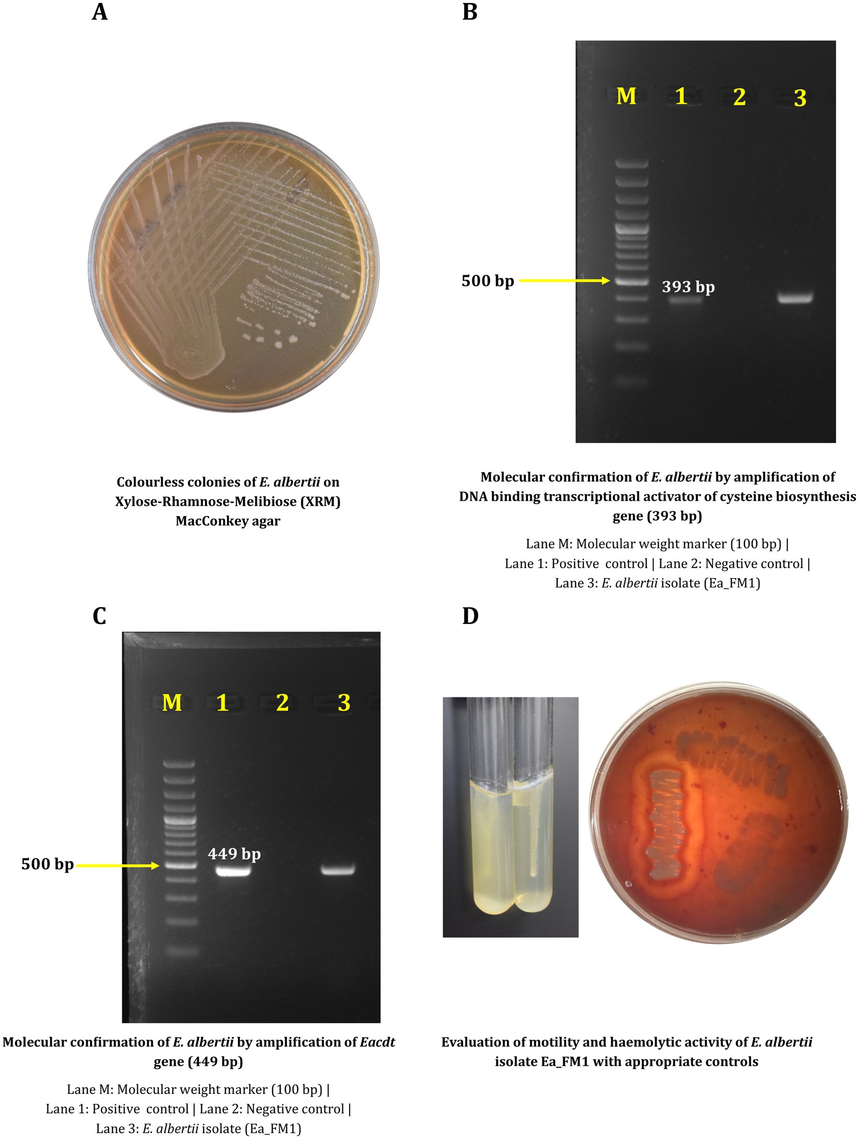
Figure 1. Phenotypic and molecular characterization of Escherichia albertii Ea_FM1 isolate. (A) Colorless colonies of E. albertii on Xylose-Rhamnose-Melibiose (XRM) MacConkey agar. (B) Molecular confirmation of E. albertii by amplification of DNA binding transcriptional activator of cysteine biosynthesis gene (393 bp). (C) Molecular confirmation of E. albertii by amplification of Eacdt gene (449 bp). (D) Evaluation of motility and haemolytic activity of E. albertii isolate Ea_FM1 with appropriate controls.
Antimicrobial susceptibility testing against 12 different antibiotics revealed resistance against gentamicin, cefotaxime, ceftriaxone and azithromycin by virtue of which the isolate is multidrug-resistant with a MAR index of 0.33 (Table 1). Experiment to assess the biofilm forming ability revealed the negligible ability of the isolate to form biofilm in comparison to E. coli DH5α. Other phenotypic characteristics include the inability to be motile at 37°C and the inability to cause hemolysis on blood agar (Figure 1D). Biotyping based on biochemical reactions categorized the isolate into Biotype 3 (Lysine and Indole positive).

Table 1. Phenotypic characteristics of Escherichia albertii isolate Ea_FM1.
The size of the draft genome was 4.7 Mb with a G + C content of 49.5% (Figure 2). Number of contigs were 111 with a coverage of 11×. Taxonomy check using CheckM confirmed the identification of the isolate as E. albertii. Genome sequence was submitted to NCBI under the accession number JBBEFQ000000000.1 under BioProject: PRJNA889995.
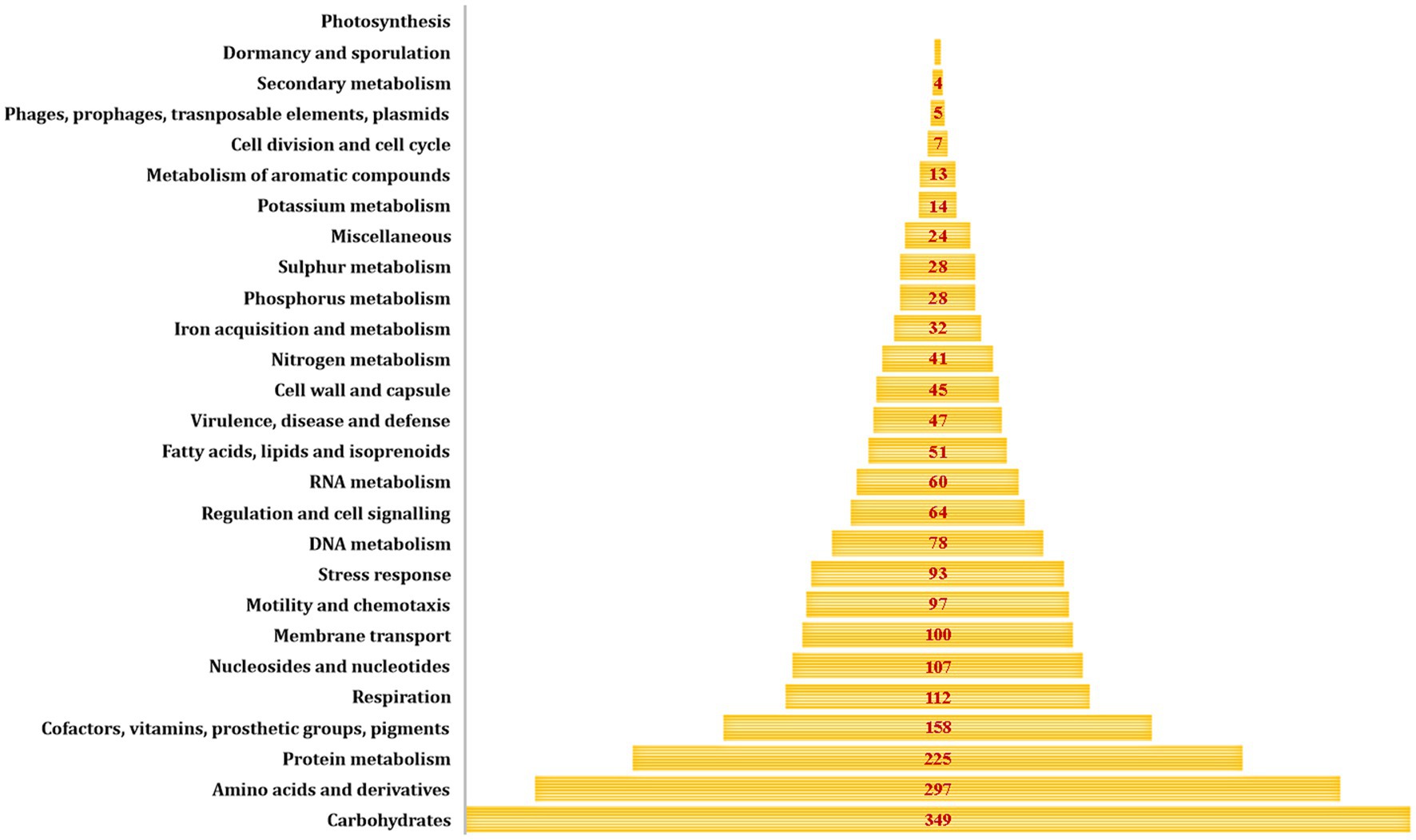
Figure 2. Classification of coding sequences according to RAST annotation of Ea_FM1 genome.
RAST annotation of the draft genome revealed the presence of 2,081 coding sequence regions with majority of them associated with carbohydrate metabolism (349/2,081, 16.77%) followed by amino acids metabolism (297/2,081, 14.27%) and protein metabolism (225/2,081, 10.81%) (Figure 3). Though, the isolate was found to be non-motile, 97 coding regions associated with motility and chemotaxis were identified in the genome. Snippy tool identified 20 instances of mutational events which could be impactful. Out of those 20 instances, 12 were of deletion type and 8 were of insertion type mutations (Supplementary Table 2).
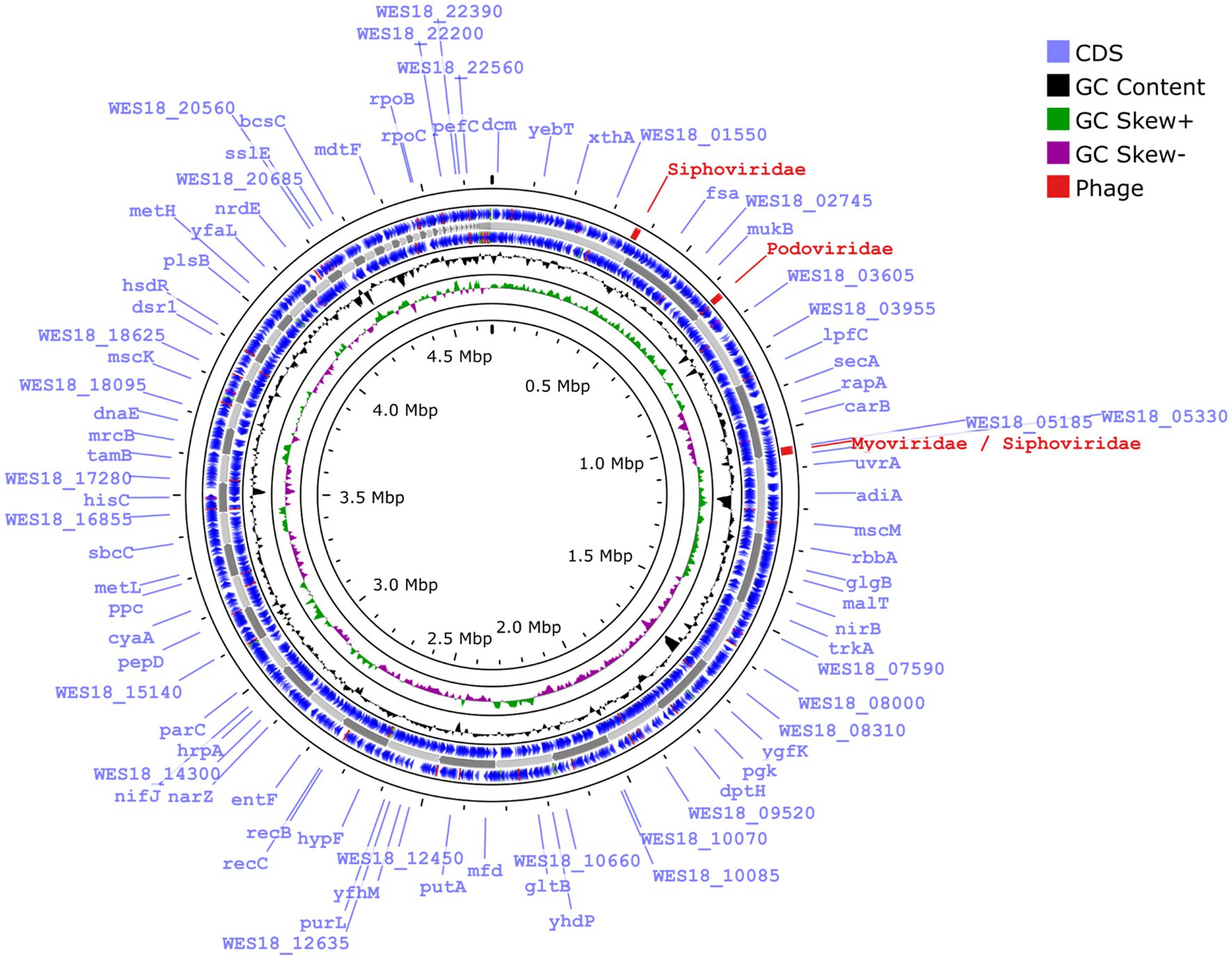
Figure 3. Genome map of Ea_FM1 with prophage elements denoted in red.
Identification of antimicrobial resistance determinants in the genome revealed the presence of 43 different resistance ontologies conferring resistance to various classes of antibiotics such as macrolides, aminoglycosides, cephalosporins, tetracyclines, peptides, nitroimidazoles, penams, fluoroquinolones, diaminopyrimidines, phenicols, glycopeptides, penems, glycylcyclines, monobactams, etc. (Table 2). Majority of the genes exert their action through antibiotic efflux mechanism (n = 32), followed by target alteration (n = 14), reduced permeability (n = 2) and inactivation (n = 1). On screening for virulence genes, a total of 113 virulence genes were identified (Table 3). Categorization the virulence genes into various virulence classes revealed that majority of the genes belonged to effector delivery system (n = 58), followed by nutritional/metabolic factor (n = 22), adherence (n = 17), regulation (n = 6), exotoxin (n = 4), invasion (n = 4), antimicrobial activity (n = 1) and immune modulation (n = 1). Pathogenic potential of the isolate toward humans was found to be on the higher side with a probability of 0.883. A total of three plasmids (IncFIA, IncFIB(pB171), IncFII(pSE11)) and two transposons (Tn3411, Tn6292) were identified from the genome; however, no integrons could be identified. Phigaro analysis detected prophages of three different phages inserted into the genome namely siphoviridae (12 virus orthologous groups), podoviridae (17 virus orthologous groups) and myoviridae/siphoviridae (23 virus orthologous groups) (Supplementary Table 3).
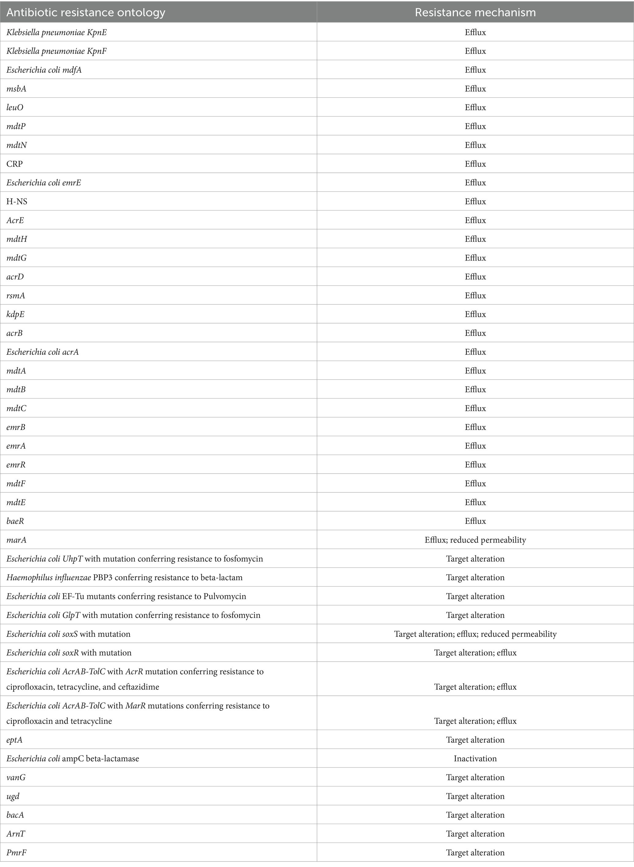
Table 2. Antimicrobial resistance genes observed in Escherichia albertii Ea_FM1 genome.
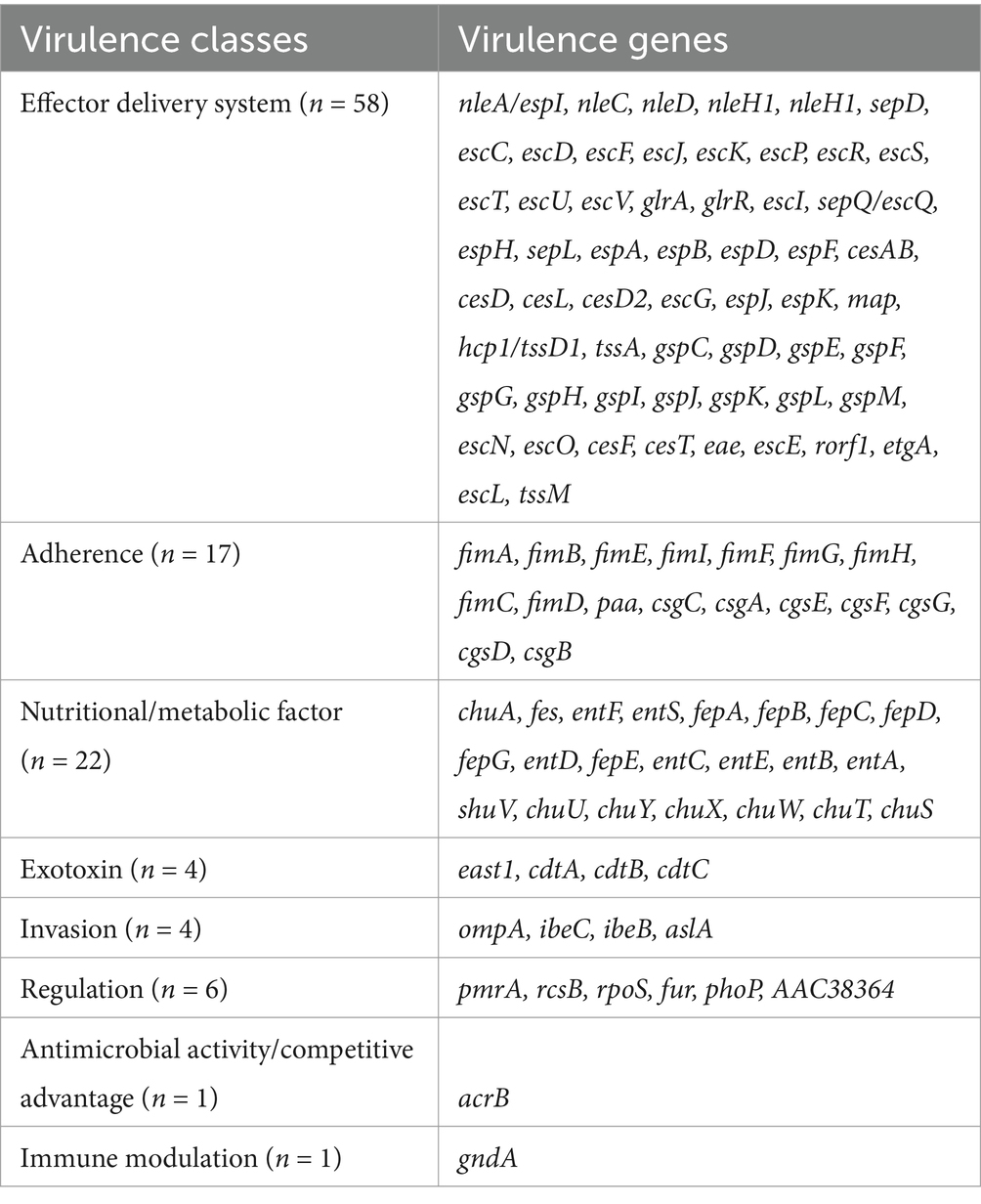
Table 3. Virulence genes in the Escherichia albertii Ea_FM1 genome.
MLS typing using E. coli PubMLST scheme matched with sequence type 4,596 with alleles adk_387, fumC_111, gyrB_408, icd_483. mdh_109, purA_149 and recA_75. Further, O and H antigen-based typing revealed that the isolate belonged to H1 and O4 genotype. Core genome based phylogenomic analysis with GTR + F + I + R6 model placed the isolate Ea_FM1 in close proximity with a Chinese isolate of unknown isolation source and an isolate PL_HUM_01. (Figure 4). The clusters were identified with a cut-off value of 0.0010146631, as identified by the tool. A total of six clusters were identified with a Dunn index of 1.421.
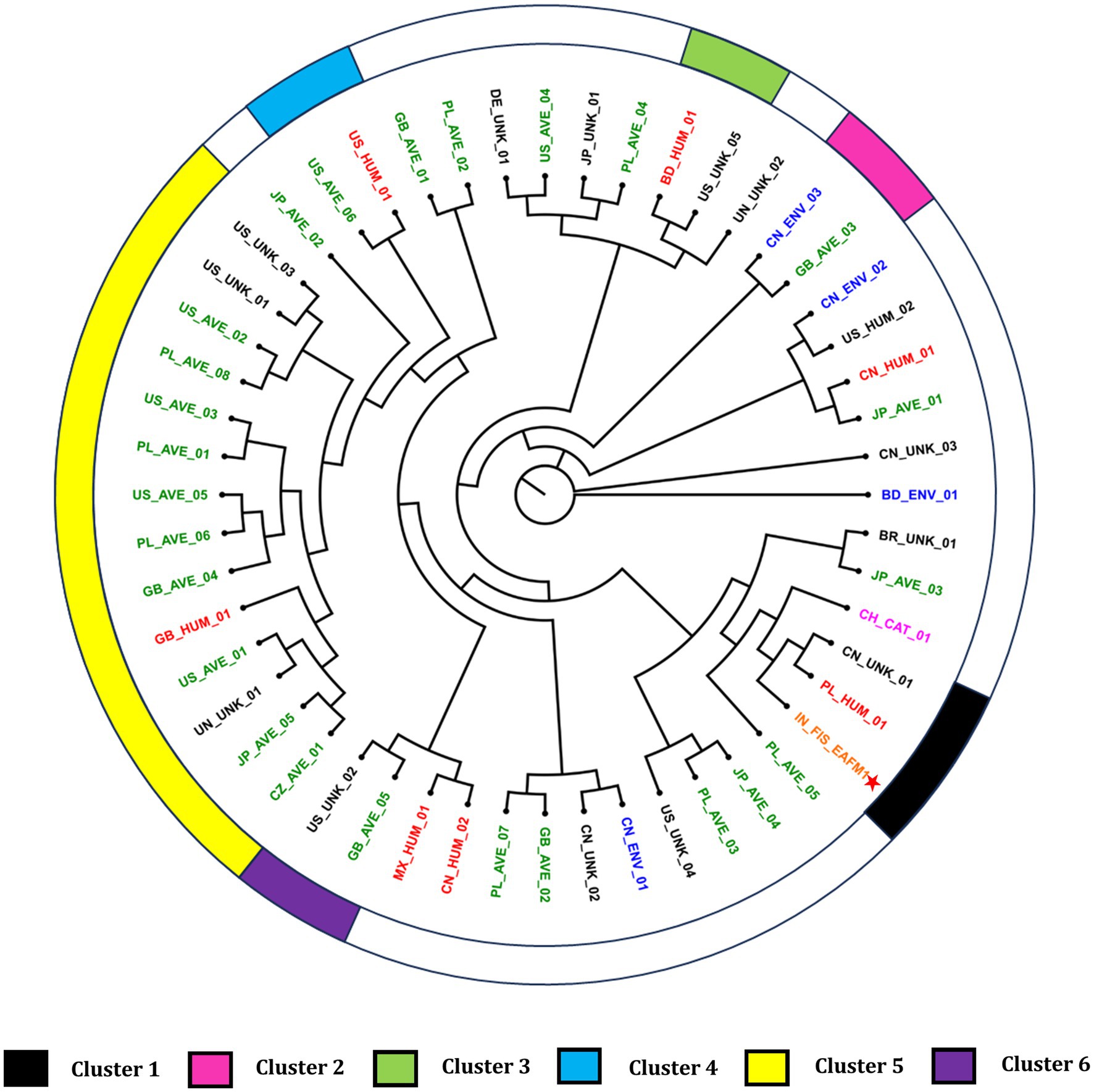
Figure 4. Phylogenomic tree of E. albertii genomes. Asterisk indicates the fish isolate Ea_FM1 sequenced in this study. The genomes were assigned codes X_Y_Z, where “X” indicates the country of isolation (ISO 3166-1 Alpha-2 code), “Y” indicates the source of isolation, and “Z” denotes the serial number of strain (i.e., BD, Bangladesh; BR, Brazil; CH: Switzerland; CN, China; CZ, Czech Republic; DE, Germany; GB, Great Britain; IN, India; JP, Japan; MX, Mexico; PL, Poland; US, United States of America; UN, unknown country of isolation; UNK, unknown source of isolation). Additionally, the coded genome names were given colors based on the source of isolation.
E. albertii is gradually reiterating its status as an emerging pathogen with an increase in spectrum in terms of geographical range, isolation source and clinical manifestations in humans (Muchaamba et al., 2022). Clinical infections implicated with E. albertii include watery to bloody diarrhea, fever, abdominal distension, dehydration and urinary tract infections (Muchaamba et al., 2022).
The reservoir of E. albertii are widely believed to be poultry, owing to its higher prevalence rates reported in various studies (Asoshima et al., 2015; Maeda et al., 2015; Wang et al., 2016; Liu et al., 2023a). However, its isolation from diverse sources such as pigs, dogs, bats, raccoons, penguins, and seals, combined with the absence of this organism in some poultry flocks, has made this claim debatable (Gordon, 2011; Wang et al., 2022; Muchaamba et al., 2022). As an enterobacterial pathogen, E. albertii is shed in the feces of animals, contaminating the environment and potentially re-entering the human food chain through contaminated food and water. Reports indicating its presence in leafy vegetables and foods of animal origin further support this hypothesis (Fiedler et al., 2018; Arai et al., 2022; Hirose et al., 2022; Muchaamba et al., 2022). Despite these findings, the transmission dynamics of E. albertii remain unclear, requiring further studies to identify its primary source. Additionally, its isolation from migratory birds provides valuable insights into the pathogen’s potential for spatial transmission across continents (Liu et al., 2023a; Barmettler et al., 2022; Carter et al., 2023).
The current study, being the first to report isolation of E. albertii from retail fish intended for consumption, has added a new entry into the list of isolation sources from which E. albertii has been isolated till date. Report from Japan on the prevalence in oysters was the closest isolation source reported from aquatic fauna (Arai et al., 2022). In addition to the earlier records of occurrence in various foods of animal origin, this addition highlights the importance of the pathogen from a food safety point of view and possible risk to fish eating population. It is to be noted that E. albertii was previously implicated in numerous food-borne outbreaks in Japan and has been infrequently isolated from water (Fiedoruk et al., 2014; Maheux et al., 2014; Masuda et al., 2020).
Enterobacteriaceae family has always been at the limelight in terms of antimicrobial resistance and has been placed in the WHO list of priority pathogens to inform research and development and public health interventions (World Health Organization, 2024). The ichthyic isolate showed phenotypic resistance to gentamicin (aminoglycoside), azithromycin (macrolide) and cephalosporins such as cefotaxime and ceftazidime. Incidentally, genomic characterization also revealed the presence of AMR genes known to confer resistance against these three classes of antibiotics. This phenotypic and genotypic correlation in terms of antimicrobial resistance was also evident in our previous study (Srinivas et al., 2023). Additionally, the isolate was also found to be multidrug-resistant which is of public health concern. MAR index of 0.33 (above the cut-off value of 0.2) hints at the probable exposure of the pathogen to environment rich in antibiotic pressure (Krumperman, 1983). Despite the presence of few earlier reports of biofilm forming E. albertii, the ichthyic isolate failed to produce biofilm on polystyrene surface (Ingle et al., 2011; Lima et al., 2019; Carter et al., 2024). Additionally, the ichthyic isolate was also found to be non-haemolytic and non-motile @ 37°C as reported earlier. Biotyping scheme as suggested by (Murakami et al., 2019) placed the isolate in Biotype 3 which is the predominant biotype in circulation as reported earlier (Murakami et al., 2019).
Mining for resistance genes identified the presence of AmpC beta-lactamase which is usually chromosomally encoded and rarely plasmid encoded (Jacoby, 2009). Presence of ampC gene among E. albertii isolates is not uncommon as it was identified to be the sole intrinsic beta-lactam resistance genes in all 12 isolates recovered from Wild birds in Switzerland (Barmettler et al., 2022). Genomic screening for virulence genes detected the presence of important virulence genes such as cdt and astA. The cytolethal distending toxin (CDT) coded by cdt gene is a AB2 toxin composed of subunits cdtA, cdtB and cdtC which results in distension of cells by disruption of the cell cycle (Scuron et al., 2016). CDT was also identified other clinically important pathogens such as Campylobacter jejuni, Shigella dysenteriae, E. coli, etc. (Scuron et al., 2016). The CDT present in E. albertii has been used as a diagnostic marker for the molecular confirmation due to the conserved nature of CDT type II gene (Hinenoya et al., 2019). Intimin coded by eae gene is an outer membrane protein which is responsible for the formation of attaching-effacing lesions on the epithelial cells (Romão et al., 2020). EAST1 encoded by astA gene shares 50% homology with heat stable STa toxin usually identified in Enterotoxigenic E. coli (ETEC) which can cause diarrhea in humans as well as animals (Dubreuil, 2019). Though not initially associated with E. albertii; recent reports have indicated presence of EAST1 among the virulence repertoires of E. albertii (Luo et al., 2021; Liu et al., 2023a). Pathogen Finder which calculates pathogenic potential, based on matching the protein families associated with disease causation, calculated a high probability for the isolate to be virulent toward humans (Cosentino et al., 2013). Analyzing the isolate using an animal model, such as Caenorhabditis elegans, would be intriguing. Notably, E. albertii is known to exhibit cytotoxic effects on various cell lines, including CHO, Vero, MDCK, and HeLa cells (Srinivas et al., 2024). Verotoxin producing strains of E. albertii could well lead to clinical manifestations such as Haemolytic Uraemic Syndrome, thrombotic microangiopathy and renal failure as identified in relation to verotoxin producing strains of E. coli (Banerjee et al., 2001; Muchaamba et al., 2022).
Mobile genetic elements in food borne pathogens have always been a matter of concern in terms of contaminating the food value chain and causing a health threat to the consumers (Partridge et al., 2018). Plasmids play an important role in harboring and dissemination of antimicrobial resistance genes, virulence genes and sometimes heavy metal resistance genes (Rodríguez-Beltrán et al., 2021). In this study, IncFIB(pB171) and IncFII(pSE11) are the most significant findings in terms of their ability to carry NDM genes as reported in earlier studies (Takayama et al., 2020; Hirabayashi et al., 2021). Transposons are genetic moieties which tend to jump from one genome to another and often carry resistance genes along with them (Babakhani and Oloomi, 2018). Tn3411 identified in this study is a composite transposon which is known to effect citrate utilization (Ishiguro et al., 1982). The other transposon, Tn6292 is a member of Tn3-family and often contained resistance genes against fluoroquinolones (Babakhani and Oloomi, 2018; Hua et al., 2021). Phage elements getting entangled in the genome of bacteria and the role played by them in the horizontal transfer of resistance genes is always a matter of concern (Rose et al., 2023). Siphoviridae, Myoviridae and Podoviridae exhibit a head and tail structure by virtue of their placement in order Caudovirales and they differ by means of the length and contractility of the tail region (Loh et al., 2020). These three phage taxons formed the predominant group of prophages in 177 Acinetobacter baumannii genomes with propensity to harbor AMR genes in a previous study (Loh et al., 2020).
Multi Locus sequence typing uses the polymorphism patterns of house-keeping genes to assign sequence type and closely bound sequence types form a clonal complex. In terms of E. albertii, MLST has been used for the identification of E. albertii and delineation from E. coli (Hyma et al., 2005). Adding to that, E. albertii does not have a scheme on its own and is always analyzed with schemes of E. coli (Luo et al., 2021; Barmettler et al., 2022). The sequence identified in this study was previously reported in a comparative genomics study from China in which the isolates with ST4596 were recovered from China and United Kingdom (Luo et al., 2021). Infrequent adoption of MLST analysis in various studies limits the utility of MLST as a suitable epidemiological tool in E. albertii. Somatic and flagellar antigen-based typing methods have been used in various bacteria to understand their epidemiology (van Belkum et al., 2007). Information on somatic “O” and flagellar “H” antigens provide sufficient discrimination about the serotypes of the outbreak and tracing the directions of the outbreak (Salazar et al., 2015; Sabat et al., 2013). Further, it also helps to understand the global epidemiology of the pathogen with a view to develop appropriate diagnostic tools and control measures (van Belkum et al., 2007). The EaOg4 type identified in this study was found to be the dominant type in a study from China (Liu et al., 2023b); however, it was absent in 47 E. albertii isolates recovered from migratory birds in China (Liu et al., 2023a). The flagellar antigen EaHg1 was identified in E. albertii isolate from the current study. However, the utility of these typing methods in the long run is yet to be ascertained.
Phylogenomic placement of the fish isolate close to the Chinese isolate could be justified because of the sharing of the land border and the rivers originating from China. Additionally, this could be attributed to the role of migratory birds as Indian subcontinent forms a major route for winter migration (Malik et al., 2021). The isolation and identification of E. albertii from migratory birds from China (Liu et al., 2023a), Switzerland (Barmettler et al., 2022) and USA (Carter et al., 2023) adds support to this argument that migratory birds can disseminate the organism. The fish isolate also clustered in close proximity with a human isolate from Poland. Additionally, human isolates clustering close to the avian isolates in various clusters highlights the zoonotic potential of E. albertii. However, elucidation of epidemiological links still needs efforts and further refinement. Based on our current understanding on the epidemiological mechanisms of the organism, adoption of strict biosecurity measures in the farms, early detection and hygiene food handling practices could serve as an important step to reduce the burden at farm level and subsequently reduce the risk of being transmitted along the food chain.
The current study is the first report of isolation and genomic characterization of E. albertii from fish origin. Presence of multidrug resistance and multiple toxin-producing virulence genes highlights the danger potential of E. albertii from a food safety point of view. Phylogenomic analysis revealed close clustering of the genome with a human isolate from Poland and a genome with unknown isolation source from China. Clustering of human isolates in close proximity with avian isolates provide cues on the mode and direction of transmission of this emerging pathogen which can serve as the starting point for future studies.
The genome sequenced in the study was deposited in the NCBI repository, accession number JBBEFQ000000000.1 under BioProject: PRJNA889995.
The animal study was approved by Institutional Animal Ethics Committee clearance no. V-11011(13)/12/2023-CPCSEA-DADF dated December 5, 2023. The study was conducted in accordance with the local legislation and institutional requirements.
KS: Formal analysis, Investigation, Visualization, Writing – original draft. SG: Conceptualization, Data curation, Formal analysis, Funding acquisition, Supervision, Writing – original draft, Writing – review & editing. AM: Funding acquisition, Resources, Supervision, Writing – review & editing, Project administration. SD: Methodology, Project administration, Resources, Writing – review & editing. K-uP: Supervision, Writing – review & editing. DP: Formal analysis, Software, Visualization, Writing – original draft. MA: Formal analysis, Investigation, Writing – review & editing. MP: Investigation, Writing – original draft. DB: Formal analysis, Investigation, Writing – original draft. NK: Formal analysis, Investigation, Writing – review & editing. VL: Formal analysis, Investigation, Writing – review & editing. HS: Formal analysis, Investigation, Writing – review & editing. JL: Investigation, Writing – review & editing.
The author(s) declare that financial support was received for the research, authorship, and/or publication of this article. KS received funding in the form of a fellowship from the Indian Council of Agricultural Research. SG received institutional funding from the ICAR Research Complex for NEH Region, Umiam, Meghalaya (IXX13959) and Indian Council of Agricultural Research (F.1-1/AINPOH/VPH/SO/2024-25/47).
The authors declare that the research was conducted in the absence of any commercial or financial relationships that could be construed as a potential conflict of interest.
The authors declare that no Generative AI was used in the creation of this manuscript.
All claims expressed in this article are solely those of the authors and do not necessarily represent those of their affiliated organizations, or those of the publisher, the editors and the reviewers. Any product that may be evaluated in this article, or claim that may be made by its manufacturer, is not guaranteed or endorsed by the publisher.
The Supplementary material for this article can be found online at: https://www.frontiersin.org/articles/10.3389/fmicb.2025.1521202/full#supplementary-material
1. ^https://github.com/rrwick/Assembly-Dereplicator
2. ^https://github.com/tseemann/abricate
3. ^https://cge.food.dtu.dk/services/PathogenFinder/
4. ^https://cge.food.dtu.dk/services/PlasmidFinder/
5. ^https://github.com/bobeobibo/phigaro
Alcock, B. P., Raphenya, A. R., Lau, T. T. Y., Tsang, K. K., Bouchard, M., Edalatmand, A., et al. (2020). CARD 2020: antibiotic resistome surveillance with the comprehensive antibiotic resistance database. Nucleic Acids Res. 48, D517–D525. doi: 10.1093/nar/gkz935
Alikhan, N. F., Petty, N. K., Ben Zakour, N. L., and Beatson, S. A. (2011). BLAST ring image generator (BRIG): simple prokaryote genome comparisons. BMC Genomics 12:402. doi: 10.1186/1471-2164-12-402
Allende, C., Sohn, E., and Little, C. (2015). Treelink: data integration, clustering and visualization of phylogenetic trees. BMC Bioinformatics 16:414. doi: 10.1186/s12859-015-0860-1
Andrews, S. (2010). FastQC: a quality control tool for high throughput sequence data. Available at: https://www.bioinformatics.babraham.ac.uk/projects/fastqc/
Arai, S., Ohtsuka, K., Konishi, N., Ohya, K., Konno, T., Tokoi, Y., et al. (2021). Evaluating methods for detecting Escherichia albertii in chicken meat. J. Food Prot. 84, 553–562. doi: 10.4315/JFP-20-206
Arai, S., Yamaya, S., Ohtsuka, K., Konishi, N., Obata, H., Ooka, T., et al. (2022). Detection of Escherichia albertii in retail oysters. J. Food Prot. 85, 173–179. doi: 10.4315/JFP-21-222
Asoshima, N., Matsuda, M., Shigemura, K., Honda, M., Yoshida, H., Oda, T., et al. (2015). Isolation of Escherichia albertii from raw chicken liver in Fukuoka City, Japan. Jpn. J. Infect. Dis. 68, 248–250. doi: 10.7883/yoken.JJID.2014.530
Babakhani, S., and Oloomi, M. (2018). Transposons: the agents of antibiotic resistance in bacteria. J. Basic Microbiol. 58, 905–917. doi: 10.1002/jobm.201800204
Banerjee, R., Kapoor, K. N., Agarwal, R., and Ghatak, S. (2001). Verotoxin-producing Escherichia coli (VTEC) in foods of animal origin. J. Food Sci. Technol. 38, 82–84.
Barmettler, K., Biggel, M., Treier, A., Muchaamba, F., Vogler, B. R., and Stephan, R. (2022). Occurrence and characteristics of Escherichia albertii in wild birds and poultry flocks in Switzerland. Microorganisms 10, 1–8. doi: 10.3390/microorganisms10112265
Bours, P. H. A., Polak, R., Hoepelman, A. I. M., Delgado, E., Jarquin, A., and Matute, A. J. (2010). Increasing resistance in community-acquired urinary tract infections in Latin America, five years after the implementation of national therapeutic guidelines. Int. J. Infect. Dis. 14, e770–e774. doi: 10.1016/j.ijid.2010.02.2264
Brandal, L. T., Tunsjø, H. S., Ranheim, T. E., Løbersli, I., Lange, H., and Wester, A. L. (2015). Shiga toxin 2a in Escherichia albertii. J. Clin. Microbiol. 53, 1454–1455. doi: 10.1128/JCM.03378-14
Carter, M. Q., Carychao, D., and Lindsey, R. L. (2024). Conditional expression of flagellar motility, curli fimbriae, and biofilms in Shiga toxin- producing Escherichia albertii. Front. Microbiol. 15:1456637. doi: 10.3389/fmicb.2024.1456637
Carter, M. Q., Quiñones, B., He, X., Pham, A., Carychao, D., Cooley, M. B., et al. (2023). Genomic and phenotypic characterization of Shiga toxin-producing Escherichia albertii strains isolated from wild birds in a major agricultural region in California. Microorganisms 11:2803. doi: 10.3390/microorganisms11112803
Clinical and Laboratory Standards Institute (2023). Performance standards for antimicrobial susceptibility testing. M100 ED33. Available at: https://clsi.org/standards/products/microbiology/documents/m100/
Cosentino, S., Voldby Larsen, M., Møller Aarestrup, F., and Lund, O. (2013). PathogenFinder - distinguishing friend from foe using bacterial whole genome sequence data. PLoS One 8:e77302. doi: 10.1371/journal.pone.0077302
Dubreuil, J. D. (2019). EAST1 toxin: an enigmatic molecule associated with sporadic episodes of diarrhea in humans and animals. J. Microbiol. 57, 541–549. doi: 10.1007/s12275-019-8651-4
Fiedler, G., Brinks, E., Böhnlein, C., Cho, G., Koberg, S., Kabisch, J., et al. (2018). Draft genome sequence of the intimin-positive from lettuce. Genome Announc. 6, 1–2. doi: 10.1128/genomeA.00255-18
Fiedoruk, K., Daniluk, T., Swiecicka, I., Murawska, E., Sciepuk, M., and Leszczynska, K. (2014). First complete genome sequence of Escherichia albertii strain KF1, a new potential human enteric pathogen. Genome Announc. 2, 9–11. doi: 10.1128/genomea.00004-14
Gordon, D. M. (2011). Reservoirs of infection: the epidemiological characteristics of an emerging pathogen, Escherichia albertii. Canberra, Australia: Research School of Biology, Australian National University.
Grillová, L., Sedláček, I., Páchníková, G., Staňková, E., Švec, P., Holochová, P., et al. (2018). Characterization of four Escherichia albertii isolates collected from animals living in Antarctica and Patagonia. J. Vet. Med. Sci. 80, 138–146. doi: 10.1292/jvms.17-0492
Guo, W., Wang, D., Wang, X., Wang, Z., Zhu, H., Hu, J., et al. (2024). Identification and characterization of a plasmid co-harboring blaCTX-M-55 and blaTEM-141 in Escherichia albertii from broiler in China. J. Integr. Agric. doi: 10.1016/j.jia.2023.12.038
Hinenoya, A., Ichimura, H., Yasuda, N., Harada, S., Yamada, K., Suzuki, M., et al. (2019). Development of a specific cytolethal distending toxin (cdt) gene (Eacdt)–based PCR assay for the detection of Escherichia albertii. Diagn. Microbiol. Infect. Dis. 95, 119–124. doi: 10.1016/j.diagmicrobio.2019.04.018
Hinenoya, A., Nagano, K., Okuno, K., Nagita, A., Hatanaka, N., Awasthi, S. P., et al. (2020). Development of XRM-MacConkey agar selective medium for the isolation of Escherichia albertii. Diagn. Microbiol. Infect. Dis. 97:115006. doi: 10.1016/j.diagmicrobio.2020.115006
Hirabayashi, A., Yahara, K., Mitsuhashi, S., Nakagawa, S., Imanishi, T., Ha, V. T. T., et al. (2021). Plasmid analysis of NDM metallo-β-lactamase producing Enterobacterales isolated in Vietnam. PLoS One 16, 1–16. doi: 10.1371/journal.pone.0231119
Hirose, S., Nakamura, Y., Arai, S., and Hara-Kudo, Y. (2022). The development and evaluation of a selective enrichment for the detection of Escherichia albertii in food. Foodborne Pathog. Dis. 19, 704–712. doi: 10.1089/fpd.2022.0048
Hua, X., Liang, Q., Deng, M., He, J., Wang, M., Hong, W., et al. (2021). BacAnt: a combination annotation server for bacterial DNA sequences to identify antibiotic resistance genes, integrons, and transposable elements. Front. Microbiol. 12, 1–11. doi: 10.3389/fmicb.2021.649969
Huys, G., Cnockaert, M., Janda, J. M., and Swings, J. (2003). Escherichia albertii sp. nov., a diarrhoeagenic species isolated from stool specimens of Bangladeshi children. Int. J. Syst. Evol. Microbiol. 53, 807–810. doi: 10.1099/ijs.0.02475-0
Hyma, K. E., Lacher, D. W., Nelson, A. M., Bumbaugh, A. C., Janda, J. M., Strockbine, N. A., et al. (2005). Evolutionary genetics of a new pathogenic Escherichia species: Escherichia albertii and related Shigella boydii strains. J. Bacteriol. 187, 619–628. doi: 10.1128/JB.187.2.619-628.2005
Iguchi, A., Takemura, T., Ogura, Y., Nguyen, T. T. H., Kikuchi, T., Okuno, M., et al. (2023). Genomic characterization of endemic diarrheagenic Escherichia coli and Escherichia albertii from infants with diarrhea in Vietnam. PLoS Negl. Trop. Dis. 17, e0011259–e0011213. doi: 10.1371/journal.pntd.0011259
Ingle, D. J., Clermont, O., Skurnik, D., Denamur, E., Walk, S. T., and Gordon, D. M. (2011). Biofilm formation by and thermal niche and virulence characteristics of Escherichia spp. Appl. Environ. Microbiol. 77, 2695–2700. doi: 10.1128/AEM.02401-10
Ishiguro, N., Sato, G., Sasakawa, C., Danbara, H., and Yoshikawa, M. (1982). Identification of citrate utilization transposon Tn3411 from a naturally occurring citrate utilization plasmid. J. Bacteriol. 149, 961–968. doi: 10.1128/jb.149.3.961-968.1982
Jacoby, G. A. (2009). AmpC Β-Lactamases. Clin. Microbiol. Rev. 22, 161–182. doi: 10.1128/CMR.00036-08
Kalyaanamoorthy, S., Minh, B. Q., Wong, T. K. F., Von Haeseler, A., and Jermiin, L. S. (2017). ModelFinder: fast model selection for accurate phylogenetic estimates. Nat. Methods 14, 587–589. doi: 10.1038/nmeth.4285
Katoh, K., and Toh, H. (2008). Recent developments in the MAFFT multiple sequence alignment program. Brief. Bioinform. 9, 286–298. doi: 10.1093/bib/bbn013
Keegan, K. P., Glass, E. M., and Meyer, F. (2016). MG-RAST, a metagenomics service for analysis of microbial community structure and function. Methods Mol. Biol. 1399, 207–233. doi: 10.1007/978-1-4939-3369-3_13
Krumperman, P. H. (1983). Multiple antibiotic resistance indexing of Escherichia coli to identify high-risk sources of fecal contamination of foods. Appl. Environ. Microbiol. 46, 165–170. doi: 10.1128/aem.46.1.165-170.1983
Leszczyńska, K., Święcicka, I., Daniluk, T., Lebensztejn, D., Chmielewska-Deptuła, S., Leszczyńska, D., et al. (2023). Escherichia albertii as a potential Enteropathogen in the light of epidemiological and genomic studies. Genes (Basel) 14:1384. doi: 10.3390/genes14071384
Li, Q., Wang, H., Xu, Y., Bai, X., Wang, J., Zhang, Z., et al. (2018). Multidrug-resistant Escherichia albertii: co-occurrence of β-lactamase and MCR-1 encoding genes. Front. Microbiol. 9, 1–8. doi: 10.3389/fmicb.2018.00258
Lima, M. P., Yamamoto, D., Santos, A. C. M., Ooka, T., Hernandes, R. T., Vieira, M. A. M., et al. (2019). Phenotypic characterization and virulence-related properties of Escherichia albertii strains isolated from children with diarrhea in Brazil. Pathog. Dis. 77:ftz014. doi: 10.1093/femspd/ftz014
Lindsey, R. L., Garcia-Toledo, L., Fasulo, D., Gladney, L. M., and Strockbine, N. (2017). Multiplex polymerase chain reaction for identification of Escherichia coli, Escherichia albertii and Escherichia fergusonii. J. Microbiol. Methods 140, 1–4. doi: 10.1016/j.mimet.2017.06.005
Liu, Q., Bai, X., Yang, X., Fan, G., Wu, K., Song, W., et al. (2023a). Identification and genomic characterization of Escherichia albertii in migratory birds from Poyang Lake, China. Pathogens 12, 1–13. doi: 10.3390/pathogens12010009
Liu, Q., Yang, X., Sun, H., Wang, H., Sui, X., Zhang, P., et al. (2023b). Genetic diversity and expression of intimin in Escherichia albertii isolated from humans, animals, and food. Microorganisms 11, 1–12. doi: 10.3390/microorganisms11122843
Loh, B., Chen, J., Manohar, P., Yu, Y., Hua, X., and Leptihn, S. (2020). A biological inventory of prophages in A. baumannii genomes reveal distinct distributions in classes, length, and genomic positions. Front. Microbiol. 11, 1–13. doi: 10.3389/fmicb.2020.579802
Luo, L., Wang, H., Payne, M. J., Liang, C., Bai, L., Zheng, H., et al. (2021). Comparative genomics of Chinese and international isolates of Escherichia albertii: population structure and evolution of virulence and antimicrobial resistance. Microb. Genom. 7:000710. doi: 10.1099/MGEN.0.000710
Maeda, E., Murakami, K., Sera, N., Ito, K., and Fujimoto, S. (2015). Detection of Escherichia albertii from chicken meat and giblets. J. Vet. Med. Sci. 77, 871–873. doi: 10.1292/jvms.14-0640
Magiorakos, A. P., Srinivasan, A., Carey, R. B., Carmeli, Y., Falagas, M. E., Giske, C. G., et al. (2012). Multidrug-resistant, extensively drug-resistant and pandrug-resistant bacteria: an international expert proposal for interim standard definitions for acquired resistance. Clin. Microbiol. Infect. 18, 268–281. doi: 10.1111/j.1469-0691.2011.03570.x
Maheux, A. F., Boudreau, D. K., Bergeron, M. G., and Rodriguez, M. J. (2014). Characterization of Escherichia fergusonii and Escherichia albertii isolated from water. J. Appl. Microbiol. 117, 597–609. doi: 10.1111/jam.12551
Malik, Y. S., Milton, A. A. P., Ghatak, S., and Ghosh, S. (2021) in Role of birds in transmitting zoonotic pathogens. ed. Y. S. Malik (Singapore: Springer).
Masuda, K., Ooka, T., Akita, H., Hiratsuka, T., Takao, S., Fukada, M., et al. (2020). Epidemiological aspects of Escherichia albertii outbreaks in Japan and genetic characteristics of the causative pathogen. Foodborne Pathog. Dis. 17, 144–150. doi: 10.1089/fpd.2019.2654
Muchaamba, F., Barmettler, K., Treier, A., Houf, K., and Stephan, R. (2022). Microbiology and epidemiology of Escherichia albertii - an emerging elusive foodborne pathogen. Microorganisms 10:875. doi: 10.3390/microorganisms10050875
Murakami, K., Maeda-Mitani, E., Kimura, H., Honda, M., Ikeda, T., Sugitani, W., et al. (2019). Non-biogroup 1 or 2 strains of the emerging zoonotic pathogen Escherichia albertii, their proposed assignment to biogroup 3, and their commonly detected characteristics. Front. Microbiol. 10, 1–9. doi: 10.3389/fmicb.2019.01543
Nakae, K., Ooka, T., Murakami, K., Hara-Kudo, Y., Imuta, N., Gotoh, Y., et al. (2021). Diversification of Escherichia albertii H-antigens and development of H-genotyping PCR. Front. Microbiol. 12, 10–15. doi: 10.3389/fmicb.2021.737979
Ooka, T., Seto, K., Ogura, Y., Nakamura, K., Iguchi, A., Gotoh, Y., et al. (2019). O-antigen biosynthesis gene clusters of Escherichia albertii: their diversity and similarity to Escherichia coli gene clusters and the development of an o-genotyping method. Microb. Genom. 5:e000314. doi: 10.1099/mgen.0.000314
Partridge, S. R., Kwong, S. M., Firth, N., and Jensen, S. O. (2018). Mobile genetic elements associated with antimicrobial resistance. Clin. Microbiol. Rev. 31:e00088-17. doi: 10.1128/CMR.00088-17
Rodríguez-Beltrán, J., DelaFuente, J., León-Sampedro, R., MacLean, R. C., and San Millán, Á. (2021). Beyond horizontal gene transfer: the role of plasmids in bacterial evolution. Nat. Rev. Microbiol. 19, 347–359. doi: 10.1038/s41579-020-00497-1
Romão, F. T., Martins, F. H., Hernandes, R. T., Ooka, T., Santos, F. F., Yamamoto, D., et al. (2020). Genomic properties and temporal analysis of the interaction of an invasive Escherichia albertii with epithelial cells. Front. Cell. Infect. Microbiol. 10, 1–14. doi: 10.3389/fcimb.2020.571088
Rose, T. F. A., Kannan, P., Ruban, S. W., Srinivas, K., Milton, A. A. P., Ghatak, S., et al. (2023). Isolation, susceptibility profiles and genomic analysis of a colistin-resistant Salmonella enterica serovar Kentucky strain COL-R. 3 Biotech 13:140. doi: 10.1007/s13205-023-03559-2
Sabat, A. J., Budimir, A., Nashev, D., Sá-Leão, R., Van Dijl, J. M., Laurent, F., et al. (2013). Overview of molecular typing methods for outbreak detection and epidemiological surveillance. Eur. Secur. 18:20380. doi: 10.2807/ese.18.04.20380-en
Salazar, J. K., Wang, Y., Yu, S., Wang, H., and Zhang, W. (2015). Polymerase chain reaction-based serotyping of pathogenic bacteria. J. Microbiol. Methods 110, 18–26. doi: 10.1016/j.mimet.2015.01.009
Scuron, M. D., Boesze-Battaglia, K., Dlakic, M., and Shenker, B. J. (2016). The cytolethal distending toxin contributes to microbial virulence and disease pathogenesis by acting as a tri-perditious toxin. Front. Cell. Infect. Microbiol. 6:168. doi: 10.3389/fcimb.2016.00168
Seemann, T. (2018). Shovill - assemble bacterial isolate genomes from Illumina paired-end reads. Available at: https://github.com/tseemann/shovill
Sonnevend, Á., Alali, W. Q., Mahmoud, S. A., Ghazawi, A., Bharathan, G., Melegh, S., et al. (2022). Molecular characterization of MCR-1 producing Enterobacterales isolated in poultry farms in the United Arab Emirates. Antibiotics 11, 1–12. doi: 10.3390/antibiotics11030305
Srinivas, K., Ghatak, S., Puro, K., Hussain, Z., Prasad, M. C. B., Milton, A. A. P., et al. (2024). Differential cytotoxic effects of cell-free supernatants of emerging pathogens Escherichia albertii and Escherichia fergusonii on four cell lines reveal Vero cells as a putative candidate for cytotoxicity analysis. Microorganisms 12:2370. doi: 10.3390/microorganisms12112370
Srinivas, K., Ghatak, S., Pyngrope, D. A., Angappan, M., Milton, A. A. P., das, S., et al. (2023). Avian strains of emerging pathogen Escherichia fergusonii are phylogenetically diverse and harbor the greatest AMR dissemination potential among different sources: comparative genomic evidence. Front. Microbiol. 13:1080677. doi: 10.3389/fmicb.2022.1080677
Takayama, Y., Sekizuka, T., Matsui, H., Adachi, Y., Eda, R., Nihonyanagi, S., et al. (2020). Characterization of the IncFII-IncFIB (pB171) plasmid carrying blaNDM-5 in Escherichia coli ST405 clinical isolate in Japan. Infect. Drug Resist. 13, 561–566. doi: 10.2147/IDR.S232943
van Belkum, A., Tassios, P. T., Dijkshoorn, L., Haeggman, S., Cookson, B., Fry, N. K., et al. (2007). Guidelines for the validation and application of typing methods for use in bacterial epidemiology. Clin. Microbiol. Infect. 13, 1–46. doi: 10.1111/j.1469-0691.2007.01786.x
Wang, H., Li, Q., Bai, X., Xu, Y., Zhao, A., Sun, H., et al. (2016). Prevalence of eae-positive, lactose non-fermenting Escherichia albertii from retail raw meat in China. Epidemiol. Infect. 144, 45–52. doi: 10.1017/S0950268815001120
Wang, H., Zhang, L., Cao, L., Zeng, X., Gillespie, B., and Lin, J. (2022). Isolation and characterization of Escherichia albertii originated from the broiler farms in Mississippi and Alabama. Vet. Microbiol. 267:109379. doi: 10.1016/j.vetmic.2022.109379
Wirth, T., Falush, D., Lan, R., Colles, F., Mensa, P., Wieler, L. H., et al. (2006). Sex and virulence in Escherichia coli: an evolutionary perspective. Mol. Microbiol. 60, 1136–1151. doi: 10.1111/j.1365-2958.2006.05172.x
Wood, D. E., Lu, J., and Langmead, B. (2019). Improved metagenomic analysis with kraken 2. Genome Biol. 20, 257–213. doi: 10.1186/s13059-019-1891-0
Keywords: Escherichia albertii , fish, first report, MDR, virulence, genomics
Citation: Srinivas K, Ghatak S, Milton AAP, Das S, Puro K-u, Pyngrope DA, Angappan M, Prasad MCB, Bhargavi D, Kader NA, Lyngdoh V, Shilla H and Lamare JP (2025) Genomic characterization of multidrug-resistant Escherichia albertii of fish origin—first isolation and insights into a potential food safety threat. Front. Microbiol. 16:1521202. doi: 10.3389/fmicb.2025.1521202
Edited by:
Md. Ashrafudoulla, National Institutes of Health (NIH), United StatesReviewed by:
Abdelaziz Ed-Dra, Université Sultan Moulay Slimane, MoroccoCopyright © 2025 Srinivas, Ghatak, Milton, Das, Puro, Pyngrope, Angappan, Prasad, Bhargavi, Kader, Lyngdoh, Shilla and Lamare. This is an open-access article distributed under the terms of the Creative Commons Attribution License (CC BY). The use, distribution or reproduction in other forums is permitted, provided the original author(s) and the copyright owner(s) are credited and that the original publication in this journal is cited, in accordance with accepted academic practice. No use, distribution or reproduction is permitted which does not comply with these terms.
*Correspondence: Sandeep Ghatak, Z2hhdGFrc25kQHJlZGlmZm1haWwuY29t; Arockiasamy Arun Prince Milton, dmV0bWlsdG9uQGdtYWlsLmNvbQ==; Samir Das, ZHJzYW1pcnZwaEBnbWFpbC5jb20=
Disclaimer: All claims expressed in this article are solely those of the authors and do not necessarily represent those of their affiliated organizations, or those of the publisher, the editors and the reviewers. Any product that may be evaluated in this article or claim that may be made by its manufacturer is not guaranteed or endorsed by the publisher.
Research integrity at Frontiers
Learn more about the work of our research integrity team to safeguard the quality of each article we publish.