
95% of researchers rate our articles as excellent or good
Learn more about the work of our research integrity team to safeguard the quality of each article we publish.
Find out more
ORIGINAL RESEARCH article
Front. Microbiol. , 11 July 2022
Sec. Virology
Volume 13 - 2022 | https://doi.org/10.3389/fmicb.2022.946463
 Yi Yang1,2*†
 Zaicheng Gong1†
 Yi Lu1†
 Xubin Lu3
 Jilei Zhang4
 Ye Meng1
 Yalan Peng1
 Shuangfeng Chu3
 Wenqiang Cao1
 Xiaoli Hao1
 Jie Sun5
 Heng Wang1
 Aijian Qin1,2
 Chengming Wang6
 Shaobin Shang1,2*
 Zhangping Yang3*
Bovine leukemia virus (BLV) is widespread in global cattle populations, but the effects of its infection on milk quantity and quality have not been clearly elucidated in animal models. In this study, 30 healthy first-lactation cows were selected from ≈2,988 cows in a BLV-free farm with the same criteria of parity, age, lactation number, as well as milk yield, SCS, and composition (fat, protein, and lactose). Subsequently, these cows were randomly assigned to the intervention (n = 15) or control (n = 15) group, and reared in different cowsheds. Cows in the intervention group were inoculated with 1 × phosphate-buffered solution (PBS) resuspended in peripheral blood mononuclear cells (PBMC) from a BLV-positive cow, while the controls were inoculated with the inactivated PBMC from the same individual. From June 2016 to July 2021, milk weight (kg) was automatically recorded by milk sensors, and milk SCS and composition were originated from monthly performed dairy herd improvement (DHI) testing. Fluorescence resonance energy transfer (FRET)–qPCR and ELISA showed that cows in the intervention group were successfully infected with BLV, while cows in the control group were free of BLV for the entire period. At 45 days post-inoculation (DPI), the numbers of whole blood cells (WBCs) (P = 0.010), lymphocytes (LYMs) (P = 0.002), and monocytes (MNCs) (P = 0.001) and the expression levels of IFN-γ (P = 0.013), IL-10 (P = 0.031), and IL-12p70 (P = 0.008) increased significantly in the BLV infected cows compared to the non-infected. In lactation numbers 2–4, the intervention group had significantly higher overall milk yield (P < 0.001), fat (P = 0.031), and protein (P = 0.050) than the control group, while milk SCS (P = 0.038) and lactose (P = 0.036) decreased significantly. Further analysis indicated that BLV infection was associated with increased milk yield at each lactation stage in lactation numbers 3–4 (P = 0.021 or P < 0.001), but not with SCS and milk composition. Together, this 4-year longitudinal study revealed that artificial inoculation of BLV increased the milk yield in cows in this BLV challenge model.
Bovine leukemia virus (BLV) is a member of deltaretroviruses evolutionarily close to human T-cell leukemia virus type 1 (HTLV-1), causing enzootic bovine leucosis (EBL) (Aida et al., 2013). Although it is a high level of infection, more than 95% of infected cows will not develop B-cell lymphoma, while ≈30% of them will suffer from non-malignant proliferation of untransformed B lymphocytes, termed persistent lymphocytosis, and fewer than 5% develop malignant lymphosarcoma after a long incubation period (Bartlett et al., 2013). These tumors can reduce an animal welfare and lead to premature death. Therefore, the possible effects of BLV infection on dairy cattle, including reduced milk yield, increased somatic cell count (SCC), and early slaughter, have attracted more attention (Erskine et al., 2012; Bartlett et al., 2013) because the positive rates are higher with the extension of feeding time than those in beef cattle. In the absence of an effective treatment and commercial vaccines, quarantine and early slaughter are the only feasible ways to control the transmission of BLV (Kuczewski et al., 2021). BLV was only eradicated from some Western European countries, Australia, and New Zealand and was endemic in other countries, including China, Brazil, and the United States (LaDronka et al., 2018; Ma et al., 2021; Ramalho et al., 2021). In contrast to the situation of Western European countries, within-herd and between-herd prevalences of BLV in Asian, North American, and South American countries were too high to be controlled through eradication programs (Kobayashi et al., 2010; Nekouei et al., 2015a; Ramalho et al., 2021).
BLV naturally infects cattle (Bos taurus and Bos indicus), buffaloes (Bubalus bubalis), and capybaras (Hydrochoerus hydrochaeris) and can experimentally challenge and generate an immune response in a wide range of animal species, including sheep, goat, pig, rabbit, rat, and chicken (Mammerickx et al., 1981; Olson et al., 1981; Altanerova et al., 1990; Kucerova et al., 1999; Krasnikova et al., 2019; Porta et al., 2019). Among these animals, sheep was most commonly employed as they develop tumors earlier and more frequently following challenge (Kuczewski et al., 2021). Most of the studies carried out the cytodynamics in BLV-infected ewes as a model; nonetheless, there are significant differences between cattle and ewes in the response to BLV infection (Florins et al., 2007). Therefore, due to the differences in the genetic background, the naturally infected hosts, dairy and beef cattle, are considered to be the best model to study the immune response, viral persistence, and pathogenesis in BLV infection. Although several previous studies have described the dynamics and transmission of BLV after experimental inoculation (Benitez et al., 2019; Hutchinson et al., 2020), to the best of our knowledge, there have been no studies so far to evaluate the effect of BLV on the dairy performance of cows in artificial inoculation models. Hence, we designed and implemented the following experiments to evaluate whether BLV infection has effects on dairy performance of cows.
This study was performed in a BLV-negative dairy farm in Jiangsu province, China. At the beginning of this study (June 2017), the farm had a total number of 2,988 cows, of which 2,898 were lactating cows, including 2,222 (76.67%) of first-lactation cows, 337 (11.63%) of second-lactation cows, and 339 (11.70%) of ≥third-lactation cows. The overall average daily milk yield of the farm was 26.6 kg/d, while the first-, second-, and ≥ third-lactation cows had average daily milk yields of 24.5, 32.6, and 33.8 kg/d, respectively. The average days in milk (DIM), voluntary waiting period, and calving interval were 247, ≈50, and 368 d, respectively. The cows in this farm had an average age of 22.83 months at first calving. The value of a 21-day pregnancy rate is 21.67%, while 25.8% of cows were not pregnant at 150 DIM. The cows were provided with standard diet formulation, comfortable housing, and adequate water (Erickson and Kalscheur, 2020).
It is well known that the performance of individual dairy cattle varies with the genetic background of cows, parity, lactation stages, seasons, and feed ingredients. To minimize the impact of factors, other than BLV infection, on dairy performance, we analyzed the pedigree, milk yield, and dairy herd improvement (DHI) records and comprehensively selected 30 cows from 2,988 candidates in the dairy farm in Jiangsu province, China. These enrolled Holstein dairy cattle were all in the same lactation number with similar weight (6-month-old weight), individual performance, and predicted calving date and randomly divided into intervention (n = 15) and control (n = 15) groups, respectively, using a random number generator function (Table 1). The sample size of 15 cows in each group can effectively reduce the error within the group without violating the requirements of animal ethics guidelines due to the abuse and waste of the number of animals and can meet the requirement of the subsequent statistical analysis (White et al., 2010). There were no significant differences in milk yield, SCS, and the contents of fat, protein, and lactose between the two groups of cows before inoculation (Table 1). All the experimental cows were determined to be free of BLV and some other pathogens, including bovine herpesvirus 1, bovine viral diarrhea virus, Mycobacterium bovis, Brucella abortus, Anaplasma spp., Babesia spp., and Theileria spp. by in-house fluorescence resonance energy transfer (FRET)–qPCRs or commercial ELISAs (Yang et al., 2014, 2018; Li et al., 2015; Johnson et al., 2020).
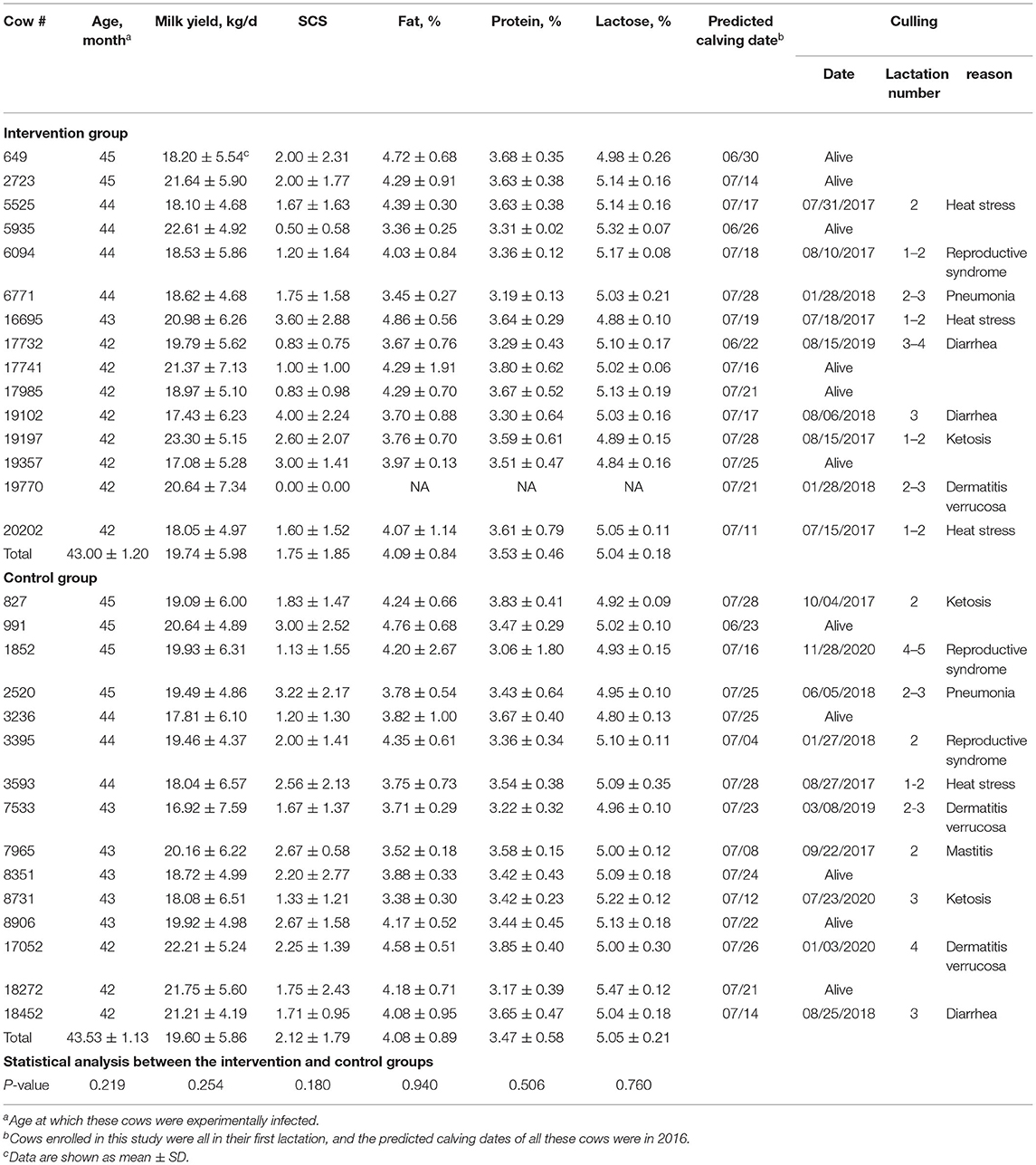
Table 1. Comparison of age, average daily milk yield, SCS, milk composition, predicted calving dates, and culling events of cows in the intervention and control groups before BLV inoculation.
After inoculation, the two groups of cows were reared separately in two cowsheds, together with ≈100 cows in each cowshed. These two cowsheds had similar context and were located in the same farm separated by ≈200 m distance. The cows in the two sheds were fed exactly the same diet by the same caregivers, who were not aware of the infection status of these cows, but they did not share the milking machines. Then the raw data of milk yield, SCS, and composition (primary objectives), as well as immune cells and immune-related cytokines in peripheral blood (secondary objectives), were collected and managed by an administrator who was unaware of the infection status of these cows, either.
Sterile whole blood was collected from a BLV-positive (B2145) cow with the combination of dipotassium ethylenediaminetetraacetic acid (EDTA), and proviral load (PVL) was determined by BLV FRET-qPCR, as described previously (Yang et al., 2016b). Subsequently, peripheral blood mononuclear cells (PBMC) were purified and resuspended in sterile 1 × phosphate-buffered solution (PBS) buffer at a concentration of 3.0 × 10⁵ cells/ml. Subsequently, half aliquot of the PBMC was inactivated by incubating at 60°C for 30 min. At the time of inoculation, each cow received a dose of 3.0 × 10⁶ activated (containing 2.7 × 10⁶ BLV proviral copies) or inactivated PBMC via intravenous inoculation through the jugular vein. The inoculations were performed by two veterinarians who were not aware of the serial numbers (ear tags were covered) of the cows and the composition of the injection (labeled A or B, respectively).
Whole blood samples were collected from the employed cows before and after inoculation. In detail, two specifications (2 and 10 ml) of evacuated tubes containing EDTA (BD, Franklin Lakes, USA) were used to hold the whole blood samples collected from all the 30 cows at 15 days before inoculation and 0 (2 h), 5, 15, 45, 75, 105, 135, and 165 days post-inoculation (DPI). The samples in the 2-ml tubes were applied for routine complete blood counts and DNA extraction, while the blood in the 10-ml tubes were used for plasma separation. In addition, the control cows were tested every 6 months to ensure that they were free of BLV. Within 2 h, these samples were smoothly transported on ice to the laboratory in Yangzhou University.
DNA was extracted from 200 μl of whole-blood samples using a Roche High Pure PCR Template Preparation Kit (Roche, Mannheim, Germany) in accordance with the manufacturer's instructions. The DNA samples were then stored at −20°C or directly used for analyzes. The FRET-qPCR targeting BLV pol gene was applied to screen BLV provirus, as described previously (sensibility: 1 copy/reaction) (Yang et al., 2016a).
Plasma was separated from whole blood samples by centrifugation at 1,500 × g for 20 min. The plasma samples were then stored at −20°C or directly used for analysis. The commercial INgezim BLV Compac 2.0 blocking ELISA kit (Ingenasa, Madrid, Spain) was also used to detect the specific antibodies to BLV gp51 protein following the manufacturer's instructions.
Routine CBC was performed on a BC-2800 Vet (Mindray, Shenzhen, China) to quantify 18 parameters, including the number of white blood cells (WBCs) (sensibility: 1 × 10⁸ cells/L), lymphocytes (LYMs) (sensibility: 1 × 10⁸ cells/L), monocytes (MNCs) (sensibility: 1 × 10⁸ cells/L), granulocytes (GRANs) (sensibility: 1 × 10⁸ cells/L), red blood cells (RBC) (sensibility: 1 × 10¹⁰ cells/L), and platelets (PLT) (sensibility: 1 × 10⁹ cells/L); the percentages of lymphocytes (LYM%) (sensibility: 0.1%), monocytes (MNC%) (sensibility: 0.1%), granulocytes (GRAN%) (sensibility: 0.1%), hematocrit (HCT) (sensibility: 0.1%), red blood cell distribution width (RDW) (sensibility: 0.1%), and plateletcrit (PCT) (sensibility: 0.001%); the content of hemoglobin (HGB) (sensibility: 1 g/L); mean corpuscular volume (MCV) (sensibility: 0.1 fl), mean corpuscular hemoglobin (MCH) (sensibility: 0.1 pg), mean corpuscular hemoglobin concentration (MCHC) (sensibility: 1 g/L), and mean platelet volume (MPV) (sensibility: 0.1 fl); and platelet distribution width (PDW) (sensibility: 0.1 CV%).
The quantification of 10 bovine cytokines (GM-CSF, IFN-γ, IL-1b, IL-2, IL-4, IL-5, IL-6, IL-10, IL-12p70, and IL-13) were performed using a commercial cytokine array (QAB-CYT-1) (RayBiotech, Norcross, USA) following the manufacturer's instructions, with sensibilities of 1 × 10⁻²-1 × 10⁻⁵ pg/ml. Briefly, 100 μl of plasma from each cow was used, with the cytokine standard mix and sample diluent as positive and negative controls, respectively. After blocking, the slides were incubated with 100 μl of samples, 80 μl of biotinylated antibody cocktail, and 80 μl of Cy3 equivalent dye–streptavidin, respectively, and washed thoroughly between each step of incubation. Water droplets were removed completely before the signals were visualized through an Axon GenePix (Molecular Devices, San Jose, USA) with a Cy3 wavelength (green channel).
The milk yield of each cow was automatically recorded by milk sensors (Afimilk, Kibbutz, Israel) three times (6.30 a.m., 2.00 and 10.00 p.m.) a day. The 20-ml milk samples collected with 0.015 g of potassium dichromate on the second day of every month were sent to Henan (Zhengzhou DHI center, Henan, China) or Nanjing (Nanjing DHI center, Jiangsu, China) DHI test center to determine the somatic cell count (sensibility: 10 thousand cells/ml) using a Fossomatic™ FC system (FOSS, Hillerod, Denmark), as well as fat (%) (performance range: 2–15%), protein (%) (performance range: 2–10%), and lactose (%) (performance range: 2–10%) using a MilkoScan™ FT+ system (FOSS, Hillerod, Denmark). Further analyses were performed with the SCS calculated as SCC [SCS = log2(SCC/100) + 3], as described previously (Yang et al., 2016a).
All statistical analyses were performed using the STATISTICA 7.0 software package (StatSoft, Oklahoma, USA). Independent Student's t-test was used to compare 18 CBC parameters, the expression levels of 10 cytokines, the milk yield (305-day milk yield and average daily milk yield), SCS, and milk composition (fat, protein, and lactose) between the intervention and control groups. The 305-day milk yield of each cow was adjusted and calculated according to the criteria developed by the Dairy Association of China (Supplementary Table S1). In addition, further analyses were performed to determine the effect of BLV infection on milk yield, SCS, and composition between the intervention and control groups in each lactation number (2, 3, and 4) stage (early, middle, and late). The reproduction performance and the incidences of six common diseases between the intervention and control groups were compared using the chi-square test. All data were presented as mean ± SD and considered statistically significant when P ≤ 0.050.
At the start of this study, 30 first-lactation cows were equally divided into the intervention and control groups, respectively. However, due to some unexpected issues and/or accidents, four cows were dead from heat stress, and 14 cows were culled from herds due to several incurable diseases, while none of the cows in the intervention group have shown one of the most common signs of BLV infection (Table 1). The culling decisions were made independently by the veterinarians. In detail, nine cows in the intervention group were dead (heat stress, n = 3) or culled (severe diarrhea, n = 2; clinical ketosis, n = 1; reproductive syndrome, n = 1; severe pneumonia, n = 1; dermatitis verrucosa, n = 1) between 15 July 2017 and 15 August 2019. Totally 10 cows in the control group were dead (heat stress, n = 1) or culled (severe diarrhea, n = 1; clinical ketosis, n = 2; incurable clinical mastitis, n = 1; reproductive syndrome, n = 2; severe pneumonia, n = 1; dermatitis verrucosa, n = 2) between 27 August 2017 and 28 November 2020.
On 07 July 2021, all the remaining cows had finished their fourth lactation. However, at that time, there were only seven and five cows remaining in the herds, respectively. It was predicted that some of these cows will be culled during the following lactation, which will significantly further reduce the power of statistical analysis. Therefore, we decided to end this study and evaluate the effects of BLV infection on cows in lactation numbers 2–4.
In order to make the employed cows in the intervention and control groups comparable, we comprehensively analyzed the parity, age, lactation number, weight, and the available records of daily milk yield (n = 8,663), SCS (n = 185), and composition [fat (n = 140), protein (n = 134), and lactose (n = 133)] of each cow. Statistical analysis showed that there were no significant differences between the intervention and control groups in parity (all in parity-1), lactation stage (all in their first lactation), age (43.00 ± 1.20 vs. 43.53 ± 1.13, month, P = 0.219), weight (345.00 ± 10.81 vs. 345.80 ± 10.63, kg, P = 0.840), milk yield (19.74 ± 5.98 vs. 19.60 ± 5.86, kg/d, P = 0.254), SCS (1.75 ± 1.85 vs. 2.12 ± 1.79, P = 0.180), fat (4.09 ± 0.84 vs. 4.08 ± 0.89, %, P = 0.940), protein (3.53 ± 0.46 vs. 3.47 ± 0.58, %, P = 0.506), and lactose (5.04 ± 0.18 vs. 5.05 ± 0.21, %, P = 0.760) before artificial inoculation (Table 1), indicating that these 30 cows employed in this study were comparable with similar dairy performance.
To evaluate the viral infection, both FRET-qPCR and commercial blocking ELISA were applied to screen BLV proviral DNA and anti-BLV antibodies. The results showed that BLV proviral DNA was available to be detected in six cows at 0 (2 h) DPI and all the cows at 15 DPI, while anti-BLV gp51 antibodies were unavailable to be detected in any cow during this period. From 45 DPI, BLV proviral DNA and anti-gp51 antibodies could be detected in all the 15 cows in the intervention group, while the control animals remained negative throughout this experiment (Figure 1).

Figure 1. Detection of BLV proviral DNA and anti-BLV antibodies. (Main) BLV FRET-qPCR showed that five and six inoculated cows were detected to be positive at 0 and 5 DPI, respectively. From 45 DPI, all cows in the intervention group were BLV-positive by FRET-qPCR and ELISA. In comparison, all the control cows were free of BLV throughout this study. (Insertion) The copy numbers of BLV pol gene of cows in the intervention and control groups were quantified by FRET-qPCR.
Between 18 May 2017 and 17 November 2017, CBC tests were performed on 241 whole blood samples collected at 15 days before inoculation and at 0, 5, 15, 45, 75, 105, 135, and 165 DPI, including 113 and 128 samples in the intervention and control groups, respectively. Before BLV inoculation, the individuals in the intervention and control groups showed similar and normal CBC parameters compared with reference values (Supplementary Figure S1, Supplementary Tables S2, S3). Our results reliably revealed that the number of WBCs, LYMs, and MNCs of the BLV-inoculated cows decreased immediately after challenge (5 DPI) and subsequently drastically increased (15 and 45 DPI). In detail, at 5 DPI, the number of WBCs, LYMs, and MNCs all decreased in the challenged cows, among which WBCs (5.89 ± 2.20 vs. 8.15 ± 1.65, 109/L, P = 0.004) and LYMs (2.27 ± 1.11 vs. 3.67 ± 1.00, 109/L, P = 0.001) showed statistical differences. At 15 DPI, there were significant higher levels of LYMs (4.56 ± 2.00 vs. 3.13 ± 1.07, 109/L, P = 0.022) and MNCs (0.91 ± 0.32 vs. 0.67 ± 0.23, 109/L, P = 0.036) in the challenged cows. At 45 DPI, significant higher numbers of WBCs (10.13 ± 3.48 vs. 6.98 ± 2.50, 109/L, P = 0.010), LYMs (5.05 ± 2.13 vs. 2.99 ± 0.90, 109/L, P = 0.002), and MNCs (1.19 ± 0.61 vs. 0.58 ± 0.19, 109/L, P = 0.001) were observed in the challenged cows (Figures 2A–C, Supplementary Tables S2, S3).
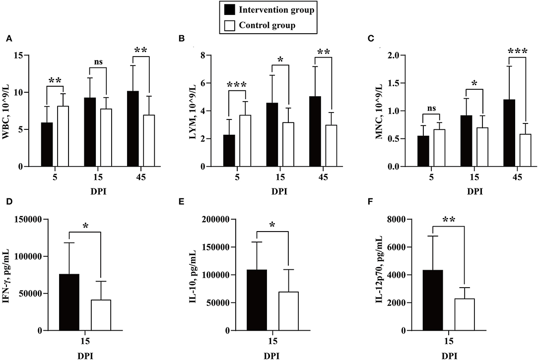
Figure 2. Quantification of CBC parameters and cytokines. The numbers of white blood cells (WBCs) (A), lymphocytes (LYM) (B), and monocytes (MNCs) (C) were compared between cows in the intervention (in black) and control (in white) groups at 5, 15, and 45 DPI. The expression levels of IFN-γ (D), IL-10 (E), and IL-12p70 (F) were compared between the cows in the inoculation (in black) and control (in black) groups at 15 DPI. Data are shown as mean ± SD, and the symbols indicate P ≤ 0.001 (***), P ≤ 0.010 (**), P ≤ 0.050 (*), and P > 0.050 (ns), respectively.
Between 18 May 2017 and 17 November 2017, the quantitation of 10 bovine cytokines was performed on 128 plasma samples collected before inoculation and at 0, 15, 75, and 165 DPI, including 61 and 67 samples in the intervention and control groups, respectively. The results showed that the expression levels of IFN-γ (75,794.73 ± 42,529.12 vs. 41,065.30 ± 25,216.67, pg/ml, P = 0.013), IL-10 (108,555.82 ± 50,554.92 vs. 69,994.84 ± 39,582.16, pg/ml, P = 0.031), and IL-12p70 (4,304.16 ± 2,483.99 vs. 2,306.02 ± 779.66, pg/ml, P = 0.008) increased significantly in the challenged cows compared with the controls at 15 DPI (Figures 2D–F, Supplementary Tables S4, S5).
Statistical analysis showed that no significant differences were noted between the two groups for the duration in the herds (827.00 ± 697.98 vs. 865.93 ± 596.59, day, P = 0.871). From 26 June 2016 to 07 July 2021, a total of 23,468 records of individual daily milk yield were automatically recorded by milk sensors (Afimilk, Kibbutz, Israel). Subsequently, these records archived after artificial inoculation (n = 14,805) were divided into nine subsets based on lactation number and stage, including subset 1 (lactation number 2, first-lactation stage, n = 2,337), subset 2 (lactation number 2, middle lactation stage, n = 2,207), subset 3 (lactation number 2, late lactation stage, n = 2,296), subset 4 (lactation number 3, first-lactation stage, n = 1,430), subset 5 (lactation number 3, middle lactation stage, n = 1,428), subset 6 (lactation number 3, late lactation stage, n = 1,432), subset 7 (lactation number 4, first-lactation stage, n = 1,220), subset 8 (lactation number 4, middle lactation stage, n = 1,216), and subset 9 (lactation number 4, first-lactation stage, n = 1,239). At the same time, a total of 1,742 of records of milk SCS and composition were obtained from monthly performed DHI tests, including 492 of milk SCS (lactation numbers 1/2/3/4: 185/133/93/81), 451 of milk fat (lactation numbers 1/2/3/4: 140/141/89/81), 440 of milk protein (lactation numbers 1/2/3/4: 134/137/95/74), and 359 of milk lactose (lactation numbers 1/2/3: 133/143/83), respectively. Then these records were divided into different subsets according to the same criteria.
When we compared the overall milk yield between the two groups, independent Student's t-test was used to compare the average daily milk yield and 305-day milk yield between the two groups. The results showed that the cows in the intervention group had significant increased average daily milk production (32.53 ± 10.79 vs. 29.74 ± 10.77, kg/d, P < 0.001) and adjusted 305-day milk yield (10,550.07 ± 1,127.20 vs. 8,979.51 ± 1,541.15, kg, P = 0.002) than the uninfected cows (Figures 3A,B). Subsequently, further analysis was carried out to determine the association between BLV infection and milk yield in cows in different lactation numbers (2, 3, and 4) and stages (first, middle, and late). Interestingly, from the early lactation stage of lactation number 3 to the end of this study, the infection of BLV was determined to be associated with significantly increased milk yield. In detail, the average daily milk yield of cows in the intervention group was significantly higher than that of the control at all lactation stages of lactation number 3 (seven intervened cows and nine or seven control cows) (early: 40.68 ± 12.33 vs. 39.32 ± 9.88, kg/d, P = 0.021; middle: 36.34 ± 9.43 vs. 32.28 ± 8.19, kg/d, P < 0.001; late: 25.26 ± 8.49 vs. 19.69 ± 9.59, kg/d, P < 0.001) and lactation number 4 (six intervened cows and seven control cows) (44.90 ± 9.86 vs. 38.62 ± 8.92, kg/d, P < 0.001; middle: 37.58 ± 6.41 vs. 31.27 ± 7.09, kg/d, P < 0.001; late: 29.12 ± 6.36 vs. 22.07 ± 9.51, kg/d, P < 0.001) (Figure 4).
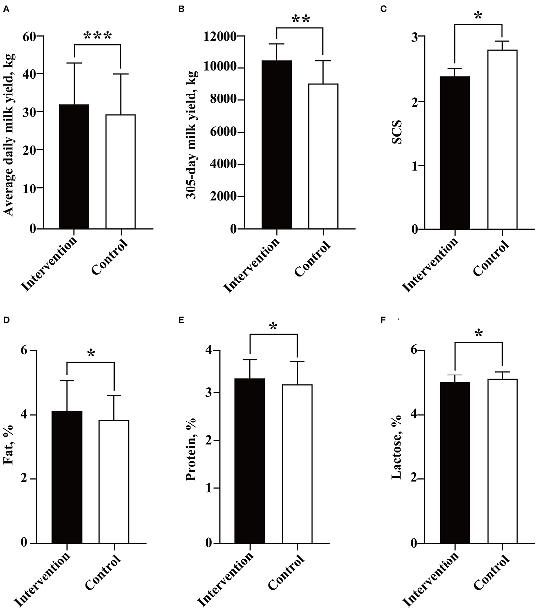
Figure 3. Association between BLV infection and dairy performance of cows. The average daily milk yield (A), 305-day milk yield (B), SCS (C), and contents of fat (D), protein (E), and lactose (F) were compared and analyzed between the intervention (in black) and control (in white) groups by Student's t-test. Data are shown as mean ± SD, and the symbols indicate P ≤ 0.001 (***), P ≤ 0.010 (**), and P ≤ 0.05 (*), respectively.
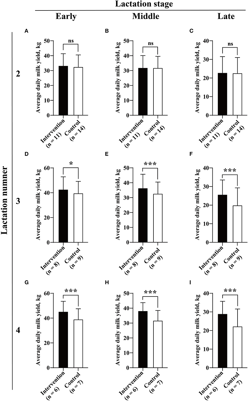
Figure 4. Association between BLV infection and milk yield. Average daily milk yield (A–I) for each of three lactation numbers (2, 3, and 4) and stages (early, middle, and late) were compared between the intervention (in black) and control (in white) groups by Student's t-test. Data are shown as mean ± SD, and the symbols indicate P ≤ 0.001 (***), P ≤ 0.050 (*), and P > 0.050 (ns), respectively.
Although the infection of BLV was determined to be associated with reduced milk SCS (2.40 ± 1.56 vs. 2.81 ± 1.87, P = 0.038) and lactose (5.04 ± 0.24 vs. 5.11 ± 0.24, %, P = 0.036), and increased milk fat (4.00 ± 0.98 vs. 3.80 ± 0.71, %, P = 0.031) and protein (3.40 ± 0.41 vs. 3.30 ± 0.51, %, P = 0.050) (Figures 3C–F), statistical differences were only observed at the late lactation stage of lactation number 4 (six intervened cows and six control cows) (2.55 ± 0.93 vs. 3.73 ± 1.58, P = 0.036) for milk SCS (Figure 5), the early lactation stage of lactation number 2 (10 intervened cows and 12 control cows) (4.16 ± 1.00 vs. 3.50 ± 0.86, %, P = 0.021), and the middle lactation stage of lactation number 4 (six intervened cows and six control cows) (4.51 ± 1.39 vs. 3.58 ± 0.63, %, P = 0.025) for milk fat and early lactation stage of lactation number 2 (nine intervened cows and 12 control cows) (3.26 ± 0.44 vs. 2.93 ± 0.27, %, P = 0.005) for milk protein (Figure 6).
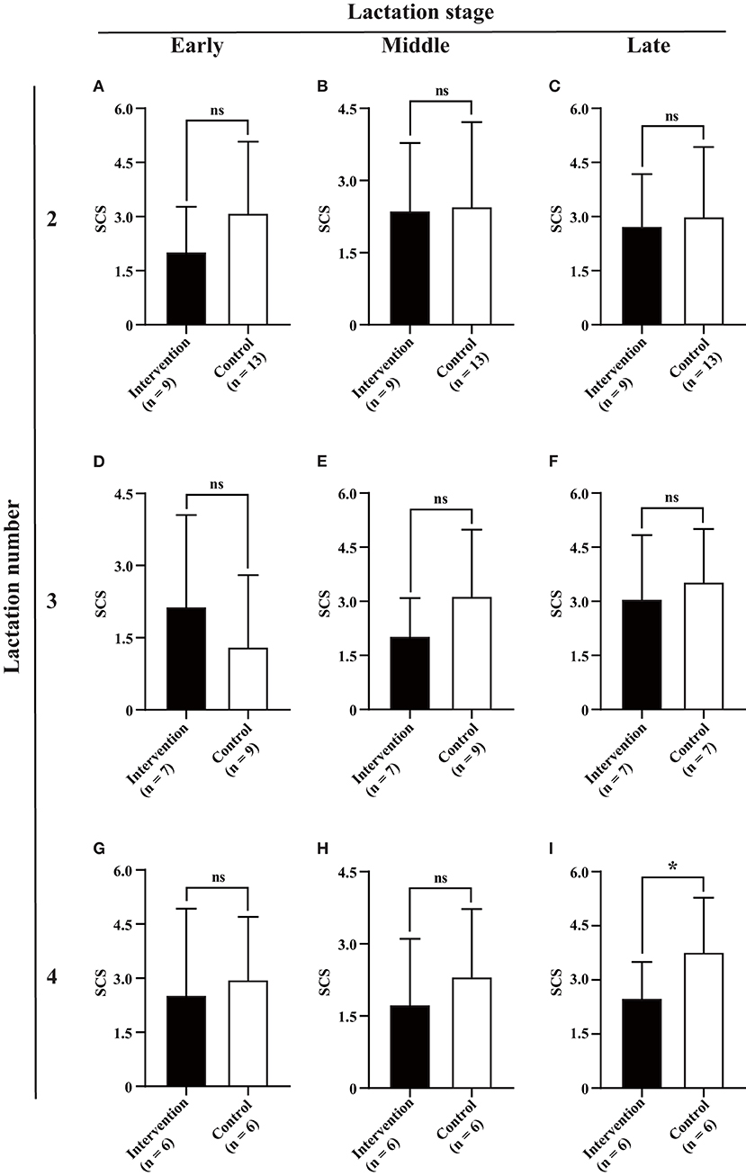
Figure 5. Association between BLV infection and milk SCS. Average monthly milk SCS (A–I) for each of three lactation numbers (2, 3, and 4) and stages (early, middle, and late) was compared between the intervention (in black) and control (in white) groups by Student's t-test. Data are shown as mean ± SD, and the symbols indicate P ≤ 0.050 (*) and P > 0.050 (ns), respectively.
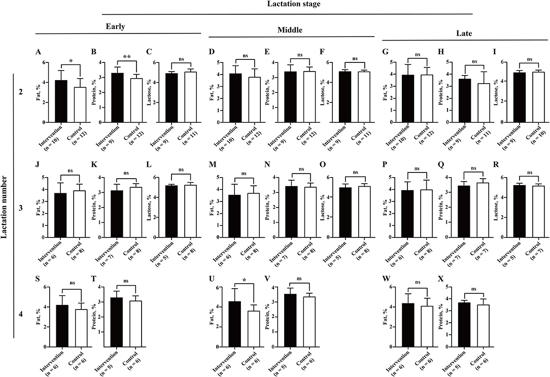
Figure 6. Association between BLV infection and milk composition (fat, protein, and lactose). Average monthly milk fat (A,D,G,J,M,P,S,U,W), protein (B,E,H,K,N,Q,T,V,X), and lactose (C,F,I,L,O,R) for each of three lactation numbers (2, 3, and 4) and stages (early, middle, and late) were compared between the intervention (in black) and control (in white) groups by Student's t-test. Data are shown as mean ± SD, and the symbols indicate P ≤ 0.050 (*) and P > 0.050 (ns), respectively.
To investigate in depth other factors that could have affected the dairy performance of cow uncertainty, four universally used KPIs in reproduction (days at first breeding, the percentage of pregnant cows at 150 DIM, and the frequencies of pregnancy loss <90 or ≥90 days of pregnancy) and the incidences of six common diseases (the frequencies of ketosis, metritis, retained placenta, clinical mastitis, lameness, and enteritis) were compared between cows in the intervention and control groups. The data of six common diseases were only available for the cows in lactation numbers 3–4, due to a breakdown on the data storage system. The results showed that in lactation numbers 1–4, cows in the intervention and control groups had similar reproduction performance, including days at first breeding, the rate of pregnancy at 150 DIM, and the rate of pregnancy loss <90 or ≥90 days of pregnancy (P > 0.05). Similarly, no significant differences were observed in the incidences of ketosis, metritis, retained placenta, clinical mastitis, lameness, and enteritis between the two groups in lactation numbers 3–4 (P > 0.05) (Table 2).
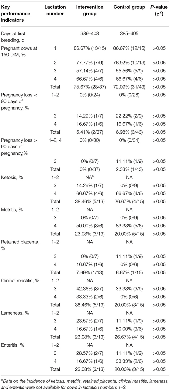
Table 2. Comparison of four key performance indicators and the incidences of six common diseases between the intervention and control groups.
It is prohibitively expensive to design and conduct a parallel-group trial with BLV infection with sufficient statistical power. Hutchinson determined the timing and described early fluctuations of BLV detection by qPCR, ELISA, and lymphocyte counts following experimental BLV inoculation with 23 Holstein steers (15 infected steers and eight control steers) (Hutchinson et al., 2020). Benitez evaluated the potential for BLV transmission during natural breeding between a BLV-infected bull and 40 uninfected heifers (20 control heifers and 20 challenged heifers) (Benitez et al., 2019). However, to the best of our knowledge, no study so far has investigated the effects of BLV infection on dairy performance based on artificially inoculated cows. To establish a BLV challenge model with comparable cows, from a BLV-free farm with 2,988 cows, 30 healthy first-lactation cows with similar performance were employed. All these cows were in their second pregnancy with the similar dairy performance in the first lactation. According to the previous large-scale molecular epidemiological studies, BLV genotype 6 was demonstrated to be the endemic strain in China, which shared a significant nucleotide/amino acid polymorphism with FLK-BLV (genotype 1) (Yang et al., 2019a,b; Yu et al., 2019). Hence, in this study, the artificial inoculation was performed with PBMC from a Holstein cow infected with high PVL of BLV genotype 6.
The detection of BLV proviral DNA and gp51 antibody showed that the BLV challenge model was successfully established, and no cross-infection occurred from the beginning to the end of the experiment. For convenience, the hematological diagnostic method based on LYM counts was widely used as the screening and disease severity monitoring tool for BLV, especially among greater parity animals (Nieto Farias et al., 2018; Wisnieski et al., 2020). In this study, we indicated that the most significant increases in WBC, LYM, and MNC counts were all observed at 45 DPI, but not at the later stage of infection. Therefore, it is strongly recommended to use real-time PCR or ELISA, instead of complete blood count for the detection of BLV infection.
The antibody-based detection tests (agar gel immunodiffusion and ELISA) for the diagnosis of BLV infection have been authorized by the World Organisation for Animal Health OIE, 2018. Klintevall et al. (1994) reported that BLV antibodies could be detected by ELISA on 26 DPI. Evermann et al. found that the experimental calves seroconverted to BLV within 8 and 14 weeks, respectively, depending on the equivalent of inoculation (Evermann et al., 1986). Moreover, it is convenient to screen for BLV infection using milk from the DHI sampling process (Nekouei et al., 2015b). In this study, the anti-gp51 antibody was available to be detected since 45 DPI, which emphasized the early diagnostic importance of real-time qPCR.
A retrospective study indicated that the mRNA expression levels of IFN-γ and IL-12 were significantly higher in PBMC from infected cattle with low and high PVL than uninfected animals (Farias et al., 2016). In this study, the significant higher expression levels of IFN-γ and IL-12p70 in plasma were observed during the early stage of BLV infection (15 DPI). In addition, the expression level of IL-10 was also significant higher in plasma of infected individuals.
The main purpose of this study was to better understand the possible association between BLV infection and the dairy performance of cows. Although several previous studies have provided evidence that BLV infection is associated with decreased milk yield at herd or individual levels (Erskine et al., 2012; Nekouei et al., 2016; Norby et al., 2016), other studies indicated that the BLV infected rate is not associated with milk production (Sorge et al., 2011). Some studies have even shown that the herd with a higher BLV-positive rate actually produce more milk (Abdalla et al., 2016). It has been well revealed that individual dairy performance of cows can be greatly influenced by a variety of factors, such as breed, parity, lactation stage, feeding conditions, and the occurrence of diseases (Inchaisri et al., 2010; Soufleri et al., 2019; Barth, 2020; Fehlberg et al., 2020; Goncalves et al., 2020; Adriaens et al., 2021). Therefore, to minimize these effects, the 30 cows enrolled in this study were comparable with the identical parity/lactation number, similar age, dairy performance, and predicted calving date before BLV inoculation. This is one of the advantages of this experiment over other observational studies. In addition, to create greater certainty in our conclusion, milk yield, SCS, and milk composition (fat, protein, and lactose) were analyzed in the intervention and control groups according to different lactation numbers (2, 3, and 4) and stages (early, middle, and late). Unexpectedly, our 4-year study showed that BLV-infected cows had significant increased milk yield compared with the uninfected cows. At the same time, there were statistical differences in milk yield between the two groups from the early lactation stage of lactation number 3 to the end of this study. Although the data of six common diseases were only available for the cows in lactation numbers 3–4, the available data indicated that differences in reproduction performance and the incidence of diseases would not contribute to the significant increased milk yield in the intervention group in lactation numbers 3–4. The Canadian and U.S. studies determined a negative association between herd-level milk production and BLV positivity (Sargeant et al., 1997; Erskine et al., 2012), based on AGID and ELISA, respectively. A recent study indicated that BLV-infected cows with two and three lactations showed significantly lower life milk productions than their negative counterparts (Nekouei et al., 2016). These results are at odds with this study, but we did not analyze the lifetime milk yield. However, our results are partially consistent with a previous study in 16 U.S. states, which indicated that selection for higher milk yield may lead to increased BLV prevalence in dairy herds (Abdalla et al., 2016). By contrast, although the overall milk SCS and composition varied between the intervention and control groups, there were almost no significant differences in the lactation number and stage-based analysis, which may be due to the limited sample size. However, it should be noted that in previous case–control studies, it is not really clear which genotype(s) of BLV had infected those enrolled cows, probably BLV genotype 1, the most widespread genotype of BLV in the world. This variability could be a source of differences in results among different studies. Our study has some inherent limitations that could have biased our results. First, the limited number of experimental animals may affect the reliability of the results. Second, the individual differences and lack of specific pathogen-free rearing conditions will interfere with the control of variables. Third, although the farm managers and workers were supposed to be blinded as to which herd of cows was challenged, they frequently figured it out. Therefore, more similar studies were recommended to be conducted to clarify and mitigate such detrimental effects.
The original contributions presented in the study are included in the article/Supplementary Material, further inquiries can be directed to the corresponding author/s.
The animal study was reviewed and approved by the Institutional Animal Care and Use Committee of the Yangzhou University College of Veterinary Medicine, China.
YY, CW, and ZY contributed to conception and design of the study. YY, ZG, YL, JZ, YP, SC, WC, and HW performed the experiments. YY, ZG, XL, YM, XH, JS, and AQ organized the database. YY, ZG, XL, and YM performed the statistical analysis. YY wrote the first draft of the manuscript. CW, SS, and ZY revised the manuscript. All authors contributed to the article and approved the submitted version.
This work was funded by the National Natural Science Foundation of China (32002263 to YY), Basic Research Program of Jiangsu Province (BK20190881 to YY), Postdoctoral Research Foundation of China (2019M650126 to YY), Natural Science Foundation of Jiangsu Higher Education Institutions of China (19KJB230001 to YY), Seed Industry Vitalization Program of Jiangsu Province (JBGS[2021]117 to YY), Jiangsu Agricultural Science and Technology Independent Innovation Fund (CX(20)3089 to YY), and Priority Academic Program Development of Jiangsu Higher Education Institutions (NA to CW, SS, and ZY). The funders had no role in study design, data collection and analysis, decision to publish, or preparation of the manuscript.
The authors declare that the research was conducted in the absence of any commercial or financial relationships that could be construed as a potential conflict of interest.
All claims expressed in this article are solely those of the authors and do not necessarily represent those of their affiliated organizations, or those of the publisher, the editors and the reviewers. Any product that may be evaluated in this article, or claim that may be made by its manufacturer, is not guaranteed or endorsed by the publisher.
We thank Huiguang Wu (College of Veterinary Medicine, Yangzhou University) for helping us with the statistical analysis in this study. We also thank Yongjiang Mao (College of Animal Science and Technology, Yangzhou University) for helping us analyze the key performance indicators of cows.
The Supplementary Material for this article can be found online at: https://www.frontiersin.org/articles/10.3389/fmicb.2022.946463/full#supplementary-material
Abdalla, E. A., Weigel, K. A., Byrem, T. M., and Rosa, G. J. M. (2016). Short communication: genetic correlation of bovine leukosis incidence with somatic cell score and milk yield in a US Holstein population. J. Dairy Sci. 99, 2005–2009. doi: 10.3168/jds.2015-9833
Adriaens, I., van den Brulle, I., D'Anvers, L., Statham, J. M. E., Geerinckx, K., De Vliegher, S., et al. (2021). Milk losses and dynamics during perturbations in dairy cows differ with parity and lactation stage. J. Dairy Sci. 104, 405–418. doi: 10.3168/jds.2020-19195
Aida, Y., Murakami, H., Takahashi, M., and Takeshima, S. N. (2013). Mechanisms of pathogenesis induced by bovine leukemia virus as a model for human T-cell leukemia virus. Front. Microbiol. 4, 328. doi: 10.3389/fmicb.2013.00328
Altanerova, V., Ban, J., Kettmann, R., and Altaner, C. (1990). Induction of leukemia in chicken by bovine leukemia virus due to insertional mutagenesis. Arch. Geschwulstforsch. 60, 89–96.
Barth, K.. (2020). Effects of suckling on milk yield and milk composition of dairy cows in cow-calf contact systems. J. Dairy Res. 87, 133–137. doi: 10.1017/S0022029920000515
Bartlett, P. C., Norby, B., Byrem, T. M., Parmelee, A., Ledergerber, J. T., and Erskine, R. J. (2013). Bovine leukemia virus and cow longevity in Michigan dairy herds. J. Dairy Sci. 96, 1591–1597. doi: 10.3168/jds.2012-5930
Benitez, O. J., Roberts, J. N., Norby, B., Bartlett, P. C., Maeroff, J. E., and Grooms, D. L. (2019). Lack of Bovine leukemia virus transmission during natural breeding of cattle. Theriogenology 126, 187–190. doi: 10.1016/j.theriogenology.2018.12.005
Erickson, P. S., and Kalscheur, K. F. (2020). Nutrition and feeding of dairy cattle. Anim. Agric. 157–180. doi: 10.1016/B978-0-12-817052-6.00009-4
Erskine, R. J., Bartlett, P. C., Byrem, T. M., Render, C. L., Febvay, C., and Houseman, J. T. (2012). Association between bovine leukemia virus, production, and population age in Michigan dairy herds. J. Dairy Sci. 95, 727–734. doi: 10.3168/jds.2011-4760
Evermann, J. F., DiGiacomo, R. F., Ferrer, J. F., and Parish, S. M. (1986). Transmission of bovine leukosis virus by blood inoculation. Am. J. Vet. Res. 47, 1885–1887.
Farias, M. V., Lendez, P. A., Marin, M., Quintana, S., Martinez-Cuesta, L., Ceriani, M. C., et al. (2016). Toll-like receptors, IFN-gamma and IL-12 expression in bovine leukemia virus-infected animals with low or high proviral load. Res. Vet. Sci. 107, 190–195. doi: 10.1016/j.rvsc.2016.06.016
Fehlberg, L. K., Guadagnin, A. R., Thomas, B. L., Sugimoto, Y., Shinzato, I., and Cardoso, F. C. (2020). Feeding rumen-protected lysine prepartum increases energy-corrected milk and milk component yields in Holstein cows during early lactation. J. Dairy Sci. 103, 11386–11400. doi: 10.3168/jds.2020-18542
Florins, A., Gillet, N., Asquith, B., Boxus, M., Burteau, C., Twizere, J. C., et al. (2007). Cell dynamics and immune response to BLV infection: a unifying model. Front. Biosci. 12, 1520–1531. doi: 10.2741/2165
Goncalves, J. L., Kamphuis, C., Vernooij, H., Araujo, J. P. Jr., Grenfell, R. C., Juliano, L., et al. (2020). Pathogen effects on milk yield and composition in chronic subclinical mastitis in dairy cows. Vet. J. 262, 105473. doi: 10.1016/j.tvjl.2020.105473
Hutchinson, H. C., Norby, B., Droscha, C. J., Sordillo, L. M., Coussens, P. M., and Bartlett, P. C. (2020). Bovine leukemia virus detection and dynamics following experimental inoculation. Res. Vet. Sci. 133, 269–275. doi: 10.1016/j.rvsc.2020.09.026
Inchaisri, C., Hogeveen, H., Vos, P. L., van der Weijden, G. C., and Jorritsma, R. (2010). Effect of milk yield characteristics, breed, and parity on success of the first insemination in Dutch dairy cows. J. Dairy Sci. 93, 5179–5187. doi: 10.3168/jds.2010-3234
Johnson, J. W., Lucas, H., King, S., Caron, T., Wang, C., and Kelly, P. J. (2020). Serosurvey for Brucella spp. and Coxiella burnetii in animals on Caribbean islands. Vet. Med. Sci. 6, 39–43. doi: 10.1002/vms3.214
Klintevall, K., Ballagi-Pordany, A., Naslund, K., and Belak, S. (1994). Bovine leukaemia virus: rapid detection of proviral DNA by nested PCR in blood and organs of experimentally infected calves. Vet. Microbiol. 42, 191–204. doi: 10.1016/0378-1135(94)90018-3
Kobayashi, S., Tsutsui, T., Yamamoto, T., Hayama, Y., Kameyama, K., Konishi, M., et al. (2010). Risk factors associated with within-herd transmission of bovine leukemia virus on dairy farms in Japan. BMC Vet. Res. 6, 1. doi: 10.1186/1746-6148-6-1
Krasnikova, E. S., Bouchemla, F., Krasnikov, A. V., Radionov, R. V., and Belyakova, A. S. (2019). The hematobiochemical status of Wistar rat line under the bovine leukemia virus experimental infection. Vet. World 12, 382–388. doi: 10.14202/vetworld.2019.382-388
Kucerova, L., Altanerova, V., Altaner, C., and Boris-Lawrie, K. (1999). Bovine leukemia virus structural gene vectors are immunogenic and lack pathogenicity in a rabbit model. J. Virol. 73, 8160–8166. doi: 10.1128/JVI.73.10.8160-8166.1999
Kuczewski, A., Orsel, K., Barkema, H. W., Mason, S., Erskine, R., and van der Meer, F. (2021). Invited review: bovine leukemia virus-transmission, control, and eradication. J. Dairy Sci. 104, 6358–6375. doi: 10.3168/jds.2020-18925
LaDronka, R. M., Ainsworth, S., Wilkins, M. J., Norby, B., Byrem, T. M., and Bartlett, P. C. (2018). Prevalence of bovine leukemia virus antibodies in US Dairy Cattle. Vet. Med. Int. 2018, 5831278. doi: 10.1155/2018/5831278
Li, J., Kelly, P., Zhang, J., Xu, C., and Wang, C. (2015). Development of a pan-Babesia FRET-qPCR and a survey of livestock from five Caribbean islands. BMC Vet. Res. 11, 246. doi: 10.1186/s12917-015-0560-0
Ma, B. Y., Gong, Q. L., Sheng, C. Y., Liu, Y., Ge, G. Y., Li, D. L., et al. (2021). Prevalence of bovine leukemia in 1983-2019 in China: a systematic review and meta-analysis. Microb. Pathog. 150, 104681. doi: 10.1016/j.micpath.2020.104681
Mammerickx, M., Portetelle, D., and Burny, A. (1981). Experimental cross-transmissions of bovine leukemia virus (BLV) between several animal species. Zentralblatt Veterinarmedizin Reihe B 28, 69–81. doi: 10.1111/j.1439-0450.1981.tb01740
Nekouei, O., Stryhn, H., VanLeeuwen, J., Kelton, D., Hanna, P., and Keefe, G. (2015a). Predicting within-herd prevalence of infection with bovine leukemia virus using bulk-tank milk antibody levels. Prev. Vet. Med. 122, 53–60. doi: 10.1016/j.prevetmed.2015.10.009
Nekouei, O., VanLeeuwen, J., Stryhn, H., Kelton, D., and Keefe, G. (2016). Lifetime effects of infection with bovine leukemia virus on longevity and milk production of dairy cows. Prev. Vet. Med. 133, 1–9. doi: 10.1016/j.prevetmed.2016.09.011
Nekouei, O. A., Sanchez, J., and Keefe, G. P. (2015b). Carryover of bovine leukemia virus antibodies in samples from shared milk meters. J. Dairy Sci. 98, 5274–5279. doi: 10.3168/jds.2014-9036
Nieto Farias, M. V., Souza, F. N., Lendez, P. A., Martinez-Cuesta, L., Santos, K. R., Della Libera, A., et al. (2018). Lymphocyte proliferation and apoptosis of lymphocyte subpopulations in bovine leukemia virus-infected dairy cows with high and low proviral load. Vet. Immunol. Immunopathol. 206, 41–48. doi: 10.1016/j.vetimm.2018.10.012
Norby, B., Bartlett, P. C., Byrem, T. M., and Erskine, R. J. (2016). Effect of infection with bovine leukemia virus on milk production in Michigan dairy cows. J. Dairy Sci. 99, 2043–2052. doi: 10.3168/jds.2015-10089
OIE. (2018). “Enzootic bovine leucosis,” in OIE Terrestrial Manual. Available online at: https://www.woah.org/fileadmin/Home/eng/Health_standards/tahm/2.04.10_EBL.pdf
Olson, C., Kettmann, R., Burny, A., and Kaja, R. (1981). Goat lymphosarcoma from bovine leukemia virus. J. Natl. Cancer Inst. 67, 671–675.
Porta, N. G., Alvarez, I., Suarez Archilla, G., Ruiz, V., Abdala, A., and Trono, K. (2019). Experimental infection of sheep with Bovine leukemia virus (BLV): Minimum dose of BLV-FLK cells and cell-free BLV and neutralization activity of natural antibodies. Rev. Argent. Microbiol. 51, 316–323. doi: 10.1016/j.ram.2019.01.004
Ramalho, G. C., Silva, M., Falcao, B. M. R., Limeira, C. H., Nogueira, D. B., Dos Santos, A. M., et al. (2021). High herd-level seroprevalence and associated factors for bovine leukemia virus in the semi-arid Paraiba state, Northeast Region of Brazil. Prev. Vet. Med. 190, 105324. doi: 10.1016/j.prevetmed.2021.105324
Sargeant, J. M., Kelton, D. F., Martin, S. W., and Mann, E. D. (1997). Associations between farm management practices, productivity, and bovine leukemia virus infection in Ontario dairy herds. Prev. Vet. Med. 31, 211–221. doi: 10.1016/s0167-5877(96)01140-3
Sorge, U. S., Lissemore, K., Cantin, R., and Kelton, D. F. (2011). Short communication: Milk ELISA status for bovine leukosis virus infection is not associated with milk production in dairy cows. J. Dairy Sci. 94, 5062–5064. doi: 10.3168/jds.2011-4339
Soufleri, A., Banos, G., Panousis, N., Fletouris, D., Arsenos, G., and Valergakis, G. E. (2019). Genetic parameters of colostrum traits in Holstein dairy cows. J. Dairy Sci. 102, 11225–11232. doi: 10.3168/jds.2019-17054
White, L. J., Schukken, Y. H., Dogan, B., Green, L., Dopfer, D., Chappell, M. J., et al. (2010). Modelling the dynamics of intramammary E. coli infections in dairy cows: understanding mechanisms that distinguish transient from persistent infections. Vet. Res. 41, 13. doi: 10.1051/vetres/2009061
Wisnieski, L., Norby, B., Gandy, J., Byrem, T. M., and Sordillo, L. M. (2020). Changes in bovine leukemia virus serological status and lymphocyte count between dry-off and early lactation in Michigan dairy cows. J. Dairy Sci. 103, 9473–9480. doi: 10.3168/jds.2019-17839
Yang, Y., Chen, L., Dong, M., Huang, W., Hao, X., Peng, Y., et al. (2019a). Molecular characterization of bovine leukemia virus reveals existence of genotype 4 in Chinese dairy cattle. Virol. J. 16, 108. doi: 10.1186/s12985-019-1207-8
Yang, Y., Chu, S., Shang, S., Yang, Z., and Wang, C. (2019b). Short communication: genotyping and single nucleotide polymorphism analysis of bovine leukemia virus in Chinese dairy cattle. J. Dairy Sci. 102, 3469–3473. doi: 10.3168/jds.2018-15481
Yang, Y., Fan, W., Mao, Y., Yang, Z., Lu, G., Zhang, R., et al. (2016a). Bovine leukemia virus infection in cattle of China: association with reduced milk production and increased somatic cell score. J. Dairy Sci. 99, 3688–3697. doi: 10.3168/jds.2015-10580
Yang, Y., Kelly, P. J., Bai, J., Zhang, R., and Wang, C. (2016b). First molecular characterization of bovine leukemia virus infections in the Caribbean. PLoS ONE 11, e0168379. doi: 10.1371/journal.pone.0168379
Yang, Y., Mao, Y., Kelly, P., Yang, Z., Luan, L., Zhang, J., et al. (2014). A pan-Theileria FRET-qPCR survey for Theileria spp. in ruminants from nine provinces of China. Parasit. Vectors 7, 413. doi: 10.1186/1756-3305-7-413
Yang, Y., Yang, Z., Kelly, P., Li, J., Ren, Y., and Wang, C. (2018). Anaplasma phagocytophilum in the highly endangered Pere David's deer Elaphurus davidianus. Parasit. Vectors 11, 25. doi: 10.1186/s13071-017-2599-1
Keywords: bovine leukemia virus, artificially inoculation, milk yield, somatic cell score, nutrition compositions
Citation: Yang Y, Gong Z, Lu Y, Lu X, Zhang J, Meng Y, Peng Y, Chu S, Cao W, Hao X, Sun J, Wang H, Qin A, Wang C, Shang S and Yang Z (2022) Dairy Cows Experimentally Infected With Bovine Leukemia Virus Showed an Increased Milk Production in Lactation Numbers 3–4: A 4-Year Longitudinal Study. Front. Microbiol. 13:946463. doi: 10.3389/fmicb.2022.946463
Received: 17 May 2022; Accepted: 20 June 2022;
Published: 11 July 2022.
Edited by:
Guo Huichen, Chinese Academy of Agricultural Sciences, ChinaReviewed by:
Paul Bartlett, Michigan State University, United StatesCopyright © 2022 Yang, Gong, Lu, Lu, Zhang, Meng, Peng, Chu, Cao, Hao, Sun, Wang, Qin, Wang, Shang and Yang. This is an open-access article distributed under the terms of the Creative Commons Attribution License (CC BY). The use, distribution or reproduction in other forums is permitted, provided the original author(s) and the copyright owner(s) are credited and that the original publication in this journal is cited, in accordance with accepted academic practice. No use, distribution or reproduction is permitted which does not comply with these terms.
*Correspondence: Yi Yang, eWFuZ3lpQHl6dS5lZHUuY24=; Shaobin Shang, c2hhb2JpbnNoYW5nQHl6dS5lZHUuY24=; Zhangping Yang, eXpwQHl6dS5lZHUuY24=
†These authors have contributed equally to this work
Disclaimer: All claims expressed in this article are solely those of the authors and do not necessarily represent those of their affiliated organizations, or those of the publisher, the editors and the reviewers. Any product that may be evaluated in this article or claim that may be made by its manufacturer is not guaranteed or endorsed by the publisher.
Research integrity at Frontiers

Learn more about the work of our research integrity team to safeguard the quality of each article we publish.