 Leila Gholami
 Shiva Shahabi
 Marzieh Jazaeri
 Mahdi Hadilou
 Reza Fekrazad
95% of researchers rate our articles as excellent or good
Learn more about the work of our research integrity team to safeguard the quality of each article we publish.
Find out more
REVIEW article
Front. Microbiol. , 05 January 2023
Sec. Infectious Agents and Disease
Volume 13 - 2022 | https://doi.org/10.3389/fmicb.2022.1020995
This article is part of the Research Topic Photonics-based Diagnosis and Treatment of Infectious and Inflammatory Diseases View all 9 articles
Given the emergence of resistant bacterial strains and novel microorganisms that globally threaten human life, moving toward new treatment modalities for microbial infections has become a priority more than ever. Antimicrobial photodynamic therapy (aPDT) has been introduced as a promising and non-invasive local and adjuvant treatment in several oral infectious diseases. Its efficacy for elimination of bacterial, fungal, and viral infections and key pathogens such as Streptococcus mutans, Porphyromonas gingivalis, Candida albicans, and Enterococcus faecalis have been investigated by many invitro and clinical studies. Researchers have also investigated methods of increasing the efficacy of such treatment modalities by amazing developments in the production of natural, nano based, and targeted photosensitizers. As clinical studies have an important role in paving the way towards evidence-based applications in oral infection treatment by this method, the current review aimed to provide an overall view of potential clinical applications in this field and summarize the data of available randomized controlled clinical studies conducted on the applications of aPDT in dentistry and investigate its future horizons in the dental practice. Four databases including PubMed (Medline), Web of Science, Scopus and Embase were searched up to September 2022 to retrieve related clinical studies. There are several clinical studies reporting aPDT as an effective adjunctive treatment modality capable of reducing pathogenic bacterial loads in periodontal and peri-implant, and persistent endodontic infections. Clinical evidence also reveals a therapeutic potential for aPDT in prevention and reduction of cariogenic organisms and treatment of infections with fungal or viral origins, however, the number of randomized clinical studies in these groups are much less. Altogether, various photosensitizers have been used and it is still not possible to recommend specific irradiation parameters due to heterogenicity among studies. Reaching effective clinical protocols and parameters of this treatment is difficult and requires further high quality randomized controlled trials focusing on specific PS and irradiation parameters that have shown to have clinical efficacy and are able to reduce pathogenic bacterial loads with sufficient follow-up periods.
Photodynamic therapy (PDT) as a non-invasive and outpatient therapeutic method is an amazing field of photonic-based treatments with various applications in the medical field. It has evolved through the years with developments in photosensitizers (PS) and more complex methodologies resulting in revolutionary treatment outcomes in areas such as cancer therapy (Shi et al., 2019), alleviation of autoimmune disease complications (Gallardo-Villagrán et al., 2019), wound healing improvement (Tardivo et al., 2014; Oyama et al., 2020), and control or elimination of viral, fungal and bacterial infections in both planktonic or biofilm forms (Hu et al., 2018).
In recent years and with the emergence of multi-drug resistant bacteria, antimicrobial photodynamic therapy (aPDT) has attracted more attention than ever in medicine (Chung and Toh, 2014; Hu et al., 2018).
The mechanism behind the occurrence of the desired photodynamic reaction relies on three components of light, a light sensitive agent/photosensitizer (PS) and ambient oxygen molecules. The PS irradiated with a compatible light wavelength converts from singlet base state to singlet exited state, then converts to base state by releasing absorbed energy or with the occurrence of an intersystem crossing, it goes through two types of reactive oxygen species (ROS) production pathways. Both pathways are oxygen dependent (Castano et al., 2005; Robertson et al., 2009). The first, is to transfer an exited electron to other substrates such as other molecules or cell structures resulting in free radicals which produce ROS by reacting with triplet state oxygen. The second pathway occurs by energy transfer directly from the PS to the base state triplet oxygen molecules to be converted to exited singlet state with extremely oxidating characteristics (Hu et al., 2018). These two pathways happen simultaneously and the dominancy of each depends on oxygen concentration, PS characteristics, pH and dielectric constant of tissue (Kwiatkowski et al., 2018).
Recently, a new mechanism has been purposed as Type III pathway, which is oxygen independent and not limited to the visible light spectrum. This mechanism may be found in anerobic/hypoxic conditions and with PSs such as psoralens and tetracyclines and with the addition of organic salts such as potassium iodide and sodium azide (Hamblin and Abrahamse, 2020). Due to the diverse intracellular metabolism pathways being hit by ROS agents, developing resistance against aPDT is highly unlikely (Azaripour et al., 2018), although, changes in virulence factor, adaptation or escape of microorganism from these light-based treatments need to be further explored (Marasini et al., 2021; Figure 1).
Figure 1. Schematic illustration of the three types of photochemical mechanisms involved in PDT.
There are other methods such as phototherapy and photothermal therapy that need to be distinguished from photodynamic therapies (Ebrahimi et al., 2021; Zhao et al., 2021).
Photosensitizers can be activated by different light sources. Lasers are a group of light sources that irradiate high density light but currently considered as expensive devices (Juzeniene et al., 2004), on the other hand, light sources such as light emitting diodes (LED) are cheaper and more compact than traditional lasers (Rajesh et al., 2011). Broadband spectrum light sources such as lamps could also be used in PDT (Brancaleon and Moseley, 2002). Most of the common photosensitizers used in medicine are activated by the near red-light spectrum (630–700 nm; Salva, 2002; Ozog et al., 2016). High wavelengths lack the energy to excite the oxygen molecules and low wavelengths lack the ability to penetrate through the skin and reach to the target regions and may have the potential to induce photodamage to the tissues. Also, being respondent to low wavelengths leads to the activation of PS by sunlight which can interfere with the treatment. Furthermore, the PS’s activation spectrum should not overlap the body’s endogenous dyes’ absorbance range such as oxyhemoglobin, hemoglobin, and melanin (Allison et al., 2004; Kwiatkowski et al., 2018).
Safety and approval of photosensitizer molecules to be used in vivo is an important aspect to be considered. The eligibility of a PS to be administered for clinical use can be evaluated in various aspects: (1) Toxicity: PSs must not be toxic or generate toxic by-products during their metabolization in the body. (2) Elimination time: They must have an appropriate half-life and be removed from the body when they are not necessary. (3) Selectivity: They must accumulate only in the desired regions of the body intended to be treated and prefer to target micro-organisms or intended tumor cells instead of normal body cells. (4) Appropriate irradiation wavelength: They should respond to appropriate light wavelengths (in the optical window range 600–800 nm). (5) Easy clinical administration. (6) Biochemistry: Sufficient solubility in water to easily be transferred in the circulatory system. (7) Availability: Their production should be feasible in a commercial scale (Allison et al., 2004; Konopka and Goslinski, 2007; Sperandio et al., 2013; Kwiatkowski et al., 2018).
The available PSs in medicine have been evolved through time. Nowadays PSs can be divided into four groups as follows: (1) Tetrapyrrole structures, (2) Synthetic dyes, (3) Natural products, and (4) Modified PSs (Abrahamse and Hamblin, 2016). It is important to use approved photosensitizers in vivo and have sufficient invitro and preclinical studies to investigate their toxicity suitable clearance rate from tissues and determine safety for clinical applications in dentistry.
Some of the Ps considered non-toxic and approved for intraoral use are Methylene blue and toluidine blue-O which are approved phenothiazinium salts from the synthetic dyes group that have the most frequent application in dentistry and aPDT (Asnaashari et al., 2017; Okamoto et al., 2020; Ramalho et al., 2021; Romero et al., 2021) Their structural cationic charge facilitates an easy penetration to the gram-negative bacteria’s outer membrane with a high affinity to bacterial cells over body cells (Gomer, 1991). Other PSs that have been considered safe and used to eradicate microorganisms involved in oral and dental diseases are synthetic fluorescent dyes such as the Indocyanine green (Nikinmaa et al., 2021; Wadhwa et al., 2021), natural compounds such as Curcumin (Mendez et al., 2021; Moradi et al., 2021), nanosized natural zeolite (Ghazi et al., 2021), Riboflavin (Moradi et al., 2021), and Rose Bengal (Li et al., 2021). Various nanoparticle-integrated PSs have also been developed and investigated for their safety and efficacy in clinical applications (Hendi et al., 2021; Shahmoradi et al., 2022). There is a potential for side effects such as teeth staining, local damage to tissues and cells, possibility of overheating of the oral tissues and also risk of damage to eye and skin with overexposure to high intensity irradiations that should be considered when conducting clinical studies (Nielsen et al., 2015; Costa et al., 2016; Afrasiabi et al., 2022; Solarte et al., 2022).
Limiting factors in clinical applications of PDT such as the necessity of sufficient oxygen molecules has been an issue in the application of PDT in treating solid tumors or deep anerobic and hypoxic tissues such as deep periodontal pockets which has been the focus of researchers in recent years. In this regard, application of H2O2 (Yang et al., 2019) and oxygen self-sufficient nanoplatforms (Sun et al., 2021) in aPDT have shown promising results, since they increase the oxygen density in the target tissues which leads to more successful and functional aPDT.
Nano or porous structure incorporated PSs have also been a step forward in PS developments. Nanostructure incorporation of PS provides different functionalities and physicochemical properties such as magnetism and luminescence which is dependent to the type of the nanostructure. They also could extend the bioavailability of PSs by preventing rapid renal and hepatic clearance (Escudero et al., 2021). Moreover, the hydrophobic PSs’ solubility in water can be improved by encapsulating them (Alonso, 2004; Couvreur et al., 2006). Nanostructures may also enable drug release in desired tissues or times (Mura et al., 2013). Moreover, photoimmunotherapy (PIT) and monoclonal antibody technology has been used to improve the selectivity of the PSs (Schmitt and Juillerat-Jeanneret, 2012).
Overall, the broad spectrum of action and low level of adverse effects associated with PDT has made researchers eager to look to antimicrobial effects of PDT as suitable alternatives to conventional methods of treating infections. Microbial biofilms are responsible for almost 80% of human body infections led by bacteria or fungi (Høiby, 2017). The biofilm structure prevents the infiltration of immune system agents and antibiotics (Stewart, 2003) due to the altered physiological and metabolic properties of bacteria in biofilms compared to planktonic cells (Donlan, 2002). Therefore, microbes occupying a biofilm are able to tolerate 10–1,000 times greater antibiotic levels compared to planktonic ones (Ceri et al., 1999). The unique and local antimicrobial treatment method of aPDT is of great value in treating infections associated with microbial biofilms. Moreover, it has no undesirable systemic effects to the liver, kidney function and the commensal microbiota of the intestine. Recent findings on the existence of a gut–lung–brain axis and the role of gut microbiota in immunomodulation, and local and long-distance health and disease homeostasis adds to the importance of local antimicrobial treatment methods as alternative to systemic drugs (Liu et al., 2015; Enaud et al., 2020). Although aPDT has been reported to be effective against microbial biofilms, however, disruption oral bacterial biofilm in niches such as deep pockets prior to aPDT is of great importance and the treatment should be considered an adjunctive to conventional treatment of conditions such as periodontal and root canal infections for optimal results (Quishida et al., 2015; Cieplik et al., 2018; Hu et al., 2018).
A considerable number of studies have been conducted on the effects of PDT on microorganisms involved with oral diseases, including Porphyromonas gingivalis (P. gingivalis; Ding et al., 2021; Ghazi et al., 2021; Oruba et al., 2021; Yoshida et al., 2021) as a gram-negative obligate anaerobe bacterium (Hajishengallis et al., 2012) which is the main representative of pathogenic bacteria involved in periodontal diseases (Ding et al., 2021), Streptococcus mutans (S. mutans; Benine-Warlet et al., 2022; Dos Santos et al., 2022; Fernandes et al., 2022) which is the main bacteria involved in caries development, and Candida albicans which is the main opportunistic fungus in the oral microbial flora (de Lapena et al., 2022; Dos Santos et al., 2022).
In recent years several clinical randomized controlled trials have also been designed to evaluate the efficacy of this method in treating oral infectious diseases such as periodontitis (Wadhwa et al., 2021; Arsic et al., 2022), periimplantitis (Shatha-Subhi et al., 2022; Shetty et al., 2022), halitosis (Llanos do Vale et al., 2021; Romero et al., 2021), recurrent herpes labialis (Ramalho et al., 2021), root canal disinfection (Asnaashari et al., 2017; Okamoto et al., 2020), oral plaque and caries control (Nikinmaa et al., 2021), and treating fungal diseases caused by candida albicans (Alves et al., 2020).
With the potential of aPDT in eradicating microbial organisms and its rapid and daily development in recent years, this study aimed to review and provide an overall map of the available evidence in different clinical application areas of aPDT in dentistry.
PubMed (Medline), Web of Science, Scopus and Embase databases were searched up to September 2022 to retrieve related clinical trials with the different combinations of manual keywords and the ones obtained from MeSH terms and Entree including “aPDT,” “antimicrobial photodynamic therapy,” “antibacterial photodynamic therapy,” “dent,” “oral,” “viral,” “bacteria,” “fungal,” “periodontitis.” Detailed search strategies in the databases are provided as Additional file 1. All reviews, in-vitro and animal studies were excluded, clinical studies were all investigated but only randomized clinical trials were included for data extraction. The studies resulted from the search were screened by the authors first based on the titles and abstracts, then based on the full texts. Finally, 89 RCT were identified and subclassified based on the type of infection they were focusing on (Additional file 1 attached). Six main oral infections/conditions were identified with randomized controlled clinical trials conducted on the application of aPDT for their treatment. The extracted data from final included papers included author name, publication year, study groups, investigated pathology, photosensitizer type/concentration, investigated microorganism, light type and irradiation parameters, follow-up periods, and the outcome of the individual studies.
Periodontal disease is characterized by the progressive loss of alveolar bone and the supporting periodontium surrounding the teeth or dental implants (Papapanou et al., 2018; Tonetti et al., 2018). The etiology of this disease is widely varied and may have different origins, such as developmental, inflammatory, traumatic, neoplastic, genetic, or metabolic conditions (Pihlstrom et al., 2005; Schwarz et al., 2018). Pathologic microorganisms in the biofilm or dental plaque adjacent to the root surface and their interactions with the host immune response are the principal cause of periodontal diseases. The most popular microorganisms involved are Aggregatibacter actinomycetemcomitans (A. actinomycetemcomitans), Fusobacterium spp., Porphyromonas gingivalis, Prevotella spp., Treponema denticola, and Streptococcus beta-hemolytic (Papapanou, 2002; Theodoro et al., 2015). Thus, the main goal of periodontal therapy is to reduce or even eliminate the biofilm (Koyanagi et al., 2013).
Scaling and root planning (SRP) is the most common therapeutic method to achieve this goal. However, SRP has limitations regarding eradicating pathogens in cases where mechanical instruments have restricted access (Umeda et al., 2004). As a result, complementary treatments are needed to help eliminate periodontal pathogens. Local and systemic antibiotics have been proven effective in intra-oral biofilm control and adjunctive to periodontal treatment; however, their application is accompanied by unwanted side effects such as antibiotic resistance and gastrointestinal disorders (Feres et al., 2015; Carcuac et al., 2016). Therefore, studies have focused on methods such as aPDT as a non-invasive alternative treatment modality that does not result in side effects.
Pathogenic microorganisms are not the only predisposing factor for periodontal diseases. Several genetic and environmental factors have also been associated with periodontal diseases, including tobacco and alcohol usage, impaired host response, stress, nutrition, osteoporosis, and diabetes (Pihlstrom et al., 2005; Van Dyke and Sheilesh, 2005). Research shows that aPDT can be an effective adjunctive treatment in such populations with HIV-associated periodontitis, diabetic patients with periodontitis, smokers and patients undergoing orthodontic treatments with appliances that make plaque control difficult (Noro Filho et al., 2012; Theodoro et al., 2018; Alshahrani et al., 2020; Cláudio et al., 2021).
Applying aPDT has raised some concerns regarding potential side effects on host cells. However, according to the literature, the intensity of the light required for bacterial eradication is much lower than the toxic limit for host cells. Furthermore, the aPDT effects on host cells have benefited the treatment process. Some studies have indicated that aPDT can inactivate the inflammatory mediators such as tumor necrosis factor-α (TNF-α) and interleukin-1β (IL-1β) that diminish the periodontal restoration and, as a result, promote angiogenesis and restorative processes (Braham et al., 2009; Séguier et al., 2010).
Numerous studies have investigated the effect of aPDT on periodontal diseases that range from in-vitro studies, animal studies to RCTs and systematic reviews and meta-analyses. In-vitro studies have focused on the susceptibility of different periodontal disease-related microorganisms to aPDT. The application of aPDT has been shown to reduce several periodontal disease-associated bacteria such as P. gingivalis, A. actinomycetemcomitans, and Fusobacterium nucleatum (F. nucleatum) in biofilms (Prasanth et al., 2014; Kranz et al., 2015; Yoshida et al., 2017; Valle et al., 2019; Oruba et al., 2021). Kikuchi et al.’s review confirmed the susceptibility of bacteria to aPDT in different forms of planktonic cultures, plaque scrapings, and biofilms. They also reported the most common PSs used in periodontal therapy as follows: methylene blue, methylene blue-loaded polymeric nanoparticles, toluidine blue O, phthalocyanine, hematoporphyrin HCl, hematoporphyrin ester, chlorin e6, indocyanine green, indocyanine green-loaded nanospheres, and safranine (Kikuchi et al., 2015). The safety of most of these PS have to be considered in vivo applications. Although many studies have suggested that indocyanine green without laser irradiation is not significantly toxic to cells, it should be considered that under light activation, indocyanine green could act differently (Ateş et al., 2018). Researchers have found that the temperature rises as the laser irradiation time increases, leading to more significant cell toxicity. Moreover, the greater the concentration of indocyanine green, the higher its cytotoxicity effect (Solarte et al., 2022).
Systematic reviews have been conducted on the results of RCTs and the adjuvant effect of aPDT on periodontal diseases. Moro et al. included 22 RCTs in a systematic review comparing SRP alone and SRP associated with aPDT with at least 3-month follow-ups. According to their results, the association between SRP and aPDT depicted a significant clinical attachment level (CAL) gain and probing pocket depth (PPD) reduction. Indocyanine green as a PS and high concentrations of phenothiazine chloride showed clinical effectiveness in adjunctive aPDT treatment (Moro et al., 2021). Zhao et al. systematically reviewed the effect of aPDT versus antibiotics in treating periodontal diseases. Although there are controversial results, overall, they concluded that aPDT could replace antibiotics in treating both peri-implantitis and periodontitis (Du et al., 2021). In another systematic review, Chambrone et al. reviewed 26 RCTs and suggested that aPDT might yield clinical improvements in PD and CAL comparable to those resulting from conventional periodontal therapy for both periodontitis and periimplantitis (Chambrone et al., 2018). In another recent systematic review, the effect of aPDT on non-surgical management of untreated periodontitis cases the number of available studies were considered low with a high level of heterogeneity. A metanalysis was conducted to investigate changes in PPD, which failed to show any statistically significant difference (Salvi et al., 2020).
Future clinical studies need to be designed carefully to reduce the heterogenicity observed among current available literature and allow systematic reviews to be conducted investigating more specific clinical questions, narrowing down inclusion criteria and performing metanalysis of data to reach evidence-based conclusions.
A considerable number of clinical studies have focused on evaluating the effect of aPDT in the treatment of various periodontal diseases, including previously termed aggressive and chronic periodontitis, necrotizing periodontitis, and the maintenance phase of periodontal therapy (Table 1). The main PSs used in the available literature were phenothiazine chloride, toluidine blue, methylene blue, and indocyanine green. Light sources with wavelengths in the range of 400–1,000 nm have been used with irradiation parameters that were different between studies, making it hard to suggest a suitable irradiation protocol.
Table 1. aPDT treatment in patients with periodontitis.
Previously Vohra et al. reviewed the results of seven clinical studies to assess the effect of adjuvant aPDT in aggressive periodontitis therapy, now mostly classified as sever stages of periodontitis or periodontitis with a molar incisor pattern. Five studies confirmed the development of aPDT as an adjuvant to SRP to manage aggressive periodontitis, while two studies revealed that antibiotic administration in association with SRP had better outcomes than adjuvant aPDT to SRP. In these studies, Diode lasers with wavelengths between 660 and 690 nm for 60 to 120 s were used (Vohra et al., 2016). aPDT has shown positive outcomes regarding chronic periodontitis treatment as well. Akram et al. has systematically reviewed the evidence regarding aPDT and laser irradiation as adjuvants to open flap debridement (OFD) in chronic periodontitis treatment. Improvements in periodontal parameters were observed when aPDT was added to OFD (Akram et al., 2020).
The effect of aPDT is not merely limited to the treatment phase. Ramanauskaite et al. have explored the studies on the impact of aPDT on patients under supportive periodontal treatment (SPT) in a systematic review. Within the limitations of assessed studies, the data indicated the following outcomes: single and multiple adjunctive usages of aPDT subsequent to the subgingival debridement (SD) yielded a substantial reduction in BOP in comparison with SD alone; multiple applications of aPDT did not improve the outcomes compared to a single application (Ramanauskaite et al., 2021). In an RCT conducted by da Cruz Andrade et al., patients with severe chronic periodontitis treated by non-surgical periodontal therapy who underwent aPDT during the maintenance phase showed reduced inflammatory mediators. aPDT was conducted using 1 ml of the PS methylene blue 0.01% and 660 nm diode laser with an energy density of 90 J/cm2 (da Cruz Andrade et al., 2017). Thus, aPDT could be beneficial to inflammation control during the maintenance phase.
Based on the search results of the current study considerable number of RCT have been conducted on the clinical efficacy of different aPDT protocols compared to conventional non-surgical methods of treatment for periodontitis with the majority reporting improvements in clinical parameters (Table 1). Many of the conducted RCTs have also compared the changes in pathogenic bacterial levels and indicating the effectiveness of aPDT therapy in reducing bacterial loads with follow-ups of at least 3 months.
Photosensitizers used in the available studies have been Phenothiazines, Toluidine blue, Methylene blue, used with wave lengths in the red range 660–690 for activation and Indocyanine green with a wavelength in the near infrared rang 808-810 nm. Interestingly in some studies although the adjunctive aPDT was shown to be effective compared to baseline, however, no significant difference could be found compared to conventional treatment methods (Monzavi et al., 2016; Tabenski et al., 2017; Theodoro et al., 2018). Some studies have compared aPDT with adjunctive antibiotics and reporting superior results for conventional antibiotics (Arweiler et al., 2014; Hokari et al., 2018). There are also studies that have reported aPDT to be more effective in improving clinical attachment levels in moderate depth pockets compared to the groups receiving adjunctive antibiotic or antimicrobial treatments (Theodoro et al., 2018; Niazi et al., 2020). Overall, it seems that most studies report superior results of adding aPDT to conventional periodontal debridement.
Some studies have evaluated adjunctive aPDT in patients with systemic conditions such as diabetes or smokers and patients undergoing orthodontic treatments and reporting additional benefits for such adjunctive treatments in controlling the periodontal status in these patients (Queiroz et al., 2015; Alshahrani et al., 2020; Al-Momani, 2021). Interestingly, Niazi et al. looked into the clinical efficacy of aPDT in treating necrotizing ulcerative periodontitis (NUP) in HIV seropositive patients. They discovered that aPDT administration (0.005% methylene blue as PS and 670 nm diode laser with an energy density of 22 J/cm2) added to SRP effectively improved clinical periodontal parameters and reduced bacterial levels among HIV-positive patients affected by NUP. Yet, the improvements were not more significant than those in HIV seronegative patients (Niazi et al., 2020). Further studies conducted in such patient groups could add to the evidence.
Irradiation parameters varied between studies and no specific setting could be recommended. Most studies use low output powers of less than 500 mW that is considered suitable for PDT. The trials presented follow-up reviews of at least 3 months in all studies and a few with long term follow ups of up to 12 months. The available evidence indicates that multiple sessions of aPDT improve clinical, immunological, and microbiological parameters more effectively than a single session (Ozog et al., 2016; Joseph et al., 2017).
Light sources such as diode lasers and LEDs have been commonly used in the periodontal field (Cieplik et al., 2014). However, these light sources are associated with drawbacks such as tissue overheating when used with incorrect power densities and a restricted wavelength spectrum (Nagata et al., 2012). It is worth mentioning that the ideal light source for aPDT should be inexpensive, easy to handle, and capable of producing a range of wavelengths without overheating the tissues (Konopka and Goslinski, 2007). The combination of visible light and water-filtered infrared-A (VIS + wIRA) has been investigated as a novel light source to fulfill the mentioned aims. Studies have shown that applying VIS + wIRA significantly reduced the amount of periodontal and intra-radicular bacteria (Karygianni et al., 2014; Al-Ahmad et al., 2016; Burchard et al., 2020). These light sources increase the oxygen partial pressure in the target tissue without overheating the external tissue layers (Jung and Grune, 2012; Künzli et al., 2013).
Overall, based on the available evidence, aPDT can be considered a safe adjunctive method to conventional mechanical therapy for treating periodontal diseases and eliminating periodontal pathogens (Chambrone et al., 2018). However, various light sources, irradiation settings, and protocols have been used in different studies and conducting future systematic reviews focusing on a certain wavelength and also on each of the several photosensitizer types may be more beneficial in investigating their therapeutic effects and identifying suitable and most effective irradiation parameters for each. Moreover, the study population and various confounding factors need to be considered to reach more heterogenous clinical studies.
One of the limitations of aPDT in treating periodontitis is the lack of oxygen in deep periodontal pockets. Since oxygen is a vital requirement in photochemical reactions of aPDT, the efficacy of this method is questionable in deep periodontal pockets. It has been argued that photothermal therapy using PSs such as indocyanine green and other PS that relay on oxygen independent mechanisms of action may be more effective in these cases (Yang et al., 2019). Limitation of tissue penetration depth of visible light is another limitation that has been tried to be addressed by designing novel PS (Shi et al., 2019) Future developments in PSs may lead to superior clinical outcomes and evidence that better supports aPDT application in treating periodontal disease and even maintaining periodontal health.
Peri-implant disease is caused by the accumulation of bacterial biofilm, which induces an inflammatory process that influences the soft and hard tissue surrounding the implant fixture (Tenenbaum et al., 2017). It can be divided into two main categories: peri-implant mucositis and peri-implantitis (Klinge et al., 2018). The inflammation at the early stage only impacts the soft tissues surrounding the implant, causing symptoms like redness and bleeding. This stage is diagnosed as peri-implant mucositis. If the biofilm does not get removed, the inflammation proceeds to the hard tissue resulting in bone loss known as peri-implantitis. If left untreated, peri-implantitis could lead to implant loosening and failure (Berglundh et al., 2018).
Similar to periodontitis, the red complex bacteria including P. gingivalis, Tannerella forsythia, and Treponema denticola are the most common associated with severe peri-implantitis (Lasserre et al., 2018). Sites with peri-implant mucositis have a significantly higher proportion of Prevotella spp., Porphyromonas spp., Treponema spp., and Alloprevotella spp. than the healthy sites (Philip et al., 2022).
When infection is limited to soft tissues, mechanical debridement of the implant’s supracrestal surface and surrounding tissues assisted by aPDT has shown higher efficacy in controlling the biofilm’s spread compared to mechanical debridement alone. Consequently, this method could be beneficial in preventing the progress of biofilm to hard tissues and implant fixture surfaces. The detoxification process becomes more complicated as the biofilm proceeds into deeper tissues (Tavares et al., 2017). The design and topography of the implant fixtures make the debridement by mechanical tools rather difficult. Furthermore, mechanical debridement might damage the implant’s surface. Therefore, less invasive therapies such as aPDT can be beneficial in completing the biofilm removal in peri-implantitis cases (Fraga et al., 2018). However, the light source and the type of applied photosensitizer during aPDT should be cautiously chosen so as to minimize the absorbed light by titanium and thus, prevent a significant increase in the implant’s body temperature (Suarez et al., 2013).
Numerous in-vitro studies have been conducted to inspect the effectiveness of aPDT on microbial biofilm on implant surfaces and have reported obliteration of bacterial biofilms and substantial reduction in periodontal pathogens such as P. gingivalis, Prevotella intermedia, and A. actinomycetemcomitans (Dörtbudak et al., 2001; Sayar et al., 2019). significant reductions in counts of P. gingivalis and Tannerella forsythia at a 6-months follow-up after aPDT (Bassetti et al., 2014).
Several clinical studies have reported successful results following aPDT in managing peri-implant infections and eliminating pathogenic bacteria (Table 2).
Table 2. aPDT treatment in patients with peri-implant diseases.
The effect of adjunctive aPDT in treating peri implant disease in patients with systemic risk factors such as smoking has also been a topic of clinical research. Al Rifaiy et al. showed that aPDT is more effective than mechanical debridement alone in treating peri-implant mucositis in e-cigarette (vaping) users (Al Rifaiy et al., 2018). Javed et al. also stated the superiority of aPDT and mechanical curettage (MC) in treating peri-implant mucositis in cigarette smokers compared with MC alone (Javed et al., 2017). According to Almohareb et al.’s study, aPDT adjuvant to mechanical debridement was as effective as conventional antimicrobial therapy in reducing severe peri-implant symptoms. aPDT was applied via a 670 nm diode laser and methylene blue as the required PS (Almohareb et al., 2020). There are also positive effects of aPDT in treating peri-implant diseases in patients with predisposing factors. Alqahtani et al. reported mechanical debridement with adjunct aPDT to effectively treat peri-implantitis in smokers. 0.005% of MB was applied into the pocket and later irradiated with a 660 nm diode laser at a power output of 150 mW and energy fluency of 0.0125 J/cm2 (Alqahtani et al., 2019). In addition to the therapeutic effects of aPDT on clinical outcomes and bacterial loads, some studies have investigated the preventive impact of aPDT on reducing inflammatory cytokines following implant insertion. Zhou et al. observed a significant reduction in cytokine levels (IL-1β, ΤΝF-α, IL-6, and ΙL-17) of the group treated with aPDT right after the completion of implants’ supra-structures (Zhou et al., 2019).
Immediate implant placement is a technique that reduces treatment time, facilitates the healing period, and improves esthetic results. Although, this method comes with the risk of infection spreading to the implant’s surrounding tissues in cases of active infection in extraction sockets (Quirynen et al., 2005). It is worth mentioning that aPDT has shown to be effective in reducing periapical infection in these cases and thus elevating the success rate of immediately inserted implants in infected sockets receiving this adjunctive treatment (Alghandour et al., 2018).
Several studies have investigated the effect of aPDT on peri-implantitis’ treatment outcomes. Romeo et al. showed that aPDT as a co-adjuvant in the treatment of peri-implantitis associated with mechanical (scaling) and surgical (grafts) treatments resulted in a better value in terms of PPD, BOP, and PI after 6 months compared with mechanical and surgical therapies alone. They used a 670 nm diode laser with an output of 75 mW/cm2 and 10 mg/ml methylene blue as the PS (Romeo et al., 2016). Two trials concluded that aPDT combined with mechanical debridement is as effective as local antibiotic delivery with mechanical debridement (Bassetti et al., 2014). However, some studies have reported that other disinfection methods showed superior results compared to aPDT. For example, Birang et al.’s study revealed the antibacterial effect of 2% chlorhexidine on biofilm of A. actinomycetemcomitans to sandblasted, large-grit, acid-etched (SLA) implant surfaces to be greater than aPDT (660 nm diode laser with an energy density of 5 J/cm2) and Er.YAG laser effects. Although, aPDT displayed higher antibacterial effects compared to disinfection with Er.YAG laser (Birang et al., 2019).
One of the advantages of using aPDT in peri-implant diseases discovered in experimental studies is that, unlike mechanical debridement, aPDT is less likely to damage the implant’s surface during the detoxification process. In a confirming study, Saffarpour et al. examined the microstructure of contaminated implants after aPDT (630 nm light-emitting diode with toluidine blue O as PS, 810 nm diode laser with indocyanine green as PS) by scanning electron microscopy and energy-dispersive x-ray spectroscopy, which depicted no alterations to the surface of treated implants (Saffarpour et al., 2018).
A network meta-analysis has been conducted on RCTs that compared aPDT and other treatments in individuals with peri-implantitis. These studies depicted a substantial reduction in the values of clinical attachment through aPDT combined with mechanical debridement in comparison with other treatments investigated. However, no statistically significant results were observed for BOP, PD, and plaque scores (Sivaramakrishnan and Sridharan, 2018). The impact of aPDT on peri-implant mucositis has also been investigated in a systematic review. The included studies revealed improved inflammation around dental implants. However, the definite conclusion was hindered by heterogeneity in laser parameters, control groups, and follow-up periods. Hence, further well-designed studies with standardized parameters are required (Albaker et al., 2018). Moreover, in a recent review of RCTs, aPDT decreases bacterial load associated with peri-implant diseases and may be considered an alternative to antibiotics (Rahman et al., 2022).
The available clinical studies have used Phenothiazine Chloride, Toluidine blue and Methylene blue activated with wavelengths of 630-660 nm. Most studies have used aPDT in only one session.
Based on the available RCT, there are some studies reporting positive effects on clinical outcomes with aPDT treatment and others only showing aPDT to be effective in improving clinical outcomes and reducing bacterial loads compared to baseline without any significant difference compared to the control groups receiving conventional treatment.
Overall, aPDT can be considered a promising treatment for peri-implant infections. Due to its local effects and non-invasive on implants. However, like most treatment procedures, cases of peri-implant diseases need to be carefully selected and administration parameters should be set appropriately. Further studies are still needed implementing similar protocols as much as possible to add the evidence that can guide clinicians.
Various bacterial strains can be associated with endodontic infections, such as Streptococcus spp., Peptostreptococcus spp., Lactobacilli spp., Propionibacterium spp., Actinomyces spp., Eubacterium spp., Veillonella parvula, Bacteroides spp., Fusobacterium spp., and Enterococcus faecalis (Siqueira and Rôças, 2014). During primary apical periodontitis, intra-radicular areas of the teeth are mainly occupied by obligate anaerobic species. On the other hand, secondary apical periodontitis is characterized by the dominant presence of both anaerobes and facultatives (Siqueira Jr. and Rôças, 2022). Microbial contamination of the root canals can proceed beyond the pulp tissue space to the dentinal tubules. Thus, mechanical decontamination and chemical irrigation methods cannot eradicate bacterial contamination, especially considering limitations such as accessory canals, anastomoses, and the root canal complex anatomy. Using PS molecules with aPDT has shown to be a promising alternative for endodontic disinfection (Abdelkarim-Elafifi et al., 2021). Commonly used PSs in treating endodontic infections are hematoporphyrin derivatives, toluidine blue O, methylene blue, cyanine, phthalocyanine, and phototherapeutic agents (Meisel and Kocher, 2005; Sigusch et al., 2005; Pinheiro et al., 2009). Among them, phenothiazine salts like toluidine blue O and methylene blue are more popular since they can stain both Gram-negative and Gram-positive bacteria responsible for most endodontic infections (Gajdács et al., 2017). The most common wavelength that activated these PSs ranged between 630 and 700 nm (Giusti et al., 2008). We should mention that a wide range of power settings (40 to 100 mW) and exposure times (60 to 240 s) have been used in the literature (Foschi et al., 2007; Garcez et al., 2008; Garcez and Hamblin, 2017). Thus, the differences in culture protocols and aPDT parameters deter comparison between studies. The available clinical studies in this field have mostly used Phenothiazinium Chloride, Methylene blue and Toluidine blue as a PS activated with LED or laser light sources in the red range wavelength. Irradiation parameters are different in each study.
It is worth mentioning that the application of aPDT has its drawbacks. The structure of dentinal tubules with 1–2 μm lumen and 2–3 mm length causes serious challenges for all disinfection methods. aPDT is not an exception as the light propagation and PS penetration inside dentinal tubules are restricted. Moreover, reports have shown that bacteria can migrate into dentinal tubules up to a depth of 1,000 μm, where oxygen as a vital component of aPDT is absent (Balhaddad et al., 2020; Rosen et al., 2020). To overcome these issues, some studies used hydrogen peroxide solution that provided pre-treatment of the biofilm, resulting in better PS penetration and increased available oxygen in the environment (Garcez et al., 2011; Garcez and Hamblin, 2017). Recently, nanoparticles (1–100 nm) have been introduced as emergent PS carriers that provide many advantages in favor of the antimicrobial efficacy of aPDT over conventional PSs. These nanoparticles can be diversely designed and conveniently penetrate into dentinal tubules (Misba et al., 2016; Alfirdous et al., 2021).
Studies have shown that aPDT makes significant bacterial elimination possible, even in cases with antibiotic-resistant species. Several in-vitro studies have confirmed the positive effect of adding aPDT to conventional irrigation methods (sodium hypochlorite or chlorhexidine) on Enterococcus faecalis reduction (Ghorbanzadeh et al., 2018; Asnaashari et al., 2020). There is also promising evidence of the lethal effect of aPDT on Enterococcus faecalis in both primary endodontic treatments and re-treatments (Tennert et al., 2014). Vendramini et al. have systematically reviewed in-vitro studies about the antimicrobial effect of aPDT on intracanal biofilm and concluded that aPDT reduced bacterial amounts in most studies, particularly when assisted by the conventional endodontic techniques to treat refractory infection (Vendramini et al., 2020).
Clinical studies on this topic recommend that aPDT can be a promising treatment modality for reducing bacterial complications (Table 3). In a recent retrospective clinical study by Conejero et al., 100 teeth were treated with conventional chemo-mechanical disinfection (CMD) on either a primary or re-treatment basis, and 114 teeth received CMD + aPDT. aPDT was applied using 0.1 mg/ml toluidine blue PS and 630 nm LED at 2000 mW/cm2. The CMD + aPDT group showed a shorter periapical healing time (15 ± 9.33 months) and higher success rate (97.2%) compared to CMD alone healing time (20.35 ± 22.1 months) and success rate (94.7%; Conejero et al., 2021).
Table 3. aPDT treatment in patients with endodontic infection.
Few RCTs on aPDT concerning endodontic treatments have been done. De Miranda et al. reached boosted healing and lower periapical index (PAI) points at the 6-month follow-up after treating necrotic teeth with aPDT. They injected 0.5 ml of 25 μg/ml MB into the canals, followed by a 660 nm diode laser irradiation at 100 mW (de Miranda and Colombo, 2018). Juric et al. investigated the value of aPDT (PS: phenothiazinium chloride; 660 nm diode laser at 100 mW power) in patients with root-filled teeth and infected root canal systems accompanied by chronic apical periodontitis. They found that conventional endodontic therapy followed by aPDT reached a significant additional reduction of intracanal microbial load (Jurič et al., 2014).
Systematic reviews on this topic have confirmed its therapeutic advantages in endodontics. Pourhajibagher et al. conducted a systematic review and meta-analysis to evaluate the effect of combined aPDT and conventional chemo-mechanical debridement of infected root canal systems in patients with endodontic infections. They found a decrease in microbial load with the adjunctive application of aPDT in all studies; nevertheless, more RCTs with robust designs to focus on coordinating applicated aPDT parameters were suggested (Pourhajibagher and Bahador, 2019).
An interesting finding of the RCTs is the benefits of aPDT in a single session. Rabello et al., who investigated the antimicrobial effect of aPDT in a single visit versus two-visit cases with calcium hydroxide intracanal medication between appointments, reported a significant bacterial reduction in single-visit patients treated with aPDT with no further improvements in the two-visit method. They applied 0.1 mg/ml of MB to root canals, which were subsequently irradiated with a 660 nm diode laser at a fluency of 129 J/cm2 (Rabello et al., 2017). Another RCT by Asnaashari et al. concerning endodontic re-treatment cases revealed even a greater microbiological elimination after using aPDT in a single session compared to a calcium hydroxide covering in two sessions. After the 0.5 ml toluidine blue (0.1 mg/ml) application into canals, they were irradiated with a 630 nm LED at the fluency of 1.2–4.4 mJ/cm2 (Asnaashari et al., 2017). Hence, a single visit completed endodontic treatment enables instant coronal restoration, decreasing potential bacterial contamination from the oral microbial flora over the waiting interval between two sessions.
aPDT may be considered an antibacterial alternative to systemic and local antibiotics in endodontics due to the lack of bacterial resistance reported with aPDT so far (Abdelkarim-Elafifi et al., 2021).
There have been debates on the necessary amount of carious dentin that needs to be removed before restorative treatment in carious dentin in recent years. Maximum tissue preservation, particularly in managing deep carious lesions, is highly recommended to prevent potential pulp exposure. Disinfection of remaining affected dentin using minimally invasive approaches such as aPDT can improve the treatment prognosis by inactivating the cariogenic bacteria while preserving tooth structure (Rolim et al., 2012; Alves et al., 2019; Bargrizan et al., 2019). The rate of success in microbial inactivation depends on factors like light dosimetry, incubation time, and PS penetration in the targeted cells, which is determined by the charge, size, and solubility of the substance (Rolim et al., 2012; Pogue et al., 2016). Many studies have investigated the phenothiazinium dyes methylene blue and toluidine blue O because of their ability to generate a high singlet oxygen amount, strong absorption in the red-light spectrum (600–680 nm), and also reducing bacterial matrix polysaccharides (Pereira et al., 2011; Vahabi et al., 2011; Felgenträger et al., 2013; Manoil et al., 2014; Cusicanqui Méndez et al., 2018). Other investigated substances are curcumin, indocyanine green, rose bengal, fotoenticine, and some other PSs which have shown a positive antimicrobial effect on dentin caries (Soria-Lozano et al., 2015; Azizi et al., 2016; Cusicanqui Méndez et al., 2018; Alfirdous et al., 2021; Table 4).
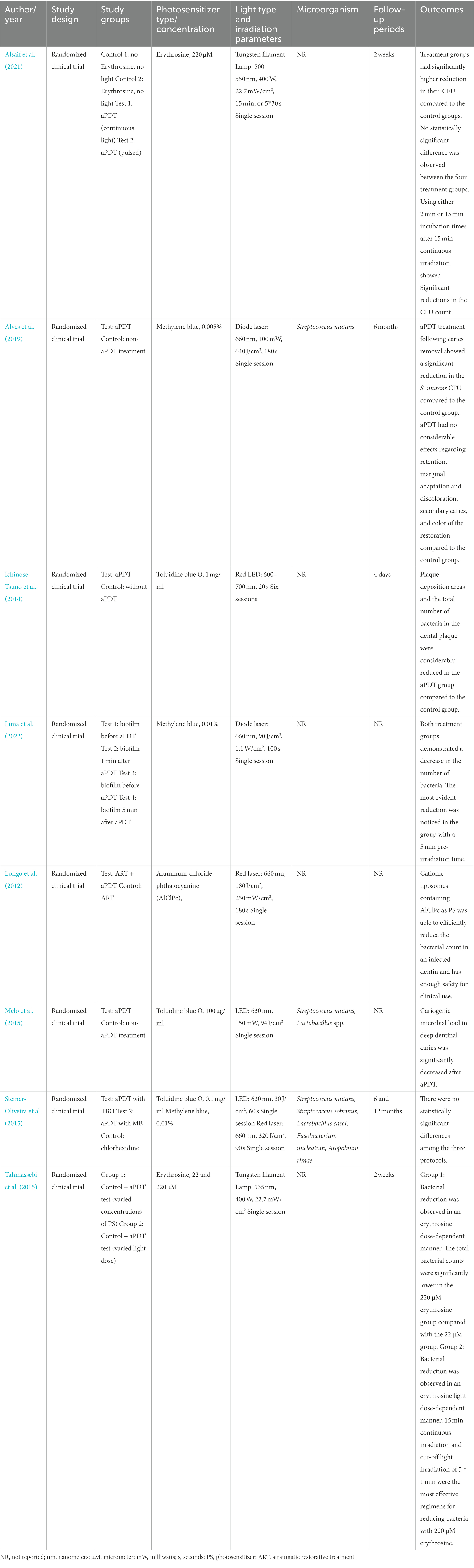
Table 4. Anticaries aPDT treatment.
In this review, eight randomized clinical studies were identified, evaluating anticaries effect of aPDT with varying clinical protocols, however, there are other clinical studies available that do not have a randomized design. Toluidine blue O, Methylene blue, Aluminum-chloride-phthalocyanine (AlClPc) and Erythrosine were used as PS in these studies.
Melo et al. performed a randomized, controlled, split-mouth clinical trial to investigate the efficacy of deep dentinal caries disinfection with aPDT. The number of viable S. mutans and Lactobacillus spp. were significantly reduced after being exposed to 100 μg/ml toluidine blue O for 5 min and 630 nm LED with a total energy density of 94 J/cm2 indicating the improvement in carious dentin disinfection following aPDT (Melo et al., 2015). A significant decrease was seen in the viability of S. mutans, L. casei, and A. viscosus in both planktonic and sessile forms in an in-vitro study by Darmani et al. using a GaAlAs laser at 670 nm and toluidine blue O as PS (Darmani et al., 2018).
Methylene blue was the subject of anticaries aPDT in some clinical studies (but not-randomized) conducted by Neves et al., Guglielmi et al., and Ornellas et al. evaluating the effect of aPDT on S. mutans and Lactobacillus ssp. resulting in controversial findings (Neves et al., 2016; Ornellas et al., 2018). Neves et al. conducted a case control study and microbiologically assessed the dentin samples obtained from the pulp wall of deciduous teeth before and after applying aPDT. They irradiated the cavity after taking the control sample and applying methylene blue using an InGaAlP laser with an energy density of 120 J/cm2. While they declared no statistically significant reduction in the count of viable carious microorganisms before and after application of PS, Guglielmi et al. reported that aPDT significantly decreased total viable bacteria of the permanent molar samples with an active deep carious lesion without pulpal involvement. Both mentioned studies used an InGaAlP laser with 660 nm wavelength and 5 min pre-irradiation time, but the energy density in the second study was higher and reported as 320 J/cm2.The energy density and the observed results in the clinical experiment of Ornellas et al. (InGaAIP, 660 nm, 300 J/cm2, 5 min) were more similar to that of Guglielmi et al.
A RCT by Steiner-Oliveria et al. compared aPDT with TB and MB using an LED 630 nm irradiation at 30 J/cm2 and a control treatment of Chlorhexidine on S. mutans, S. sobrinus, and Lactobacilus casei. The results revealed no statistically significant difference among study groups (Steiner-Oliveira et al., 2015). However, another RCT using TB as a PS and application of LED 600–700 nm irradiation in six sessions, reported considerable reduction in the total number of bacteria and plaque deposition in the aPDT treated group vs. controls without any aPDT (Ichinose-Tsuno et al., 2014).
Reducing the S. mutans count in the oral cavity by performing aPDT and before restoration placement may decrease the risk of caries reoccurrence and some studies have focused on these clinical applications of aPDT. Effect of aPDT on salivary S. mutans in 5- to 6-year-old children aPDT with severe early childhood caries using toluidine blue O and 633 nm diode laser was assessed in a case–control study by Bargrizan et al. Two sessions of aPDT were performed (20 mW, 6 J/cm2), and salivary samples were collected to be compared against the other groups. They concluded that the efficacy of toluidine blue O plus diode laser in reducing S. mutans count was higher than other groups that used only toluidine blue O, only laser, or none, and also the treatment was more durable after receiving two doses of aPDT (Bargrizan et al., 2019). aPDT has been demonstrated to prevent enamel demineralization even with the presence of a cariogenic diet, that indicates its effectiveness in caries prevention (Baptista et al., 2012).
Mendez et al. analyzed the influence of methylene blue on the viability of carious microorganisms and their lactic acid production. The highest reduction in the vitality of intact biofilms and the number of microorganisms was measured after using methylene blue with 75 J/cm2 fluence, and all treatment groups had significantly lower lactic acid production except when the methylene blue was used without illumination (Cusicanqui Méndez et al., 2018). In another study by Mendez et al., the combination of curcumin and LED laser irradiation significantly reduced the number of colony-forming units and vitality of intact biofilms, but it did not show any considerable drop in their acidogenicity feature (Cusicanqui Méndez et al., 2018).
Pereira et al. aimed to explore the effect of polyacrylic acid (PA) 11.5% containing 0.3% methylene blue as a PS to reduce the microbial load before restoration placement. Treatment with methylene blue and methylene blue + PA showed the most reduction in S. mutans growth, respectively, depicting that it can be used as a PS to diminish S. mutans carious dentin (Pereira et al., 2020). Pinheiro et al. also examined the use of a dental acid etchant containing 37% phosphoric acid and methylene blue (DAE) as a sensitizing agent in aPDT of dentinal caries.
The specimens were exposed to a 660 nm light irradiation with 4 J/cm2 energy density. The most significant relative reduction in the number of S. mutans was obtained in the PDT group and then the DAE group making it a potential PS to be used in future clinical studies (Pinheiro et al., 2019).
One of the investigation areas in restorative dentistry is the effect of aPDT on the bond stability of materials and the durability of the treatment. The less collagen content, loss of peritubular dentin matrix, and increased water in the carious affected dentine are some of the characteristics that compromise the success of the treatment by causing gap or leakage in the interface of the tooth and restoration and lowering the bond strength (Nakajima et al., 2011; Pinna et al., 2015), thus, it is strongly suggested to efficiently disinfect this layer in order to achieve suitable bond integrity and successful restoration (Perdigão et al., 2021). The conventional disinfection methods such as exploiting chlorhexidine gluconate, sodium hypochlorite, ethylene diamine tetra acetic acid, and hydrogen peroxide in the cavity have been shown to jeopardize the stability of the bond over time (Tulunoglu et al., 1998; Shafiei and Memarpour, 2012; Coelho et al., 2021).
Recently, aPDT has been investigated by many researchers and is considered an alternative non-invasive treatment for treating deep carious lesions for its promising results (Borgia et al., 2018; Alrahlah et al., 2020; Alshami et al., 2021; Hashemikamangar et al., 2022). In a study by Alrahlah et al. evaluating the effect of methylene blue, curcumin, indocyanine green, and H2O2 on shear bond strength (SBS) of composite resin restorations on carious dentin samples, the highest SBS value was detected in the samples disinfected by curcumin. Curcumin and indocyanine green demonstrated the potential to be used as PS since they can improve the SBS of restoration to carious tissues (Alrahlah et al., 2020). Keskin et al. compared the microtensile bond strength (μTBS) of giomer to carious dentin when disinfected with CHX, NaOCl, aPDT, or Er, Cr: YSGG laser before restoration placement (Keskin et al., 2021). Faria et al. clinically evaluated the performance of composite restorations after caries removal (SCR) associated with aPDT (Faria et al., 2022). The marginal adaptation of the restoration was significantly better in aPDT group compared to the control group after a 12-month follow-up.
Although all the disinfection protocols reduced the bond strength to caries-affected dentin, The aPDT and laser groups showed more μTBS values than the CHX and NaOCl (Keskin et al., 2021). Their results were in agreement with Vellappally et al.’s research indicating the effectiveness of aPDT in augmenting the bond strength. However, using aPDT as effective treatment has raised scientific debates as there have been controversial outcomes regarding the bond strength. Al saffan et al. reported the highest bond strength with CHX compared to using methylene blue as an aPDT agent or Nd:YAG laser (Al Saffan et al., 2021). Also, in another study conducted by Alshahrani et al., resin-modified glass ionomer cement bonded to CHX disinfected caries-affected dentin displayed the maximum shear bond strength, while methylene blue mediated aPDT had the lowest SBS. The results of LED application with curcumin and Er, Cr:YSGG laser irradiation were comparable to the CHX group (Alshahrani et al., 2020). These controversial reports necessitate more experiments on the adhesive bond strength of resin composites following different disinfection protocols.
Most of this assessed clinical studies recruited a laser with 630–660 nm wavelength with methylene blue or TB as a PS, some reporting a significant reduction in the count of S. mutans after treatment compared to the control. All in all, many in-vitro and clinical studies indicate the efficacy of aPDT in prevention and treatment of dental caries and increasing the bond strength between restoration and tooth structure; however, in order to obtain conclusive results, more clinical studies with standardized methodology are required. Considerable heterogeneity exists in irradiation protocols and study deigns which need to be considered and addressed in future studies.
Candida species are natural members of a healthy microbial flora in the oral cavity and are in commensalism with other members of the microbial flora of mouth; however, they can become pathogenic and irritate the mucosa, especially when the host’s immune system is dysfunctional (Giannini and Shetty, 2011; Singh et al., 2014). Candidiasis is one of the most prevalent diseases in oral mucosa that is mainly caused by Candida albicans (C. albicans). This microorganism has also been proposed as the main pathogen isolated from the denture of the patients suffering from denture stomatitis (DS). Practicing an appropriate dental and prosthetic hygiene routine is necessary to prevent fungal infections and stomatitis; nevertheless, it is difficult for people with disabilities or hospitalized elderlies to effectively clean their mouth or disinfect their prostheses which can lead to oral infections and the use of antifungals (Papadiochou and Polyzois, 2018; Khadka et al., 2020).
As the known situation with antibiotics against bacteria, there has been increasing resistance to antifungal treatments due to the widespread use of these drugs, and the conduction of inadequate therapies in time or doses, is making the conventional therapies less and less effective. In addition, these medications have a limited range of action, and they can be toxic (Morio et al., 2017; Rodríguez-Cerdeira et al., 2021). Thus, other therapeutic approaches have been investigated against oral fungal infections in recent years, e.g., using oregano oil, tea tree oil, ozone therapy, nanoparticles, and light therapy (Ninomiya et al., 2013; Szweda et al., 2015; Maciel et al., 2016; Bhat et al., 2018; Monzillo et al., 2020; Pérez-Laguna et al., 2021). Many studies have introduced aPDT as a promising approach instead of the conventional antifungal treatments in managing oral candidiasis and denture stomatitis (Kato et al., 2013; Azizi et al., 2016; Janeth Rimachi Hidalgo et al., 2019; Alves et al., 2020). Several systematic reviews have been published on the effects of utilizing aPDT indicating the ever-increasing potential of aPDT as an effective antifungal therapy, but the consistency between the different study protocols is low. A summary of some of these studies can be found in Table 5.

Table 5. aPDT treatment in patients with fungal lesions.
Yeasts are less sensitive to PS agents compared to bacteria because of their size and the presence of a thick cell wall, so only a few PSs and light sources can be used in order to successfully eliminate them (Sousa et al., 2016; Wiench et al., 2019). The thin channels in the cell wall prevent the PS from passing through the wall; therefore, cationic PS and more extended contacts with the wall than the gram-negative bacteria are needed to ensure the yeast’s death. The Main examined PSs for fungal inactivation in the literature are methylene blue, toluidine blue O, indocyanine green, and Photogem®. Wiench et al. systematically reviewed toluidine blue O mediated aPDT on Candida spp. (Wiench et al., 2021). Analyzing the 21 included studies showed the following results: In the experiments with planktonic cells, one study showed complete annihilation of C. albicans, and others were partially effective. Also, one study did not show any significant difference (Merigo et al., 2019). Experiments conducted on the yeast biofilm indicated not complete but statically significant reduction in the cell number and growth. Reduced adhesion of C. albicans to epithelial cells and inhibited penetration ability into the epithelium have been reported (Dai et al., 2011; Sherwani et al., 2015).
Based on the findings of a systematic review and meta-analysis of 5 articles by Vila-Nova et al., the aPDT can be beneficial for reducing the colony-forming units on the palate and denture, but the conventional antifungal treatment revealed better performance after 15 and 30 days probably because the drugs can penetrate the pores in the denture and remain in them (Vila-Nova et al., 2022). Boltes Cecatto et al. also did a systematic review on methylene blue-mediated aPDT in human clinical studies (Boltes Cecatto et al., 2020). Of the five selected studies, two were on onychomycosis, one about oral candidiasis in HIV patients, and two about infected diabetic feet. In the oral candidiasis article, three approaches were used: Conventional antifungal therapy, phototherapy, and photodynamic therapy by methylene blue with the same irradiation parameters (660 nm, 30 mW, 7.5 J/cm2). While photobiomodulation did not show any candida spp. reduction, both aPDT and conventional medication decreased the number of cells. Complete elimination of fungus colonies without any reoccurrence was observed in aPDT group, but medication did not prevent the return of candidiasis (Scwingel et al., 2012).
Du et al. practiced aPDT using methylene blue plus potassium iodide (KI) in adult patients suffering from acquired immune deficiency syndrome (AIDS). They divided the patients into two groups with 400 and 600 μM methylene blue concentration and LED light with 633 nm wavelength and energy density of 37.29 J/cm2, however, there were no evidence of randomization in the study protocol. According to their results, although there was no significant difference between the 400 and 600 μM methylene blue concentrations, both protocols alleviated the clinical symptoms between 50% and 75% and reduced the number of the fungal cells and control opportunistic fungus. One or two aPDT sessions did not significantly affect the biofilm formation capacity of C. albicans (Ajmal, 2021). In another study, effectiveness of total mouth aPDT in individuals with AIDS was evaluated using 50 μg/ml porphyrin as PS and 660 nm LED. Though the treatment was able to reduce the general count of microorganisms in the oral cavity, the reduction in the number of Log10 CFU/ml of Candida spp. was not significant (da Silva et al., 2022). Wiench et al. introduced aPDT using toluidine blue O and a 635 nm diode laser with the energy density of 24 J/cm2, 400 mW power, and 30 s irradiation time as a possible therapeutic approach in future clinical studies (Wiench et al., 2019).
Dias et al. reported that using aPDT (660 nm, 18 J/cm2, 34 mW/cm2) successively three times using Photodithazine® 25 mg/L could completely inactivate the C. albicans in planktonic cultures; however, to prevent the re-cultivation of the cells in the biofilm model 5 sessions of aPDT was required (Bhat et al., 2018). Biofilm makes C. albicans more resistant to antimicrobial photodynamic therapy agents by creating extra protective layers. In compound biofilms containing more than one species, there is a higher possibility of developing a more resistant polymeric extracellular matrix as a result of a mutual coaggregation of the fungal species hampering the inactivation process (Falsetta et al., 2014; Kim et al., 2018). In the mentioned research, fluconazole was presented as an agent that can potentiate the aPDT regardless of the presence of biofilm. To destroy a biofilm model, the concentration of the PS must be 100 times greater than the one used in a planktonic model (Rodríguez-Cerdeira et al., 2021).
A randomized controlled trial by Alrabiah et al. compared the efficacy of aPDT and local nystatin therapy for denture stomatitis treatment. Thirty-six individuals were divided to two groups, one utilizing GaAlAs diode laser (660 nm, 100 mW, 28 J/cm2) and methylene blue (450 μg/ml) as PS. The other group used topical nystatin oral suspension of 100,000 IU four times a day for 15 days. They stated that the recorded CFU/ml values were not different between the groups throughout the study, and aPDT was as effective as nystatin for treating denture stomatitis (Alrabiah et al., 2019). This result was also confirmed by Mima et al. examining Photogem 500 mg/L irradiated by LED in a clinical trial comparing the effects of conventional antifungal therapy with aPDT (Mima et al., 2012). Afroozi et al. investigated the effect of indocyanine green-mediated aPDT (1 mg/ml) in combination with nystatin (100,000 U) in the management of denture stomatitis in comparison with the conventional nystatin therapy. Sixty-six patients were assigned into two groups and received nystatin mouthwash three times a day for 15 days, but the aPDT group also got laser irradiation (810 nm, 56 J/cm2) twice a day once a week. Evaluations showed that the mean reduction in the number of Candida spp. was markedly higher in the aPDT + nystatin group suggesting it as an alternative to the currently available antifungal therapies (Afroozi et al., 2019).
Oral mucositis is a common side effect, of chemotherapy and radiotherapy in the process of cancer treatment, which makes the patient susceptible to infections caused by opportunistic microorganisms such as Candida spp. (Trotti et al., 2003). Several protocols have been suggested to alleviate the pain and the inflammation like promoting oral hygiene, using antibiotics, analgesics, growth factors, anti-inflammatory agents, photobiomodulation, and antimicrobial photodynamic therapy (Fekrazad and Chiniforush, 2014). Andrade et al. compared the efficacy of photobiomodulation and Curcumin mediated aPDT as an adjuvant therapy of oral mucositis in oncologic patients using a 450 nm LED with 20.1 J/cm2 energy density. Their results illustrated a lower number of CFUs and lower degree of mucositis in the aPDT group on 21th and 30th days of follow-up compared to the control group. There was no statically significant difference between the aPDT and PBM groups indicating that they are both effective therapies in the management of oral mucositis resulted from chemotherapy and radiotherapy (de Cássia Dias Viana Andrade et al., 2022). As been said, one of the primary factors affecting the results of photodynamic therapy is the light parameter, including the wavelength, energy density, power density, and irradiation time. Most of the reviewed articles used a wavelength ranging between 630 and 660 nm and an incubation time of 5 to 20 min. It is essential to use a light source that induces the most absorption of PS in the cells, which also depends on the type and concentration of the PS solution (D'Ilario and Martinelli, 2006). Photodithazine derivatives were the most used sensitizing agents that displayed no clinical adverse effects and are considered safe substances to be used in treating superficial fungal infections in a controlled procedure. Of all the discussed pieces of research, most of them reported an improvement in the clinical features of the evaluated fungal disease or a reduction in the CFUs of samples.
Even though the effectiveness of aPDT can be supported to some extent as an adjunct fungal therapy, the quality of many of them is not satisfactory; therefore, more clinical trials are needed in order to determine the ideal amount of efficacious factors and the safety of the approach (Boltes Cecatto et al., 2020).
We were able to identify several studies have evaluated the effect of aPDT on oral fungi such as C. albicans compared with treatments with Nystatin as an antifungal reporting it to be able to result in higher reduction in the aPDT treated groups. These studies have mostly used Methylene blue as a PS and wavelengths of 660 nm either with diode lasers or LED devices were used. ICG was used in one study with a wavelength of 810 nm that is suitable for its activation and reported higher reduction of candida CFU in patients with denture stomatitis (Afroozi et al., 2019).
Photogem which is a hematoporphyrin derivative mostly used in photodynamic drug therapy of malignant tumors, was used in one study. The PS was activated with 455 nm LED and resulted in clinical success rate of 53% compared to 45% in the control group that used topical Nystatin as their treatment. They concluded that aPDT can be almost as equally effective in treatment of denture stomatitis as Nystatin (Mima et al. 2012).
aPDT has been introduced as a potential treatment against the viral diseases when used as an adjunctive treatment alongside the antiviral medications with promising results by the recently published RCTs (Ajmal, 2021; Ramalho et al., 2021; Shetty et al., 2022; Table 6). These studies have all used methylene blue as a PS activated with a red wavelength of 660 nm. Viral infections usually have manifestations such as blister or ulcers in the oral cavity that can be very irritating for patients. They have also been considered to play a role in periodontal disease and some oral cancers (Healy and Moran, 2019; Puletic et al., 2020; Tsuchida, 2020; Sarkar et al., 2021; Hajishengallis, 2022). The conventional treatment for viral infections is the use of antiviral medications such as acyclovir which is a synthetic acyclic purine-nucleoside analog in the case of herpes simplex virus (HSV) infections as the etiology of herpetic gingivostomatitis (Whitley and Roizman, 2001). However, these infections are prone to get resistant against antiviral drugs after a long-term topical, oral and intravenous use which reduces the effect of antiviral medications over time. Also, the oral or intravenous application is only for the severe or high-rate recurrence of viral infections (Frobert et al., 2014). The mechanisms leading to the viral infection resistance have been explained by the mutations occurred in the virus genes responsible for encoding thymidine kinase, generating thymidine-kinase-deficient mutants which cannot phosphorylate acyclovir (Ramalho et al., 2021). It has been shown that aPDT can photo-inactivate both DNA and RNA viruses. Further, photo-inactivation of HSV by methylene blue as a cationic charged PS has shown promising outcomes, especially that the viral infections are unable to become resistant against aPDT (de Paula Eduardo et al., 2014).
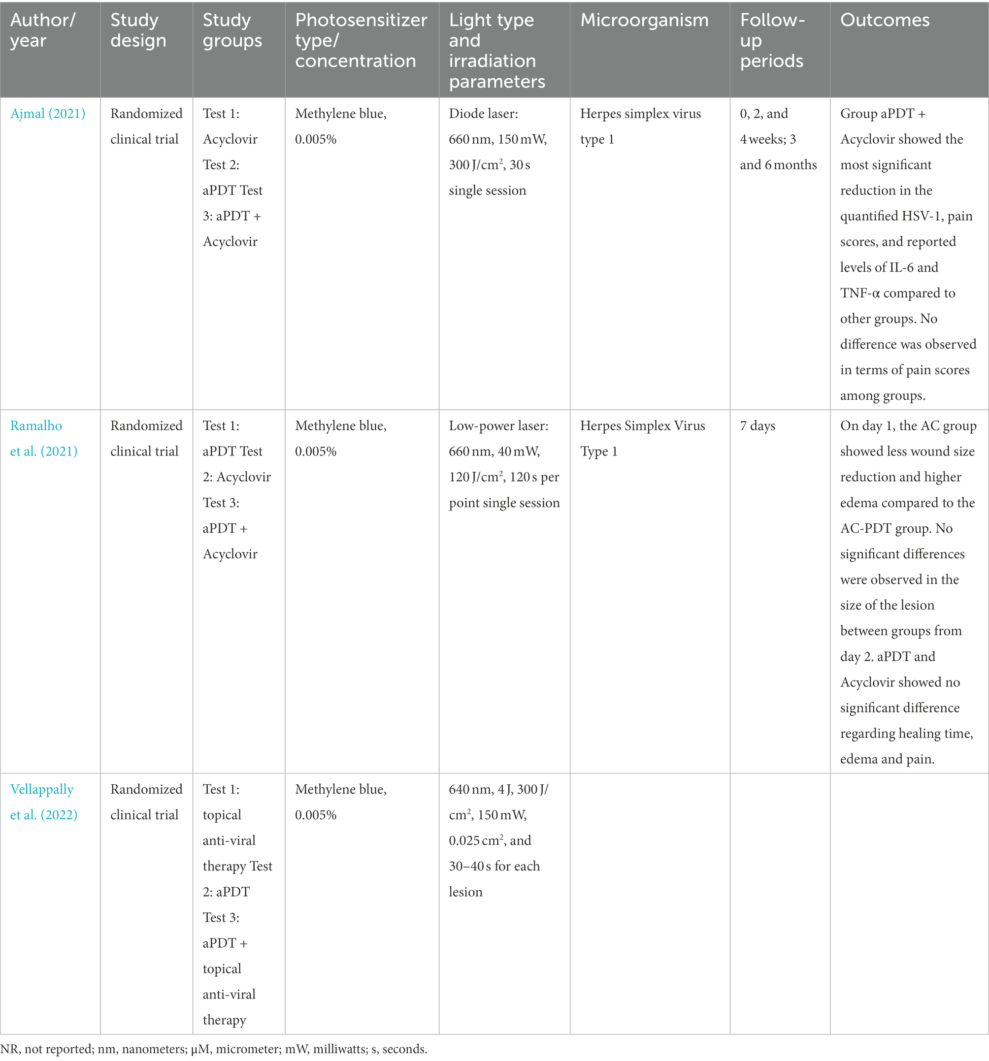
Table 6. aPDT treatment in patients with oral viral infections.
There is a lack of robust RCTs investigating the treatment of oral viral infections with aPDT as the main or adjunct treatments. Vellappally et al. and Ajmal investigated the effect of aPDT (methylene blue 0.005% as PS) for the treatment of herpes labialis in adolescent population. Both demonstrated that using aPDT in adjunct with acyclovir reduced the pain scores as a parameter mostly important for patients in addition to the molecular level parameters including quantified HSV-1, IL-6, and TNF-α (Ajmal, 2021; Shetty et al., 2022). In another RCT by Ramalho et al., with similar methodology on the adult population, application of adjunctive aPDT yielded no substantial difference compared to the acyclovir in terms of lesion healing time, edema and pain. Furthermore, no side effects were reported in the groups containing aPDT treatment by the participants (Ramalho et al., 2021). Given the promising but scarce evidence concerning the effect of aPDT in the treatment of oral viral infection, it is suggested to conduct more RCTs to investigate the efficiency of this method to treat oral viral conditions associated with viruses such as Epstein–Barr virus, cytomegalovirus, human herpesviruses and herpes zoster and even SARS-CoV-2, in healthy and/or immunocompromised patients (Patel and Woolley, 2021; Thakkar et al., 2022).
Although the efficacy of aPDT is approved by many studies, there are some downsides of using it as an adjuvant in clinical therapies. Poor target selection, an uncontrollable manner in drug releasing, poor water solubility, high cost of the PSs, and low oxygen concentrations in deeper parts of the targeted tissues are some factors limiting PS usage in future clinical trials (Yin et al., 2015). In order to deal with these factors and improve their antimicrobial performance, researchers have been working on novel approaches such as utilizing nanoparticles (NPs) as vehicles to transfer hydrophobic PS into the microorganisms, using nanomaterials with similar properties as PSs or conjugating PSs with monoclonal antibodies that possess better targeting characteristics than the PS alone (Schmitt and Juillerat-Jeanneret, 2012). With the development of various nanomaterials, it is of great importance to consider the potential long-term toxicity of not only the tested NPs but also the safe drugs that have not shown any toxic effects in previous studies with short-term follow-ups since the long-term adverse manifestations may be observed after a long interval (Qi et al., 2019). Further, pigmenting the patient’s teeth and gums is another issue with PSs that can be minimized by using nanocarriers that protect the treated area from being colored (Silvestre et al., 2021).
Studies on novel designs of PSs evaluating their antimicrobial effects on S. mutans have shown more effective bacterial reduction and PSs’ characteristics, when conjugated tolouidine blue O with silver nanoparticles (AgNPs) and nanocarriers containing Graphene Oxide-Carnosine/Hydroxyapatite loaded with indocyanine green has been used (Misba et al., 2016; Gholibegloo et al., 2018). Also, photoactivation-independent PSs like Rose bengal-functionalized chitosan NPs (CSRBnp) can significantly reduce the inflammatory marker expressed from macrophages and efficiently inactivate the endotoxins and lipopolysaccharides (Shrestha et al., 2015).
Novel designs of PSs such as Nano-Graphene oxide conjugated with indocyanine green are able to increase the bactericidal characteristics against biofilm formation of Enterocucus faecalis with lower effective concentrations than indocyanine green alone (Akbari et al., 2017). Also, new PS structure designing techniques, can make the previously not efficient PSs effective, like indocyanine green -loaded NPs covered with chitosan which is able to significantly reduce the viability and the load of P. gingivalis whereas indocyanine green alone could not affect the P. gingivalis due to its positive surface charge (Nagahara et al., 2013). Porphyromonas gingivalis is considered as a keystone pathogen and the major culprit not only in periodontal destruction but also in the relationship between periodontitis and systemic conditions such as diabetes, cardiovascular diseases (Liccardo et al., 2019), preterm low birth weight (Teshome and Yitayeh, 2016) and Alzheimer’s (Borsa et al., 2021; Ding et al., 2021). Therefore, a local and non-invasive method to effectively reduce and control the load inherent oral environment bacteria such as P. gingivalis, can be considered as promising modalities of overcoming its related adverse effects on end organ systemic diseases.
Considering the promising results of aPDT against viruses (Namvar et al., 2019), and recently emerged SARS-CoV-2, there is so much to exploit from this method in future studies specifically in the field of virology and dentistry. Also developing anticaries vaccines and modulating the oral microbiota to non-pathogen species is not impossible (Ghazi et al., 2021).
Considering the effectiveness of aPDT against a wide range of micro-organisms and its promising results demonstrated in some clinical studies; it is highly suggested that further in-vivo studies and clinical trials be conducted with more detailed and homogenous study designs to optimize irradiation protocols and wavelengths suitable for PS activation. Systematic reviews on the different clinical application areas with metanalysis of the results are needed to pave the way toward evidence-based application of aPDT in dentistry. Moreover, novel methods of PS structure design improved by carriers and adjuvants to enhance the current conventional therapies’ safety, efficacy, targeting and cost-effectiveness can help clinicians reach the desired therapeutic goals. Patient satisfaction and safety/adverse effects are also an important aspect that needs to be considered and evaluated in future clinical studies.
MH: methodology, investigation, data curation, and writing—original draft. SS and MJ: investigation, data curation, and writing—review and editing. LG: supervision and writing—review and editing. RF: conceptualization, validation, supervision, project administration, and review and editing. All authors contributed to the article and approved the submitted version.
The authors declare that the research was conducted in the absence of any commercial or financial relationships that could be construed as a potential conflict of interest.
All claims expressed in this article are solely those of the authors and do not necessarily represent those of their affiliated organizations, or those of the publisher, the editors and the reviewers. Any product that may be evaluated in this article, or claim that may be made by its manufacturer, is not guaranteed or endorsed by the publisher.
The Supplementary material for this article can be found online at: https://www.frontiersin.org/articles/10.3389/fmicb.2022.1020995/full#supplementary-material
Abdelkarim-Elafifi, H., Parada-Avendaño, I., and Arnabat-Dominguez, J. (2021). Photodynamic therapy in Endodontics: a helpful tool to combat antibiotic resistance? A literature review. Antibiotics 10:1106. doi: 10.3390/antibiotics10091106
Abrahamse, H., and Hamblin, M. R. (2016). New photosensitizers for photodynamic therapy. Biochem. J. 473, 347–364. doi: 10.1042/BJ20150942
Afrasiabi, S., Partoazar, A., Chiniforush, N., and Goudarzi, R. (2022). The potential application of natural photosensitizers used in antimicrobial photodynamic therapy against Oral infections. Pharmaceuticals 15:767. doi: 10.3390/ph15060767
Afroozi, B., Zomorodian, K., Lavaee, F., Zare Shahrabadi, Z., and Mardani, M. (2019). Comparison of the efficacy of indocyanine green-mediated photodynamic therapy and nystatin therapy in treatment of denture stomatitis. Photodiagnosis Photodyn. Ther. 27, 193–197. doi: 10.1016/j.pdpdt.2019.06.005
Ahangari, Z., Mojtahed Bidabadi, M., Asnaashari, M., Rahmati, A., and Tabatabaei, F. S. (2017). Comparison of the antimicrobial efficacy of calcium hydroxide and photodynamic therapy against enterococcus faecalis and Candida albicans in teeth with Periapical lesions; an in vivo study. J Lasers Med. Sci. 8, 72–78. doi: 10.15171/jlms.2017.13
Ajmal, M. (2021). Effectiveness of photodynamic therapy as an adjunct to topical antiviral therapy in the treatment of herpes labialis: a randomized controlled clinical trial. Photodiagnosis Photodyn. Ther. 34:102302. doi: 10.1016/j.pdpdt.2021.102302
Akbari, T., Pourhajibagher, M., Hosseini, F., Chiniforush, N., Gholibegloo, E., Khoobi, M., et al. (2017). The effect of indocyanine green loaded on a novel nano-graphene oxide for high performance of photodynamic therapy against enterococcus faecalis. Photodiagnosis Photodyn. Ther. 20, 148–153. doi: 10.1016/j.pdpdt.2017.08.017
Akram, Z., Shafqat, S. S., Niaz, M. O., Raza, A., and Naseem, M. (2020). Clinical efficacy of photodynamic therapy and laser irradiation as an adjunct to open flap debridement in the treatment of chronic periodontitis: a systematic review and meta-analysis. Photodermatol. Photoimmunol. Photomed. 36, 3–13. doi: 10.1111/phpp.12499
Al Rifaiy, M. Q., Qutub, O. A., Alasqah, M. N., Al-Sowygh, Z. H., Mokeem, S. A., and Alrahlah, A. (2018). Effectiveness of adjunctive antimicrobial photodynamic therapy in reducing peri-implant inflammatory response in individuals vaping electronic cigarettes: a randomized controlled clinical trial. Photodiagnosis Photodyn. Ther. 22, 132–136. doi: 10.1016/j.pdpdt.2018.03.002
Al Saffan, A., Dewan, H., Chohan, H., Mustafa, M., Kotha, S. B., and Al Kheraif, A. A. (2021). Efficacy of photodynamic therapy, photobiomodulation and antimicrobial agent on the shear bond strength of composite restorations to hypomineralized teeth. Photodiagnosis Photodyn. Ther. 33:102188. doi: 10.1016/j.pdpdt.2021.102188
Al-Ahmad, A., Walankiewicz, A., Hellwig, E., Follo, M., Tennert, C., Wittmer, A., et al. (2016). Photoinactivation using visible light plus water-filtered infrared-a (Vis+wIRA) and chlorine e6 (Ce6) eradicates planktonic periodontal pathogens and subgingival biofilms. Front. Microbiol. 7:1900. doi: 10.3389/fmicb.2016.01900
Albaker, A. M., ArRejaie, A. S., Alrabiah, M., and Abduljabbar, T. (2018). Effect of photodynamic and laser therapy in the treatment of peri-implant mucositis: a systematic review. Photodiagnosis Photodyn. Ther. 21, 147–152. doi: 10.1016/j.pdpdt.2017.11.011
Alfirdous, R. A., Garcia, I. M., Balhaddad, A. A., Collares, F. M., Martinho, F. C., and Melo, M. A. S. (2021). Advancing photodynamic therapy for endodontic disinfection with nanoparticles: present evidence and upcoming approaches. Appl. Sci. 11:4759. doi: 10.3390/app11114759
Alghandour, A., Elsharkawy, T., Elshalkamy, M., and Abdollah, A. (2018). Immediate implants placed in infected sockets using photodynamic therapy. Sch. J. Appl. Sci. Res. 1, 43–48.
Allison, R. R., Downie, G. H., Cuenca, R., Hu, X.-H., Childs, C. J. H., and Sibata, C. H. (2004). Photosensitizers in clinical PDT. Photodiagnosis Photodyn. Ther. 1, 27–42. doi: 10.1016/S1572-1000(04)00007-9
Almohareb, T., Alhamoudi, N., Al Deeb, M., Bin-Shuwaish, M. S., Mokeem, S. A., Shafqat, S. S., et al. (2020). Clinical efficacy of photodynamic therapy as an adjunct to mechanical debridement in the treatment of per-implantitis with abscess. Photodiagnosis Photodyn. Ther. 30. doi: 10.1016/j.pdpdt.2020.101750
Al-Momani, M. M. (2021). Indocyanine-mediated antimicrobial photodynamic therapy promotes superior clinical effects in stage III and grade C chronic periodontitis among controlled and uncontrolled diabetes mellitus: a randomized controlled clinical trial. Photodiagnosis Photodyn. Ther. 35:102379. doi: 10.1016/j.pdpdt.2021.102379
Alonso, M. J. (2004). Nanomedicines for overcoming biological barriers. Biomed. Pharmacother. 58, 168–172. doi: 10.1016/j.biopha.2004.01.007
Alqahtani, F., Alqhtani, N., Alkhtani, F., Divakar, D. D., Al-Kheraif, A. A., and Javed, F. (2019). Efficacy of mechanical debridement with and without adjunct antimicrobial photodynamic therapy in the treatment of peri-implantitis among moderate cigarette-smokers and waterpipe-users. Photodiagnosis Photodyn. Ther. 28, 153–158. doi: 10.1016/j.pdpdt.2019.09.003
Alqerban, A. (2020). Efficacy of antimicrobial photodynamic and photobiomodulation therapy against Treponema denticola, fusobacterium nucleatum and human beta defensin-2 levels in patients with gingivitis undergoing fixed orthodontic treatment: a clinic-laboratory study. Photodiagnosis Photodyn. Ther. 29:101659. doi: 10.1016/j.pdpdt.2020.101659
Alrabiah, M., Alsahhaf, A., Alofi, R. S., Al-Aali, K. A., Abduljabbar, T., and Vohra, F. (2019). Efficacy of photodynamic therapy versus local nystatin in the treatment of denture stomatitis: a randomized clinical study. Photodiagnosis Photodyn. Ther. 28, 98–101. doi: 10.1016/j.pdpdt.2019.08.028
Alrahlah, A., Niaz, M. O., Abrar, E., Vohra, F., and Rashid, H. (2020). Treatment of caries affected dentin with different photosensitizers and its effect on adhesive bond integrity to resin composite. Photodiagnosis Photodyn. Ther. 31:101865. doi: 10.1016/j.pdpdt.2020.101865
Alsaif, A., Tahmassebi, J. F., and Wood, S. R. (2021). Treatment of dental plaque biofilms using photodynamic therapy: a randomised controlled study. Eur. Arch. Paediatr. Dent. 22, 791–800. doi: 10.1007/s40368-021-00637-y
AlSarhan, M. A., Altammami, M. A., Alaqeely, R. S., AlEbdi, A., Jasser, R. A., Otaibi, D. A., et al. (2021). Short-term improvement of clinical parameters and microbial diversity in periodontitis patients following Indocyanine green-based antimicrobial photodynamic therapy: a randomized single-blind split-mouth cohort. Photodiagnosis Photodyn. Ther. 35. doi: 10.1016/j.pdpdt.2021.102349
Alshahrani, A., Abrar, E., Maawadh, A. M., Al-Hamdan, R. S., Almohareb, T., AlFawaz, Y., et al. (2020). Management of caries affected dentin (CAD) with resin modified glass ionomer cement (RMGIC) in the presence of different caries disinfectants and photosensitizers. Photodiagnosis Photodyn. Ther. 32:101978. doi: 10.1016/j.pdpdt.2020.101978
Alshami, A. A., Sawan, N. M., Alhamed, S. A., Helmi, M., Mustafa, O. S. E., and Alsagob, E. I. (2021). Shear bond strength of resin modified glass ionomer cement following photodynamic therapy, Er, Cr: YSGG and chlorhexidine in hypoplastic primary teeth. Photodiagnosis Photodyn. Ther. 36:102593. doi: 10.1016/j.pdpdt.2021.102593
Alvarenga, L. H., Gomes, A. C., Carribeiro, P., Godoy-Miranda, B., Noschese, G., Simões Ribeiro, M., et al. (2019). Parameters for antimicrobial photodynamic therapy on periodontal pocket—randomized clinical trial. Photodiagnosis Photodyn. Ther. 27, 132–136. doi: 10.1016/j.pdpdt.2019.05.035
Alves, F., Carmello, J. C., Alonso, G. C., Mima, E. G. D. O., Bagnato, V. S., and Pavarina, A. C. (2020). A randomized clinical trial evaluating Photodithazine-mediated antimicrobial photodynamic therapy as a treatment for denture stomatitis. Photodiagnosis Photodyn. Ther. 32:102041. doi: 10.1016/j.pdpdt.2020.102041
Alves, L. V. G. L., Curylofo-Zotti, F. A., Borsatto, M. C., Salvador, S. L. D. S., Valério, R. A., Souza-Gabriel, A. E., et al. (2019). Influence of antimicrobial photodynamic therapy in carious lesion. Randomized split-mouth clinical trial in primary molars. Photodiagnosis Photodyn. Ther. 26, 124–130. doi: 10.1016/j.pdpdt.2019.02.018
Alves-Silva, E. G., Arruda-Vasconcelos, R., Louzada, L. M., De-Jesus-Soares, A., Ferraz, C. C. R., Almeida, J. F. A., et al. (2022). The effect of photodynamic therapy on postoperative pain in teeth with primary endodontic infection. Photodiagnosis Photodyn. Ther. 37:102700. doi: 10.1016/j.pdpdt.2021.102700
Arsic, Z., Jovanovic, R., Djordjevic, A., Sehalic, M., Marjanovic, D., Mikic, M., et al. (2022). Clinical and microbiological effects of photodynamic therapy applied in non-surgical treatment of periodontitis. Vojnosanit. Pregl. 79, 17–24. doi: 10.2298/VSP200304060A
Arweiler, N. B., Pietruska, M., Pietruski, J., Skurska, A., Dolińska, E., Heumann, C., et al. (2014). Six-month results following treatment of aggressive periodontitis with antimicrobial photodynamic therapy or amoxicillin and metronidazole. Clin. Oral Investig. 18, 2129–2135. doi: 10.1007/s00784-014-1193-6
Asnaashari, M., Ashraf, H., Rahmati, A., and Amini, N. (2017). A comparison between effect of photodynamic therapy by LED and calcium hydroxide therapy for root canal disinfection against enterococcus faecalis: a randomized controlled trial. Photodiagnosis Photodyn. Ther. 17, 226–232. doi: 10.1016/j.pdpdt.2016.12.009
Asnaashari, M., Kooshki, N., Salehi, M. M., Azari-Marhabi, S., and Amin Moghadassi, H. (2020). Comparison of antibacterial effects of photodynamic therapy and an irrigation activation system on root canals infected with enterococcus faecalis: an in vitro study. J. Lasers Med. Sci. 11, 243–248. doi: 10.34172/jlms.2020.41
Ateş, G. B., Ak, A., Garipcan, B., and Gülsoy, M. (2018). Indocyanine green-mediated photobiomodulation on human osteoblast cells. Lasers Med. Sci. 33, 1591–1599. doi: 10.1007/s10103-018-2530-9
Azaripour, A., Dittrich, S., Van Noorden, C. J. F., and Willershausen, B. (2018). Efficacy of photodynamic therapy as adjunct treatment of chronic periodontitis: a systematic review and meta-analysis. Lasers Med. Sci. 33, 407–423. doi: 10.1007/s10103-017-2383-7
Azizi, A., Amirzadeh, Z., Rezai, M., Lawaf, S., and Rahimi, A. (2016). Effect of photodynamic therapy with two photosensitizers on Candida albicans. J. Photochem. Photobiol. B Biol. 158, 267–273. doi: 10.1016/j.jphotobiol.2016.02.027
Baeshen, H. A., Alshahrani, A., Kamran, M. A., Alnazeh, A. A., Alhaizaey, A., and Alshahrani, I. (2020). Effectiveness of antimicrobial photodynamic therapy in restoring clinical, microbial, proinflammatory cytokines and pain scores in adolescent patients having generalized gingivitis and undergoing fixed orthodontic treatment. Photodiagnosis Photodyn. Ther. 32. doi: 10.1016/j.pdpdt.2020.101998
Balhaddad, A. A., Garcia, I. M., Ibrahim, M. S., Rolim, J., Gomes, E. A. B., Martinho, F. C., et al. (2020). Prospects on Nano-based platforms for antimicrobial photodynamic therapy against Oral biofilms. Photobiomodul. Photomed. Laser Surg. 38, 481–496. doi: 10.1089/photob.2020.4815
Baptista, A., Kato, I. T., Prates, R. A., Suzuki, L. C., Raele, M. P., Freitas, A. Z., et al. (2012). Antimicrobial photodynamic therapy as a strategy to arrest enamel demineralization: a short-term study on incipient caries in a rat model†. Photochem. Photobiol. 88, 584–589. doi: 10.1111/j.1751-1097.2011.01059.x
Bargrizan, M., Fekrazad, R., Goudarzi, N., and Goudarzi, N. (2019). Effects of antibacterial photodynamic therapy on salivary mutans streptococci in 5- to 6-year-olds with severe early childhood caries. Lasers Med. Sci. 34, 433–440. doi: 10.1007/s10103-018-2650-2
Bassetti, M., Schär, D., Wicki, B., Eick, S., Ramseier, C. A., Arweiler, N. B., et al. (2014). Anti-infective therapy of peri-implantitis with adjunctive local drug delivery or photodynamic therapy: 12-month outcomes of a randomized controlled clinical trial. Clin. Oral Implants Res. 25, 279–287. doi: 10.1111/clr.12155
Bechara Andere, N. M. R., Dos Santos, N. C. C., Araujo, C. F., Mathias, I. F., Rossato, A., de Marco, A. C., et al. (2018). Evaluation of the local effect of nonsurgical periodontal treatment with and without systemic antibiotic and photodynamic therapy in generalized aggressive periodontitis. A randomized clinical trial. Photodiagnosis Photodyn. Ther. 24, 115–120. doi: 10.1016/j.pdpdt.2018.09.002
Benine-Warlet, J., Brenes-Alvarado, A., and Steiner-Oliveira, C. (2022). Potassium iodide enhances inactivation of Streptococcus mutans biofilm in antimicrobial photodynamic therapy with red laser. Photodiagnosis Photodyn. Ther. 37:102622. doi: 10.1016/j.pdpdt.2021.102622
Berglundh, T., Armitage, G., Araujo, M. G., Avila-Ortiz, G., Blanco, J., Camargo, P. M., et al. (2018). Peri-implant diseases and conditions: consensus report of workgroup 4 of the 2017 world workshop on the classification of periodontal and Peri-implant diseases and conditions. J. Clin. Periodontol. 45, S286–s291. doi: 10.1111/jcpe.12957
Betsy, J., Prasanth, C. S., Baiju, K. V., Prasanthila, J., and Subhash, N. (2014). Efficacy of antimicrobial photodynamic therapy in the management of chronic periodontitis: a randomized controlled clinical trial. J. Clin. Periodontol. 41, 573–581. doi: 10.1111/jcpe.12249
Bhat, V., Sharma, S. M., Shetty, V., Shastry, C. S., Rao, C. V., Shenoy, S., et al. (2018). Characterization of herbal antifungal agent, Origanum vulgare against Oral Candida spp. isolated from patients with Candida-associated denture stomatitis: an in vitro study. Contempor. Clin. Dentistr. 9, S3–S10. doi: 10.4103/ccd.ccd_537_17
Birang, E., Birang, R., Narimani, T., Tolouei, A., and Fekrazad, R. (2019). Investigation of the antibacterial effect of laser irradiation and chemical agent on human oral biofilms contaminated titanium discs. Photodiagnosis Photodyn. Ther. 25, 259–264. doi: 10.1016/j.pdpdt.2018.12.008
Boltes Cecatto, R., Siqueira de Magalhães, L., Rodrigues, M. F. S. D., Pavani, C., Lino-dos-Santos-Franco, A., Gomes, M. T., et al. (2020). Methylene blue mediated antimicrobial photodynamic therapy in clinical human studies: the state of the art. Photodiagnosis Photodyn. Ther. 31:101828. doi: 10.1016/j.pdpdt.2020.101828
Borgia, F., Giuffrida, R., Caradonna, E., Vaccaro, M., Guarneri, F., and Cannavò, S. P. (2018). Early and late onset side effects of photodynamic therapy. Biomedicine 6:12. doi: 10.3390/biomedicines6010012
Borsa, L., Dubois, M., Sacco, G., and Lupi, L. (2021). Analysis the link between periodontal diseases and Alzheimer's disease: a systematic review. Int. J. Environ. Res. Public Health 18:9312. doi: 10.3390/ijerph18179312
Braham, P., Herron, C., Street, C., and Darveau, R. (2009). Antimicrobial photodynamic therapy may promote periodontal healing through multiple mechanisms. J. Periodontol. 80, 1790–1798. doi: 10.1902/jop.2009.090214
Brancaleon, L., and Moseley, H. (2002). Laser and non-laser light sources for photodynamic therapy. Lasers Med. Sci. 17, 173–186. doi: 10.1007/s101030200027
Burchard, T., Karygianni, L., Hellwig, E., Wittmer, A., and Al-Ahmad, A. (2020). Microbial composition of Oral biofilms after visible light and water-filtered infrared a radiation (VIS+wIRA) in combination with Indocyanine green (ICG) as photosensitizer. Antibiotics 9:532. doi: 10.3390/antibiotics9090532
Cadore, U. B., Reis, M. B. L., Martins, S. H. L., Invernici, M. M., Novaes, A. B. Jr., Taba, M. Jr., et al. (2019). Multiple sessions of antimicrobial photodynamic therapy associated with surgical periodontal treatment in patients with chronic periodontitis. J. Periodontol. 90, 339–349. doi: 10.1002/JPER.18-0373
Carcuac, O., Derks, J., Charalampakis, G., Abrahamsson, I., Wennström, J., and Berglundh, T. (2016). Adjunctive systemic and local antimicrobial therapy in the surgical treatment of Peri-implantitis: a randomized controlled clinical trial. J. Dent. Res. 95, 50–57. doi: 10.1177/0022034515601961
Carvalho, V. F., Andrade, P. V. C., Rodrigues, M. F., Hirata, M. H., Hirata, R. D. C., Pannuti, C. M., et al. (2015). Antimicrobial photodynamic effect to treat residual pockets in periodontal patients: a randomized controlled clinical trial. J. Clin. Periodontol. 42, 440–447. doi: 10.1111/jcpe.12393
Castano, A. P., Demidova, T. N., and Hamblin, M. R. (2005). Mechanisms in photodynamic therapy: part two—cellular signaling, cell metabolism and modes of cell death. Photodiagnosis Photodyn. Ther. 2, 1–23. doi: 10.1016/S1572-1000(05)00030-X
Castro Dos Santos, N. C., Andere, N. M., Araujo, C. F., de Marco, A. C., Dos Santos, L. M., Jardini, M. A., et al. (2016). Local adjunct effect of antimicrobial photodynamic therapy for the treatment of chronic periodontitis in type 2 diabetics: split-mouth double-blind randomized controlled clinical trial. Lasers Med. Sci. 31, 1633–1640. doi: 10.1007/s10103-016-2030-8
Ceri, H., Olson, M. E., Stremick, C., Read, R. R., Morck, D., and Buret, A. (1999). The Calgary biofilm device: new technology for rapid determination of antibiotic susceptibilities of bacterial biofilms. J. Clin. Microbiol. 37, 1771–1776. doi: 10.1128/JCM.37.6.1771-1776.1999
Chambrone, L., Wang, H. L., and Romanos, G. E. (2018). Antimicrobial photodynamic therapy for the treatment of periodontitis and peri-implantitis: an American Academy of periodontology best evidence review. J. Periodontol. 89, 783–803. doi: 10.1902/jop.2017.170172
Chitsazi, M. T., Shirmohammadi, A., Pourabbas, R., Abolfazli, N., Farhoudi, I., Daghigh Azar, B., et al. (2014). Clinical and microbiological effects of photodynamic therapy associated with non-surgical treatment in aggressive periodontitis. J. Dent. Res. Dent. Clin. Dent. Prospects 8, 153–159. doi: 10.5681/joddd.2014.028
Chung, P. Y., and Toh, Y. S. (2014). Anti-biofilm agents: recent breakthrough against multi-drug resistant Staphylococcus aureus. Pathog. Dis. 70, 231–239. doi: 10.1111/2049-632X.12141
Cieplik, F., Deng, D., Crielaard, W., Buchalla, W., Hellwig, E., Al-Ahmad, A., et al. (2018). Antimicrobial photodynamic therapy - what we know and what we don't. Crit. Rev. Microbiol. 44, 571–589. doi: 10.1080/1040841X.2018.1467876
Cieplik, F., Tabenski, L., Buchalla, W., and Maisch, T. (2014). Antimicrobial photodynamic therapy for inactivation of biofilms formed by oral key pathogens. Front. Microbiol. 5:405. doi: 10.3389/fmicb.2014.00405
Cláudio, M. M., Nuernberg, M. A. A., Rodrigues, J. V. S., Belizário, L. C. G., Batista, J. A., Duque, C., et al. (2021). Effects of multiple sessions of antimicrobial photodynamic therapy (aPDT) in the treatment of periodontitis in patients with uncompensated type 2 diabetes: a randomized controlled clinical study. Photodiagnosis Photodyn. Ther. 35:102451. doi: 10.1016/j.pdpdt.2021.102451
Coelho, A., Amaro, I., Rascão, B., Marcelino, I., Paula, A., Saraiva, J., et al. (2021). Effect of cavity disinfectants on dentin bond strength and clinical success of composite restorations—a systematic review of in vitro, in situ and clinical studies. Int. J. Mol. Sci. 22:353. doi: 10.3390/ijms22010353
Coelho, M. S., Vilas-Boas, L., and Tawil, P. Z. (2019). The effects of photodynamic therapy on postoperative pain in teeth with necrotic pulps. Photodiagnosis Photodyn. Ther. 27, 396–401. doi: 10.1016/j.pdpdt.2019.07.002
Conejero, M. J., Almenar, A., Forner, L., Sanz, J. L., and Llena, C. (2021). Retrospective clinical evaluation of root canal treatment with or without photodynamic therapy for necrotic teeth and teeth subjected to retreatment. J. Oral Sci. 63, 163–166. doi: 10.2334/josnusd.20-0429
Cosgarea, R., Eick, S., Batori-Andronescu, I., Jepsen, S., Arweiler, N. B., Rößler, R., et al. (2021). Clinical and microbiological evaluation of local doxycycline and antimicrobial photodynamic therapy during supportive periodontal therapy: a randomized clinical trial. Antibiotics 10:277. doi: 10.3390/antibiotics10030277
Cosgarea, R., Ramseier, C. A., Jepsen, S., Arweiler, N. B., Jervøe-Storm, P. M., Batori-Andronescu, I., et al. (2022). One-year clinical, microbiological and immunological results of local doxycycline or antimicrobial photodynamic therapy for recurrent/persisting periodontal pockets: a randomized clinical trial. Antibiotics 11:738. doi: 10.3390/antibiotics11060738
Costa, L. M., Matos, F. D. S., Correia, A. M. D. O., Carvalho, N. C., Faria-e-Silva, A. L., Paranhos, L. R., et al. (2016). Tooth color change caused by photosensitizers after photodynamic therapy: an in vitro study. J. Photochem. Photobiol. B Biol. 160, 225–228. doi: 10.1016/j.jphotobiol.2016.04.019
Couvreur, P., Gref, R., Andrieux, K., and Malvy, C. (2006). Nanotechnologies for drug delivery: application to cancer and autoimmune diseases. Prog. Solid State Chem. 34, 231–235. doi: 10.1016/j.progsolidstchem.2005.11.009
Cusicanqui Méndez, D. A., Gutierres, E., José Dionisio, E., Afonso Rabelo Buzalaf, M., Cardoso Oliveira, R., Andrade Moreira Machado, M. A., et al. (2018). Curcumin-mediated antimicrobial photodynamic therapy reduces the viability and vitality of infected dentin caries microcosms. Photodiagnosis Photodyn. Ther. 24, 102–108. doi: 10.1016/j.pdpdt.2018.09.007
da Cruz Andrade, P. V., Euzebio Alves, V. T., de Carvalho, V. F., De Franco Rodrigues, M., Pannuti, C. M., Holzhausen, M., et al. (2017). Photodynamic therapy decrease immune-inflammatory mediators levels during periodontal maintenance. Lasers Med. Sci. 32, 9–17. doi: 10.1007/s10103-016-2076-7
da Silva, C. C., Chaves Júnior, S. P., Pereira, G. L. D., Fontes, K., Antunes, L. A. A., Póvoa, H. C. C., et al. (2018). Antimicrobial photodynamic therapy associated with conventional endodontic treatment: a clinical and molecular microbiological study. Photochem. Photobiol. 94, 351–356. doi: 10.1111/php.12869
da Silva, F. C., Rosa, L. P., de Jesus, I. M., de Oliveira Santos, G. P., Inada, N. M., Blanco, K. C., et al. (2022). Total mouth photodynamic therapy mediated by red LED and porphyrin in individuals with AIDS. Lasers Med. Sci. 37, 1227–1234. doi: 10.1007/s10103-021-03377-z
Dai, T., Arce, V. J. B. D., Tegos, G. P., and Hamblin, M. R. (2011). Blue dye and red light, a dynamic combination for prophylaxis and treatment of cutaneous Candida albicans infections in mice. Antimicrob. Agents Chemother. 55, 5710–5717. doi: 10.1128/AAC.05404-11
Darmani, H., Tawalbeh, K. H., Al-Hiyasat, A. S., and Al-Akhras, M.-A. (2018). Comparison of the photosensitivity of biofilms of different genera of cariogenic bacteria in tooth slices. Pol. J. Microbiol. 67, 455–462. doi: 10.21307/pjm-2018-053
De Angelis, N., Felice, P., Grusovin, M. G., Camurati, A., and Esposito, M. (2012). The effectiveness of adjunctive light-activated disinfection (LAD) in the treatment of peri-implantitis: 4-month results from a multicentre pragmatic randomised controlled trial. Eur. J. Oral Implantol. 5, 321–331.
de Cássia Dias Viana Andrade, R., Azevedo Reis, T., Rosa, L. P., de Oliveira Santos, G. P., and da Cristina Silva, F. (2022). Comparative randomized trial study about the efficacy of photobiomodulation and curcumin antimicrobial photodynamic therapy as a coadjuvant treatment of oral mucositis in oncologic patients: antimicrobial, analgesic, and degree alteration effect. Support. Care Cancer 30:7365. doi: 10.1007/s00520-022-07127-x
de Lapena, S. A. B., Terra-Garcia, M., Ward, R., Rossoni, R. D., Melo, V. M. M., and Junqueira, J. C. (2022). Enhancing effect of chitosan on methylene blue-mediated photodynamic therapy against C. albicans: a study in planktonic growth, biofilms, and persister cells. Photodiagnosis Photodyn. Ther. 38:102837. doi: 10.1016/j.pdpdt.2022.102837
de Melo Soares, M. S., D’Almeida Borges, C., de Mendonça Invernici, M., Frantz, F. G., de Figueiredo, L. C., de Souza, S. L. S., et al. (2019). Antimicrobial photodynamic therapy as adjunct to non-surgical periodontal treatment in smokers: a randomized clinical trial. Clin. Oral Investig. 23, 3173–3182. doi: 10.1007/s00784-018-2740-3
de Miranda, R. G., and Colombo, A. P. V. (2018). Clinical and microbiological effectiveness of photodynamic therapy on primary endodontic infections: a 6-month randomized clinical trial. Clin. Oral Investig. 22, 1751–1761. doi: 10.1007/s00784-017-2270-4
de Paula Eduardo, C., Aranha, A. C. C., Simões, A., Bello-Silva, M. S., Ramalho, K. M., Esteves-Oliveira, M., et al. (2014). Laser treatment of recurrent herpes labialis: a literature review. Lasers Med. Sci. 29, 1517–1529. doi: 10.1007/s10103-013-1311-8
de Senna, A. M., Vieira, M. M. F., Machado-de-Sena, R. M., Bertolin, A. O., Núñez, S. C., and Ribeiro, M. S. (2018). Photodynamic inactivation of Candida ssp. on denture stomatitis. A clinical trial involving palatal mucosa and prosthesis disinfection. Photodiagnosis Photodyn. Ther. 22, 212–216. doi: 10.1016/j.pdpdt.2018.04.008
Di Taranto, V., Libonati, A., Montemurro, E., Gallusi, G., and Campanella, V. (2022). Antimicrobial effects of photodynamic and high-power laser endodontic therapy on patients with necrotic pulp and periapical lesion. J. Biol. Regul. Homeost. Agents 36, 41–48.
D'Ilario, L., and Martinelli, A. (2006). Toluidine blue: aggregation properties and structural aspects. Model. Simul. Mater. Sci. Eng. 14, 581–595. doi: 10.1088/0965-0393/14/4/003
Ding, C., Zhang, F., Gao, Y., Li, Y., Cheng, D., Wang, J., et al. (2021). Antibacterial photodynamic treatment of Porphyromonas gingivalis with toluidine blue O and a non laser red light source enhanced by Dihydroartemisinin. Photochem. Photobiol. 97, 377–384. doi: 10.1111/php.13333
Donlan, R. (2002). Biofilms: microbial life on surfaces. Emerg. Infect. Dis. J. 8:881. doi: 10.3201/eid0809.020063
Dörtbudak, O., Haas, R., Bernhart, T., and Mailath-Pokorny, G. (2001). Lethal photosensitization for decontamination of implant surfaces in the treatment of peri-implantitis. Clin. Oral Implants Res. 12, 104–108. doi: 10.1034/j.1600-0501.2001.012002104.x
Dos Santos, D. D. L., Besegato, J. F., de Melo, P. B. G., Junior, J. A. O., Chorilli, M., Deng, D., et al. (2022). Effect of curcumin-encapsulated Pluronic(®) F-127 over duo-species biofilm of Streptococcus mutans and Candida albicans. Lasers Med. Sci. 37, 1775–1786. doi: 10.1007/s10103-021-03432-9
Du, M., Xuan, W., Zhen, X., He, L., Lan, L., Yang, S., et al. (2021). Antimicrobial photodynamic therapy for oral Candida infection in adult AIDS patients: a pilot clinical trial. Photodiagnosis Photodyn. Ther. 34:102310. doi: 10.1016/j.pdpdt.2021.102310
Ebrahimi, P., Hadilou, M., Naserneysari, F., Dolatabadi, A., Tarzemany, R., Vahed, N., et al. (2021). Effect of photobiomodulation in secondary intention gingival wound healing—a systematic review and meta-analysis. BMC Oral Health 21:258. doi: 10.21203/rs.3.rs-198716/v1
Enaud, R., Prevel, R., Ciarlo, E., Beaufils, F., Wieërs, G., Guery, B., et al. (2020). The gut-lung Axis in health and respiratory diseases: a place for inter-organ and inter-kingdom Crosstalks. Front. Cell. Infect. Microbiol. 10:9. doi: 10.3389/fcimb.2020.00009
Escudero, A., Carrillo-Carrión, C., Castillejos, M. C., Romero-Ben, E., Rosales-Barrios, C., and Khiar, N. (2021). Photodynamic therapy: photosensitizers and nanostructures. Materials Chem. Front. 5, 3788–3812. doi: 10.1039/D0QM00922A
Falsetta, M. L., Klein, M. I., Colonne, P. M., Scott-Anne, K., Gregoire, S., Pai, C.-H., et al. (2014). Symbiotic relationship between Streptococcus mutans and Candida albicans synergizes virulence of plaque biofilms <i>in vivo</i>. Infect. Immun. 82, 1968–1981. doi: 10.1128/IAI.00087-14
Faria, L. V., Antunes, L. S., Pio, L. R. R., Dias, J. C., Pinheiro, L. H. M., Reis, C. L. B., et al. (2022). Evaluation of composite restorations in primary molars subjected to selective caries removal associated with antimicrobial photodynamic therapy: a randomized controlled trial. Int. J. Paediatr. Dent. 32, 585–597. doi: 10.1111/ipd.12937
Fekrazad, R., and Chiniforush, N. (2014). Oral mucositis prevention and management by therapeutic laser in head and neck cancers. J. Lasers Med. Sci. 5, 1–7.
Felgenträger, A., Maisch, T., Dobler, D., and Späth, A. (2013). Hydrogen bond acceptors and additional cationic charges in methylene blue derivatives: Photophysics and antimicrobial efficiency. Biol. Med. Res. Int. 2013:482167. doi: 10.1155/2013/482167
Feres, M., Figueiredo, L. C., Soares, G. M., and Faveri, M. (2015). Systemic antibiotics in the treatment of periodontitis. Periodontol. 2000 67, 131–186. doi: 10.1111/prd.12075
Fernandes, F. G. L., de Moraes, F. B., De Cezare, J. A., Degasperi, G. R., Fontana, C. E., Grandizoli, D. R. P., et al. (2022). In vitro evaluation of EDTA combined with photodynamic therapy to reduce Streptococcus mutans in carious dentin. Photodiagnosis Photodyn. Ther. 37:102718. doi: 10.1016/j.pdpdt.2022.102718
Foschi, F., Fontana, C. R., Ruggiero, K., Riahi, R., Vera, A., Doukas, A. G., et al. (2007). Photodynamic inactivation of enterococcus faecalis in dental root canals in vitro. Lasers Surg. Med. 39, 782–787. doi: 10.1002/lsm.20579
Fraga, R. S., Antunes, L. A. A., Fontes, K., Küchler, E. C., Iorio, N., and Antunes, L. S. (2018). Is antimicrobial photodynamic therapy effective for microbial load reduction in Peri-implantitis treatment? A systematic review and meta-analysis. Photochem. Photobiol. 94, 752–759. doi: 10.1111/php.12901
Frobert, E., Burrel, S., Ducastelle-Lepretre, S., Billaud, G., Ader, F., Casalegno, J.-S., et al. (2014). Resistance of herpes simplex viruses to acyclovir: an update from a ten-year survey in France. Antiviral Res. 111, 36–41. doi: 10.1016/j.antiviral.2014.08.013
Gajdács, M., Spengler, G., and Urbán, E. (2017). Identification and antimicrobial susceptibility testing of anaerobic bacteria: Rubik's cube of clinical microbiology? Antibiotics 6:25. doi: 10.3390/antibiotics6040025
Gallardo-Villagrán, M., Leger, D. Y., Liagre, B., and Therrien, B. (2019). Photosensitizers used in the photodynamic therapy of rheumatoid arthritis. Int. J. Mol. Sci. 20:3339. doi: 10.3390/ijms20133339
Garcez, A. S., and Hamblin, M. R. (2017). Methylene blue and hydrogen peroxide for photodynamic inactivation in root canal - a new protocol for use in Endodontics. Eur. Endod. J. 2, 1–7. doi: 10.14744/eej.2017.17023
Garcez, A. S., Núñez, S. C., Baptista, M. S., Daghastanli, N. A., Itri, R., Hamblin, M. R., et al. (2011). Antimicrobial mechanisms behind photodynamic effect in the presence of hydrogen peroxide. Photochem. Photobiol. Sci. 10, 483–490. doi: 10.1039/c0pp00082e
Garcez, A. S., Nuñez, S. C., Hamblin, M. R., and Ribeiro, M. S. (2008). Antimicrobial effects of photodynamic therapy on patients with necrotic pulps and periapical lesion. J. Endod. 34, 138–142. doi: 10.1016/j.joen.2007.10.020
Ghazi, M., Pourhajibagher, M., Bahador, A., Chiniforush, N., Dadpour, S., and Dadpour, Y. (2021). Evaluation of adding nanosized natural zeolite to photodynamic therapy against P. gingivalis biofilm on titanium disks. Photodiagnosis Photodyn. Ther. 36:102519. doi: 10.1016/j.pdpdt.2021.102519
Gholibegloo, E., Karbasi, A., Pourhajibagher, M., Chiniforush, N., Ramazani, A., Akbari, T., et al. (2018). Carnosine-graphene oxide conjugates decorated with hydroxyapatite as promising nanocarrier for ICG loading with enhanced antibacterial effects in photodynamic therapy against Streptococcus mutans. J. Photochem. Photobiol. B Biol. 181, 14–22. doi: 10.1016/j.jphotobiol.2018.02.004
Ghorbanzadeh, A., Fekrazad, R., Bahador, A., Ayar, R., Tabatabai, S., and Asefi, S. (2018). Evaluation of the antibacterial efficacy of various root canal disinfection methods against enterococcus faecalis biofilm. An ex-vivo study. Photodiagnosis Photodyn. Ther. 24, 44–51. doi: 10.1016/j.pdpdt.2018.08.010
Giannini, P. J., and Shetty, K. V. (2011). Diagnosis and management of oral candidiasis. Otolaryngol. Clin. North Am. 44, 231–240. doi: 10.1016/j.otc.2010.09.010
Giusti, J. S., Santos-Pinto, L., Pizzolito, A. C., Helmerson, K., Carvalho-Filho, E., Kurachi, C., et al. (2008). Antimicrobial photodynamic action on dentin using a light-emitting diode light source. Photomed. Laser Surg. 26, 281–287. doi: 10.1089/pho.2007.2149
Gomer, C. J. (1991). Preclinical examination of first and second generation photosensitizers used in photodynamic therapy. Photochem. Photobiol. 54, 1093–1107. doi: 10.1111/j.1751-1097.1991.tb02133.x
Grzech-Leśniak, K., Gaspirc, B., and Sculean, A. (2019). Clinical and microbiological effects of multiple applications of antibacterial photodynamic therapy in periodontal maintenance patients. A randomized controlled clinical study. Photodiagnosis Photodyn. Ther. 27, 44–50. doi: 10.1016/j.pdpdt.2019.05.028
Guimaraes, L. D. S., da Silva, E. A. B., Hespanhol, F. G., Fontes, K. B. F. D. C., Antunes, L. A. A., and Antunes, L. S. (2021). Effect of photobiomodulation on post-operative symptoms in teeth with asymptomatic apical periodontitis treated with foraminal enlargement: a randomized clinical trial. Int. Endod. J. 54, 1708–1719. doi: 10.1111/iej.13593
Hajishengallis, G. (2022). Interconnection of periodontal disease and comorbidities: evidence, mechanisms, and implications. Periodontol. 2000 89, 9–18. doi: 10.1111/prd.12430
Hajishengallis, G., Darveau, R. P., and Curtis, M. A. (2012). The keystone-pathogen hypothesis. Nat. Rev. Microbiol. 10, 717–725. doi: 10.1038/nrmicro2873
Hamblin, M. R., and Abrahamse, H. (2020). Oxygen-independent antimicrobial Photoinactivation: type III photochemical mechanism? Antibiotics 9:53. doi: 10.3390/antibiotics9020053
Hashemikamangar, S. S., Alsaedi, R. J. F., Chiniforush, N., and Motevaselian, F. (2022). Effect of antimicrobial photodynamic therapy with different photosensitizers and adhesion protocol on the bond strength of resin composite to sound dentin. Clin. Oral Investig. 26, 4011–4019. doi: 10.1007/s00784-022-04370-8
Healy, C. M., and Moran, G. P. (2019). The microbiome and oral cancer: more questions than answers. Oral Oncol. 89, 30–33. doi: 10.1016/j.oraloncology.2018.12.003
Hendi, S. S., Shiri, M., Poormoradi, B., Alikhani, M. Y., Afshar, S., and Farmani, A. (2021). Antibacterial effects of a 940 nm diode laser with/without silver nanoparticles against enterococcus faecalis. J. Lasers Med. Sci. 12:e73. doi: 10.34172/jlms.2021.73
Hill, G., Dehn, C., Hinze, A. V., Frentzen, M., and Meister, J. (2019). Indocyanine green-based adjunctive antimicrobial photodynamic therapy for treating chronic periodontitis: a randomized clinical trial. Photodiagnosis Photodyn. Ther. 26, 29–35. doi: 10.1016/j.pdpdt.2019.02.019
Høiby, N. (2017). A short history of microbial biofilms and biofilm infections. APMIS 125, 272–275. doi: 10.1111/apm.12686
Hokari, T., Morozumi, T., Komatsu, Y., Shimizu, T., Yoshino, T., Tanaka, M., et al. (2018). Effects of antimicrobial photodynamic therapy and local Administration of Minocycline on clinical, microbiological, and inflammatory markers of periodontal pockets: a pilot study. Int. J. Dent. 2018:1748584. doi: 10.1155/2018/1748584
Hu, X., Huang, Y. Y., Wang, Y., Wang, X., and Hamblin, M. R. (2018). Antimicrobial photodynamic therapy to control clinically relevant biofilm infections. Front. Microbiol. 9:1299. doi: 10.3389/fmicb.2018.01299
Husejnagic, S., Lettner, S., Laky, M., Georgopoulos, A., Moritz, A., and Rausch-Fan, X. (2019). Photoactivated disinfection in periodontal treatment: a randomized controlled clinical split-mouth trial. J. Periodontol. 90, 1260–1269. doi: 10.1002/JPER.18-0576
Ichinose-Tsuno, A., Aoki, A., Takeuchi, Y., Kirikae, T., Shimbo, T., Lee, M.-C.-I., et al. (2014). Antimicrobial photodynamic therapy suppresses dental plaque formation in healthy adults: a randomized controlled clinical trial. BMC Oral Health 14:152. doi: 10.1186/1472-6831-14-152
Janeth Rimachi Hidalgo, K., Carmello, J. C., Jordão, C. C., Barbugli, P. A., de Sousa Costa, C. A., de Oliveira Mima, E. G., et al. (2019). Antimicrobial photodynamic therapy in combination with Nystatin in the treatment of experimental Oral candidiasis induced by Candida albicans resistant to fluconazole. Pharmaceuticals 12:140. doi: 10.3390/ph12030140
Javed, F., Bin Shabaib, M. S., Alharthi, S. S., and Qadri, T. (2017). Role of mechanical curettage with and without adjunct antimicrobial photodynamic therapy in the treatment of peri-implant mucositis in cigarette smokers: a randomized controlled clinical trial. Photodiagnosis Photodyn. Ther. 18, 331–334. doi: 10.1016/j.pdpdt.2017.04.015
Joseph, B., Janam, P., Narayanan, S., and Anil, S. (2017). Is antimicrobial photodynamic therapy effective as an adjunct to scaling and root Planing in patients with chronic periodontitis? A systematic review. Biomol. Ther. 7:79. doi: 10.3390/biom7040079
Joshi, K., Baiju, C. S., Khashu, H., and Bansal, S. (2020). Clinical effectiveness of indocyanine green mediated antimicrobial photodynamic therapy as an adjunct to scaling root planing in treatment of chronic periodontitis- a randomized controlled clinical trial. Photodiagnosis Photodyn. Ther. 29:101591. doi: 10.1016/j.pdpdt.2019.101591
Jung, T., and Grune, T. (2012). Experimental basis for discriminating between thermal and athermal effects of water-filtered infrared a irradiation. Ann. N. Y. Acad. Sci. 1259, 33–38. doi: 10.1111/j.1749-6632.2012.06581.x
Jurič, I. B., Plečko, V., Pandurić, D. G., and Anić, I. (2014). The antimicrobial effectiveness of photodynamic therapy used as an addition to the conventional endodontic re-treatment: a clinical study. Photodiagnosis Photodyn. Ther. 11, 549–555. doi: 10.1016/j.pdpdt.2014.10.004
Juzeniene, A., Juzenas, P., Ma, L. W., Iani, V., and Moan, J. (2004). Effectiveness of different light sources for 5-aminolevulinic acid photodynamic therapy. Lasers Med. Sci. 19, 139–149. doi: 10.1007/s10103-004-0314-x
Karimi, M. R., Hasani, A., and Khosroshahian, S. (2016). Efficacy of antimicrobial photodynamic therapy as an adjunctive to mechanical debridement in the treatment of Peri-implant diseases: a randomized controlled clinical trial. J. Lasers Med. Sci. 7, 139–145. doi: 10.15171/jlms.2016.24
Karygianni, L., Ruf, S., Follo, M., Hellwig, E., Bucher, M., Anderson, A. C., et al. (2014). Novel broad-Spectrum antimicrobial Photoinactivation of in situ Oral biofilms by visible light plus water-filtered infrared A. Appl. Environ. Microbiol. 80, 7324–7336. doi: 10.1128/AEM.02490-14
Kato, I. T., Prates, R. A., Sabino, C. P., Fuchs, B. B., Tegos, G. P., Mylonakis, E., et al. (2013). Antimicrobial photodynamic inactivation inhibits Candida albicans virulence factors and reduces in vivo pathogenicity. Antimicrob. Agents Chemother. 57, 445–451. doi: 10.1128/AAC.01451-12
Katsikanis, F., Strakas, D., and Vouros, I. (2020). The application of antimicrobial photodynamic therapy (aPDT, 670 nm) and diode laser (940 nm) as adjunctive approach in the conventional cause-related treatment of chronic periodontal disease: a randomized controlled split-mouth clinical trial. Clin. Oral Investig. 24, 1821–1827. doi: 10.1007/s00784-019-03045-1
Keskin, G., Uçar Gündoğar, Z., and Yaman, M. (2021). Bonding of an ion-releasing restorative material to caries-affected dentin disinfected with photodynamic therapy, Er, Cr: YSGG laser, and chemical disinfectants. Photodiagnosis Photodyn. Ther. 34:102261. doi: 10.1016/j.pdpdt.2021.102261
Khadka, S., Khan, S., King, A., Goldberg, L. R., Crocombe, L., and Bettiol, S. (2020). Poor oral hygiene, oral microorganisms and aspiration pneumonia risk in older people in residential aged care: a systematic review. Age Ageing 50, 81–87. doi: 10.1093/ageing/afaa102
Kikuchi, T., Mogi, M., Okabe, I., Okada, K., Goto, H., Sasaki, Y., et al. (2015). Adjunctive application of antimicrobial photodynamic therapy in nonsurgical periodontal treatment: a review of literature. Int. J. Mol. Sci. 16, 24111–24126. doi: 10.3390/ijms161024111
Kim, D., Liu, Y., Benhamou, R. I., Sanchez, H., Simón-Soro, Á., Li, Y., et al. (2018). Bacterial-derived exopolysaccharides enhance antifungal drug tolerance in a cross-kingdom oral biofilm. ISME J. 12, 1427–1442. doi: 10.1038/s41396-018-0113-1
Klinge, B., Klinge, A., Bertl, K., and Stavropoulos, A. (2018). Peri-implant diseases. Eur. J. Oral Sci. 126, 88–94. doi: 10.1111/eos.12529
Konopka, K., and Goslinski, T. (2007). Photodynamic therapy in dentistry. J. Dent. Res. 86, 694–707. doi: 10.1177/154405910708600803
Koyanagi, T., Sakamoto, M., Takeuchi, Y., Maruyama, N., Ohkuma, M., and Izumi, Y. (2013). Comprehensive microbiological findings in peri-implantitis and periodontitis. J. Clin. Periodontol. 40, 218–226. doi: 10.1111/jcpe.12047
Kranz, S., Huebsch, M., Guellmar, A., Voelpel, A., Tonndorf-Martini, S., and Sigusch, B. W. (2015). Antibacterial photodynamic treatment of periodontopathogenic bacteria with indocyanine green and near-infrared laser light enhanced by Trolox (TM). Lasers Surg. Med. 47, 350–360. doi: 10.1002/lsm.22336
Künzli, B. M., Liebl, F., Nuhn, P., Schuster, T., Friess, H., and Hartel, M. (2013). Impact of preoperative local water-filtered infrared a irradiation on postoperative wound healing: a randomized patient- and observer-blinded controlled clinical trial. Ann. Surg. 258, 887–894. doi: 10.1097/SLA.0000000000000235
Kwiatkowski, S., Knap, B., Przystupski, D., Saczko, J., Kędzierska, E., Knap-Czop, K., et al. (2018). Photodynamic therapy - mechanisms, photosensitizers and combinations. Biomed. Pharmacother. 106, 1098–1107. doi: 10.1016/j.biopha.2018.07.049
Lasserre, J. F., Brecx, M. C., and Toma, S. (2018). Oral microbes, biofilms and their role in periodontal and Peri-implant diseases. Materials 11:1802. doi: 10.3390/ma11101802
Li, R. J., Yuan, L. T., Jia, W. Q., Qin, M., and Wang, Y. G. (2021). Effects of rose Bengal- and methylene blue-mediated potassium iodide-potentiated photodynamic therapy on enterococcus faecalis: a comparative study. Lasers Surg. Med. 53, 400–410. doi: 10.1002/lsm.23299
Liccardo, D., Cannavo, A., Spagnuolo, G., Ferrara, N., Cittadini, A., Rengo, C., et al. (2019). Periodontal disease: a risk factor for diabetes and cardiovascular disease. Int. J. Mol. Sci. 20:1414. doi: 10.3390/ijms20061414
Lima, N. G., Monteiro, R. M., Torres, C. P., de Souza-Gabriel, A. E., Watanabe, E., and Borsatto, M. C. (2022). Influence of antimicrobial photodynamic therapy with different pre-irradiation times on children’s dental biofilm: Randomized clinical trial. Eur. Arch. Paediatr. Dent. 23, 897–904. doi: 10.1007/s40368-022-00716-8
Liu, Y., Qin, R., Zaat, S. A. J., Breukink, E., and Heger, M. (2015). Antibacterial photodynamic therapy: overview of a promising approach to fight antibiotic-resistant bacterial infections. J. Clin. Transl. Res. 1, 140–167.
Llanos do Vale, K., Horliana, A. C. R. T., Santos, S. R. D., Schalch, T. O., de Ana, A. M., Ferrari, R. A. M., et al. (2021). Treatment of halitosis with photodynamic therapy in older adults with complete dentures: a randomized, controlled, clinical trial. Photodiagnosis Photodyn. Ther. 33:102128. doi: 10.1016/j.pdpdt.2020.102128
Longo, J. P. F., Leal, S. C., Simioni, A. R., de Fátima Menezes, M., Almeida-Santos, A. C. T., and Azevedo, R. B. (2012). Photodynamic therapy disinfection of carious tissue mediated by aluminum-chloride-phthalocyanine entrapped in cationic liposomes: an in vitro and clinical study. Lasers Med. Sci. 27, 575–584. doi: 10.1007/s10103-011-0962-6
Maciel, C. M., Piva, M. R., Ribeiro, M. A. G., de Santana Santos, T., Ribeiro, C. F., and Martins-Filho, P. R. S. (2016). Methylene blue-mediated photodynamic inactivation followed by low-laser therapy versus Miconazole gel in the treatment of denture stomatitis. J. Prosthodont. 25, 28–32. doi: 10.1111/jopr.12284
Manoil, D., Filieri, A., Gameiro, C., Lange, N., Schrenzel, J., Wataha, J. C., et al. (2014). Flow cytometric assessment of Streptococcus mutans viability after exposure to blue light-activated curcumin. Photodiagnosis Photodyn. Ther. 11, 372–379. doi: 10.1016/j.pdpdt.2014.06.003
Marasini, S., Leanse, L. G., and Dai, T. (2021). Can microorganisms develop resistance against light based anti-infective agents? Adv. Drug Deliv. Rev. 175:113822. doi: 10.1016/j.addr.2021.05.032
Martins, S. H. L., Novaes, A. B. Jr., Taba, M. Jr., Palioto, D. B., Messora, M. R., Reino, D. M., et al. (2017). Effect of surgical periodontal treatment associated to antimicrobial photodynamic therapy on chronic periodontitis: a randomized controlled clinical trial. J. Clin. Periodontol. 44, 717–728. doi: 10.1111/jcpe.12744
Meisel, P., and Kocher, T. (2005). Photodynamic therapy for periodontal diseases: state of the art. J. Photochem. Photobiol. B 79, 159–170. doi: 10.1016/j.jphotobiol.2004.11.023
Melo, M. A. S., Rolim, J. P. M. L., Passos, V. F., Lima, R. A., Zanin, I. C. J., Codes, B. M., et al. (2015). Photodynamic antimicrobial chemotherapy and ultraconservative caries removal linked for management of deep caries lesions. Photodiagnosis Photodyn. Ther. 12, 581–586. doi: 10.1016/j.pdpdt.2015.09.005
Mendez, D. A. C., Cuellar, M. R. C., Pedrinha, V. F., Espedilla, E. G. V., de Andrade, F. B., Rodrigues, P. D., et al. (2021). Effects of curcumin-mediated antimicrobial photodynamic therapy associated to different chelators against enterococcus faecalis biofilms. Photodiagnosis Photodyn. Ther. 35:102464. doi: 10.1016/j.pdpdt.2021.102464
Merigo, E., Chevalier, M., Conti, S., Ciociola, T., Fornaini, C., Manfredi, M., et al. (2019). Antimicrobial effect on <i>Candida albicans</i> biofilm by application of different wavelengths and dyes and the synthetic killer decapeptide KP. Laser Ther. 28, 180–186. doi: 10.5978/islsm.28_19-OR-14
Mima, E. G., Vergani, C. E., Machado, A. L., Massucato, E. M. S., Colombo, A. L., Bagnato, V. S., et al. (2012). Comparison of photodynamic therapy versus conventional antifungal therapy for the treatment of denture stomatitis: a randomized clinical trial. Clin. Microbiol. Infect. 18, E380–E388. doi: 10.1111/j.1469-0691.2012.03933.x
Misba, L., Kulshrestha, S., and Khan, A. U. (2016). Antibiofilm action of a toluidine blue O-silver nanoparticle conjugate on Streptococcus mutans: a mechanism of type I photodynamic therapy. Biofouling 32, 313–328. doi: 10.1080/08927014.2016.1141899
Mocanu, R. C., Martu, M.-A., Luchian, I., Sufaru, I. G., Maftei, G. A., Ioanid, N., et al. (2021). Microbiologic profiles of patients with dental prosthetic treatment and periodontitis before and after Photoactivation therapy—randomized clinical trial. Microorganisms 9:713. doi: 10.3390/microorganisms9040713
Mongardini, C., Di Tanna, G. L., and Pilloni, A. (2014). Light-activated disinfection using a light-emitting diode lamp in the red spectrum: clinical and microbiological short-term findings on periodontitis patients in maintenance. A randomized controlled split-mouth clinical trial. Lasers Med. Sci. 29, 1–8. doi: 10.1007/s10103-012-1225-x
Monzavi, A., Chinipardaz, Z., Mousavi, M., Fekrazad, R., Moslemi, N., Azaripour, A., et al. (2016). Antimicrobial photodynamic therapy using diode laser activated indocyanine green as an adjunct in the treatment of chronic periodontitis: a randomized clinical trial. Photodiagnosis Photodyn. Ther. 14, 93–97. doi: 10.1016/j.pdpdt.2016.02.007
Monzillo, V., Lallitto, F., Russo, A., Poggio, C., Scribante, A., Arciola, C. R., et al. (2020). Ozonized gel against four Candida species: a pilot study and clinical perspectives. Materials 13:1731. doi: 10.3390/ma13071731
Moradi, M., Fazlyab, M., Pourhajibagher, M., and Chiniforush, N. (2021). Antimicrobial action of photodynamic therapy on enterococcus faecalis biofilm using curing light, curcumin and riboflavin. Aust. Endod. J. 48, 274–282. doi: 10.1111/aej.12565
Moreira, A. L., Novaes, A. B. Jr., Grisi, M. F., Taba, M. Jr., Souza, S. L., Palioto, D. B., et al. (2015). Antimicrobial photodynamic therapy as an adjunct to non-surgical treatment of aggressive periodontitis: a split-mouth randomized controlled trial. J. Periodontol. 86, 376–386. doi: 10.1902/jop.2014.140392
Moreira, S. D. A., Nunes, J. B., Colombo, F. A., Fonseca, N. D. S. M., and Viola, N. V. (2021). Radiographic and antimicrobial evaluation of enterococcus Faecalis and Actinomyces Israelii micro-organisms after photodynamic therapy (aPDT). Photodiagnosis Photodyn. Ther. 35:102433. doi: 10.1016/j.pdpdt.2021.102433
Morio, F., Jensen, R. H., Le Pape, P., and Arendrup, M. C. (2017). Molecular basis of antifungal drug resistance in yeasts. Int. J. Antimicrob. Agents 50, 599–606. doi: 10.1016/j.ijantimicag.2017.05.012
Moro, M. G., de Carvalho, V. F., Godoy-Miranda, B. A., Kassa, C. T., Horliana, A., and Prates, R. A. (2021). Efficacy of antimicrobial photodynamic therapy (aPDT) for nonsurgical treatment of periodontal disease: a systematic review. Lasers Med. Sci. 36, 1573–1590. doi: 10.1007/s10103-020-03238-1
Müller Campanile, V. S., Giannopoulou, C., Campanile, G., Cancela, J. A., and Mombelli, A. (2015). Single or repeated antimicrobial photodynamic therapy as adjunct to ultrasonic debridement in residual periodontal pockets: clinical, microbiological, and local biological effects. Lasers Med. Sci. 30, 27–34. doi: 10.1007/s10103-013-1337-y
Mura, S., Nicolas, J., and Couvreur, P. (2013). Stimuli-responsive nanocarriers for drug delivery. Nat. Mater. 12, 991–1003. doi: 10.1038/nmat3776
Nagahara, A., Mitani, A., Fukuda, M., Yamamoto, H., Tahara, K., Morita, I., et al. (2013). Antimicrobial photodynamic therapy using a diode laser with a potential new photosensitizer, indocyanine green-loaded nanospheres, may be effective for the clearance of Porphyromonas gingivalis. J. Periodontal Res. 48, 591–599. doi: 10.1111/jre.12042
Nagata, J. Y., Hioka, N., Kimura, E., Batistela, V. R., Terada, R. S., Graciano, A. X., et al. (2012). Antibacterial photodynamic therapy for dental caries: evaluation of the photosensitizers used and light source properties. Photodiagnosis Photodyn. Ther. 9, 122–131. doi: 10.1016/j.pdpdt.2011.11.006
Nakajima, M., Kunawarote, S., Prasansuttiporn, T., and Tagami, J. (2011). Bonding to caries-affected dentin. Japan. Dental Sci. Rev. 47, 102–114. doi: 10.1016/j.jdsr.2011.03.002
Namvar, M. A., Vahedi, M., Abdolsamadi, H. R., Mirzaei, A., Mohammadi, Y., and Jalilian, F. A. (2019). Effect of photodynamic therapy by 810 and 940 nm diode laser on herpes simplex virus 1: an in vitro study. Photodiagnosis Photodyn. Ther. 25, 87–91. doi: 10.1016/j.pdpdt.2018.11.011
Neves, P. A. M., Lima, L. A., Rodrigues, F. C. N., Leitao, T. J., and Ribeiro, C. C. C. (2016). Clinical effect of photodynamic therapy on primary carious dentin after partial caries removal. Braz. Oral Res. 30. doi: 10.1590/1807-3107BOR-2016.vol30.0047
Niazi, F. H., Koppolu, P., Tanvir, S. B., Samran, A., and Alqerban, A. (2020). Clinical efficacy of photodynamic therapy in the treatment of necrotizing ulcerative periodontitis among HIV seropositive patients: a randomized controlled clinical trial. Photodiagnosis Photodyn. Ther. 29:101608. doi: 10.1016/j.pdpdt.2019.101608
Niazi, F. H., Noushad, M., Tanvir, S. B., Ali, S., Al-Khalifa, K. S., Qamar, Z., et al. (2020). Antimicrobial efficacy of indocyanine green-mediated photodynamic therapy compared with Salvadora persica gel application in the treatment of moderate and deep pockets in periodontitis. Photodiagnosis Photodyn. Ther. 29:101665. doi: 10.1016/j.pdpdt.2020.101665
Nielsen, H. K., Garcia, J., Væth, M., and Schlafer, S. (2015). Comparison of riboflavin and toluidine blue O as photosensitizers for Photoactivated disinfection on endodontic and periodontal pathogens in vitro. PLoS One 10:e0140720. doi: 10.1371/journal.pone.0140720
Nikinmaa, S., Moilanen, N., Sorsa, T., Rantala, J., Alapulli, H., Kotiranta, A., et al. (2021). Indocyanine green-assisted and LED-light-activated antibacterial photodynamic therapy reduces dental plaque. Dent. J. 9:52. doi: 10.3390/dj9050052
Ninomiya, K., Hayama, K., Ishijima, S. A., Maruyama, N., Irie, H., Kurihara, J., et al. (2013). Suppression of inflammatory reactions by Terpinen-4-ol, a Main constituent of tea tree oil, in a murine model of Oral candidiasis and its suppressive activity to cytokine production of macrophages in vitro. Biol. Pharm. Bull. 36, 838–844. doi: 10.1248/bpb.b13-00033
Noro Filho, G. A., Casarin, R. C., Casati, M. Z., and Giovani, E. M. (2012). PDT in non-surgical treatment of periodontitis in HIV patients: a split-mouth, randomized clinical trial. Lasers Surg. Med. 44, 296–302. doi: 10.1002/lsm.22016
Novaes, A. B. Jr., Schwartz-Filho, H. O., de Oliveira, R. R., Feres, M., Sato, S., and Figueiredo, L. C. (2012). Antimicrobial photodynamic therapy in the non-surgical treatment of aggressive periodontitis: microbiological profile. Lasers Med. Sci. 27, 389–395. doi: 10.1007/s10103-011-0901-6
Okamoto, C. B., Bussadori, S. K., Prates, R. A., da Mota, A. C. C., Tempestini Horliana, A. C. R., Fernandes, K. P. S., et al. (2020). Photodynamic therapy for endodontic treatment of primary teeth: a randomized controlled clinical trial. Photodiagnosis Photodyn. Ther. 30:101732. doi: 10.1016/j.pdpdt.2020.101732
Ornellas, P. O., Antunes, L. S., Motta, P. C., Mendonça, C., Póvoa, H., Fontes, K., et al. (2018). Antimicrobial photodynamic therapy as an adjunct for clinical partial removal of deciduous carious tissue: a minimally invasive approach. Photochem. Photobiol. 94, 1240–1248. doi: 10.1111/php.12966
Oruba, Z., Gawron, K., Bereta, G. P., Sroka, A., Potempa, J., and Chomyszyn-Gajewska, M. (2021). Antimicrobial photodynamic therapy effectively reduces Porphyromonas gingivalis infection in gingival fibroblasts and keratinocytes: an in vitro study. Photodiagnosis Photodyn. Ther. 34:102330. doi: 10.1016/j.pdpdt.2021.102330
Oyama, J., Fernandes Herculano Ramos-Milaré, Á. C., Lopes Lera-Nonose, D. S. S., Nesi-Reis, V., Galhardo Demarchi, I., Alessi Aristides, S. M., et al. (2020). Photodynamic therapy in wound healing in vivo: a systematic review. Photodiagnosis Photodyn. Ther. 30:101682. doi: 10.1016/j.pdpdt.2020.101682
Ozog, D. M., Rkein, A. M., Fabi, S. G., Gold, M. H., Goldman, M. P., Lowe, N. J., et al. (2016). Photodynamic therapy: a clinical consensus guide. Dermatol. Surg. 42, 804–827. doi: 10.1097/DSS.0000000000000800
Papadiochou, S., and Polyzois, G. (2018). Hygiene practices in removable prosthodontics: a systematic review. Int. J. Dent. Hyg. 16, 179–201. doi: 10.1111/idh.12323
Papapanou, P. N. (2002). Population studies of microbial ecology in periodontal health and disease. Ann. Periodontol. 7, 54–61. doi: 10.1902/annals.2002.7.1.54
Papapanou, P. N., Sanz, M., Buduneli, N., Dietrich, T., Feres, M., Fine, D. H., et al. (2018). Periodontitis: consensus report of workgroup 2 of the 2017 world workshop on the classification of periodontal and Peri-implant diseases and conditions. J. Clin. Periodontol. 45, S162–s170. doi: 10.1111/jcpe.12946
Patel, J., and Woolley, J. (2021). Necrotizing periodontal disease: Oral manifestation of COVID-19. Oral Dis. 27, 768–769. doi: 10.1111/odi.13462
Perdigão, J., Araujo, E., Ramos, R. Q., Gomes, G., and Pizzolotto, L. (2021). Adhesive dentistry: current concepts and clinical considerations. J. Esthet. Restor. Dent. 33, 51–68. doi: 10.1111/jerd.12692
Pereira, L. M., Estevam, L. R., da Silva, M. F., and Pinheiro, S. L. (2020). Polyacrylic acid with methylene blue dye as a sensitizing agent for photodynamic therapy to reduce Streptococcus mutans in dentinal caries. Photobiomodulat. Photomed. Laser Surg. 38, 687–693. doi: 10.1089/photob.2019.4736
Pereira, C. A., Romeiro, R. L., Costa, A. C. B. P., Machado, A. K. S., Junqueira, J. C., and Jorge, A. O. C. (2011). Susceptibility of Candida albicans, Staphylococcus aureus, and Streptococcus mutans biofilms to photodynamic inactivation: an in vitro study. Lasers Med. Sci. 26, 341–348. doi: 10.1007/s10103-010-0852-3
Pérez-Laguna, V., Barrena-López, Y., Gilaberte, Y., and Rezusta, A. (2021). In vitro effect of photodynamic therapy with different lights and combined or uncombined with Chlorhexidine on Candida spp. Pharmaceutics 13:1176. doi: 10.3390/pharmaceutics13081176
Petelin, M., Perkič, K., Seme, K., and Gašpirc, B. (2015). Effect of repeated adjunctive antimicrobial photodynamic therapy on subgingival periodontal pathogens in the treatment of chronic periodontitis. Lasers Med. Sci. 30, 1647–1656. doi: 10.1007/s10103-014-1632-2
Philip, J., Buijs, M. J., Pappalardo, V. Y., Crielaard, W., Brandt, B. W., and Zaura, E. (2022). The microbiome of dental and peri-implant subgingival plaque during peri-implant mucositis therapy: a randomized clinical trial. J. Clin. Periodontol. 49, 28–38. doi: 10.1111/jcpe.13566
Pihlstrom, B. L., Michalowicz, B. S., and Johnson, N. W. (2005). Periodontal diseases. Lancet 366, 1809–1820. doi: 10.1016/S0140-6736(05)67728-8
Pinheiro, S. L., Schenka, A. A., Neto, A. A., de Souza, C. P., Rodriguez, H. M., and Ribeiro, M. C. (2009). Photodynamic therapy in endodontic treatment of deciduous teeth. Lasers Med. Sci. 24, 521–526. doi: 10.1007/s10103-008-0562-2
Pinheiro, S. L., Segatti, B., Pucca, D. S., and Dutra, P. T. (2019). Dental acid etchant as a sensitizing agent in photodynamic therapy to reduce S. mutans in dentinal carious lesions. Lasers Med. Sci. 34, 305–309. doi: 10.1007/s10103-018-2590-x
Pinna, R., Maioli, M., Eramo, S., Mura, I., and Milia, E. (2015). Carious affected dentine: its behaviour in adhesive bonding. Aust. Dent. J. 60, 276–293. doi: 10.1111/adj.12309
Pogue, B. W., Elliott, J. T., Kanick, S. C., Davis, S. C., Samkoe, K. S., Maytin, E. V., et al. (2016). Revisiting photodynamic therapy dosimetry: reductionist & surrogate approaches to facilitate clinical success. Phys. Med. Biol. 61:R57. doi: 10.1088/0031-9155/61/7/R57
Pourhajibagher, M., and Bahador, A. (2018). An in vivo evaluation of microbial diversity before and after the photo-activated disinfection in primary endodontic infections: traditional phenotypic and molecular approaches. Photodiagnosis Photodyn. Ther. 22, 19–25. doi: 10.1016/j.pdpdt.2018.02.016
Pourhajibagher, M., and Bahador, A. (2019). Adjunctive antimicrobial photodynamic therapy to conventional chemo-mechanical debridement of infected root canal systems: a systematic review and meta-analysis. Photodiagnosis Photodyn. Ther. 26, 19–26. doi: 10.1016/j.pdpdt.2019.02.009
Prasanth, C. S., Karunakaran, S. C., Paul, A. K., Kussovski, V., Mantareva, V., Ramaiah, D., et al. (2014). Antimicrobial photodynamic efficiency of novel cationic porphyrins towards periodontal gram-positive and gram-negative pathogenic bacteria. Photochem. Photobiol. 90, 628–640. doi: 10.1111/php.12198
Puletic, M., Popovic, B., Jankovic, S., and Brajovic, G. (2020). Detection rates of periodontal bacteria and herpesviruses in different forms of periodontal disease. Microbiol. Immunol. 64, 815–824. doi: 10.1111/1348-0421.12857
Qi, M., Chi, M., Sun, X., Xie, X., Weir, M. D., Oates, T. W., et al. (2019). Novel nanomaterial-based antibacterial photodynamic therapies to combat oral bacterial biofilms and infectious diseases. Int. J. Nanomedicine 14, 6937–6956. doi: 10.2147/IJN.S212807
Queiroz, A. C., Suaid, F. A., de Andrade, P. F., Oliveira, F. S., Novaes, A. B., Taba, M., et al. (2015). Adjunctive effect of antimicrobial photodynamic therapy to nonsurgical periodontal treatment in smokers: a randomized clinical trial. Lasers Med. Sci. 30, 617–625. doi: 10.1007/s10103-013-1379-1
Quirynen, M., Vogels, R., Alsaadi, G., Naert, I., Jacobs, R., and van Steenberghe, D. (2005). Predisposing conditions for retrograde peri-implantitis, and treatment suggestions. Clin. Oral Implants Res. 16, 599–608. doi: 10.1111/j.1600-0501.2005.01147.x
Quishida, C. C., Carmello, J. C., Mima, E. G., Bagnato, V. S., Machado, A. L., and Pavarina, A. C. (2015). Susceptibility of multispecies biofilm to photodynamic therapy using Photodithazine®. Lasers Med. Sci. 30, 685–694. doi: 10.1007/s10103-013-1397-z
Rabello, D. G. D., Corazza, B. J. M., Ferreira, L. L., Santamaria, M. P., Gomes, A. P. M., and Martinho, F. C. (2017). Does supplemental photodynamic therapy optimize the disinfection of bacteria and endotoxins in one-visit and two-visit root canal therapy? A randomized clinical trial. Photodiagnosis Photodyn. Ther. 19, 205–211. doi: 10.1016/j.pdpdt.2017.06.005
Rahman, B., Acharya, A. B., Siddiqui, R., Verron, E., and Badran, Z. (2022). Photodynamic therapy for Peri-implant diseases. Antibiotics 11:918. doi: 10.3390/antibiotics11070918
Rajesh, S., Koshi, E., Philip, K., and Mohan, A. (2011). Antimicrobial photodynamic therapy: an overview. J. Indian Soc. Periodontol. 15, 323–327. doi: 10.4103/0972-124X.92563
Ramalho, K. M., Cunha, S. R., Gonçalves, F., Escudeiro, G. S., Steiner-Oliveira, C., Horliana, A., et al. (2021). Photodynamic therapy and acyclovir in the treatment of recurrent herpes labialis: a controlled randomized clinical trial. Photodiagnosis Photodyn. Ther. 33:102093. doi: 10.1016/j.pdpdt.2020.102093
Ramanauskaite, E., Moraschini, V., Machiulskiene, V., and Sculean, A. (2021). Clinical efficacy of single and multiple applications of antimicrobial photodynamic therapy in periodontal maintenance: a systematic review and network meta-analysis. Photodiagnosis Photodyn. Ther. 36:102435. doi: 10.1016/j.pdpdt.2021.102435
Robertson, C. A., Evans, D. H., and Abrahamse, H. (2009). Photodynamic therapy (PDT): a short review on cellular mechanisms and cancer research applications for PDT. J. Photochem. Photobiol. B Biol. 96, 1–8. doi: 10.1016/j.jphotobiol.2009.04.001
Rodríguez-Cerdeira, C., Martínez-Herrera, E., Fabbrocini, G., Sanchez-Blanco, B., López-Barcenas, A., EL-Samahy, M., et al. (2021). New applications of photodynamic therapy in the Management of Candidiasis. J. Fungi 7:1025. doi: 10.3390/jof7121025
Rolim, J. P. M. L., De-Melo, M. A. S., Guedes, S. F., Albuquerque-Filho, F. B., de Souza, J. R., Nogueira, N. A. P., et al. (2012). The antimicrobial activity of photodynamic therapy against Streptococcus mutans using different photosensitizers. J. Photochem. Photobiol. B Biol. 106, 40–46. doi: 10.1016/j.jphotobiol.2011.10.001
Romeo, U., Nardi, G. M., Libotte, F., Sabatini, S., Palaia, G., and Grassi, F. R. (2016). The antimicrobial photodynamic therapy in the treatment of Peri-Implantitis. Int. J. Dent. 2016:7692387. doi: 10.1155/2016/7692387
Romero, S. S., do Vale, K. L., Remolina, V. G., Silva, T. G., Schalch, T. O., Ramalho, K. M., et al. (2021). Oral hygiene associated with antimicrobial photodynamic therapy or lingual scraper in the reduction of halitosis after 90 days follow up: a randomized, controlled, single-blinded trial. Photodiagnosis Photodyn. Ther. 33:102057. doi: 10.1016/j.pdpdt.2020.102057
Rosen, E., Elbahary, S., Haj-Yahya, S., Jammal, L., Shemesh, H., and Tsesis, I. (2020). The invasion of bacterial biofilms into the dentinal tubules of extracted teeth retrofilled with fluorescently labeled retrograde filling materials. Appl. Sci. 10:6996. doi: 10.3390/app10196996
Saffarpour, A., Nozari, A., Fekrazad, R., Saffarpour, A., Heibati, M. N., and Iranparvar, K. (2018). Microstructural evaluation of contaminated implant surface treated by laser, photodynamic therapy, and Chlorhexidine 2 percent. Int. J. Oral Maxillofac. Implants 33, 1019–1026. doi: 10.11607/jomi.6325
Salva, K. A. (2002). Photodynamic therapy: unapproved uses, dosages, or indications. Clin. Dermatol. 20, 571–581. doi: 10.1016/S0738-081X(02)00266-3
Salvi, G. E., Stähli, A., Schmidt, J. C., Ramseier, C. A., Sculean, A., and Walter, C. (2020). Adjunctive laser or antimicrobial photodynamic therapy to non-surgical mechanical instrumentation in patients with untreated periodontitis: a systematic review and meta-analysis. J. Clin. Periodontol. 47, 176–198. doi: 10.1111/jcpe.13236
Sarkar, P., Malik, S., Laha, S., Das, S., Bunk, S., Ray, J. G., et al. (2021). Dysbiosis of Oral microbiota during Oral squamous cell carcinoma development. Front. Oncol. 11:614448. doi: 10.3389/fonc.2021.614448
Sayar, F., Chiniforush, N., Bahador, A., Etemadi, A., Akhondi, N., and Azimi, C. (2019). Efficacy of antimicrobial photodynamic therapy for elimination of Aggregatibacter actinomycetemcomitans biofilm on laser-Lok titanium discs. Photodiagnosis Photodyn. Ther. 27, 462–466. doi: 10.1016/j.pdpdt.2019.07.012
Schmitt, F., and Juillerat-Jeanneret, L. (2012). Drug targeting strategies for photodynamic therapy. Anticancer Agents Med Chem. 12, 500–525. doi: 10.1016/j.pdpdt.2019.07.012
Schwarz, F., Derks, J., Monje, A., and Wang, H. L. (2018). Peri-implantitis. J. Clin. Periodontol. 45, S246–s266. doi: 10.1111/jcpe.12954
Scwingel, A. R., Barcessat, A. R. P., Núnez, S. C., and Ribeiro, M. S. (2012). Antimicrobial photodynamic therapy in the treatment of oral candidiasis in HIV-infected patients. Photomed. Laser Surg. 30, 429–432. doi: 10.1089/pho.2012.3225
Séguier, S., Souza, S. L., Sverzut, A. C., Simioni, A. R., Primo, F. L., Bodineau, A., et al. (2010). Impact of photodynamic therapy on inflammatory cells during human chronic periodontitis. J. Photochem. Photobiol. B 101, 348–354. doi: 10.1016/j.jphotobiol.2010.08.007
Shafiei, F., and Memarpour, M. (2012). Antibacterial activity in adhesive dentistry: a literature review. Gen. Dent. 60, e346–e356.
Shahmoradi, S., Shariati, A., Amini, S. M., Zargar, N., Yadegari, Z., and Darban-Sarokhalil, D. (2022). The application of selenium nanoparticles for enhancing the efficacy of photodynamic inactivation of planktonic communities and the biofilm of Streptococcus mutans. BMC. Res. Notes 15:84. doi: 10.1186/s13104-022-05973-w
Shatha-Subhi, A. L., Alamry, N. Z., and Shabaib, M. S. B. (2022). Effect of multiple sessions of photodynamic therapy on bone regeneration around dental implants among patients with peri-implantitis. Photodiagnosis Photodyn. Ther. 37:102612. doi: 10.1016/j.pdpdt.2021.102612
Sherwani, M. A., Tufail, S., Khan, A. A., and Owais, M. (2015). Gold nanoparticle-photosensitizer conjugate based photodynamic inactivation of biofilm producing cells: potential for treatment of C. albicans infection in BALB/c mice. PLoS One 10:e0131684. doi: 10.1371/journal.pone.0131684
Shetty, B., Ali, D., Ahmed, S., Ibraheem, W. I., Preethanath, R. S., Vellappally, S., et al. (2022). Role of antimicrobial photodynamic therapy in reducing subgingival oral yeasts colonization in patients with peri-implant mucositis. Photodiagnosis Photodyn. Ther. 38:102803. doi: 10.1016/j.pdpdt.2022.102803
Shi, X., Zhang, C. Y., Gao, J., and Wang, Z. (2019). Recent advances in photodynamic therapy for cancer and infectious diseases. Wiley Interdiscip. Rev. Nanomed. Nanobiotechnol. 11:e 1560. doi: 10.1002/wnan.1560
Shrestha, A., Cordova, M., and Kishen, A. (2015). Photoactivated Polycationic bioactive chitosan nanoparticles inactivate bacterial endotoxins. J. Endod. 41, 686–691. doi: 10.1016/j.joen.2014.12.007
Sigusch, B. W., Pfitzner, A., Albrecht, V., and Glockmann, E. (2005). Efficacy of photodynamic therapy on inflammatory signs and two selected periodontopathogenic species in a beagle dog model. J. Periodontol. 76, 1100–1105. doi: 10.1902/jop.2005.76.7.1100
Silvestre, A. L. P., Di Filippo, L. D., Besegato, J. F., de Annunzio, S. R., de Camargo, B. A. F., de Melo, P. B. G., et al. (2021). Current applications of drug delivery nanosystems associated with antimicrobial photodynamic therapy for oral infections. Int. J. Pharm. 592:120078. doi: 10.1016/j.ijpharm.2020.120078
Singh, A., Verma, R., Murari, A., and Agrawal, A. (2014). Oral candidiasis: an overview. J. Oral Maxillofacial Pathol. 18, S81–S85. doi: 10.4103/0973-029X.141325
Siqueira, J. F. Jr., and Rôças, I. N. (2022). Present status and future directions: microbiology of endodontic infections. Int. Endod. J. 55, 512–530. doi: 10.1111/iej.13677
Siqueira, J. R. J. F., and Rôças, I. N. (2014). Present status and future directions in endodontic microbiology. Endod. Top. 30, 3–22. doi: 10.1111/etp.12060
Sivaramakrishnan, G., and Sridharan, K. (2018). Photodynamic therapy for the treatment of peri-implant diseases: a network meta-analysis of randomized controlled trials. Photodiagnosis Photodyn. Ther. 21, 1–9. doi: 10.1016/j.pdpdt.2017.10.013
Solarte, D. L. G., Rau, S. J., Hellwig, E., Vach, K., and Al-Ahmad, A. (2022). Antimicrobial behavior and cytotoxicity of Indocyanine green in combination with visible light and water-filtered infrared a radiation against periodontal bacteria and subgingival biofilm. Biomedicine 10:956. doi: 10.3390/biomedicines10050956
Soria-Lozano, P., Gilaberte, Y., Paz-Cristobal, M. P., Pérez-Artiaga, L., Lampaya-Pérez, V., Aporta, J., et al. (2015). In vitro effect photodynamic therapy with differents photosensitizers on cariogenic microorganisms. BMC Microbiol. 15:187. doi: 10.1186/s12866-015-0524-3
Soundarajan, S., and Rajasekar, A. (2022). Comparative evaluation of combined efficacy of methylene blue mediated antimicrobial photodynamic therapy (a-PDT) using 660 nm diode laser versus erbium-chromium-yttrium-scandium-gallium-garnet (Er, Cr: YSGG) laser as an adjunct to scaling and root planing on clinical parameters in supportive periodontal therapy: a randomized split-mouth trial. Photodiagnosis Photodyn. Ther. 39:102971. doi: 10.1016/j.pdpdt.2022.102971
Sousa, A. S., Prates, R. A., de Santi, M. E. S. O., Lopes, R. G., Bussadori, S. K., Ferreira, L. R., et al. (2016). Photodynamic inactivation of Candida albicans biofilm: influence of the radiant energy and photosensitizer charge. Photodiagnosis Photodyn. Ther. 14, 111–114. doi: 10.1016/j.pdpdt.2016.03.004
Sperandio, F. F., Huang, Y.-Y., and Hamblin, M. R. (2013). Antimicrobial photodynamic therapy to kill gram-negative bacteria. Recent Pat. Antiinfect. Drug Discov. 8, 108–120. doi: 10.2174/1574891x113089990012
Sreedhar, A., Sarkar, I., Rajan, P., Pai, J., Malagi, S., Kamath, V., et al. (2015). Comparative evaluation of the efficacy of curcumin gel with and without photo activation as an adjunct to scaling and root planing in the treatment of chronic periodontitis: a split mouth clinical and microbiological study. J. Nat. Sci. Biol. Med. 6, S102–S109. doi: 10.4103/0976-9668.166100
Srikanth, K., Chandra, R. V., Reddy, A. A., Reddy, B. H., Reddy, C., and Naveen, A. (2015). Effect of a single session of antimicrobial photodynamic therapy using indocyanine green in the treatment of chronic periodontitis: a randomized controlled pilot trial. Quintessence Int. 46, 391–400. doi: 10.3290/j.qi.a33532
Steiner-Oliveira, C., Longo, P. L., Aranha, A. C. C., Ramalho, K. M., Mayer, M. P. A., and de Paula Eduardo, C. (2015). Randomized in vivo evaluation of photodynamic antimicrobial chemotherapy on deciduous carious dentin. J. Biomed. Opt. 20:108003. doi: 10.1117/1.JBO.20.10.108003
Stewart, P. S. (2003). New ways to stop biofilm infections. Lancet 361:97. doi: 10.1016/S0140-6736(03)12245-3
Suarez, F., Monje, A., Galindo-Moreno, P., and Wang, H. L. (2013). Implant surface detoxification: a comprehensive review. Implant Dent. 22, 465–473. doi: 10.1097/ID.0b013e3182a2b8f4
Sufaru, I.-G., Martu, M.-A., Luchian, I., Stoleriu, S., Diaconu-Popa, D., Martu, C., et al. (2022). The effects of 810 nm diode laser and Indocyanine green on periodontal parameters and HbA1c in patients with periodontitis and type II diabetes mellitus: a randomized controlled study. Diagnostics 12:1614. doi: 10.3390/diagnostics12071614
Sun, X., Sun, J., Sun, Y., Li, C., Fang, J., Zhang, T., et al. (2021). Oxygen self-sufficient Nanoplatform for enhanced and selective antibacterial photodynamic therapy against anaerobe-induced periodontal disease. Adv. Funct. Mater. 31:2101040. doi: 10.1002/adfm.202101040
Szweda, P., Gucwa, K., Kurzyk, E., Romanowska, E., Dzierżanowska-Fangrat, K., Zielińska Jurek, A., et al. (2015). Essential oils, silver nanoparticles and Propolis as alternative agents against fluconazole resistant Candida albicans, Candida glabrata and Candida krusei clinical isolates. Indian J. Microbiol. 55, 175–183. doi: 10.1007/s12088-014-0508-2
Tabenski, L., Moder, D., Cieplik, F., Schenke, F., Hiller, K. A., Buchalla, W., et al. (2017). Antimicrobial photodynamic therapy vs. local minocycline in addition to non-surgical therapy of deep periodontal pockets: a controlled randomized clinical trial. Clin. Oral Investig. 21, 2253–2264. doi: 10.1007/s00784-016-2018-6
Tahmassebi, J. F., Drogkari, E., and Wood, S. R. (2015). A study of the control of oral plaque biofilms via antibacterial photodynamic therapy. Eur. Arch. Paediatr. Dent. 16, 433–440. doi: 10.1007/s40368-014-0165-5
Tardivo, J. P., Adami, F., Correa, J. A., Pinhal, M. A., and Baptista, M. S. (2014). A clinical trial testing the efficacy of PDT in preventing amputation in diabetic patients. Photodiagnosis Photodyn. Ther. 11, 342–350. doi: 10.1016/j.pdpdt.2014.04.007
Tavares, L. J., Pavarina, A. C., Vergani, C. E., and de Avila, E. D. (2017). The impact of antimicrobial photodynamic therapy on peri-implant disease: what mechanisms are involved in this novel treatment? Photodiagnosis Photodyn. Ther. 17, 236–244. doi: 10.1016/j.pdpdt.2016.11.016
Tenenbaum, H., Bogen, O., Séverac, F., Elkaim, R., Davideau, J. L., and Huck, O. (2017). Long-term prospective cohort study on dental implants: clinical and microbiological parameters. Clin. Oral Implants Res. 28, 86–94. doi: 10.1111/clr.12764
Tennert, C., Feldmann, K., Haamann, E., Al-Ahmad, A., Follo, M., Wrbas, K. T., et al. (2014). Effect of photodynamic therapy (PDT) on enterococcus faecalis biofilm in experimental primary and secondary endodontic infections. BMC Oral Health 14:132. doi: 10.1186/1472-6831-14-132
Teshome, A., and Yitayeh, A. (2016). Relationship between periodontal disease and preterm low birth weight: systematic review. Pan Afr. Med. J. 24:215. doi: 10.11604/pamj.2016.24.215.8727
Thakkar, P., Banks, J. M., Rahat, R., Brandini, D. A., and Naqvi, A. R. (2022). Viruses of the oral cavity: prevalence, pathobiology and association with oral diseases. Rev. Med. Virol. 32:e 2311. doi: 10.1002/rmv.2311
Theodoro, L. H., Assem, N. Z., Longo, M., Alves, M. L. F., Duque, C., Stipp, R. N., et al. (2018). Treatment of periodontitis in smokers with multiple sessions of antimicrobial photodynamic therapy or systemic antibiotics: a randomized clinical trial. Photodiagnosis Photodyn. Ther. 22, 217–222. doi: 10.1016/j.pdpdt.2018.04.003
Theodoro, L. H., Lopes, A. B., Nuernberg, M. A. A., Cláudio, M. M., Miessi, D. M. J., Alves, M. L. F., et al. (2017). Comparison of repeated applications of aPDT with amoxicillin and metronidazole in the treatment of chronic periodontitis: a short-term study. J. Photochem. Photobiol. B 174, 364–369. doi: 10.1016/j.jphotobiol.2017.08.012
Theodoro, L. H., Pires, J. R., Fernandes, L. A., Gualberto Júnior, E. C., Longo, M., de Almeida, J. M., et al. (2015). Effect of antimicrobial photodynamic therapy on periodontally infected tooth sockets in rats. Lasers Med. Sci. 30, 677–683. doi: 10.1007/s10103-013-1400-8
Tonetti, M. S., Greenwell, H., and Kornman, K. S. (2018). Staging and grading of periodontitis: framework and proposal of a new classification and case definition. J. Clin. Periodontol. 45, S149–s161. doi: 10.1111/jcpe.12945
Trotti, A., Bellm, L. A., Epstein, J. B., Frame, D., Fuchs, H. J., Gwede, C. K., et al. (2003). Mucositis incidence, severity and associated outcomes in patients with head and neck cancer receiving radiotherapy with or without chemotherapy: a systematic literature review. Radiother. Oncol. 66, 253–262. doi: 10.1016/s0167-8140(02)00404-8
Tsuchida, S. (2020). Proteome analysis of molecular events in Oral pathogenesis and virus: a review with a particular focus on periodontitis. Int. J. Mol. Sci. 21:5184. doi: 10.3390/ijms21155184
Tulunoglu, O., Ayhan, H., Olmez, A., and Bodur, H. (1998). The effect of cavity disinfectants on microleakage in dentin bonding systems. J. Clin. Pediatr. Dent. 22, 299–305.
Umeda, M., Takeuchi, Y., Noguchi, K., Huang, Y., Koshy, G., and Ishikawa, I. (2004). Effects of nonsurgical periodontal therapy on the microbiota. Periodontol. 2000 2000, 98–120. doi: 10.1111/j.1600-0757.2004.03675.x
Vahabi, S., Fekrazad, R., Ayremlou, S., Taheri, S., and Zangeneh, N. (2011). The effect of antimicrobial photodynamic therapy with radachlorin and toluidine blue on streptococcus mutans: an in vitro study. J. Dentist. 8, 48–54.
Valle, L. A., Lopes, M. M. R., Zangrando, M. S. R., Sant'Ana, A. C. P., Greghi, S. L. A., de Rezende, M. L. R., et al. (2019). Blue photosensitizers for aPDT eliminate Aggregatibacter actinomycetemcomitans in the absence of light: an in vitro study. J. Photochem. Photobiol. B 194, 56–60. doi: 10.1016/j.jphotobiol.2019.03.005
Van Dyke, T. E., and Sheilesh, D. (2005). Risk factors for periodontitis. J. Int. Acad. Periodontol. 7, 3–7.
Vellappally, S., Mahmoud, M. H., Alaqeel, S. M., Alotaibi, R. N., Almansour, H., Alageel, O., et al. (2022). Efficacy of antimicrobial photodynamic therapy versus antiviral therapy in the treatment of herpetic gingivostomatitis among children: Aa randomized controlled clinical trial. Photodiagnosis Photodyn. Ther. 39:102895. doi: 10.1016/j.pdpdt.2022.102895
Vendramini, Y., Salles, A., Portella, F. F., Brew, M. C., Steier, L., de Figueiredo, J. A. P., et al. (2020). Antimicrobial effect of photodynamic therapy on intracanal biofilm: a systematic review of in vitro studies. Photodiagnosis Photodyn. Ther. 32:102025. doi: 10.1016/j.pdpdt.2020.102025 (in press).
Vila-Nova, T. E. L., Leão, R. D. S., Junior, J. F. S., Pellizzer, E. P., Vasconcelos, B. C. D. E., and Moraes, S. L. D. (2022). Photodynamic therapy in the treatment of denture stomatitis: a systematic review and meta-analysis. J. Prosthet. Dent. doi: 10.1016/j.prosdent.2021.11.028 (in press).
Vohra, F., Akram, Z., Bukhari, I. A., Sheikh, S. A., and Javed, F. (2018). Short-term effects of adjunctive antimicrobial photodynamic therapy in obese patients with chronic periodontitis: a randomized controlled clinical trial. Photodiagnosis Photodyn. Ther. 21, 10–15. doi: 10.1111/j.1600-051X.2008.01303.x
Vohra, F., Akram, Z., Safii, S. H., Vaithilingam, R. D., Ghanem, A., Sergis, K., et al. (2016). Role of antimicrobial photodynamic therapy in the treatment of aggressive periodontitis: a systematic review. Photodiagnosis Photodyn. Ther. 13, 139–147. doi: 10.1016/j.pdpdt.2015.06.010
Wadhwa, A., Mallapragada, S., and Sharma, P. (2021). Novel indocyanine green mediated antimicrobial photodynamic therapy in the management of chronic periodontitis - a randomized controlled clinico-microbiological pilot study. J. Oral Biol. Craniofac. Res. 11, 57–62. doi: 10.1016/j.jobcr.2020.11.005
Whitley, R. J., and Roizman, B. (2001). Herpes simplex virus infections. Lancet 357, 1513–1518. doi: 10.1016/S0140-6736(00)04638-9
Wiench, R., Skaba, D., Matys, J., and Grzech-Leśniak, K. (2021). Efficacy of toluidine blue—mediated antimicrobial photodynamic therapy on Candida spp. a systematic review. Antibiotics 10:349. doi: 10.3390/antibiotics10040349
Wiench, R., Skaba, D., Stefanik, N., Kępa, M., Gilowski, Ł., Cieślar, G., et al. (2019). Assessment of sensitivity of selected Candida strains on antimicrobial photodynamic therapy using diode laser 635 nm and toluidine blue – in vitro research. Photodiagnosis Photodyn. Ther. 27, 241–247. doi: 10.1016/j.pdpdt.2019.06.007
Yang, S. M., Lee, D. W., Park, H. J., Kwak, M. H., Park, J. M., and Choi, M.-G. (2019). Hydrogen peroxide enhances the antibacterial effect of methylene blue-based photodynamic therapy on biofilm-forming bacteria. Photochem. Photobiol. 95, 833–838. doi: 10.1111/php.13056
Yin, R., Agrawal, T., Khan, U., Gupta, G. K., Rai, V., Huang, Y.-Y., et al. (2015). Antimicrobial photodynamic inactivation in nanomedicine: small light strides against bad bugs. Nanomedicine 10, 2379–2404. doi: 10.2217/nnm.15.67
Yoshida, A., Inaba, K., Sasaki, H., Hamada, N., and Yoshino, F. (2021). Impact on Porphyromonas gingivalis of antimicrobial photodynamic therapy with blue light and rose Bengal in plaque-disclosing solution. Photodiagnosis Photodyn. Ther. 36:102576. doi: 10.1016/j.pdpdt.2021.102576
Yoshida, A., Sasaki, H., Toyama, T., Araki, M., Fujioka, J., Tsukiyama, K., et al. (2017). Antimicrobial effect of blue light using Porphyromonas gingivalis pigment. Sci. Rep. 7:5225. doi: 10.1038/s41598-017-05706-1
Zeza, B., Farina, R., Pilloni, A., and Mongardini, C. (2018). Clinical outcomes of experimental gingivitis and peri-implant mucositis treatment with professionally administered plaque removal and photodynamic therapy. Int. J. Dent. Hyg. 16, e58–e64. doi: 10.1111/idh.12302
Zhao, L., Zhang, X., Wang, X., Guan, X., Zhang, W., and Ma, J. (2021). Recent advances in selective photothermal therapy of tumor. J. Nanobiotechnol. 19:335. doi: 10.1186/s12951-021-01080-3
Keywords: antimicrobial photodynamic therapy, photo disinfection, photo activated disinfection, photodynamic therapy, caries, dentistry, periodontitis
Citation: Gholami L, Shahabi S, Jazaeri M, Hadilou M and Fekrazad R (2023) Clinical applications of antimicrobial photodynamic therapy in dentistry. Front. Microbiol. 13:1020995. doi: 10.3389/fmicb.2022.1020995
Edited by:
Leon G. Leanse, Harvard Medical School, United StatesReviewed by:
Yasuo Takeuchi, Tokyo Medical and Dental University, JapanCopyright © 2023 Gholami, Shahabi, Jazaeri, Hadilou and Fekrazad. This is an open-access article distributed under the terms of the Creative Commons Attribution License (CC BY). The use, distribution or reproduction in other forums is permitted, provided the original author(s) and the copyright owner(s) are credited and that the original publication in this journal is cited, in accordance with accepted academic practice. No use, distribution or reproduction is permitted which does not comply with these terms.
*Correspondence: Reza Fekrazad, ✉ cmV6YWZla3JhemFkQGdtYWlsLmNvbQ==
†ORCID: Leila Gholami https://orcid.org/0000-0001-5287-1754
Shiva Shahabi https://orcid.org/0000-0001-7458-9629
Marzieh Jazaeri https://orcid.org/0000-0002-3634-3895
Mahdi Hadilou https://orcid.org/0000-0001-9018-3450
Reza Fekrazad https://orcid.org/0000-0001-5188-8829
Disclaimer: All claims expressed in this article are solely those of the authors and do not necessarily represent those of their affiliated organizations, or those of the publisher, the editors and the reviewers. Any product that may be evaluated in this article or claim that may be made by its manufacturer is not guaranteed or endorsed by the publisher.
Research integrity at Frontiers
Learn more about the work of our research integrity team to safeguard the quality of each article we publish.