95% of researchers rate our articles as excellent or good
Learn more about the work of our research integrity team to safeguard the quality of each article we publish.
Find out more
ORIGINAL RESEARCH article
Front. Microbiol. , 03 February 2021
Sec. Microbial Symbioses
Volume 11 - 2020 | https://doi.org/10.3389/fmicb.2020.617696
This article is part of the Research Topic Evolution of Animal Microbial Communities in Response to Environmental Stress View all 11 articles
 Yanwu Chen1†
 Chong Xu2†
 Chongfa Zhong2
 Zhitang Lyu3
 Junlian Liu2
 Zhanghuang Chen2
 Huanhuan Dun2
 Bingmu Xin1,2*
 Qiong Xie2*
Confined experiments are carried out to simulate the closed environment of space capsule on the ground. The Chinese Controlled Ecological Life Support System (CELSS) is designed including a closed-loop system supporting 4 healthy volunteers surviving for 180 days, and we aim to reveal the temporal characteristics of the oropharyngeal and nasal microbiota structure in crewmembers stayed 180 days in the CELSS, so as to accumulate the information about microbiota balance associated with respiratory health for estimating health risk in future spaceflight. We investigated the distribution of microorganisms and their dynamic characteristics in the nasal cavity and oropharynx of occupants with prolonged confinement. Based on the 16S rDNA v3–v4 regions using Illumina high-throughput sequencing technology, the oropharyngeal and nasal microbiota were monitored at eight time points during confinement. There were significant differences between oropharyngeal and nasal microbiota, and there were also individual differences among the same site of different volunteers. Analysis on the structure of the microbiota showed that, in the phylum taxon, the nasal bacteria mainly belonged to Actinobacteria, Firmicutes, Proteobacteria, Bacteroidetes, etc. In addition to the above phyla, in oropharyngeal bacteria Fusobacterial accounted for a relatively high proportion. In the genus taxon, the nasal and oropharyngeal bacteria were independent. Corynebacterium and Staphylococcus were dominant in nasal cavity, and Corynebacterium, Streptococcus, and Neisseria were dominant in oropharynx. With the extension of the confinement time, the abundance of Staphylococcus in the nasal cavity and Neisseria in the oropharynx increased, and the index Chao fluctuated greatly from 30 to 90 days after the volunteers entered the CELSS.
Conclusion: The structure and diversity of the nasal and oropharyngeal microbiota changed in the CELSS, and there was the phenomenon of migration between occupants, suggesting that the microbiota structure and health of the respiratory tract could be affected by living in a closed environment for a long time.
In the human respiratory tract, there is a complex microbiota and microecological balance, and the disruption of the balance at specific sites may lead to the overgrowth of pathogens and the increased susceptibility to infection. Infection is generally associated with altered microbial diversity and microbiota structure and that had been reported in a variety of respiratory diseases, including upper respiratory tract infection accompanied by acute otitis media (AOM) (Chonmaitree et al., 2017), pharyngitis (González-Andrade et al., 2017), asthma (Katsoulis et al., 2019), and pneumonia (Morinaga et al., 2019). Nasal cavity and oropharynx locate the entrance of the upper respiratory tract, which serve as the physical barrier to the invasion of the pathogens and also important habitats colonized by a large number of conditional pathogen. Clinical pathogenic bacterium, such as Staphylococcus aureus, Streptococcus pneumoniae, Haemophilus influenzae, and Moraxella catarrhalis are generally colonized in the nasal cavity. Thus, it is also a main position of the viral infection (de Steenhuijsen Piters et al., 2015; Brugger et al., 2016; Bomar et al., 2018). The oropharynx is an important site for the colonization of pathogenic bacteria. Metagenomic sequencing has proved that the pharynx of a healthy adult is colonized by pathogenic bacteria such as Streptococcus, Haemophilus and Neisseria (Segata et al., 2012; Ver Heul et al., 2019). Normally, these pathogenic microorganisms live in the host as part of the microecosystem, occupying an ecological niche and even resisting the infection of other exogenous pathogenic bacteria. However, in a few cases, they cause respiratory diseases, which need to be triggered by various exogenous or endogenous stimuli (Bogaert et al., 2004). Dynamic surveillance of oropharyngeal and nasal microbiota structure may be crucial in predicting inflammatory lung disease and guiding medical treatment (Lee et al., 2019).
During space flight, the astronauts will have to live in the spacecraft for a long time. Factors like confined living environment, lifestyle changes, stress, and biological rhythm changes may have a significant impact on the physiological environment and the body's immune function. On the other hand, the changes of environmental stress factors, such as oxygen, pH, humidity, and nutrient, may cause the dynamic response of symbiotic bacteria. The uncontrollable increase of pathogenic bacteria in the bacterial community may lead to infection. The experience of the manned space flight of the United States and Russia has proved that with the extension of flight time, the accumulation of microorganisms in the cockpit became more and more serious, and the increase of pathogens in the living environment was more likely to cause infection and allergy and affect human health. Dynamic studies of microbes in confined spaces have shown that harmful bacteria accumulated in the environment with prolonged confined time and it weakened the immune system of humans exposed to stress and extreme environmental conditions during space flight, leading to increased susceptibility. In aerospace health events, upper respiratory symptoms (0.97 per flight year) and other (non-respiratory) infectious events are among the most prevalent, second only to rashes (1.12 per flight year) (Crucian et al., 2016a).
Detection and analysis of on-orbit microorganisms can provide a comprehensive understanding of the microbial composition and changes in pathogenic microorganisms on the space station, which is conducive to the prevention of infectious diseases (Ichijo et al., 2016; Blachowicz et al., 2017; Lang et al., 2017).
In this study, the CELSS was used to simulate space environment on the ground, and four crewmembers were confined for 180 days in the experiment. With Illumina 16S rDNA V3–V4 high-throughput sequencing technologies, the systemic research of human source bacteria microbiota was carried out, in order to research microbial characteristics of the upper respiratory tract of the crew living in a closed environment for a long time. The risk of suffering from respiratory disease for humans living in an airtight environment was assessed, and we aimed to provide reference basis for infection control on orbit.
The crewmembers consisted of 3 males and 1 female (age 34.2 ± 6.6 years, weight 64.5 ± 6.1 kg, height 169.3 ± 5.1 cm) that were selected from 2,110 volunteers through qualification examination and physical and psychological examinations. In this study, infectious disease including chronic pharyngitis, asthma, and pneumonia would be causes for rejection. Crewmembers were adequately trained to perform the related test tasks before the experiment.
The research program was approved by the Ethical Committee of the ACC (China Astronaut Research and Training Center, Beijing, China) and complied with all the guidelines in the Declaration of Helsinki. Each volunteer was informed of the content and schedule of the study, and the informed consent form was signed. Participants can withdraw from the study at any time.
The CELSS platform consists of six interconnected modules and eight compartments, including two crew pods, four greenhouses, a resource pod, and a recovery and purification system to treat waste (feces, urine, plant residues, wastewater, exhaust gas) and produce CO2 for plants, as well as a life support pod for storing and processing food. The crew cabin consists of a single bedroom, a working area, a medical monitoring area, a cafeteria, and a gym. The life support system is controlled by an automatic feedback network.
The four crewmembers follow a strict diet and working schedule. Meanwhile, they control water treatment units, plant culture, garbage disposal, life support, and air control systems, and they perform cleaning and maintenance tasks (Yuan et al., 2019). In addition, they actively engage in scientific experiments in which they are the subjects of many psychological and physical tests. The project described in this paper is one of these experiments, named “Confined Habitat and Microbial Ecology of Human Health” experiment, which aims to obtain detailed data on microbiota changes in human from confined environments. Nasal and oropharyngeal samples were taken from four crew members during the experiment which lasted 180 days. Monthly sampling was conducted on the same day or one day before the delivery so that all the samples could be treated within 48 h. After sampling, the samples were marked, sealed, and put into the cold storage device for inspection.
The samples were collected during the 180-days experiment, which was conducted in the CELSS platform located in Space Science and Technology Institute (Shenzhen), Shenzhen, Guangdong Province, China. The four crewmembers were quarantined for 180 days swabs. Sterile swabs were used for sampling from oropharynx and nasal cavity eight times. The swabs (155C conventional swab, Copan, Italy; ethylene oxide sterilization) were held on the handle and gently inserted into the oropharynx or nasal cavity, gently rotated 3–5 times, and then were removed slowly. Put the extracted samples into the sample collection tubes, break the handles, seal them and store them in the −80°C refrigerator to complete the sampling. Samples were taken 7 days before the confined experiment and 15, 30, 60, 90, 120, 150, and 180 days during the confined experiment. On a manned space station, in order to ensure that samples could truly reflect the growth of human microorganisms, on-orbit microbial samples were generally collected 1–2 days before the separation of the spacecraft and the space station (Novikova et al., 2006). So, nasal and oropharyngeal samples were collected on the same day or one day before delivery. Delivery modules happened once a month, and the samples were delivered from the capsule on that day. After sampling, the samples were marked, sealed, and stored in the cold storage device. The time between sampling and detection should not exceed 48 h. Then, DNA was extracted from samples, and microbiota analysis was carried out by using the molecular biological and genomics method.
DNA was extracted with QIAGEN PowerSoil DNA Isolation Kit. DNA quality was determined by 1% agarose gel electrophoresis. The concentration of the extracted DNA was detected and adjusted. The DNA working solution was stored at 4°C, and the storage solution was stored at −20°C.
PCR amplification was performed on the V3–V4 regions of the 16S rRNA gene of samples. The PCR reaction (30 μl) was in triplets, including 22.4 μl ultra-pure water and 6 μl Taq&Go™ Mastermix (Biomedmix, Heidelburg MP, Germany), a forward and reverse primer of 0.3 μl, respectively (10 μM), and a 1-μl extracted DNA template. Amplification was performed in 35 cycles on the PCR instrument, which was set as follows: 95°C 45 s, 55°C, 45 s, 72°C 90 s, including initial denaturation at 95°C for 5 min and final elongation at 72°C for 10 min. Gel recovery and purification: the target strip was recovered by cutting, and purified samples were obtained. Quantification of each sample: a Qubit fluorescence quantitative analyzer was used to quantify each sample. Illumina TruSeq DNA Sample Preparation Guide was used to construct DNA library. Illumina MiSeq PE300 was used for sequencing.
Sequence reads were analyzed with QIIME 1.9.1 (Caporaso et al., 2010) according to tutorials provided by the QIIME developers. After quality checking with Fastqc, the reads were assigned to each sample according to barcodes. Reads were merged to tags, and they are clustered into OTUs at 97% similarity. Based on OTUs and annotation results, composition difference analysis and microbial diversity were analyzed.
The comparisons of the microbial diversity were performed using R software (version 3.6.3). The Kruskal–Wallis test was used to compare the mean values of different groups, and p < 0.05 was considered statistically significant. The vegan package of R software was used to make principal coordinate analysis and draw the PCoA analysis chart.
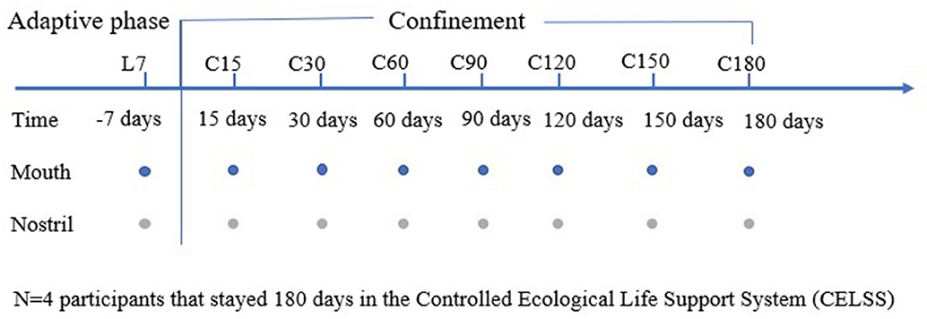
The 64 swab samples were collected from the oropharynx and nasal cavity of the four crew members during isolation in the CELSS at eight time points over a 180-days period. The implementation of the whole experiment conforms to the design of the experiment (Yuan et al., 2019). The schematic diagram of the research experiment design is shown in Figure 1. The outline of the CELSS and the living and working places of the volunteers are shown in Supplementary Figure 1.

Figure 1. Schematic diagram of research design. The sampling time point is shown at the top. The numbers represent the number of days before (L-) and during (C-) confinement, and−7 days represents the 7th day before the confinement experiment. The colored circles represent the time points that were used to collect the sample specified on the left side of the figure.
A total of 1,050,859 sequence readings were obtained from the nasal sample, and the median number of reads in the nasal sample was 31,266. A total of 666,675 sequence readings were obtained from oropharyngeal samples, and the median number of reads in oropharyngeal samples was 18,830.
The overall structure of microbial community composition was characterized by principal coordinate analysis (PCoA) of diversity differences. The results showed that the largest factor affecting the differences between samples was the sampling site (Supplementary Figure 2A). The microbial composition in the nasal cavity and oropharynx was distinctly different at the taxon of phylum (Supplementary Figure 2B). The nasal bacteria mainly belonged to Actinobacteria, Firmicutes, Proteobacteria, and Bacteroidetes, while Fusobacterial occupied a relatively high proportion in oropharyngeal microbiota in addition to the above-mentioned phyla. The microbiota of the same site had an individual difference in relative numbers (Supplementary Figures 2D,E). The index Chao of the oropharyngeal microbial community was higher than the nasal cavity, and it fluctuated more wildly in the nasal microbial community, but there was no statistical difference (P = 0.63) (Supplementary Figure 2C).
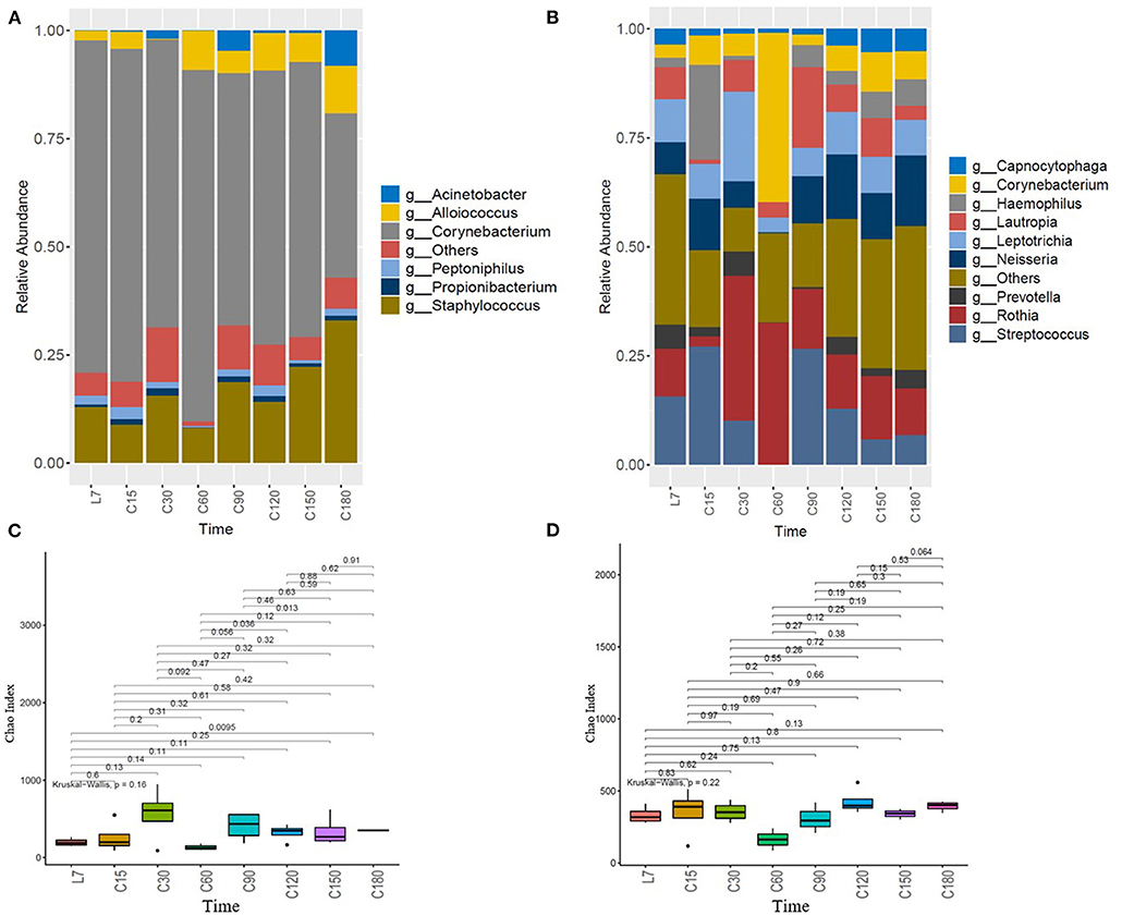
QIIME 1.9.1 was used to generate the species abundance distribution diagram of multiple samples. According to the classification results, bacteria in the top six positions of richness detected in the nose samples mainly belong to Corynebacterium, Staphylococcus, Alloiococcus, Peptoniphilus, Propionibacterium, and Acinetobacter. Analysis results of nasal bacterial composition (Figure 2A, Supplementary Figure 3) showed that Corynebacterium and Staphylococcus were dominant, and with the extension of confined time, the relative abundance of Corynebacterium showed a downward trend (P = 0.083), and Staphylococcus and Alloiococcus increased (P = 0.48, P = 0.92).

Figure 2. Dynamic analysis of the changes in the structure of microbiota over time. (A) Temporal characteristic of nasal microbiota, (B) temporal characteristic of oropharyngeal microbiota, (C) changes of nasal microbial index Chao with time, and (D) changes of oropharyngeal microbial index Chao with time. The horizontal axis represents the time point. L7 represents 7 days before the confined experiment, and C15, C30, C60, C90, C120, C150, and C180, respectively represent 15, 30, 60, 90, 120, 150, and 180 days during the confined experiment. The vertical axis represents the relative abundance of the genera.
The analysis results of the oropharyngeal microbial composition showed that (Figure 2B, Supplementary Figure 3) the dominant bacteria in oropharyngeal samples belonged to Corynebacterium, Streptococcus, Neisseria, Rothia, Prevotella, Leptotrichia, Haemophilus, Capnocytophaga, etc. The evenness of oropharyngeal dominant bacteria was higher than that of nasal dominant bacteria. On the whole, Prevotella showed a decreasing trend (P = 0.18), Neisseria showed an increasing trend (P = 0.34), Capnocytophaga decreased at first and then increased, while Leptotrichia showed little change.
The index Chao showed no obvious trend (Figures 2C,D), but it fluctuated greatly over time, especially at the time points of C30, C60, and C90. The index was relatively stable at the beginning and end of confinement.
It is worth mentioning that at the time point of C60, Corynebacterium has increased in the nasal cavity and oropharynx synchronously, the index Chao reduced and Staphylococcus in the nasal cavity and Streptococcus in oropharynx reduced.
There is some microbial specificity between individuals, so we analyzed the changes of individual bacterial community over time (Supplementary Figure 4). With the extension of the isolation time, as a whole, the nasal microbiota showed a decreasing trend of Corynebacterium (P = 0.083) and an increasing trend of Staphylococcus (P = 0.48), which was very obvious in volunteer 3 and 4. It should be noted that the abundance of Alloiococcus was higher and increased in the nasal cavity of volunteer 1, but it was rare in other volunteers. Acinetobacter was characterized abnormally high periodically in the nasal cavity of volunteer 4. Propionibacterium occupied a relatively low proportion in these dominant bacteria. Staphylococcus was very rarely in the nasal bacterial community of volunteers 2 and 4 before the confined experiment, while it was higher in the nasal bacterial community of volunteers 1 and 3. An interesting higher proportion of Staphylococcus was detected in the nasal bacterial community of all the volunteers during the confined experiment. The DNA extraction of nasal samples from volunteer 2 on day 180 and volunteer 4 on day 60 failed.
Neisseria in oropharyngiae (except occupant 1) showed an increased trend in the study (Supplementary Figure 5). An outbreak of Rossella happened 1–2 months after isolation and then returned to normal level. DNA was failed to be extracted from the oropharyngeal samples of volunteer 1 at day 60 and 90 and volunteer 4 at day 60.
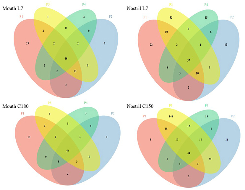
With 97% similarity, the OTU number of each sample was obtained. The Venn diagram was used to show the number of common and unique OTU numbers of multiple samples, and the OTU overlap among samples was visually displayed to reflect the microbial crossover between samples. We selected two time points of pre-confinement (L7) and confinement time 180 days (C180) for the Venn diagram analysis. Since it was failed to extract the DNA of nasal sample C180 from Volunteer 2, we chosed the nasal sample of confinement time 150 days (C150) instead of C180 for analysis (Figure 3). The number of common and unique OTU numbers of oropharyngeal samples was consistent before and after confinement. The common OTU numbers of nasal samples increased after confinement, which indicated that the nasal microbiota had the dynamic characteristic of evolving in the same direction after confinement, and the reason may be that airtight environmental factors caused the change of microbiota, while some microorganisms in the nasal cavity spread through the air and transfered between individuals in an airtight space.

Figure 3. Venn diagram of oropharyngeal and nasal samples before and after confinement. Mouth L7 and Mouth C180 represent oropharyngeal samples taken 7 days before and 180 days after confinement, and Nostril L7 and Nostril C150 represent nasal samples taken 7 days before and 150 days after confinement, respectively. P1, P2, P3, and P4 are the numbers of four volunteers. Circles of different colors are used to represent different individual samples, and the numbers of overlapping areas represent the numbers of common OTUs among samples. The common OUT numbers increase after confinement in Nostril samples Nostril L7 and Nostril C150.
Some genera of the dominant bacteria in the oropharyngeal and nasal microbial community contain pathogenic or opportunistic pathogens. According to the literature reports (Chonmaitree et al., 2017; González-Andrade et al., 2017; Katsoulis et al., 2019; Morinaga et al., 2019), several genera were selected from the dominant bacteria, which contained opportunistic pathogens. For example, Staphylococcus contains staphylococcus aureus, which produces enterotoxins that are harmful to health. Neisseria contains N. meningitidis which is a pathogenic bacterium of epidemic cerebrospinal meningitis, and it is mainly transmitted through the respiratory tract. S. pyogenes attached to Streptococcus can cause a variety of suppurative inflammation and hypersensitivity diseases, and S. pneumoniae can cause respiratory infections in human beings. The A. otitis attached to Alloiococcus is a pathogenic factor in adult secretome otitis media. P. acnes in Propionibacterium can cause skin inflammation and is the pathogen of acne.
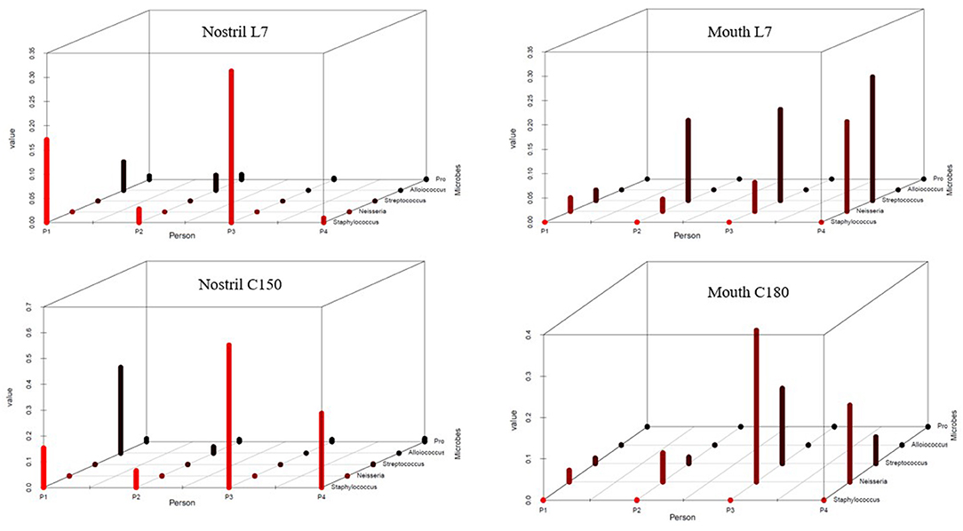
To reveal the changes of the above opportunistic pathogens in the human body in an isolation environment, we tried to compare the difference of the genera containing pathogen bacteria before and after the isolation (Figure 4, Supplementary Table 2). It was worth mentioning that the sequencing sequence of the 16s rRNA gene V3–V4 area could not distinguish the species level; however, the genera containing potentially pathogenic bacteria that we had listed could reflect the increased risk of infection by opportunistic pathogens from the side. It was failed to extract DNA from the nasal sample of C180 of Volunteer 2, and C150 was used for alternative analysis. The figure showed obvious increase in Staphylococcus of the nasal cavity and in Neisseria of the oropharynx after confinement, suggesting an increased risk of infection.

Figure 4. Changes in the content of potential pathogenic bacteria before and after confinement. The bacterial distribution 7 days before confinement (L7) and 180 days after confinement (C180) in different individuals was statistically analyzed. The horizontal axis P1, P2, P3, and P4 represented volunteers, the vertical axis represented the content value of bacteria, and the oblique axis represents the names of the bacterial genera. Nostril L7 and Nostril C180 represented the nasal samples taken 7 days before and 180 days after confinement. It was failed to extract in which the DNA of 180 days' nasal sample of Volunteer 2 and it was replaced by 150 days' nasal sample of Volunteer 2. Mouth L7 and Mouth C180 represented oropharyngeal samples taken 7 days before and 180 days after confinement, respectively. Note that the scale values used on the vertical axis were different and need to be distinguished.
In space, the special environmental factors may change bacterial community structure in the human body of astronauts, and break the microbiota balance. Working on-orbit results in decreased immunity and increases the risk of pathogenic infections. These infections may occur in the respiratory tract, the digestive tract, urinary tract, and skin, affecting the individual health and subjective experience. At the same time, these could affect other individuals, even the whole crew, through the migration of bacteria. Therefore, the study of the human bacterial community structure in the confined environment is of great significance for the prevention of infectious diseases during long-term flight on-orbit in the future. In our study, the temporal characteristics of the oropharyngeal and nasal microbiota structure in crewmembers were researched, in order to provide reference for revealing possible effects of microbiota balance on respiratory health in a closed environment.
The culture method is the most commonly used method for studying microbes on the space station, and although there have been attempts to sequence them on-orbit, conditions on the space station have limited the use of sequencing technology on-orbit. Currently, on-orbit microbial samples are returned to the earth for high-throughput sequencing and analysis for microbiota studies on space station. However, due to the limitation of return time, dynamic analysis of microbiota cannot be carried out immediately, and long-term preservation and space transportation may lead to the risk of analysis error. Therefore, many scholars simulate the space environment on the ground to dynamically study the changes of environment and human microbiota. In order to monitor the microbial characteristics of the closed system on the ground in real time, the researchers designed different experiments and simulated buildings to conduct research. Russia conducted a confined Mars500 habitat, and simulated flight and landing on Mars, using high-throughput sequencing technology to study the dynamic changes of the intestinal microbiota structure over time in six volunteers (Schwendner et al., 2017). Then, in the expansion of the lunar/Mars habitat simulation test was carried out, high-throughput sequencing technology was used to survey bacterial community in the air, and the results showed that in closed habitats, bacterial structure of air had a close relationship with human existence (Mayer et al., 2016).
The CELSS system is an excellent platform to simulate the closed environment of the space capsule on the ground. The confined experiment of four people for 180 days provided an opportunity to dynamically track the changes of the crews' own microbiota and the microbiota of others living in close proximity for a long time. During the 180-days confinement simulation experiment, human respiratory tract microbial samples were collected at eight time points, and the microbiota were continuously observed over time. This is the first time to study the changes of respiratory microbiota over time through CELSS.
This study aimed to reveal the effect of long-term confinement on the composition and diversity of nasal and oropharyngeal microbiota. We found that nasal cavity and oropharynx were independent in terms of microbiota structure and diversity (Supplementary Figures 2A,B), and this independence did not disappear with the extension of confinement time, indicating that the physiological environment and microecological environment of the growth site of the microbiota were the most important influential factors on the microbiota. Studies had investigated the temporal stability and diversity of skin microbiota, regardless of the sampling interval, and despite the skin community's constant exposure to external factors, the stability of the microbiota still existed, while the nature and degree of this stability was highly individualized (Oh et al., 2014).
In terms of bacterial composition, the nasal bacteria mainly belong to Actinobacteria, Firmicutes, Proteobacteria, Bacteroidetes, etc. In addition to the abovementioned phyla, the Fusobacterial occupies a relatively high proportion in oropharyngeal bacteria. At the level of genus, the dominant bacteria of nasal cavity are Corynebacterium, Staphylococcus, Diaphylococcus, Propionibacterium, etc., while the dominant bacteria of oropharynx are Corynebacterium, Streptococcus, Neisseria, and Prevotella. Previous studies have shown that the main members of the microbial ecosystem in the nasal cavity are usually Actinobacteria (containing Corynebacterium and Propionibacterium) and Firmicutes (Streptococcus in children and Staphylococcus in adults), while the abundance of anaerobic bacteria in Bacteroidetes is low (Camarinha-Silva et al., 2014; Oh et al., 2014). At the generic level, Corynebacterium, Propionibacterium, and Staphylococcus are the most common genera in the nasal cavity (Zhou et al., 2014). Previous studies have shown that bacterial biodiversity and uniformity vary greatly in the upper respiratory tract; thus, the oropharynx and oral cavity have the highest biodiversity and uniformity (Charlson et al., 2010; Zhou et al., 2013). On the contrary, similar to other areas covered by human skin epithelium, the nasal cavity shows low biodiversity (Grice and Segre, 2011).
During 30–90 days of confinement, the structure of nasal and oropharyngeal microbiota changed (Figures 2A,B), and the index Chao of the microbiota fluctuated greatly (Figures 2C,D). The confinement environment disturbed the nasal and oropharyngeal microbiota, especially after 30 days of confinement. In addition, the oropharyngeal microbial diversity was higher than the nasal cavity, and the index Chao of nasal cavity fluctuated more, but there was no statistical difference (P = 0.63) (Supplementary Figure 2C). The results of behavioral monitoring and psychological state assessment in this project showed that after 1 month of detention, behavioral flow reflecting global activity decreased 1.5- to 2-fold. Psychological questionnaires revealed a decrease in hostility and negative emotions but an increase in emotional adaptation suggesting boredom and monotony (Biesbroek et al., 2014). Physiological and psychological changes in volunteers in the isolation environment may trigger a series of physiological stress changes, such as immunity and secretion of respiratory antibacterial substances, which may affect the respiratory microbiota. According to NASA's research data, the exchange or migration of pathogenic microorganisms between passenger groups occurred in flight missions over 18 days (Crucian et al., 2016a). This is another explanation for the change in the microbiota after 30 days of confinement.
Dynamic analysis of the microbiota structure over time found that Staphylococcus and Alloiococcus showed an upward trend, while Corynebacterium showed a downward trend (Figure 2A). Neisseria showed an upward trend in oropharyngeal microbiota, while Corynebacterium showed a large fluctuation (Figure 2B). Early nasopharyngeal microbial studies showed that high abundance ratios of Corynebacterium and Moraxella marked a more stable structure, and poor stability was characterized by high abundance of Streptococcus and acquired H. influenzae, and the microbiota characteristics were correlative with reported higher incidence of respiratory tract infections and asthma in the early stages of life (Stubbendieck et al., 2019). Microbial communities characterized by high abundance of Corynebacterium and lack of the presence of H. influenzae and S. pneumoniae, suggesting colonization resistance to these potential pathogens. In this study, the abundance of Corynebacterium was the highest, indicating that the structure of the bacterial community was stable. However, during the confined period, Corynebacterium showed a downward trend, while Staphylococcus showed an increasing trend, suggesting that confinement had an adverse effect on the structural stability of the nasal bacterial community.
Worth noting is that although Corynebacterium showed a trend of decrease in the nasal cavity, but at the C60 time point, there was a dramatic rise of Corynebacterium in nasal and oropharyngeal microbiota collaboratively (Figures 2A,B), together with much lower index Chao (Figures 2C,D), at the same time Staphylococcus in the nasal cavity and Streptococcus in oropharynx greatly reduced. Studies have shown that Corynebacterium competes for limited iron resources in the nasal cavity by producing the iron-chelating vector, which is related to the ability to inhibit Staphylococcus. At the same time, the lipase secreted by Corynebacterium can lyse the triacylglycerol in the nasal cavity and produce the free fatty acid which is resistant to microorganisms, thus inhibiting the growth of Streptococcus in vitro (Marik, 2001).
Oropharynx connects the mouth, nasopharynx, larynx, lower respiratory tract, and gastrointestinal tract and it is exposed to exogenous and endogenous microorganisms. Thus, the species pool contained in oropharyngeal microecology is usually large and a highly diverse of bacterial community can be observed in adults. The oropharynx is also the niche of potentially pathogenic bacteria that may cause local (pharyngitis) or diffuse (lung) diseases (Mermel, 2013). Neisseria contains N. meningitidis which is a pathogenic bacterium of epidemic cerebrospinal meningitis, and it is mainly transmitted through the respiratory tract. Rothia is a class of symbiotic bacteria that is widespread in the mouth. In this study, Neisseria in the oropharynx (except volunteer 1) showed an increasing trend. An outbreak of Rossella happened 1–2 months after confinement, then it returned to normal (Supplementary Figure 5). Disturbance of the microbiota may lead to increased risk of infection. The analysis suggests that it may be related to the inadaptability to the confined environment at the initial stage of experiment, the insufficient proficiency of each item, the tension of the volunteers, and the great physical and mental pressure.
We respectively analyzed the changes in the microbiota of four volunteers over time and compared them crosswise. Nasal Staphylococcus in volunteers 2 and 4 makes up only a very small percentage, while in volunteers 1 and 3 they makes up a bigger percentage before confined experiment. Nasal Staphylococcus accounted for a bigger percentage in all volunteers after 180 days confined experiment (Supplementary Figure 4). To explore this phenomenon, we used the Venn diagram to show the common and unique OTU numbers of samples before and after confinement, and there is a tendency of convergence in the nasal cavity (Figure 3), so we speculated that four volunteers had a long and close contact in a confined space, resulting in a microbial transfer between them. The microorganisms carried on the surface of the volunteers are in direct contact with the environment and can have an impact on the environment and then can be transferred. According to NASA's research data, the exchange or migration of pathogenic microorganisms between the crew takes place in a flight mission of more than 18 days (Crucian et al., 2016a). The understanding of the population status of the nasal cavity and oropharyngeal microorganisms is beneficial to the prediction and prevention of the occurrence of on-orbit infectious diseases. Studies have shown that the microbiota on the internal surface of the International Space Station is similar to the microbiota on the crew's skin. These data provide reference for future microbial monitoring work of the ISS and crew and the control of microbial contamination of manned spacecraft (Avila-Herrera et al., 2020).
To reveal the changes of pathogenic bacteria over time during confinement, considering our sequencing method could not accurately locate the level of species, so we chose genera for symbolic judgment (Figure 4). It could be seen that Staphylococcus in the nasal cavity and Neisseria in the oropharynx increases and spread with the extension of the confinement time. Venn diagrams also reflected a similar situation (Figure 3), and common OTU numbers of nasal samples increased after confinement. Nasal microbiota had the same direction of evolutionary dynamic characteristic, and the reason may be that closed environmental factors caused the change of bacterial community; some nasal microorganisms in airtight space spread through the air and transferred between individuals.
The results of the bacterial census of the International Space Station showed that Staphylococcus, Bacillus, and Corynebacterium were among the top three in the detection rate of environmental microbial samples from International Space Station (Venkateswaran et al., 2014). Staphylococcus are a bacteria of human origin, and it is also pathogen that induce infectious diseases. Staphylococcus aureus are a conditional pathogenic bacterium that NASA attaches great importance to, and crew need medical treatment whose naval S. aureus is resistant to drugs in preflight medical examination. The purpose is to reduce infectious diseases caused by migration and exchange of conditional pathogenic bacteria between susceptible people (individual difference) during long-term living closely (Ramakrishnan et al., 2013). A large number of studies have found that chronic nasosinusitis in the upper respiratory tract of patients is associated with higher abundance of Staphylococcus accompanied by decreased bacterial diversity (Feazel et al., 2012; Jervis Bardy and Psaltis, 2016; Muluk et al., 2018), and Staphylococcus is also positively correlated with other respiratory diseases (such as allergic rhinitis and asthma) (Voorhies et al., 2019). Although other factors experienced by ISS crew members may play a role in the development of upper respiratory symptoms, the increased relative abundance of Staphylococcus in the nasal bacterial community is consistent with these symptoms (Crucian et al., 2016; Paetzold et al., 2019).
The bacterial index Chao did not show an obvious trend (Figures 2C,D), but it fluctuated greatly over time, especially at the time points of C30, C60, and C90. The index Chao was relatively stable at the beginning of confinement and before confinement ended, showing a certain stability. Host state and complex interactions of the disturbance of environment may influence the local change of bacterial community, and some species could flow in some individuals and parts and influence the structure and function of the microbiota in the short term, while microbiota maintain certain stability in the long run. As with the study of skin microbiota, the most typical example is that the human use probiotics to improve skin health (Oh et al., 2016), but skin microbiota seem to have inherent stability (Crucian et al., 2016).
Two respiratory sites were detected in this study; in another article, we also discussed the changes of the skin microbiota (Bingmu et al., 2018). According to the on-orbit disease investigation and analysis, intestinal tract, urinary tract, and eye are also common sites of infectious disease, in addition to the respiratory disease, diarrhea, urinary tract infection with aeruginosa, and eye sty caused by S. aureus also happened on-orbit (Hurlbert et al., 2010; Hodkinson et al., 2017). These infectious diseases prompted us to analyze microbial microbiota of these parts and carry out astronaut individualized microbiota characteristic analysis, especially for long-term orbits. Some research directions such as real-time acquisition and detect microbial samples observe the change of environmental and the astronaut's microbiota, prevent infectious disease, also have important research value for future long-term orbit, and these works still need to continue to explore.
In this study, 16S rDNA Illumina Miseq high-throughput sequencing was used to analyze the effects of nasal and oropharyngeal microbiota from crewmembers' long-term confined in the CELSS. The results showed that the structure of the nasal and oropharyngeal microbiota varied greatly, the individual bacterial community structure and the diversity changed with time. Staphylococcus in the nasal cavity increased and showed the characteristics of inter-individual transfer, suggesting that the microbiota structure and health of the respiratory tract could be affected by living in a closed environment for a long time.
The imbalance of the respiratory microbial community and the increase of opportunistic pathogens may be the inducement of respiratory diseases, but it does not mean that the increase of opportunistic pathogens will cause diseases; in addition, it is closely related to individual immune function. In the future, we will continue to carry out relevant studies on opportunistic pathogens. In the following work, we will also use a higher-resolution sequencing method to locate the species level and conduct in-depth analysis on pathogenic bacteria that may cause respiratory diseases.
The datasets presented in this article are not readily available because of the data confidentiality clause of the project sponsor. Requests to access the datasets should be directed to Space Science and Technology Institute (Shenzhen, http://www.szsisc.com/).
The research program was approved by the Ethical Committee of the ACC (China Astronaut Reasearch and Training Center Beijing, China) and complied with all the guidelines in the Declaration of Helsinki. Each volunteer was informed of the content and schedule of the study and signed an informed consent form. Participants can withdraw from the study at any time.
YC and CX: experimental design. YC, BX, and CZ: methodology. ZL, ZC, and JL: investigation. YC and BX: writing—original draft. CX, QX, and ZL: writing—review and editing. BX and QX: funding acquisition. All authors agree to be accountable for the content of the work.
This work was supported by the Key Program of Logistics Research of China. Grant number BWS17J030, Advanced space medico-engineering research project of China. Grant number 010101, and National Science and Technology Major Project for Major New Drugs Innovation and Development. Grant number 2015zx09j15102-002.
The authors declare that the research was conducted in the absence of any commercial or financial relationships that could be construed as a potential conflict of interest.
We are grateful to Cheng Zhang who checked for language errors. We thank Bin Xiao for graph processing and valuable advice. We thank Bin Wu, Zhiqi Fan, and Ying Chen for help during the experiments, as well as other members of the microbiology lab for suggestions.
The Supplementary Material for this article can be found online at: https://www.frontiersin.org/articles/10.3389/fmicb.2020.617696/full#supplementary-material
Avila-Herrera, A., Thissen, J., Urbaniak, C., Be, N. A., Smith, D. J., Karouia, F., et al. (2020). Crewmember microbiome may influence microbial composition of ISS habitable surfaces. PLoS ONE 15:e0231838. doi: 10.1371/journal.pone.0231838
Biesbroek, G., Tsivtsivadze, E., Sanders, E. A., Montijn, R., Veenhoven, R. H., Keijser, B. J., et al. (2014). Early respiratory microbiota composition determines bacterial succession patterns and respiratory health in children. Am. J. Respir. Crit. Care Med. 190, 1283–1292. doi: 10.1164/rccm.201407-1240OC
Bingmu, X., Heng, W., Yuanliang, W., Chongfa, Z., Junlian, L., Zhanghuang, C., et al. (2018). Analysis on body microbiota of people surviving in controlled ecological life support system of 180 days experiment. Space Med. Med. Eng. 31, 282–288.
Blachowicz, A., Mayer, T., Bashir, M., Pieber, T. R., De León, P., and Venkateswaran, K. (2017). Human presence impacts fungal diversity of inflated lunar/mars analog habitat. Microbiome 5:62. doi: 10.1186/s40168-017-0280-8
Bogaert, D., De Groot, R., and Hermans, P. W. (2004). Streptococcus pneumoniae colonisation: the key to pneumococcal disease. Lancet Infect. Dis. 4, 144–154. doi: 10.1016/S1473-3099(04)00938-7
Bomar, L., Brugger, S. D., and Lemon, K. P. (2018). Bacterial microbiota of the nasal passages across the span of human life. Curr. Opin. Microbiol. 41, 8–14. doi: 10.1016/j.mib.2017.10.023
Brugger, S. D., Bomar, L., and Lemon, K. P. (2016). Commensal-pathogen interactions along the human nasal passages. PLoS Pathog. 12:e1005633. doi: 10.1371/journal.ppat.1005633
Camarinha-Silva, A., Jáuregui, R., Chaves-Moreno, D., Oxley, A. P., Schaumburg, F., Becker, K., et al. (2014). Comparing the anterior nare bacterial community of two discrete human populations using illumina amplicon sequencing. Environ. Microbiol. 16, 2939–2952. doi: 10.1111/1462-2920.12362
Caporaso, J. G., Kuczynski, J., Stombaugh, J., Bittinger, K., Bushman, F. D., Costello, E. K., et al. (2010). QIIME allows analysis of high-throughput community sequencing data. Nat. Methods 7, 335–336. doi: 10.1038/nmeth.f.303
Charlson, E. S., Chen, J., Custers-Allen, R., Bittinger, K., Li, H., Sinha, R., et al. (2010). Disordered microbial communities in the upper respiratory tract of cigarette smokers. PLoS ONE 5:e15216. doi: 10.1371/journal.pone.0015216
Chonmaitree, T., Jennings, K., Golovko, G., Khanipov, K., Pimenova, M., Patel, J. A., et al. (2017). Nasopharyngeal microbiota in infants and changes during viral upper respiratory tract infection and acute otitis media. PLoS ONE 12:e0180630. doi: 10.1371/journal.pone.0180630
Crucian, B., Babiak-Vazquez, A., Johnston, S., Pierson, D. L., Ott, C. M., and Sams, C. (2016). Incidence of clinical symptoms during long-duration orbital spaceflight. Int. J. Gen. Med. 9, 383–391. doi: 10.2147/IJGM.S114188
de Steenhuijsen Piters, W. A., Sanders, E. A., and Bogaert, D. (2015). The role of the local microbial ecosystem in respiratory health and disease. Philos. Trans. R. Soc. Lond. Series B Biol. Sci. 370:20140294. doi: 10.1098/rstb.2014.0294
Feazel, L. M., Robertson, C. E., Ramakrishnan, V. R., and Frank, D. N. (2012). Microbiome complexity and Staphylococcus aureus in chronic rhinosinusitis. Laryngoscope 122, 467–472. doi: 10.1002/lary.22398
González-Andrade, B., Santos-Lartigue, R., Flores-Treviño, S., Ramirez-Ochoa, N. S., Bocanegra-Ibarias, P., Huerta-Torres, F. J., et al. (2017). The carriage of interleukin-1B-31*C allele plus Staphylococcus aureus and Haemophilus influenzae increases the risk of recurrent tonsillitis in a Mexican population. PLoS ONE 12:e0178115. doi: 10.1371/journal.pone.0178115
Grice, E. A., and Segre, J. A. (2011). The skin microbiome. Nat. Rev. Microbiol. 9, 244–253. doi: 10.1038/nrmicro2537
Hodkinson, P. D., Anderton, R. A., Posselt, B. N., and Fong, K. J. (2017). An overview of space medicine. Br. J. Anaesth. 119(Suppl.1), i143–i153. doi: 10.1093/bja/aex336
Hurlbert, K., Bagdigian, B., Carroll, C., et al. (2010). DRAFT Human Health, Life Support, and Habitation System. NASA Headquarters Washington, 2010:TA06-18.
Ichijo, T., Yamaguchi, N., Tanigaki, F., Shirakawa, M., and Nasu, M. (2016). Four-year bacterial monitoring in the international space station-japanese experiment module “Kibo” with culture-independent approach. NPJ Microgr. 2:16007. doi: 10.1038/npjmgrav.2016.7
Jervis Bardy, J., and Psaltis, A. J. (2016). Next generation sequencing and the microbiome of chronic rhinosinusitis: a primer for clinicians and review of current research, its limitations, and future directions. Ann. Otol. Rhinol. Laryngol. 125, 613–621. doi: 10.1177/0003489416641429
Katsoulis, K., Ismailos, G., Kipourou, M., and Kostikas, K. (2019). Microbiota and asthma: clinical implications. Respir. Med. 146, 28–35. doi: 10.1016/j.rmed.2018.11.016
Lang, J. M., Coil, D. A., Neches, R. Y., Brown, W. E., Cavalier, D., Severance, M., et al. (2017). A microbial survey of the international space station (ISS). PeerJ 5:e4029. doi: 10.7717/peerj.4029
Lee, J. T., Kim, C. M., and Ramakrishnan, V. (2019). Microbiome and disease in the upper airway. Curr. Opin. Allergy Clin. Immunol. 19, 1–6. doi: 10.1097/ACI.0000000000000495
Marik, P. E. (2001). Aspiration pneumonitis and aspiration pneumonia. N. Engl. J. Med. 344, 665–671. doi: 10.1056/NEJM200103013440908
Mayer, T., Blachowicz, A., Probst, A. J., Vaishampayan, P., Checinska, A., Swarmer, T., et al. (2016). Microbial succession in an inflated lunar/mars analog habitat during a 30-day human occupation. Microbiome 4:22. doi: 10.1186/s40168-016-0167-0
Mermel, L. A. (2013). Infection prevention and control during prolonged human space travel. Clin. Infect. Dis. 56, 123–130. doi: 10.1093/cid/cis861
Morinaga, Y., Take, Y., Sasaki, D., Ota, K., Kaku, N., Uno, N., et al. (2019). Exploring the microbiota of upper respiratory tract during the development of pneumonia in a mouse model. PLoS ONE 14:e0222589. doi: 10.1371/journal.pone.0222589
Muluk, N. B., Altin, F., and Cingi, C. (2018). Role of superantigens in allergic inflammation: their relationship to allergic rhinitis, chronic rhinosinusitis, asthma, and atopic dermatitis. Am. J. Rhinol. Allergy 32, 502–517. doi: 10.1177/1945892418801083
Novikova, N., De Boever, P., Poddubko, S., Deshevaya, E., Polikarpov, N., Rakova, N., et al. (2006). Survey of environmental biocontamination on board the international space station. Res. Microbiol. 157, 5–12. doi: 10.1016/j.resmic.2005.07.010
Oh, J., Byrd, A. L., Deming, C., Conlan, S., NISC Comparative Sequencing Program, Kong, H. H., et al. (2014). Biogeography and individuality shape function in the human skin metagenome. Nature 514, 59–64. doi: 10.1038/nature13786
Oh, J., Byrd, A. L., Park, M., NISC Comparative Sequencing Program, Kong, H. H., and Segre, J. A. (2016). Temporal stability of the human skin microbiome. Cell 165, 854–866. doi: 10.1016/j.cell.2016.04.008
Paetzold, B., Willis, J. R., Pereira de Lima, J., Knödlseder, N., Brüggemann, H., Quist, S. R., et al. (2019). Skin microbiome modulation induced by probiotic solutions. Microbiome 7:95. doi: 10.1186/s40168-019-0709-3
Ramakrishnan, V. R., Feazel, L. M., Abrass, L. J., and Frank, D. N. (2013). Prevalence and abundance of Staphylococcus aureus in the middle meatus of patients with chronic rhinosinusitis, nasal polyps, and asthma. Int. Forum Allergy Rhinol. 3, 267–271. doi: 10.1002/alr.21101
Schwendner, P., Mahnert, A., Koskinen, K., Moissl-Eichinger, C., Barczyk, S., Wirth, R., et al. (2017). Preparing for the crewed mars journey: microbiota dynamics in the confined Mars500 habitat during simulated mars flight and landing. Microbiome 5:129. doi: 10.1186/s40168-017-0345-8
Segata, N., Haake, S. K., Mannon, P., Lemon, K. P., Waldron, L., Gevers, D., et al. (2012). Composition of the adult digestive tract bacterial microbiome based on seven mouth surfaces, tonsils, throat and stool samples. Genome Biol. 13:R42. doi: 10.1186/gb-2012-13-6-r42
Stubbendieck, R. M., May, D. S., Chevrette, M. G., Temkin, M. I., Wendt-Pienkowski, E., Cagnazzo, J., et al. (2019). Competition among nasal bacteria suggests a role for siderophore-mediated interactions in shaping the human nasal microbiota. Appl. Environ. Microbiol. 85, e02406–e02418. doi: 10.1128/AEM.02406-18
Venkateswaran, K., La Duc, M. T., and Horneck, G. (2014). Microbial existence in controlled habitats and their resistance to space conditions. Microb. Environ. 29, 243–249. doi: 10.1264/jsme2.ME14032
Ver Heul, A., Planer, J., and Kau, A. L. (2019). The human microbiota and asthma. Clin. Rev. Allergy Immunol. 57, 350–363. doi: 10.1007/s12016-018-8719-7
Voorhies, A. A., Mark Ott, C., Mehta, S., Pierson, D. L., Crucian, B. E., Feiveson, A., et al. (2019). Study of the impact of long-duration space missions at the international space station on the astronaut microbiome. Sci. Rep. 9:9911. doi: 10.1038/s41598-019-46303-8
Yuan, M., Custaud, M. A., Xu, Z., Wang, J., Yuan, M., Tafforin, C., et al. (2019). Multi-system adaptation to confinement during the 180-day controlled ecological life support system (CELSS) experiment. Front. Physiol. 10:575. doi: 10.3389/fphys.2019.00575
Zhou, Y., Gao, H., Mihindukulasuriya, K. A., La Rosa, P. S., Wylie, K. M., Vishnivetskaya, T., et al. (2013). Biogeography of the ecosystems of the healthy human body. Genome Biol. 14:R1. doi: 10.1186/gb-2013-14-1-r1
Keywords: controlled ecological life support system, microbial community, space flight, oropharynx, nasal cavity, microbiota
Citation: Chen Y, Xu C, Zhong C, Lyu Z, Liu J, Chen Z, Dun H, Xin B and Xie Q (2021) Temporal Characteristics of the Oropharyngeal and Nasal Microbiota Structure in Crewmembers Stayed 180 Days in the Controlled Ecological Life Support System. Front. Microbiol. 11:617696. doi: 10.3389/fmicb.2020.617696
Received: 15 October 2020; Accepted: 16 December 2020;
Published: 03 February 2021.
Edited by:
Teresa Nogueira, Instituto Nacional Investigaciao Agraria e Veterinaria (INIAV), PortugalReviewed by:
Ebrahim Kouhsari, Iran University of Medical Sciences, IranCopyright © 2021 Chen, Xu, Zhong, Lyu, Liu, Chen, Dun, Xin and Xie. This is an open-access article distributed under the terms of the Creative Commons Attribution License (CC BY). The use, distribution or reproduction in other forums is permitted, provided the original author(s) and the copyright owner(s) are credited and that the original publication in this journal is cited, in accordance with accepted academic practice. No use, distribution or reproduction is permitted which does not comply with these terms.
*Correspondence: Bingmu Xin, xin_bm@163.com; Qiong Xie, eGllcWlvQHNpbmEuY29t
†These authors have contributed equally to this work and share first authorship
Disclaimer: All claims expressed in this article are solely those of the authors and do not necessarily represent those of their affiliated organizations, or those of the publisher, the editors and the reviewers. Any product that may be evaluated in this article or claim that may be made by its manufacturer is not guaranteed or endorsed by the publisher.
Research integrity at Frontiers

Learn more about the work of our research integrity team to safeguard the quality of each article we publish.