95% of researchers rate our articles as excellent or good
Learn more about the work of our research integrity team to safeguard the quality of each article we publish.
Find out more
ORIGINAL RESEARCH article
Front. Microbiol. , 28 July 2020
Sec. Fungi and Their Interactions
Volume 11 - 2020 | https://doi.org/10.3389/fmicb.2020.01672
 Shu-Ying Yu1,2,3
 Li-Na Guo1,3*
 Meng Xiao1,3
 Meng-Lan Zhou1,2,3
 Ying Yuan1,3
 Yao Wang1,3
 Li Zhang1,3
 Tian-Shu Sun3,4
 Ya-Ting Ning1,2,3
 Pei-Yao Jia1,2,3
 Wei Kang1,3
 Fanrong Kong5
 Sharon C.-A. Chen5
 Yanan Zhao6,7
 Ying-Chun Xu1,3*
Invasive aspergillosis (IA) due to Aspergillus lentulus is associated with high mortality. In this study, we investigated the clinical and microbiological characteristics of 6 fatal cases of proven or probable IA caused by A. lentulus in China. Underlying immunosuppression, prior antifungal exposure, and intensive care unit (ICU) hospitalization were important risk factors for invasive A. lentulus infection. Phenotypic differences were observed for A. lentulus isolates including slower growth, reduced sporulation, and inability to grow at 48°C, compared with Aspergillus fumigatus complex. ITS sequencing was unable to distinguish A. lentulus from A. fumigatus, but sequencing of the benA, CaM, and rod A loci enabled reliable distinction of these closely related species. Phylogenetic analysis further confirmed that the ITS region had little variation within the Aspergillus section Fumigati while the benA gene offered the highest intraspecific discrimination. Microsatellite typing results revealed that only loci on chromosomes 1, 3, 5, and 6b generated detectable amplicons for identification. All A. lentulus isolates showed in vitro resistance to multiple antifungal drugs including amphotericin B (MIC range 4 to 8 μg/ml), itraconazole (MIC 2 μg/ml), voriconazole (MIC of 4–16 μg/ml), and posaconazole (MIC of 0.5–1 μg/ml). However, MECs for the echinocandin drugs ranged from 0.03–0.25, ≤0.008–0.015, and ≤0.015–0.03 μg/ml for caspofungin, micafungin, and anidulafungin, respectively. A. lentulus is an emerging fungal pathogen in China, causing fatal disease, and clinicians as well as laboratories should be alert to their increasing presence.
Invasive aspergillosis (IA) remains a major invasive fungal infection with serious clinical consequences among immunocompromised patients (Sugui et al., 2014), of which the Aspergillus fumigatus complex is the most common cause. Notably, other causative Aspergillus spp. including A. flavus, A. terreus, and A. niger and cryptic species within the A. fumigatus complex have been increasingly recognized (Gurcan et al., 2013; Sugui et al., 2014). Of the last, in particular Aspergillus lentulus has become a significant pathogen in many countries including America, Spain, Argentina, Denmark, France, Germany, Turkey, Switzerland, Brazil, Japan, Australia, and Korea (Balajee et al., 2004, 2005a, 2009; Alcazar-Fuoli et al., 2008; Montenegro et al., 2009; Symoens et al., 2010; Mortensen et al., 2011; Zbinden et al., 2012; Datta et al., 2013; Escribano et al., 2013; Gurcan et al., 2013; Alastruey-Izquierdo et al., 2014; Lago et al., 2014; Bastos et al., 2015; Kidd et al., 2015; Tamiya et al., 2015; Yoshida et al., 2015; Tetsuka et al., 2017; Won et al., 2018).
Aspergillus lentulus was first described in 2004 as an opportunistic pathogen responsible for fatal infections in four hematopoietic stem cell transplant patients (Balajee et al., 2004; Balajee et al., 2005b). It is a sibling species of A. fumigatus within the A. fumigatus complex but with poor sporulating capacity, leading to diagnostic difficulty (Sugui et al., 2014; Lamoth, 2016). It tends to cause infection associated with higher mortality (over 60%) and poorer clinical outcome compared with that of A. fumigatus (Sugui et al., 2014; Won et al., 2018).
As such, in the clinical laboratory, A. lentulus often is misidentified as another Aspergillus species or simply identified only to genus level, by conventional morphological analysis (Symoens et al., 2010). Molecular methods are required for definitive species identification. Analysis of partial DNA sequences of various genes, such as the internal transcribed spacer (ITS) rDNA region, β-tubulin (benA), calmodulin (CaM), and rodlet A (rodA), have been reported as markers to differentiate different species within the Aspergillus section Fumigati (Lamoth, 2016). In addition, microsatellites [or short tandem repeats (STR)] have been considered as useful genetic tools in outbreak investigation and to delineate transmission routes of the A. fumigatus species complex with reproducibility and high discrimination power (Araujo et al., 2009, 2012). Compared to multi-locus sequence typing (MLST), microsatellite-based typing seemed to be more reproducible and cost-effective for A. fumigatus identification (Klaassen, 2009; Vanhee et al., 2009). Unfortunately, there are very few data on these methods in the characterization and study of genetic diversity in A. lentulus (Araujo et al., 2012).
The present study is the first to examine the clinical, microbiological, and molecular characteristics of A. lentulus and its infections from China.
The study was approved by the Human Research Ethics Committee of Peking Union Medical College Hospital (No. S-263). Written informed consent was obtained from all patients in the study and for permission to study the isolates cultured from them for scientific research.
A total of six non-duplicate Aspergillus isolates were recovered from the respiratory tract of six patients diagnosed with proven or probable IA (De Pauw et al., 2008; Cornely et al., 2019) under the China Hospital Invasive Fungal Surveillance Net (CHIF-NET; Wang et al., 2012)—Northern China program from January 2016 to December 2017. This program is a laboratory-based, multicenter study of invasive fungal diseases (De Pauw et al., 2008) including those caused by yeasts and filamentous fungi. A total of 80 hospitals in 6 provinces in the north part of China participated. The 580 isolates of filamentous fungi were collecting in the period, and the six Aspergillus isolates were confirmed as A. lentulus by rRNA sequencing at the central laboratory. Patients’ charts were reviewed to determine patient demographics, clinical features of infection including underlying disease/risk condition, prior antifungal therapy, treatment, and outcomes. All isolates were subcultured onto potato dextrose agar (PDA) and incubated at 28°C for 7–30 days prior to study. Isolates were identified to species complex level based on morphological characteristics (Larone, 2005) as well as growth at 48°C.
The identification of all the isolates was also undertaken using two Matrix-assisted laser desorption ionization–time of flight mass spectrometry (MALDI-TOF MS) systems—the bioMérieux Vitek MS (bioMérieux) and Bruker Autoflex Speed (Bruker Daltonics, Bremen, Germany). Preparation of A. lentulus isolates for MALDI-TOF MS identification was performed according to the manufacturer’s instructions with some modifications (Li et al., 2017). The acquisition and analysis of mass spectra were handled using software Myla (for Vitek MS, database version 3.2.0, bioMérieux) and Biotyper version 3.1 with the Filamentous Fungi Library 1.0 (for Autoflex Speed, Bruker Daltonics), again following the manufacturer’s instructions. All identification results displaying a single result with a confidence score ≥ 1.700 or a confidence value of 99.9% were considered acceptable for Bruker Biotyper MS and Vitek MS, respectively (Wang et al., 2016; Zhou et al., 2016).
Genomic DNA extraction was performed with QIAamp DNA Mini Kit (QIAGEN 51306; Qiagen, Hilden, Germany) in accordance with the manufacturer’s instructions. For all isolates, DNA amplification of the ITS, benA, CaM, and rodA sequences was performed as previously described (Li et al., 2017). In order to evaluate genetic polymorphisms among A. lentulus isolates, eight primer pairs targeting eight microsatellite loci located on chromosomes 1, 2, 3, 5, 6a, 6b, 7, and 8 were selected (Araujo et al., 2009). The PCR products were sequenced in both directions using the ABI 3730XL system (Applied Biosystems, Foster City, CA, United States). Obtained ITS, benA, CaM, and rodA sequences were queried against those contained in the GenBank database under accession numbers NR135407, EF669825, EF669895, and HQ127311, using the nucleotide Basic Local Alignment Search Tool (BLASTn, http://blast.ncbi.nlm. nih.gov).
Nucleotide sequences of species closely related to A. lentulus in the Aspergillus section Fumigati including A. fumigatus sensu stricto, A. udagawae, A. viridinutans, A. thermomutatus (Neosartorya pseudofischeri), A. novofumigatus, and A. hiratsukae available in GenBank as of 30th August 2019 were downloaded (Lamoth, 2016). Phylogenetic analysis was performed with software MEGA (Molecular Evolutionary Genetic Analysis software, version 6.0; http://www.megasoftware.net) using the Neighbor-Joining (NJ) method, with all positions containing gaps and missing data eliminated from the data set. The significance of the cluster nodes was determined by bootstrapping with 1,000 randomizations A. clavatus sequences (GenBank accession NR121482, AF057312, HE661596, and AB524404) were used as outgroups.
The in vitro susceptibility to nine antifungal drugs [fluconazole (FLZ), voriconazole (VOR), itraconazole (ITR), posaconazole (POS), caspofungin (CAS), micafungin (MCF), anidulafungin (AND), amphotericin B (AMB), and 5-flucytosine (5-FC)] was determined by broth microdilution methodology according to the Clinical and Laboratory Standards Institute (CLSI) M38-A3 protocol (Clinical and Laboratory Standards Institute [CLSI], 2017) and by Sensititre YeastOneTM YO10 (SYO) methodology (Thermo Scientific, United States) following the manufacturer’s instructions. The MICs of AMB and azoles were read as the lowest concentration that resulted in no discernible growth following 48 h of incubation (Clinical and Laboratory Standards Institute [CLSI], 2017). The minimum effective concentrations (MECs) of CAS, AND, and MCF were read in accordance with the CLSI M38-A3 protocol (Clinical and Laboratory Standards Institute [CLSI], 2017). The quality control organisms were Candida parapsilosis ATCC22019 and Candida krusei ATCC6258. Due to the lack of clinical breakpoints or epidemiologic cutoff values (ECVs) for A. lentulus, current CLSI ECVs established for at the A. fumigatus species complex were used to classify the isolates as wild type (WT) or non–wild type (non-WT; Clinical and Laboratory Standards Institute [CLSI], 2018; Won et al., 2018).
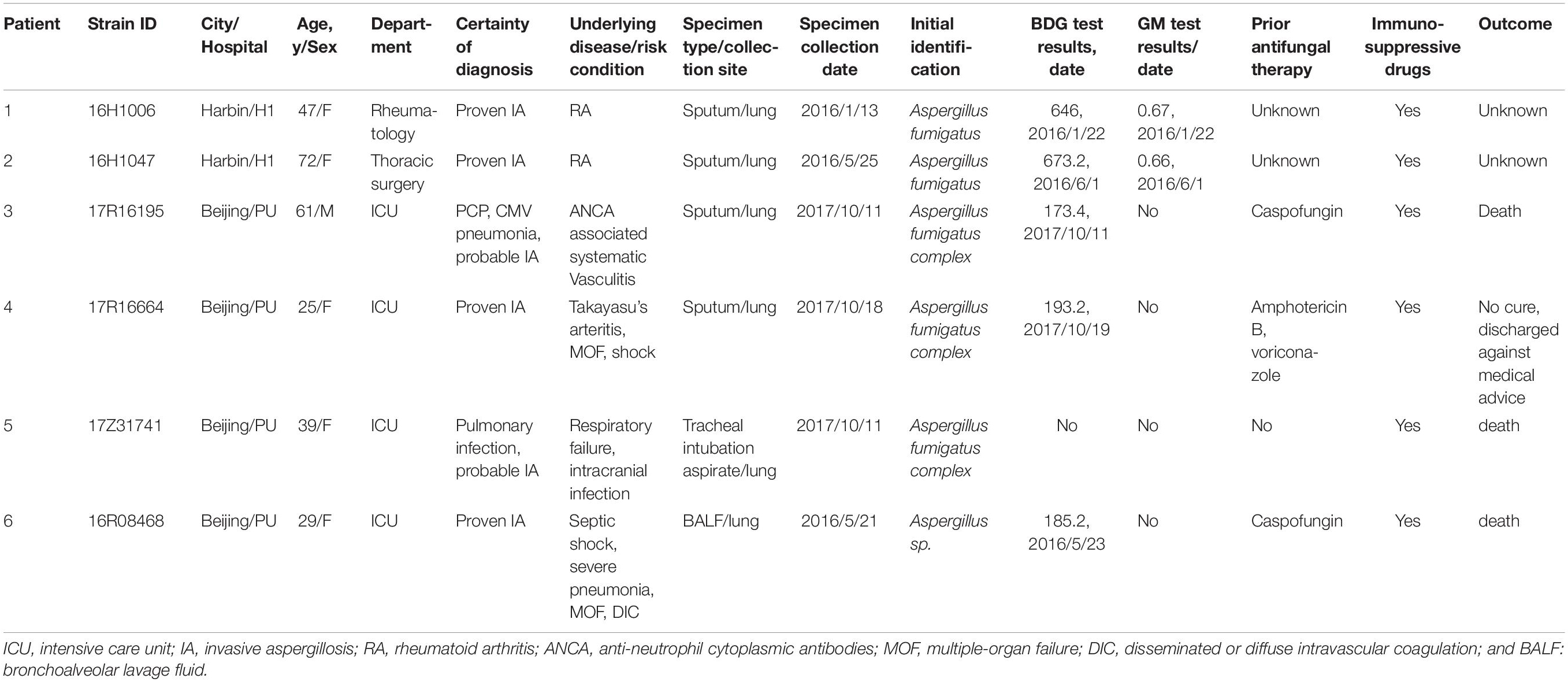
The ITS region, benA, CaM, and rodA gene sequences of strain 16R08468 (isolated from patient 6), 16H1047 (isolated from patient 2), 16H1006 (isolated from patient 1), 17R16664 (isolated from patient 4), 17Z31741 (isolated from patient 5), and 17R16195 (isolated from patient 3) have been deposited in GenBank with accession numbers MN235864 to MN235869, MN275499 to MN275504, MN275505 to MN275510, and MN275511 to MN275516, respectively, (see Table 1).

Table 1. Main clinical aspects of invasive aspergillosis caused by Aspergillus lentulus in this study.
All six patients with proven or probable A. lentulus infection were immunocompromised or had significant comorbidities such as rheumatoid arthritis and anti-neutrophil cytoplasmic antibodies (ANCA)-associated systemic vasculitis and/or were suffering from multiple-organ failure (MOF), shock, and disseminated intravascular coagulation (DIC; Table 1). Five patients were females, and four were in ICU when infection developed. All patients had received immunosuppressive therapy comprising corticosteroids and/or cyclophosphamide. There was no relevant data on prior antifungal exposure or clinical outcome information for patients 1 and 2; the other four were considered to have poor prognosis (Table 1). Three patients received prior antifungal therapy including caspofungin in two patients and amphotericin B combined with voriconazole in the other. Because the first sign, symptom, or finding of invasive fungal infection was occurring before antifungal drug treatment, they should not be classified as breakthrough IFI (Cornely et al., 2019). Five patients had obviously increased (1,3)-β-d-glucan (BDG test, Dynamiker, China), all with O.D > readings of over 100 pg/ml. Galactomannan (GM) was either unknown or not tested in these patients.
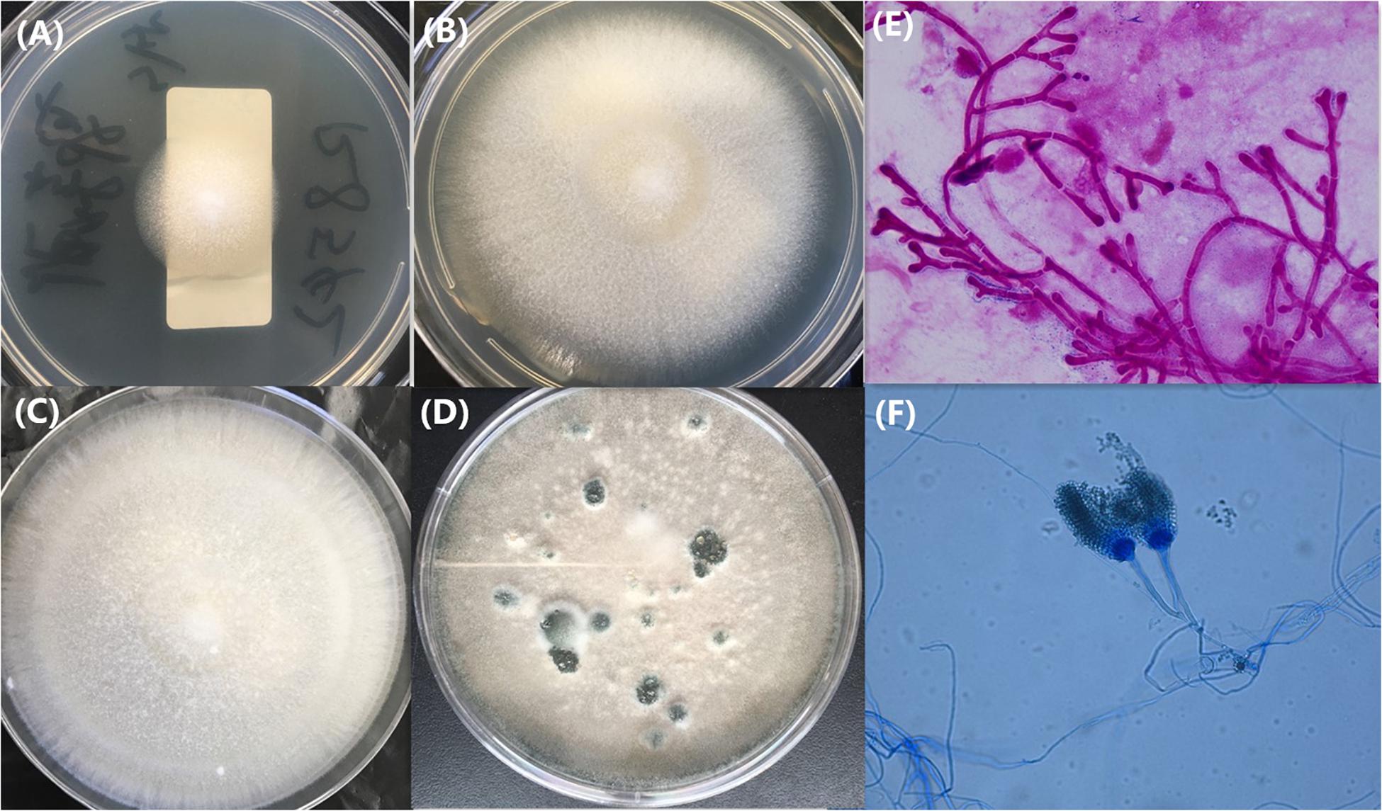
All six isolates were slow growers and poor sporulators on PDA at 28 and 35°C and failed to grow at 48°C. After 7 days of incubation on PDA, all isolates grew as colored white and velvety colonies mainly consisting of hyphae interspersed with sporadic gray-green spores (Figure 1). After prolonged incubation (8 to 21 days) on PDA, the colonies began sporulating, which had the same appearance as typical colonies of A. fumigatus (Figure 1). Direct microscopic analysis of sputum or bronchoalveolar lavage fluid revealed abundant septate fungal hyphae septum with acute angle branching, arranged radially or coral like (Figure 1E). Microscopic examination on day 3 to day 21 showed stipes, head, and conidia of A. lentulus. Uniseriate conidial heads are columnar, and conidia are produced in basipetal succession forming long chains and are globose to broadly ellipsoidal. Conidiophore stipes are smooth-walled and have subglobose vesicles. Because of near identical features to A. fumigatus sensu stricto, the two may be confused and misidentified by microscopic examination (James et al., 2011; Figure 1F).

Figure 1. Colony appearance of A. lentulus isolates on Potato Dextrose Agar (A–D) and Gram’s staining of the bronchoalveolar lavage fluid specimen (E) and microscopic appearance of the branched conidiophores on Potato Dextrose Agar by lactophenol cotton blue staining (F). Incubation conditions: 1A, 28°C, 2 days; 1B, 28°C, 5 days; 1C, 28°C, 7 days; 1D, 28 days; and 1F, 28°C, 7 days. The isolate of (A–F) selected the isolates of 16R08468.
All isolates had identical ITS and CaM sequences, but there were three single-nucleotide variations including C183T, C268T, and C347T of the benA sequence for two of the isolates. In addition, isolate 16H1047 had a unique rod A sequence with 7-base nucleotide changes including C to G in position 257; T to C in position 260, 283, 347, and 403; G to T in position 383; and C to T in position 365 compared with other isolates. All isolates were identified by ITS sequencing as “A. lentulus” or “A. fumigatus” with 100% sequence identity. Of note, Ben A and CaM gene sequencing analysis successfully identified all isolates as A. lentulus with 100% sequence identity. Except for isolate 16H1047 which was identified as A. lentulus or as A. fumigatus with 100% sequence identity, the other five isolates were successfully identified as A. lentulus by rod A sequencing.
Among eight microsatellite loci used, only four (located on chromosomes 1, 3, 5, and 6b) were detected by standard electrophoresis. PCR amplification of the other four microsatellite loci located in chromosome 2, 6a, 7, and 8 did not generate any detectable amplicon. All six A. lentulus isolates had identical peaks at positions 830, 206, 240, and 208 bp corresponding to each microsatellite locus of chromosomes 1, 3, 5, and 6b. This result clearly differs from that of A. fumigatus, which shows an expected electrophoretic profile of 8 peaks, corresponding to each microsatellite locus (Araujo et al., 2012). Furthermore, the results may indicate that the set of eight microsatellite loci had high ability to distinguish A. fumigatus and A. lentulus but low discrimination power or poor polymorphism to genotype in the A. lentulus species.
Mass spectrometry spectra of A. lentulus were not contained in the Bruker Biotyper database but were in the Vitek MS database. Hence, the Bruker Biotyper system provided “no identification” (log score < 1.70) for all A. lentulus isolates, while the Vitek MS system correctly identified all the isolates to species level with a confidence value of 99.9 (Table 2).

Table 2. Results of MALDI-TOF MS identification and antifungal susceptibility for Aspergillus lentulus isolates.
Table 2 also shows the MIC or MEC values for the six isolates. The MEC range for CAS, MCF, and AND were 0.03 to 0.25 or 0.03 to 0.12, ≤0.008 to 0.015 or ≤0.008, and ≤0.0015 to 0.03 μg/ml by CLSI or SYO, respectively. According to CLSI ECVs (Clinical and Laboratory Standards Institute [CLSI], 2018), all were classified as wild type (WT) for the echinocandins. In contrast, all the isolates showed in vitro resistance to multiple drugs in other antifungal classes based on both methods, including AMB (MIC range 4 to 8 μg/ml), ITR (MIC 2 μg/ml), VOR (MIC range 4 to 16 μg/ml), POS (MIC range 0.5 to 1 μg/ml), and FLZ (MIC > 256 μg/ml), according to CLSI ECVs, for A. fumigatus.
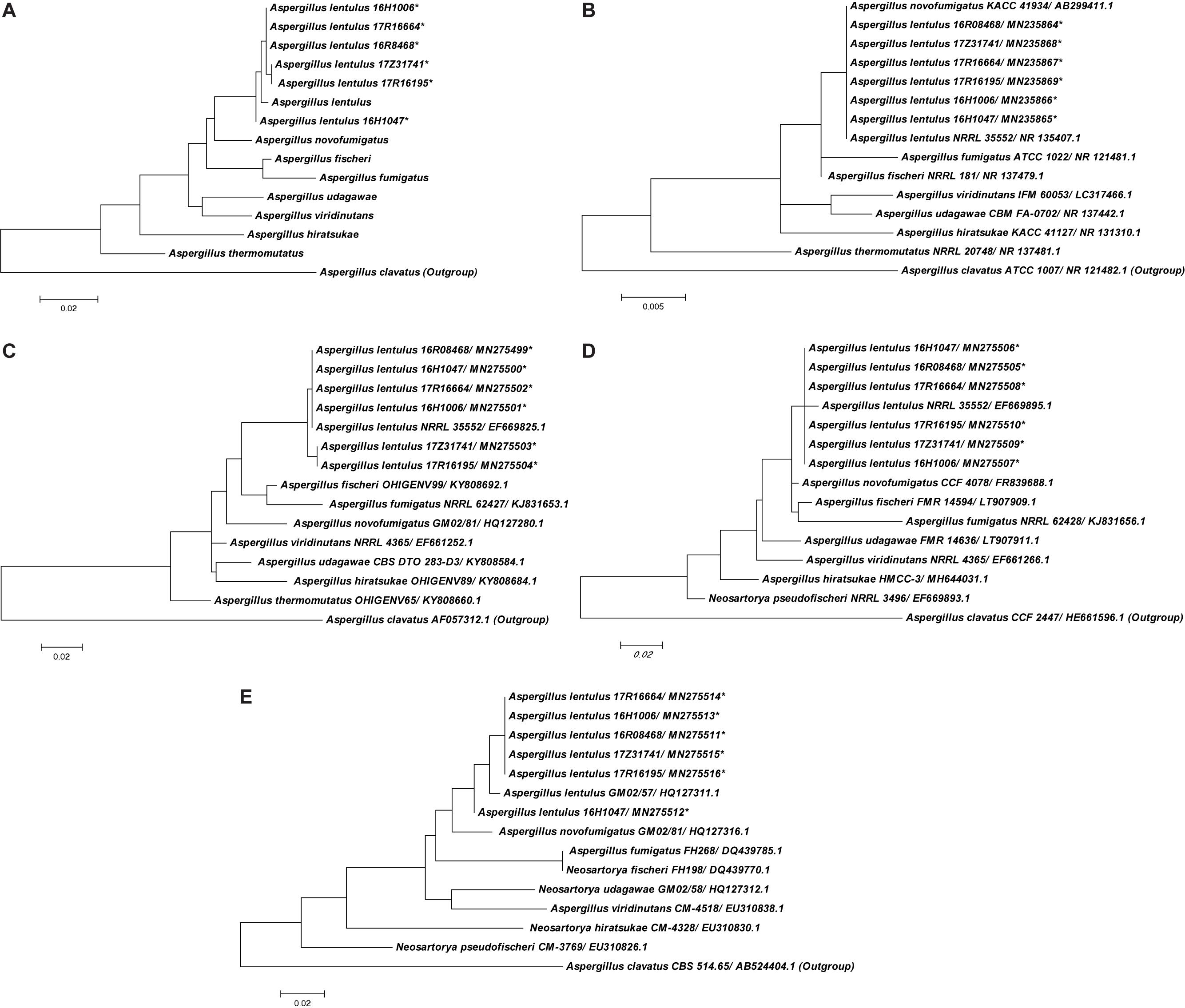
A total of nine closely related species in the Aspergillus section Fumigati were employed to construct phylogenetic trees (Figure 2). Phylogenetic analysis based on gene sequences from the combination of the ITS region and benA, CaM, and rodA loci together or each single gene alone showed that the six A. lentulus isolates in our study form a monophyletic cluster with the A. lentulus-type strain, confirming their original identification. Focusing on the phylogenetic tree constructed from all four loci, A. lentulus isolates were not only clearly distinguished from other species in the Aspergillus section Fumigati but had intraspecies polymorphisms. It was observed that 17Z31741 and 17R16195 were more closely related and 16H1047 was the most isolated, compared to other isolates in the A. lentulus species in the four loci phylogenetic tree. This was due to diverse ben A sequences of 17Z31741 and 17R16195 and a unique rod A sequence of 16H1047. Single-gene phylogenetic analysis revealed that ITS sequencing was unable to distinguish A. lentulus from A. novofumigatus, but benA, CaM, and rodA sequences were all able to separate A. lentulus from the above species in a single monophyletic cluster. Of the three loci, benA had the highest intraspecific discrimination in the nine closely related species in the Aspergillus section Fumigati.

Figure 2. The Neighbor-Joining (NJ) tree of A. lentulus generated from A. lentulus for collaboration with ITS, ben A, CaM, and rod A sequences (A) and individual gene of ITS (B), ben A (C), CaM (D), and rod A (E) from this study and other six closely related isolates in the Aspergillus section Fumigati which are most frequently recovered in clinical specimens and associated with invasive fungal diseases available in GenBank. A. clavatus (GenBank accession NR121482, AF057312, HE661596, and AB524404) were used as outgroups. *The isolates were collected from this study.
Aspergillus lentulus is described as being inherently resistant to azole drugs, and infections due to this cryptic species are rising globally. Most isolates of this species have been recovered from hematopoietic stem cell transplant, heart transplant, and kidney transplant recipients (Bastos et al., 2015). Here we present for the first time six cases of A. lentulus infection from China.
In our study, all the patients were diagnosed as proven or probable IA (De Pauw et al., 2008). All suffered from autoimmune disease and/or were severely ill. The majority of patients were ICU patients with prior antifungal therapy and had fatal outcome. We found that immunocompromised condition, prior antifungal therapy, and ICU hospitalization were important risk factors for invasive A. lentulus infection, consistent with previous findings (Yagi et al., 2019).
Phenotypically, A. lentulus isolates exhibited differences in growth characteristics compared to A. fumigatus, including slower growth, reduced sporulation, and inability of growth at 48°C (Montenegro et al., 2009; Zbinden et al., 2012). Recent improvement in MALDI-TOF MS-based databases has provided a promising alternative for routine identification of A. lentulus and other A. fumigatus-related species within a limited time frame (Verwer et al., 2014; Lamoth, 2016). In our study, the Vitek MS platform (bioMérieux) enabled identification of A. lentulus, whereas the Bruker Biotyper system did not, because currently the reference mass spectra of A. lentulus are not available. Further investigations for pretreatment procedures of filamentous fungi, spectra characterization, and the establishment of reference database are required (Lamoth, 2016).
We found that accurate identification to species level of A. lentulus can be achieved by sequencing of genetic markers. However, ITS sequencing was not sufficiently discriminatory to distinguish A. lentulus from A. fumigatus sensu stricto. In contrast, both benA and CaM were good markers for A. lentulus species identification. This finding also supports previous comparative sequence analyses of the ITS region for intersection identification of Aspergillus spp. and of the beta-tubulin or calmodulin genes for intrasection identification at the species level (Lamoth, 2016). Interestingly, the rodA locus alone was not reliable for A. lentulus identification. Isolate 16H1047 harbored a 7-SNP difference in rodA compared to other isolates in this study, resulting in an ambiguous identification of either “A. lentulus” or “A. fumigatus” using rodA. The higher variance of the rod A sequences of A. lentulus may result in a lower discriminatory resolution of this gene. Phylogenetic analysis of nine closely related species in the Aspergillus section Fumigati based on each single gene further confirmed that the ITS sequence was highly conserved among Aspergillus section Fumigati, while the benA gene had the highest intraspecific discrimination among the four genes.
The employment of microsatellite typing may be appropriate for resolving the origin of outbreak episodes and investigation of phylogenetic patterns (de Valk et al., 2007; Thierry et al., 2010). Thierry et al. (2010) have proposed a panel of eight microsatellites with high discriminatory power for genotyping A. fumigatus. Our study revealed that only loci in chromosomes 1, 3, 5, and 6b generated detectable amplicons for identification. Our results also demonstrated that microsatellite markers on chromosomes 1, 3, 5, and 6b seem to be discriminatory in differentiating A. lentulus from other species within section Fumigati, but it may lack intraspecies variation to distinguish different origins of A. lentulus. In order to determine the discrimination capacity of all eight microsatellite markers among section Fumigati, even within A. lentulus species, incorporating more species Aspergillus section Fumigati or a larger number of each species is necessary.
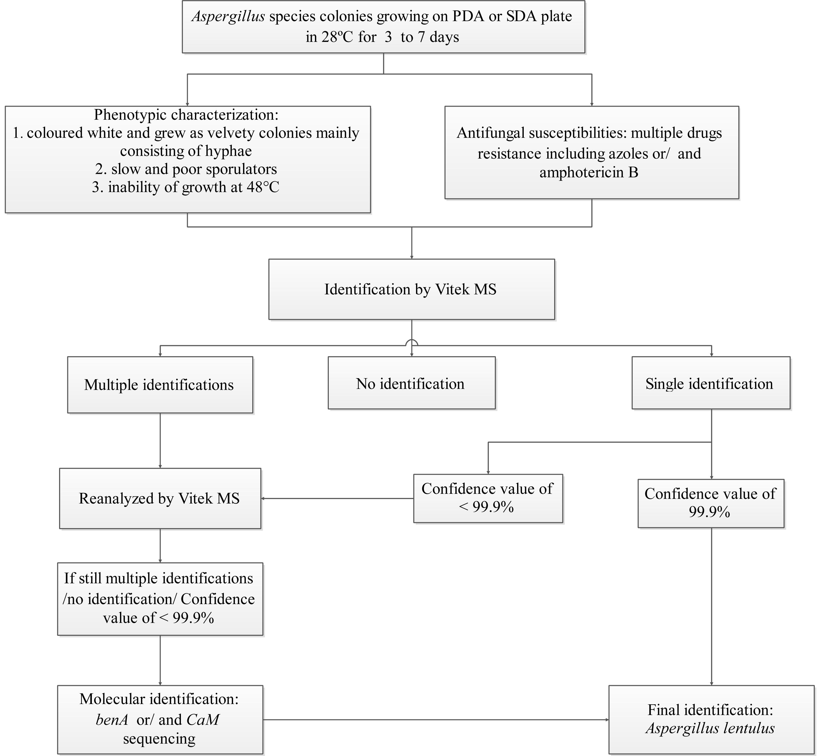
Based on our findings, we designed an identification algorithm for clinical laboratories to identify A. lentulus species with high accuracy (Figure 3). The trigger to adopt this algorithm is when colonies are suspected for A. lentulus, i.e., colonies are colored white, are of a velvety texture, mainly consist of hyphae, and grow slowly with poor sporulation. Further, inability to grow at 48°C and exhibiting multidrug resistance to azoles or/and amphotericin B increase the likelihood of A. lentulus. MALDI-TOF MS using the Vitek MS system (bioMérieux) and molecular identification are needed for definitive identification (see Figure 3 for conditions required for MALDI TOF MS identification). Ben A or/and CaM sequencing are recommended as the preferred gene loci for molecular diagnostics. It needs to be pointed out that the molecular method enables final identification for A. lentulus, regardless of MALDI TOF MS results.

Figure 3. An identification algorithm for A. lentulus species based on the Vitek MS and selective molecular identification. Abbreviation: PDA, Potato Dextrose Agar; SDA, Sabouraud Dextrose Agar.
Similar to A. fumigatus, A. lentulus showed high resistance to fluconazole, with MIC over 256 μg/ml. Moreover, in vitro susceptibility results showed that all A. lentulus isolates were resistant to amphotericin B and azole drugs yet remain susceptible to echinocandins. The MIC values of the azoles (mainly voriconazole and itraconazole) and amphotericin B in our study were higher than those in previous studies (Balajee et al., 2004, 2009; Montenegro et al., 2009; Symoens et al., 2010; Zbinden et al., 2012; Bastos et al., 2015; Tamiya et al., 2015; Won et al., 2018), while the MEC values of echinocandins were either similar to Kidd et al. (2015), Yoshida et al. (2015), and Won et al. (2018), or lower than those previously reported (Balajee et al., 2004; Alhambra et al., 2008; Montenegro et al., 2009; Symoens et al., 2010). The 2016 Infectious Diseases Society of America (IDSA) guidelines establish voriconazole as the primary antifungal choice for IA treatment; voriconazole combined with an echinocandin may be considered in documented IA patients (Patterson et al., 2016). However, A. lentulus has generally proven to possess evaluated MIC values to voriconazole and other azoles (Balajee et al., 2004, 2009; Montenegro et al., 2009; Symoens et al., 2010; Zbinden et al., 2012; Bastos et al., 2015; Tamiya et al., 2015; Won et al., 2018). More importantly, poor clinical outcome has been associated with azole therapy administered to patients with IPA caused by A. lentulus (Yoshida et al., 2015; Tetsuka et al., 2017). Tetsuka et al. (2017) chose micafungin to cure A. lentulus infection after no clinical response was observed with voriconazole use. Of note, even though all A. lentulus isolates in our study were susceptible to echinocandins in vitro, two patients died after receiving caspofungin treatment. The mechanisms of azole resistance in A. lentulus are Cyp51A dependent but are different from what has been described previously for A. fumigatus (Mellado et al., 2011). Molecular dynamics modeling revealed that some critical differences were observed in the putative closed form adopted by the proteins upon voriconazole binding in Cyp51A. Some major differences in the protein’s BC loop could differentially affect the lockup of voriconazole, which in turn could correlate with A. lentulus differences in azole susceptibility (Alcazar-Fuoli et al., 2011).
In summary, we evaluated the clinical, microbiological, and molecular features of invasive A. lentulus infections in Chinese patients. In particular, we highlight the importance of accurate identification with susceptibility testing of Aspergillus species for appropriate therapy. Molecular methods are needed for accurate speciation and further characterization of this fungal species.
The datasets generated for this study are available on request to the corresponding author.
The study was approved by the Human Research Ethics Committee of Peking Union Medical College Hospital (No. S-263). Written informed consent was obtained from all patients in the study, and for permission to study the isolates cultured from them for scientific research. Written, informed consent was obtained from the individual(s) and next of kin for the publication of any potentially identifiable images or data included in this article.
S-YY, L-NG, MX, and Y-CX conceived and designed the experiments. S-YY, YY, YW, LZ, T-SS, Y-TN, P-YJ, and WK performed the experiments. S-YY, M-LZ, and YZ performed the data analysis and wrote the manuscript. FK, YZ, and SC revised the manuscript critically for important intellectual content. All authors participated in the critical review of this manuscript.
This work was supported by the National Nature Science Foundation of China (81802049 and 81801989); the National Major Science and Technology Project for the Control and Prevention of Major Infectious Diseases of China (2017ZX10103004-003 and 2018ZX10712001); the Fundamental Research Funds for the Central Universities (3332018035, 3332018041, and 3332018024); and Innovation Fund of Peking Union Medical College (No. 2018-1002-01-02).
The authors declare that the research was conducted in the absence of any commercial or financial relationships that could be construed as a potential conflict of interest.
The reviewer MH declared past co-authorship with one of the authors SC to the handling editor.
We thank all the laboratories who participated in the CHIF-NET program in 2016–2017.
Alastruey-Izquierdo, A., Alcazar-Fuoli, L., and Cuenca-Estrella, M. (2014). Antifungal susceptibility profile of cryptic species of Aspergillus. Mycopathologia 178, 427–433. doi: 10.1007/s11046-014-9775-z
Alcazar-Fuoli, L., Cuesta, I., Rodriguez-Tudela, J. L., Cuenca-Estrella, M., Sanglard, D., and Mellado, E. (2011). Three-dimensional models of 14alpha-sterol demethylase (Cyp51A) from Aspergillus lentulus and Aspergillus fumigatus: an insight into differences in voriconazole interaction. Int. J. Antimicrob. Agents 38, 426–434. doi: 10.1016/j.ijantimicag.2011.06.005
Alcazar-Fuoli, L., Mellado, E., Alastruey-Izquierdo, A., Cuenca-Estrella, M., and Rodriguez-Tudela, J. L. (2008). Aspergillus section Fumigati: antifungal susceptibility patterns and sequence-based identification. Antimicrob. Agents Chemother. 52, 1244–1251. doi: 10.1128/AAC.00942-07
Alhambra, A., Catalan, M., Moragues, M. D., Brena, S., Ponton, J., Montejo, J. C., et al. (2008). Isolation of Aspergillus lentulus in Spain from a critically ill patient with chronic obstructive pulmonary disease. Rev. Iberoam. Micol. 25, 246–249. doi: 10.1016/s1130-1406(08)70058-5
Araujo, R., Amorim, A., and Gusmao, L. (2012). Diversity and specificity of microsatellites within Aspergillus section fumigati. BMC Microbiol. 12:154. doi: 10.1186/1471-2180-12-154
Araujo, R., Pina-Vaz, C., Rodrigues, A. G., Amorim, A., and Gusmao, L. (2009). Simple and highly discriminatory microsatellite-based multiplex PCR for Aspergillus fumigatus strain typing. Clin. Microbiol. Infect. 15, 260–266. doi: 10.1111/j.1469-0691.2008.02661.x
Balajee, S. A., Gribskov, J., Brandt, M., Ito, J., Fothergill, A., and Marr, K. A. (2005a). Mistaken identity: neosartorya pseudofischeri and its anamorph masquerading as Aspergillus fumigatus. J. Clin. Microbiol. 43, 5996–5999. doi: 10.1128/JCM.43.12.5996-5999.2005
Balajee, S. A., Gribskov, J. L., Hanley, E., Nickle, D., and Marr, K. A. (2005b). Aspergillus lentulus sp. nov., a new sibling species of A. fumigatus. Eukaryot. Cell. 4, 625–632. doi: 10.1128/EC.4.3.625-632.2005
Balajee, S. A., Kano, R., Baddley, J. W., Moser, S. A., Marr, K. A., Alexander, B. D., et al. (2009). Molecular identification of Aspergillus species collected for the transplant-associated infection surveillance network. J. Clin. Microbiol. 47, 3138–3141. doi: 10.1128/JCM.01070-1079
Balajee, S. A., Weaver, M., Imhof, A., Gribskov, J., and Marr, K. A. (2004). Aspergillus fumigatus variant with decreased susceptibility to multiple antifungals. Antimicrob. Agents Chemother. 48, 1197–1203. doi: 10.1128/aac.48.4.1197-1203.2004
Bastos, V. R., Santos, D. W., Padovan, A. C., Melo, A. S., Mazzolin Mde, A., Camargo, L. F., et al. (2015). Early invasive pulmonary aspergillosis in a kidney transplant recipient caused by Aspergillus lentulus: first Brazilian report. Mycopathologia 179, 299–305. doi: 10.1007/s11046-014-9840-7
Clinical and Laboratory Standards Institute [CLSI] (2017). Reference Method for Broth Dilution Antifungal Susceptibility Testing of Filamentous Fungi, 3rd ed. CLSI standard M38. Wayne, PA: Clinical and Laboratory Standards Institute.
Clinical and Laboratory Standards Institute [CLSI] (2018). Epidemiological Cutoff Values for Antifungal Susceptibility Testing. 2nd ed. CLSI supplement M59. Wayne, PA: Clinical and Laboratory Standards Institute.
Cornely, O. A., Hoenigl, M., Lass-Florl, C., Chen, S. C., Kontoyiannis, D. P., Morrissey, C. O., et al. (2019). Defining breakthrough invasive fungal infection-Position paper of the mycoses study group education and research consortium and the European Confederation of Medical Mycology. Mycoses 62, 716–729. doi: 10.1111/myc.12960
Datta, K., Rhee, P., Byrnes, E. III, Garcia-Effron, G., Perlin, D. S., Staab, J. F., et al. (2013). Isavuconazole activity against Aspergillus lentulus, Neosartorya udagawae, and Cryptococcus gattii, emerging fungal pathogens with reduced azole susceptibility. J. Clin. Microbiol. 51, 3090–3093. doi: 10.1128/JCM.01190-13
De Pauw, B., Walsh, T. J., Donnelly, J. P., Stevens, D. A., Edwards, J. E., Calandra, T., et al. (2008). Revised definitions of invasive fungal disease from the European Organization for research and treatment of cancer/invasive fungal infections cooperative group and the national institute of allergy and infectious diseases mycoses Study Group (EORTC/MSG) consensus group. Clin. Infect. Dis. 46, 1813–1821. doi: 10.1086/588660
de Valk, H. A., Meis, J. F., de Pauw, B. E., Donnelly, P. J., and Klaassen, C. H. (2007). Comparison of two highly discriminatory molecular fingerprinting assays for analysis of multiple Aspergillus fumigatus isolates from patients with invasive aspergillosis. J. Clin. Microbiol. 45, 1415–1419. doi: 10.1128/JCM.02423-06
Escribano, P., Pelaez, T., Munoz, P., Bouza, E., and Guinea, J. (2013). Is azole resistance in Aspergillus fumigatus a problem in Spain? Antimicrob. Agents Chemother. 57, 2815–2820. doi: 10.1128/AAC.02487-12
Gurcan, S., Tikvesli, M., Ustundag, S., and Ener, B. (2013). A case report on Aspergillus lentulus Pneumonia. Balkan Med. J. 30, 429–431. doi: 10.5152/balkanmedj.2013.8572
James, V., Karen, C., Guido, F., Jorgensen, J. H., Landry, M. L., Warnock, D. W., et al. (2011). Manual of Clinical Microbiology, 10th Edn. Washington, DC: ASM Press.
Kidd, S. E., Goeman, E., Meis, J. F., Slavin, M. A., and Verweij, P. E. (2015). Multi-triazole-resistant Aspergillus fumigatus infections in Australia. Mycoses 58, 350–355. doi: 10.1111/myc.12324
Klaassen, C. H. (2009). MLST versus microsatellites for typing Aspergillus fumigatus isolates. Med. Mycol. 47(Suppl. 1), S27–S33. doi: 10.1080/13693780802382244
Lago, M., Aguiar, A., Natario, A., Fernandes, C., Faria, M., and Pinto, E. (2014). Does fungicide application in vineyards induce resistance to medical azoles in Aspergillus species? Environ. Monit. Assess. 186, 5581–5593. doi: 10.1007/s10661-014-3804-8
Lamoth, F. (2016). Aspergillus fumigatus-related species in clinical practice. Front. Microbiol. 7:683. doi: 10.3389/fmicb.2016.00683
Larone, D. H. (2005). Medically Important Fungi: A Guide to Identification, 3 Edn. Washington DC: American Society for Microbiology.
Li, Y., Wang, H., Zhao, Y. P., Xu, Y. C., and Hsueh, P. R. (2017). Evaluation of the bruker biotyper matrix-assisted laser desorption/ionization time-of-flight mass spectrometry system for identification of aspergillus species directly from growth on solid agar media. Front. Microbiol. 8:1209. doi: 10.3389/fmicb.2017.01209
Mellado, E., Alcazar-Fuoli, L., Cuenca-Estrella, M., and Rodriguez-Tudela, J. L. (2011). Role of Aspergillus lentulus 14-alpha sterol demethylase (Cyp51A) in azole drug susceptibility. Antimicrob. Agents Chemother. 55, 5459–5468. doi: 10.1128/AAC.05178-11
Montenegro, G., Sanchez Puch, S., Jewtuchowicz, V. M., Pinoni, M. V., Relloso, S., Temporitti, E., et al. (2009). Phenotypic and genotypic characterization of Aspergillus lentulus and Aspergillus fumigatus isolates in a patient with probable invasive aspergillosis. J. Med. Microbiol. 58(Pt 3), 391–395. doi: 10.1099/jmm.0.005942-0
Mortensen, K. L., Johansen, H. K., Fuursted, K., Knudsen, J. D., Gahrn-Hansen, B., Jensen, R. H., et al. (2011). A prospective survey of Aspergillus spp. in respiratory tract samples: prevalence, clinical impact and antifungal susceptibility. Eur. J. Clin. Microbiol. Infect. Dis. 30, 1355–1363. doi: 10.1007/s10096-011-1229-7
Patterson, T. F., Thompson, G. R. III, Denning, D. W., Fishman, J. A., Hadley, S., Herbrecht, R., et al. (2016). Practice guidelines for the diagnosis and management of aspergillosis: 2016 update by the infectious diseases society of America. Clin. Infect. Dis. 63, e1–e60. doi: 10.1093/cid/ciw326
Sugui, J. A., Peterson, S. W., Figat, A., Hansen, B., Samson, R. A., Mellado, E., et al. (2014). Genetic relatedness versus biological compatibility between Aspergillus fumigatus and related species. J. Clin. Microbiol. 52, 3707–3721. doi: 10.1128/JCM.01704-14
Symoens, F., Haase, G., Pihet, M., Carrere, J., Beguin, H., Degand, N., et al. (2010). Unusual Aspergillus species in patients with cystic fibrosis. Med. Mycol. 48(Suppl. 1), S10–S16. doi: 10.3109/13693786.2010.501345
Tamiya, H., Ochiai, E., Kikuchi, K., Yahiro, M., Toyotome, T., Watanabe, A., et al. (2015). Secondary metabolite profiles and antifungal drug susceptibility of Aspergillus fumigatus and closely related species, Aspergillus lentulus, Aspergillus udagawae, and Aspergillus viridinutans. J. Infect. Chemother. 21, 385–391. doi: 10.1016/j.jiac.2015.01.005
Tetsuka, N., Yaguchi, T., Machida, H., Ito, S., and Miyairi, I. (2017). Invasive pulmonary aspergillosis due to azole-resistant Aspergillus lentulus. Pediatr. Int. 59, 362–363. doi: 10.1111/ped.13193
Thierry, S., Wang, D., Arne, P., Deville, M., De Bruin, B., Nieguitsila, A., et al. (2010). Multiple-locus variable-number tandem repeat analysis for molecular typing of Aspergillus fumigatus. BMC Microbiol. 10:315. doi: 10.1186/1471-2180-10-315
Vanhee, L. M., Symoens, F., Jacobsen, M. D., Nelis, H. J., and Coenye, T. (2009). Comparison of multiple typing methods for Aspergillus fumigatus. Clin. Microbiol. Infect. 15, 643–650. doi: 10.1111/j.1469-0691.2009.02844.x
Verwer, P. E., van Leeuwen, W. B., Girard, V., Monnin, V., van Belkum, A., Staab, J. F., et al. (2014). Discrimination of Aspergillus lentulus from Aspergillus fumigatus by raman spectroscopy and MALDI-TOF MS. Eur. J. Clin. Microbiol. Infect. Dis. 33, 245–251. doi: 10.1007/s10096-013-1951-4
Wang, H., Fan, Y. Y., Kudinha, T., Xu, Z. P., Xiao, M., Zhang, L., et al. (2016). A comprehensive evaluation of the bruker biotyper MS and Vitek MS matrix-assisted laser desorption ionization-time of flight mass spectrometry systems for identification of yeasts, part of the national china hospital invasive fungal surveillance net (CHIF-NET) Study, 2012 to 2013. J. Clin. Microbiol. 54, 1376–1380. doi: 10.1128/JCM.00162-16
Wang, H., Xiao, M., Chen, S. C., Kong, F., Sun, Z. Y., Liao, K., et al. (2012). In vitro susceptibilities of yeast species to fluconazole and voriconazole as determined by the 2010 National China Hospital Invasive Fungal Surveillance Net (CHIF-NET) study. J. Clin. Microbiol. 50, 3952–3959. doi: 10.1128/JCM.01130-12
Won, E. J., Shin, J. H., Kim, S. H., Choi, M. J., Byun, S. A., Kim, M. N., et al. (2018). Antifungal susceptibilities to amphotericin B, triazoles and echinocandins of 77 clinical isolates of cryptic Aspergillus species in multicenter surveillance in Korea. Med. Mycol. 56, 501–505. doi: 10.1093/mmy/myx067
Yagi, K., Ushikubo, M., Maeshima, A., Konishi, M., Fujimoto, K., Tsukamoto, M., et al. (2019). Invasive pulmonary aspergillosis due to Aspergillus lentulus in an adult patient: a case report and literature review. J. Infect. Chemother. 25, 547–551. doi: 10.1016/j.jiac.2019.02.003
Yoshida, H., Seki, M., Umeyama, T., Urai, M., Kinjo, Y., Nishi, I., et al. (2015). Invasive pulmonary aspergillosis due to Aspergillus lentulus: successful treatment of a liver transplant patient. J. Infect. Chemother. 21, 479–481. doi: 10.1016/j.jiac.2015.02.010
Zbinden, A., Imhof, A., Wilhelm, M. J., Ruschitzka, F., Wild, P., Bloemberg, G. V., et al. (2012). Fatal outcome after heart transplantation caused by Aspergillus lentulus. Transpl. Infect. Dis. 14, E60–E63. doi: 10.1111/j.1399-3062.2012.00779.x
Zhou, M., Yang, Q., Kudinha, T., Zhang, L., Xiao, M., Kong, F., et al. (2016). Using matrix-assisted laser desorption ionization-time of flight (MALDI-TOF) complemented with selected 16S rRNA and gyrB genes sequencing to practically identify clinical important viridans Group Streptococci (VGS). Front. Microbiol. 7:1328. doi: 10.3389/fmicb.2016.01328
Keywords: Aspergillus lentulus, accurate identification, multi-drug resistance, fatal infections, China
Citation: Yu S-Y, Guo L-N, Xiao M, Zhou M-L, Yuan Y, Wang Y, Zhang L, Sun T-S, Ning Y-T, Jia P-Y, Kang W, Kong F, Chen SC-A, Zhao Y and Xu Y-C (2020) Clinical and Microbiological Characterization of Invasive Pulmonary Aspergillosis Caused by Aspergillus lentulus in China. Front. Microbiol. 11:1672. doi: 10.3389/fmicb.2020.01672
Received: 10 February 2020; Accepted: 25 June 2020;
Published: 28 July 2020.
Edited by:
Juergen Prattes, Medical University of Graz, AustriaReviewed by:
Joerg Steinmann, Paracelsus Medical Private University, Nuremberg, GermanyCopyright © 2020 Yu, Guo, Xiao, Zhou, Yuan, Wang, Zhang, Sun, Ning, Jia, Kang, Kong, Chen, Zhao and Xu. This is an open-access article distributed under the terms of the Creative Commons Attribution License (CC BY). The use, distribution or reproduction in other forums is permitted, provided the original author(s) and the copyright owner(s) are credited and that the original publication in this journal is cited, in accordance with accepted academic practice. No use, distribution or reproduction is permitted which does not comply with these terms.
*Correspondence: Li-Na Guo, Z3VvMDIwMTIwNUAxMjYuY29t; Ying-Chun Xu, eHljcHVtY2hAMTM5LmNvbQ==
Disclaimer: All claims expressed in this article are solely those of the authors and do not necessarily represent those of their affiliated organizations, or those of the publisher, the editors and the reviewers. Any product that may be evaluated in this article or claim that may be made by its manufacturer is not guaranteed or endorsed by the publisher.
Research integrity at Frontiers

Learn more about the work of our research integrity team to safeguard the quality of each article we publish.