 Cedric Blötz
 Neil Singh
 Roger Dumke
 Jörg Stülke
- 1Department of General Microbiology, Göttingen Center for Molecular Biosciences, University of Göttingen, Göttingen, Germany
- 2Medical Faculty Carl Gustav Carus, Institute of Medical Microbiology and Hygiene, Technical University Dresden, Dresden, Germany
Bacteria evolved many ways to invade, colonize and survive in the host tissue. Such complex infection strategies of other bacteria are not present in the cell-wall less Mycoplasmas. Due to their strongly reduced genomes, these bacteria have only a minimal metabolism. Mycoplasma pneumoniae is a pathogenic bacterium using its virulence repertoire very efficiently, infecting the human lung. M. pneumoniae can cause a variety of conditions including fever, inflammation, atypical pneumoniae, and even death. Due to its strongly reduced metabolism, M. pneumoniae is dependent on nutrients from the host and aims to persist as long as possible, resulting in chronic diseases. Mycoplasmas evolved strategies to subvert the host immune system which involve proteins fending off immunoglobulins (Igs). In this study, we investigated the role of MPN400 as the putative factor responsible for Ig-binding and host immune evasion. MPN400 is a cell-surface localized protein which binds strongly to human IgG, IgA, and IgM. We therefore named the protein MPN400 immunoglobulin binding protein of Mycoplasma (IbpM). A strain devoid of IbpM is slightly compromised in cytotoxicity. Taken together, our study indicates that M. pneumoniae uses a refined mechanism for immune evasion.
Introduction
The cell-wall less bacteria of the genus Mycoplasma are commensal, opportunistic or pathogenic bacteria that colonize diverse hosts including plants, animals and humans (Parrott et al., 2016). Mycoplasmas belong to the group of Mollicutes, which is characterized by their strongly reduced genomes (Dandekar et al., 2000), accompanied by limited metabolic capabilities of these bacteria. The reduction in metabolic pathways is caused by adaptation to their hosts, resulting in the strong dependency on the acquisition of nutrients (Waites and Talkington, 2004; Halbedel et al., 2007). Due to their reduced genomes and their experimental amenability, Mycoplasma pneumoniae and Mycoplasma mycoides are model organisms for systems and synthetic biology, respectively. Furthermore, both are important pathogens in medical research due to their virulence in their respective hosts, man and cattle. Mycoplasma species can cause a broad range of symptoms in various hosts, e.g., fever, inflammation, autoimmune responses, or atypical pneumoniae (Citti and Blanchard, 2013), followed more often by a chronic disease state rather than killing the host (Atkinson et al., 2008). The strong dependency on the host’s survival is a good explanation for the occurrence of chronical diseases caused by Mycoplasma infections. Despite their reduced genomes and consequently the limited metabolism Mycoplasmas can infect their hosts efficiently.
Remarkably, only few candidate virulence factors of Mycoplasma are known or well described. For M. pneumoniae, only the community acquired respiratory distress syndrome (CARDS) toxin is described as a typical toxin representative (Kannan and Baseman, 2006; Kannan et al., 2010; Johnson et al., 2011; Becker et al., 2015). Hydrogen peroxide produced during glycerol utilization has been proposed to be the major virulence factor for M. pneumoniae and other Mycoplasmas (Hames et al., 2009; Blötz and Stülke, 2017). In addition, hydrogen sulfide was also identified to play a significant role in the cytotoxicity of M. pneumoniae (Großhennig et al., 2016). Moreover, in some Mycoplasma species, e.g., Mycoplasma fermentans, M. pneumoniae, M. genitalium, or M. mycoides, immunomodulatory proteins have been identified (Mühlradt et al., 1997; Into et al., 2004, 2007; Okusawa et al., 2004; Arfi et al., 2016; Campos et al., 2018).
In Gram-positive and Gram-negative bacteria, such as Yersinia spp., Listeria spp., Salmonella spp., or enterohemorrhagic Escherichia spp., many different mechanisms which influence the host immune response are well-described (Bhavsar et al., 2007). Overall, the cytadherence or attachment to host cells is a prerequisite for the growth of pathogenic bacteria and concomitant infection (Rottem, 2003; Catrein et al., 2004). The link between attachment and virulence is exemplified by non-adherent Mycoplasma mutants which are nearly non-pathogenic (Waldo and Krause, 2006; Chaudhry et al., 2007). M. pneumoniae and M. genitalium encode a very complex protein network, the attachment organelle, which is responsible for their gliding motility and attachment to human epithelial cells (Balish and Krause, 2006; Kenri et al., 2018; Krause et al., 2018; Seybert et al., 2018). Proteins encoding subunits of this so-called tip structure seem to be responsible for enhanced survival of M. genitalium by antigenic and phase variation, a strategy described for many bacteria (van der Woude and Bäumler, 2004; Burgos et al., 2018).
In several Gram-positive bacteria surface proteins are known to bind to human surface proteins. One important class of such binding proteins are immunoglobulin binding proteins (IBPs) (Boyle, 1990; Sidorin and Solov’eva, 2011). The IBPs can bind to different immunoglobulins (Igs) without the requirement of antigen-binding sites. This non-immune binding mechanism is thought to protect bacteria from the action of the complement system. This system is responsible for the phagocyte-independent immune defense in vertebrates, decreasing phagocytosis and finally promoting the evasion of the bacteria from the host’s immune system. IBPs are classified into functional groups according to their ability to bind to human or animal Igs. The most intensively studied IBPs are Protein A (Staphylococcus aureus), Protein G (group C Streptococci), and the M-protein (group A Streptococci) that bind to the Fc region (crystallizable fragment of Igs) (Fischetti, 1989; Sjöbring et al., 1991; Graille et al., 2000), and the Protein L (Finegoldia magna) that binds the light chain of Igs (Akerström and Björck, 1989; Graille et al., 2002). Interestingly, the M-protein not only binds to Igs, but it can also bind factor H in serum, thereby blocking the innate immune response (Horstmann et al., 1988).
Recently, IBPs were identified in M. genitalium (Protein M) and Mycoplasma mycoides subsp. capri (Mycoplasma immunoglobulin binding protein, MIB) (Grover et al., 2014; Arfi et al., 2016). Interestingly, the 50 kDa IBP from M. genitalium differs in its tertiary architecture from all available structures in the Protein Data Bank (PDB). Moreover, its structure is different compared to well-known IBPs (Grover et al., 2014). Protein M and homologs of IBPs from other Mycoplasmas, not to be confused with the M-protein from Streptococci, seem to have evolved convergently (Frick et al., 1992; Arfi et al., 2016). Regarding the reduced genomes of Mycoplasmas, the evolution of a putative immune evasion system is striking. Even more striking, M. mycoides possesses multiple paralogous pairs of proteins that bind Igs (MIP) and proteolytically cleave off the variable heavy chain (Mycoplasma immunoglobulin protease, MIP). Furthermore, in Ureaplasma urealyticum an immunoglobulin A (IgA) specific protease was identified and characterized (Robertson et al., 1984; Spooner et al., 1992). Similarly, an IgG protease was identified in Mycoplasma synoviae and Mycoplasma gallisepticum (Cizelj et al., 2011). However, the MIB-MIP system present in M. mycoides and protein M homologs as found in M. genitalium, seem to be conserved mutually exclusive. For M. pneumoniae, the presence of a protein M has been predicted (Arfi et al., 2016). The two types of Ig binding proteins do not appear to occur in one genome at the same time. Furthermore, the MIB-MIP system is present in Mycoplasmas infecting animals and humans, but not in plant pathogens (Arfi et al., 2016).
In this work, we addressed the localization and function of a putative IBP from M. pneumoniae, encoded by mpn400. Furthermore, we identified putative proteases and investigated their activity to cleave Igs. Our results demonstrate the surface localization of MPN400 that allows the interaction with external factors. Moreover, we produced recombinant MPN400 in E. coli and showed that purified rMPN400 can bind different Igs. A M. pneumoniae strain lacking MPN400 exhibits reduced cytotoxicity thus supporting the idea that MPN400 plays an important role for the virulence of M. pneumoniae.
Materials and Methods
Bacterial Strains, Transformation, and Growth Conditions
The M. pneumoniae strains used in this study were M. pneumoniae M129 (ATCC 29342) and its isogenic mutant derivative GPM113 (mpn400:Tn4001). M. pneumoniae was grown at 37°C in 175 cm2 tissue culture flasks containing 100 ml of modified Hayflick medium with glucose (1%, w/v) as described previously (Halbedel et al., 2004). Surface-attached mycoplasmas were washed four times with phosphate-buffered saline (PBS; pH 7.2). Strains harboring transposon insertions or plasmids were cultivated in the presence of 80 mg/ml gentamicin and/or 2.5 mg/ml tetracycline. Plasmids were introduced into M. pneumoniae by electroporation using Gene Pulser (Bio-Rad, Hercules, CA, United States) and the transformants were selected by incubation at 37°C on agar plates containing appropriate antibiotics (Halbedel et al., 2004). For molecular cloning, Escherichia coli strain XL1-Blue (Stratagene, San Diego, CA, United States) was grown at 37°C in lysogeny broth (LB) medium containing the appropriate antibiotics (100 mg/ml ampicillin, 5 mg/ml tetracycline).
Plasmid Construction
Plasmids for the overexpression and purification of MPN400, MPN400 lacking the C-terminus (A446Stop), and MPN641 (negative control) were constructed as follows. The gene of interest was amplified by PCR from M129 M. pneumoniae wild type genomic DNA using specifically designed oligonucleotides. The vectors pBSKII(-) (Stratagene) and pGP172 (Merzbacher et al., 2004) were digested with KpnI/BamHI for mpn400 as well as its truncated version and SacI/BamHI for mpn641, respectively. The gene mpn400 was ligated into pBSKII, resulting in pGP2743. This plasmid served as the template for the multiple mutation reaction (Hames et al., 2005) to replace TGA (opal stop codon in E. coli) by TGG (tryptophan) codons. The codon optimized mpn400 amplicon was used to obtain fragments encoding the protein lacking the trans-membrane domain and the C-terminally truncated protein. The products were cloned into pGP172 yielding pGP3215 and pGP3217, respectively. Similarly, the mpn641 gene was ligated into pGP172. The resulting plasmid was pGP3235. The plasmids were verified by determination of the DNA sequence of the inserts.
Isolation of Mutant Strains
For the isolation of M. pneumoniae mutants, we used an M. pneumoniae transposon library carrying insertions of Tn4001 (Halbedel et al., 2006). The presence of the desired mutant was assayed by a PCR screen using one oligonucleotide that hybridizes to the transposon (directed outward, SH30, 5′ CAATACGCAAACCGCCTC), and a second oligonucleotide specific for the gene of interest (CB37, 5′ GAGAAGAACACTATATCTTTAATAGGTG).
Southern Blot Analysis
Chromosomal DNA was isolated using the Bacterial DNA Kit (PEQLAB, Erlangen) according to the manufacturer’s instructions. For both strains, M129 and GPM113, cells were grown in T75-flasks and harvested for DNA isolation. 2 μg of each DNA sample was pre-incubated with RNase A (1 μl; 20 mg ml–1) and digested with 3 μl SacI for at least 5 h at 37°C. Digests were separated by agarose gel electrophoresis (1%), transferred onto positively charged nylon membranes (Roche, Munich) and probed with digoxigenin-labeled riboprobes as described earlier (Sambrook et al., 1989; Halbedel et al., 2006).
Expression and Purification of Recombinant Proteins
The Strep-tagged proteins were overexpressed in and purified from E. coli BL21(DE3) as described previously (Schmidl et al., 2011). Briefly, cells were cultivated in two-fold LB medium and the expression (1 l culture, 37°C, 200 rpm, baffled flasks, 3 h) was induced by the addition of 1 mM IPTG to exponentially growing cultures (OD600 of 0.6 to 0.8). The cells were pelleted and washed once with buffer W (100 mM Tris-HCl pH8.0, 150 mM NaCl, 1 mM EDTA). Cells lysis was carried out using a French press (18,000 p.s.i., 138,000 kPa, three passes, SLM Aminco, United States), subsequently followed by centrifugation of the crude extracts at 35,000 rpm for 30 min. Purification was done at RT using StrepTrap columns (2 × 5 ml, GE Healthcare). The crude extract was loaded onto the column (flow rate 0.5 ml min–1, max. 0.5 MPa). StrepTraps were washed with buffer W until the 280 nm absorbance reached the base line. We used 2.5 mM D-desthiobiotin for elution (5 ml fractions; flow rate 1–1.5 ml min–1, max. 0.5 MPa). Purification and purity of proteins (>98%) were checked with SDS-PAGE and Colloidal Coomassie staining. Proteins were dialyzed against buffer W using VivaSpin columns (MCO 5 kDa, Sartorius, Göttingen). The pure proteins were frozen in aliquots in liquid nitrogen and stored at −80°C.
Production of Guinea Pig Polyclonal Antibodies
Polyclonal antisera were produced in guinea pigs (Charles River). The animal experiments were approved by the Ethical Board of Landesdirektion Sachsen, Dresden, Germany (permit no. 24-9168.25-1/2011/1). Primary subcutaneous immunization of animals with recombinant proteins, booster immunizations and serum collection were performed as reported (Thomas et al., 2013).
Western Blotting
For Western blotting, M. pneumoniae GPM113 and the wild type were grown till 95% confluency. Cells were collected and once washed with PBS. Cells were lysed in a tissue lyser with 0.1 mm glass beads (2 × 2.5 min, 30 Hz, cooled block) followed by centrifugation for 10 min at 14,000 rpm and 4°C. From each sample 20 μg total protein was mixed with SDS-loading buffer, boiled for 10 min at 95°C and separated in 12% SDS-PAGE. After electrophoresis, the proteins were transferred to a polyvinylidene difluoride (PVDF) membrane (Bio-Rad) by electroblotting (80 mA/membrane, 1 h). Proteins were detected using antibodies recognizing MPN400 (1:250). The blots were developed with α-guinea pig IgG (Dako; 1:1 000) and visualized by using anti-guinea pig IgG-AP secondary antibodies and the CDP∗ detection system (Roche Diagnostics) as described previously (Diethmaier et al., 2011).
Localization of MPN400
Colony blotting was used to specify the localization of MPN400 as described earlier (Thomas et al., 2013). Briefly, diluted M. pneumoniae cells were grown for 10 days on PPLO agar plates and colonies were covered with a nitrocellulose membrane for 5 min. The membrane was dried for 10 min at room temperature, washed and blocked three times (for 10 min, 10% FCS in PBS/Tween). The reactivity of the blotted proteins was tested either with α-MPN400, α-Nox, and α-P14 (1:250 each). The NADH-oxidase (Nox; Pollack et al., 1997; Hagemann et al., 2017) and the C-terminal part of the main P1 adhesin (P14; Schurwanz et al., 2009) were used as cytosolic and surface-localized reference proteins, respectively.
As a further method, mild surface proteolysis of M. pneumoniae cells was carried out. Mycoplasma cells were grown as described above, harvested, and the protein concentration was adjusted to 200 μg ml–1. The cells were centrifuged for 5 min at 13,000 × g and subsequently incubated with 10 μg ml–1, 40 μg ml–1, and 100 μg ml–1 or without trypsin (Sigma) in PBS for 30 min at 37°C. The samples were centrifuged (13,000 × g, 10 min) and pellets resuspended in 100 μl protein sample buffer followed by 10 min boiling (95°C) and separation by SDS-PAGE. Subsequently, proteins were blotted onto nitrocellulose membranes by standard procedure. The blots were incubated with α-MPN400, α-Nox, and α-P14 (1:250 each). α-guinea pig IgG (Dako; 1:1,000) was used to detect the proteins.
Enzyme-Linked Immunosorbent Assay (ELISA)
The binding of immunoglobulins and other human serum proteins by MPN400 was quantified in ELISA experiments. Recombinant MPN641 was used as negative control. MPN641 is a lipoprotein with unknown function and unlikely to bind immunoglobulins. Furthermore, we used human serum albumin and buffer as additional negative controls. MPN400 and a truncated version lacking the predicted C-terminal domain (A446Stop) were used to address Ig binding. Frozen aliquots were thawed and diluted (2, 3, 4, 5, 10, 50 μg ml–1) and subsequently coated onto 96-well plates. The plates were incubated overnight at 4°C and mild shaking (50 rpm). Afterward, wells were washed (buffer + 0.05% Tween20) and blocked (1% skim milk, 1 h, 50 rpm, 4°C) three times, followed by a last washing step. The antibodies [IgA/IgM/IgG (Sigma-Aldrich, Germany); 0.2 ng ml–1] were added to the wells with immobilized recombinant proteins and incubated overnight at 4°C (50 rpm). The wells were washed three times before detection. For detection we used an antibody with affinity to human Igs conjugated to horseradish peroxidase (goat α-Ig HRP detecting IgG, IgM, IgA; Thermo Fisher 0.5 ng ml–1). For detection we incubated wells with 100 μl of α-Ig HRP for 1 h at 4°C (50 rpm). After washing three times, ABTS solution (1-Step ABTS, Thermo Fisher) was added and incubated (20 min, dark, RT). Absorbance detection of bound antibodies was indicated by green color formation, measured in a plate reader (395–415 nm, λmax = 405 nm).
Pulldown of MPN400 Binding Proteins From Human Serum
To isolate MPN400-binding proteins from human serum (Thermo Scientific; H4522), we cultivated E. coli strains harboring either pGP3215 to express Strep-MPN400 or the empty vector pGP172. Protein extracts were prepared as described above. StrepTactin columns (CV = 250 μl) were saturated with MPN400 (four columns) or the empty vector crude extract (one column). After extensive washing (10 times 2 ml buffer W), human serum was applied to all columns. For the empty vector column and one with bound MPN400 we used 5 μg of human serum. The remaining MPN400 columns were incubated with 3, 2, and 1 μg of human serum each. After 10 washing steps we eluted the bound proteins four times with buffer W containing D-desthiobiotin (2.5 mM). From elution fractions we used 20 μl for SDS-PAGE analysis, followed by silver staining. Significant bands were further processed by LC-MS/MS analysis.
Protein Digestion With Trypsin and Protein Identification With LC-MS/MS
Trypsin digestion of proteins was performed as described earlier (Shevchenko et al., 1996). Briefly, the Stage TipStageTip method was used to purify peptides (Rappsilber et al., 2007), which were subsequently separated by reversed-phase liquid chromatography. For analysis an RSLCnano Ultimate 3000 system (Thermo Scientific) followed by mass analysis with an Orbitrap Velos ProHybrid mass spectrometer (Thermo Scientific) was applied as described in more detail elsewhere (Lin et al., 2015; Schmitt et al., 2017). MS/MS2 data processing for peptide analysis and protein identification was performed with the Proteome Discoverer 1.4 software (Thermo Scientific) and the Mascot and SequestHT search algorithms. Proteins identified in empty vector control (pGP172 with human serum) were regarded as unspecific binding and excluded.
HeLa Cell Cytotoxicity Assay
Infection of HeLa cultures with M. pneumoniae cells was done as described previously (Hames et al., 2009; Schmidl et al., 2010). DMEM medium complemented with 10% FBS was used to grow cells. After 4 days post infection, HeLa cultures were stained with crystal violet (10 min fixation in 10% formalin; 150 μl 0.1% crystal violet solution for 30 min at RT; three times washing) and photographed. For quantification the surviving cells, disruption was carried out with 0.5% SDS solution. The OD595 served as indication for cell survival and therefore the cytotoxicity of M. pneumoniae.
Results
Homology Comparison of Potential Ig-Binding Proteins
In order to identify possible Ig-binding proteins in M. pneumoniae, we searched for homologs of the known mollicute Ig-binding proteins, protein M from M. genitalium and MIB from M. mycoides in M. pneumoniae. For this purpose, we used BLASTp search with standard settings. We identified MPN400, a protein with 55% amino acid identity to protein M (MG281), whereas no protein with similarity to MIB was identified. The alignment of potential Ig-binding proteins in other Mycoplasma species revealed only very decent sequence conservation (Mycoplasma iowae 19.7%, Mycoplasma imitans 19.9%, Mycoplasma gallisepticum 21%). For further analysis, structural modeling was used to analyze the structure of MPN400 in comparison to the crystal structure of Protein M of M. genitalium (PDB: 4NZR, Grover et al., 2014). The calculated structural alignment was visualized using PYMOL (Omasits et al., 2014). As shown in Figure 1A, the calculated structure seems to be highly similar comparing both IBPs. The sequence and structural comparison revealed a very similar domain architecture as described for Protein M (Figure 1B). The analysis predicts that MPN400 is a membrane protein which is embedded into the membrane via a single trans-membrane helix (amino acids 21–40), and that the main part of the protein is exposed to the cell surface.
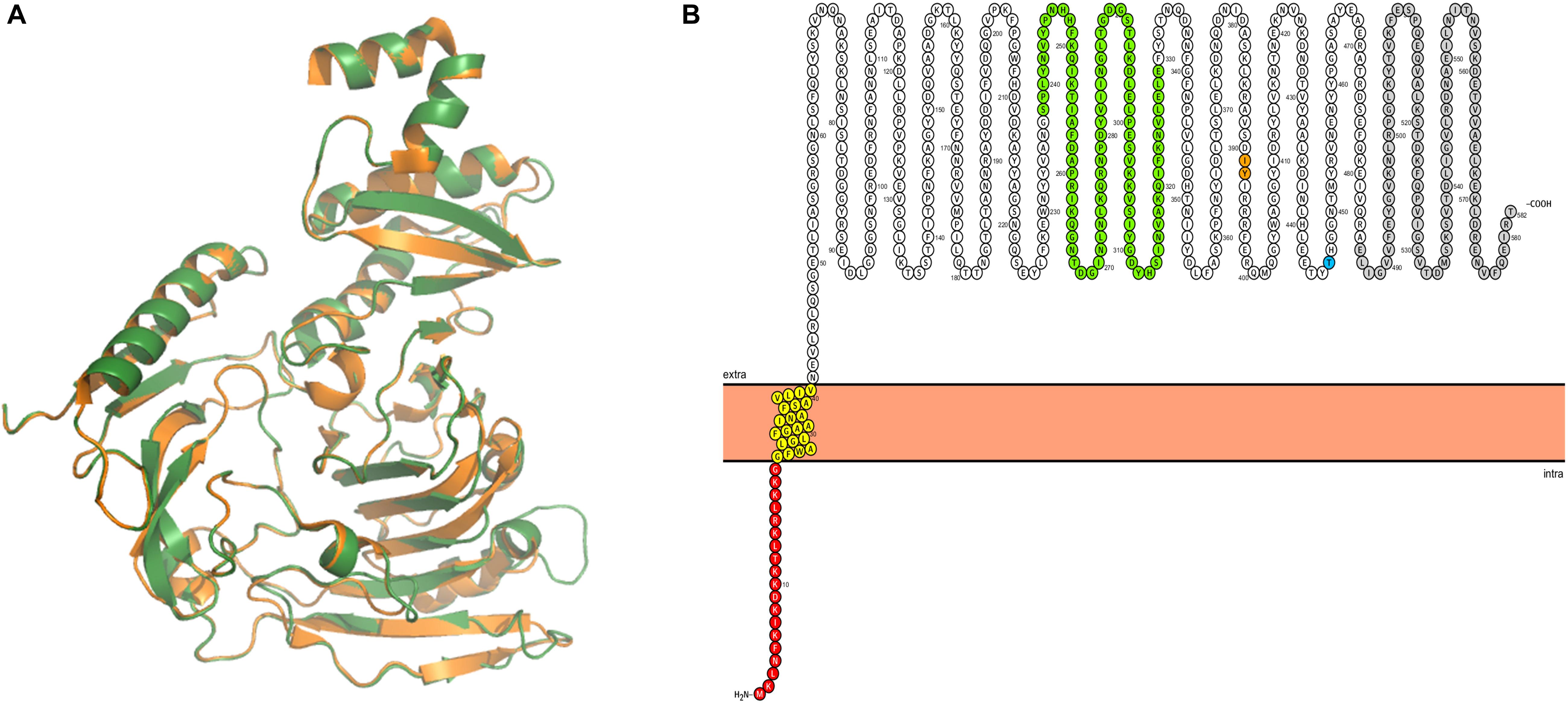
Figure 1. Structural analysis of IbpM (MPN400). (A) Structural alignment of the predicted tertiary structure of IbpM from M. pneumoniae (green) to the resolved crystal structure of the antibody-binding region of Protein M (PDB: 4NZR, Grover et al., 2014) from M. genitalium (orange). The figure was created by PyMOL. (B) The ProtterBlot shows the domain architecture of IbpM (using UniProt accession number P75383) anchored in the plasma membrane. Amino acids in red indicate the signal peptide and yellow the predicted transmembrane domain. Colored residues in green indicate a leucine rich region-like domain (LRR domain) and gray the C-terminal disordered domain. Thr insertion point for the mini-transposon is indicated in orange (aa 391–392) and the truncation point for the recombinant mutant in blue (aa 446).
Surface Localization of MPN400
The analysis described above as well as the described activity of homologs on the cell surface in other Mycoplasma species (Grover et al., 2014; Arfi et al., 2016) suggests that MPN400 is also localized to the cell surface. The surface localization was corroborated with colony blotting (Figure 2A) and by mild proteolysis (Figure 2B). In the colony blotting experiment strong signals could be detected for MPN400 and the surface localized protein P14 (positive control), whereas the cytosolic NADH oxidase Nox was not detected exposed to the surface. Further, upon addition of increasing amounts of trypsin, the corresponding bands for surface protein P14 and MPN400 were less prominent and fainter. From this observation, we conclude stronger degradation for these two proteins in contrast to Nox which showed identical bands despite increasing trypsin concentrations. Collectively, these results clearly demonstrate the surface localization of MPN400.
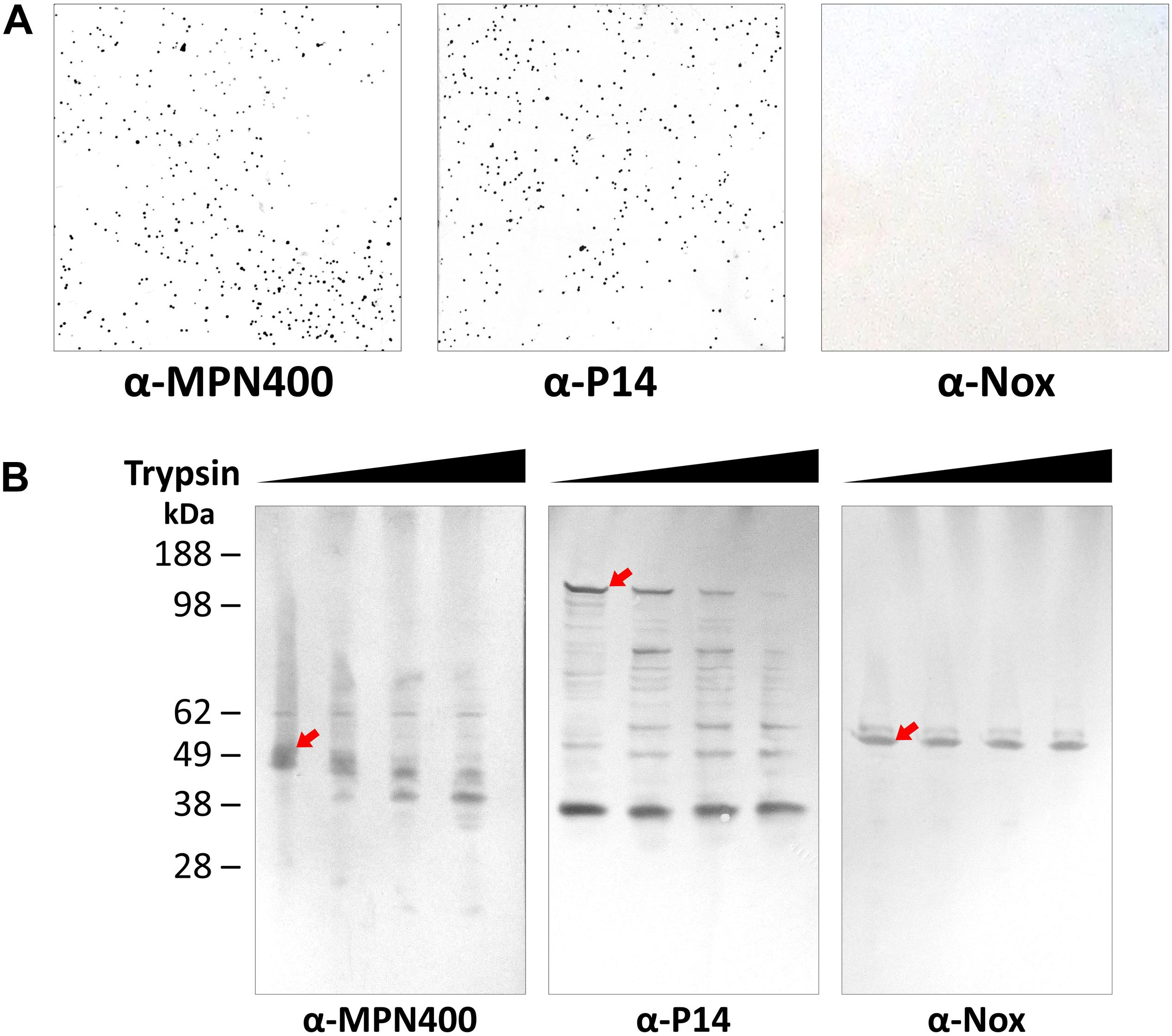
Figure 2. Localization of IbpM (MPN400) on the surface of M. pneumoniae cells. (A) Results of colony blot of freshly grown M. pneumoniae colonies. Blotted colonies were treated with guinea pig α-MPN400, α-P14 (positive control), and α-NADH oxidase (Nox, negative control), respectively. (B) Reactivity of SDS-PAGE-separated and blotted whole M. pneumoniae proteins after mild treatment with increasing concentrations of trypsin. Lane 1: 0 μg ml– 1, lane 2: 10 μg ml– 1, lane 3: 40 μg ml– 1 and lane 4: 100 μg ml– 1 trypsin. Western blots were incubated with guinea pig α-MPN400, α-Nox and α-P14, respectively. The bands corresponding to the non-degraded proteins of interest are highlighted by red arrows. Guinea pig antibodies were detected using rabbit α-guinea pig HRP conjugated antibody.
Isolation of an mpn400 Mutant
A recent analysis of the essential gene set of M. pneumoniae revealed that mpn400 is dispensable (Lluch-Senar et al., 2015). Therefore, we attempted to isolate an mpn400 mutant from an ordered transposon mutant library as described earlier (Halbedel et al., 2006). The pooled M. pneumoniae mutants were screened within a PCR reaction to detect the presence of the mini-transposon disrupting mpn400 (Figure 3A). The successful disruption of mpn400 and the single transposon integration were verified by Southern blot analysis (Figure 3B). As only one single band could be detected for the aac-aphD probe in the mutant (red arrow), we can conclude that the mutant carries a single copy of the transposon which disrupts the mpn400 gene. For the mpn400 specific probe a clear shift is visible compared with the wild type signal (indicated by red arrows, Figure 3B); and this shift corresponds to the increased size of the fragment due to transposon integration. The precise integration site was determined by DNA sequencing, showing that mpn400 is interrupted at its 1178th nucleotide, resulting in a C-terminal truncation of 191 amino acid (of total 582 amino acids). The isolated mutant was named GPM113 (mpn400:Tn4001).
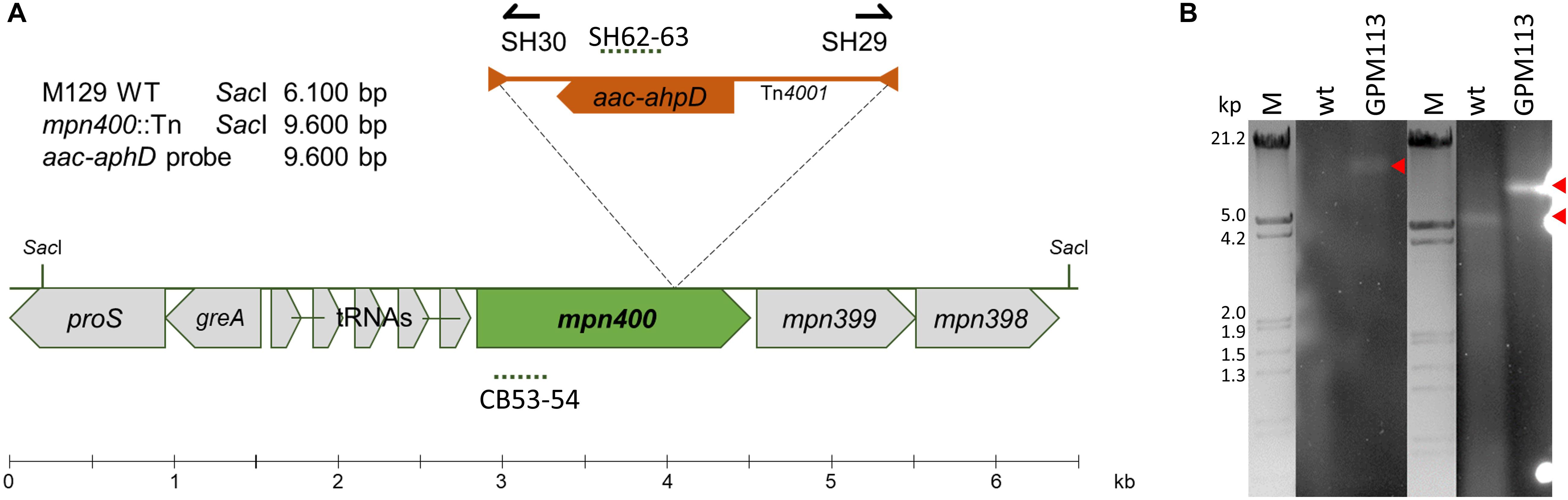
Figure 3. Isolation of a mpn400 transposon insertion mutant. (A) Schematic representation of the genomic region of mpn400 in M. pneumoniae and the transposon insertion site in the mpn400:Tn-4001 mutant GPM113. The location of the probes is indicated by dashed lines. (B) Southern blot analysis to confirm single transposon integration using chromosomal DNA of the wild type (wt) and strain GPM113 were digested using SacI. Detection was carried out with a probe specific for the aac-ahpD resistance cassette and a probe hybridizing to the gene mpn400 (right) which is upshifted after transposon integration. The relevant bands are highlighted by red arrows. λ-marker (HindIII/EcoRI) served as a size standard.
Characterization of the mpn400 Mutant
When the growth of the mutant strain GPM113 was compared with the wild type M129, no changes could be observed, irrespectively of the carbon source (glucose or glycerol). In agreement with the deletion of the mpn400 gene, the MPN400 protein was not detectable by colony blotting with GPM113 (data not shown). A Western blot using crude extracts of the wild type and the mutant strain GPM113 showed a faint band with reduced size (∼55 kDa) compared to the wild type MPN400 band at 65 kDa (data not shown). To test the role of MPN400 in virulence, we performed a HeLa cytotoxicity assay (see Figure 4A). This assay revealed a slight, but significant reduction of cytotoxicity of the mutant after 48 h but not 96 h post infection (see Figure 4B). After 48 h post infection wild type cells caused ∼50% cytotoxicity while GPM113 caused less than 35%. Thus, MPN400 plays only a minor role in the direct interaction of M. pneumoniae with the host cells.

Figure 4. Crystal violet stain of HeLa cells after mycoplasmal infection. Cytotoxicity of M. pneumoniae strains toward HeLa cell cultures. Confluent HeLa cell culture without infection or with M129 wild type cells and with GPM113 (mpn400:Tn) mutant cells. (A) After 20 h, 48 h, and 96 h post infection HeLa cells were stained with crystal violet and photographed. (B) OD595 measurements were plotted and statistical significance of the cytotoxicities in different strains at the two time points was calculated. For the original data, see Supplementary Data Sheet S1.
Pulldown of Human Serum Proteins Bound by MPN400
We hypothesized that the main function of MPN400 is an interaction with proteins of the human immune system, in order to allow immune evasion. In order to identify potential interaction partners in human serum in an unbiased manner, we performed a pull-down experiment with MPN400 as the bait. For this purpose, recombinant Strep-tagged MPN400 was immobilized on StrepTactin columns and incubated with different concentrations of human serum. The bound proteins were analyzed with silver stained SDS-PAGE (see Figure 5) and LC-MS/MS analysis. We analyzed proteins larger than 50 kDa for the empty vector control and for MPN400-bound proteins. Among the proteins copurified with MPN400 we identified 1,510 unique peptide-spectrum matches (PSMs) (see Supplementary Table S1 for a complete list). Among the 30 most abundant PSMs were the apolipoprotein B-100 and 17 peptides from immunoglobulins. Non-specific immunoglobulin heavy chains were the most abundant peptides, followed by anti-Pseudomonas aeruginosa IgG and anti-rabies virus S057 (Fab) IgG antibodies. Constant regions from the heavy chain were also found along with monoclonal antibodies. Taken together this directed us to test the ability of MPN400 to bind monoclonal and polyclonal antibodies.
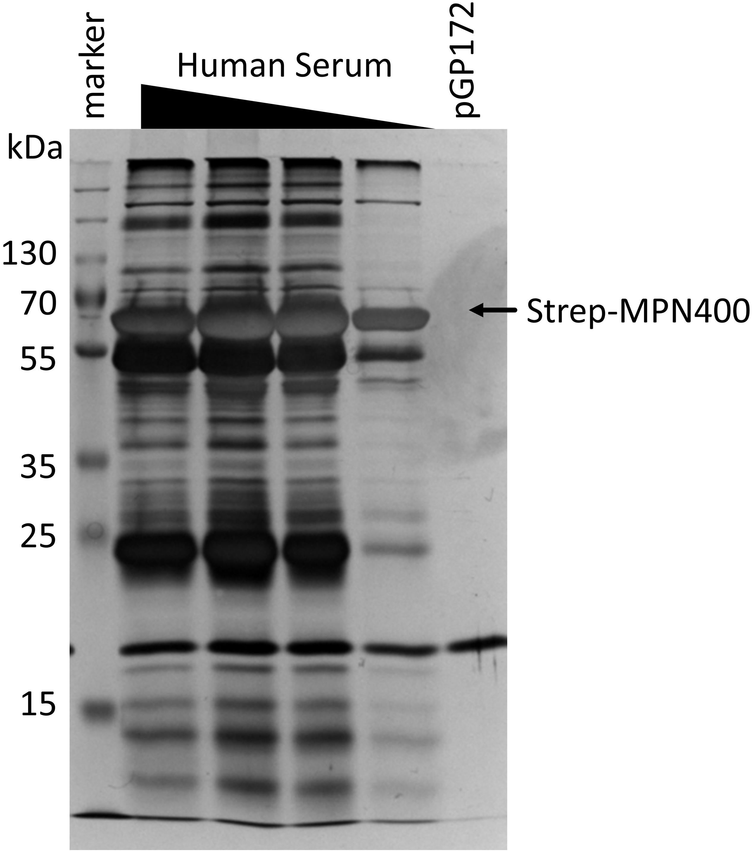
Figure 5. Detection of proteins bound by immobilized MPN400. The binding was performed with recombinant MPN400 (from E. coli BL21:pGP3215) immobilized to StrepTactin matrix, which was incubated with different concentrations of human serum (HuSe). Columns were washed extensively, and bound proteins eluted four times with D-desthiobiotin. We used the fractions from elution 3 to analyze the protein content by 12% SDS-PAGE stained with silver. Lane 1, pre-stained protein ladder plus (Thermo Fisher); lanes 2–5, rMPN400 incubated with 5, 3, 2, and 1 μg protein from HuSe, respectively; lane 6, empty vector crude extract from E. coli incubated with 5 μg HuSe.
Binding of Immunoglobulins to MPN400
To study the binding of Igs to MPN400 in more detail, we performed an enzyme linked immunosorbent assay (ELISA). Recombinantly expressed proteins (MPN400 and negative control lipoprotein MPN641) were coated onto 96-well plates. Coated wells were incubated with different Igs in various concentrations (see Figure 6A). The negative controls (human serum albumin coated or MPN641-coated as well as blank wells with only buffer) showed no binding to the Igs. For MPN400 coated wells we detected strong binding of the tested Igs (IgG, IgA, and IgM) (see Figure 6B for comparative binding across Igs tested, Supplementary Figure S1 for individual binding profiles of the Igs). Thus, MPN400 specifically binds human immunoglobulins.
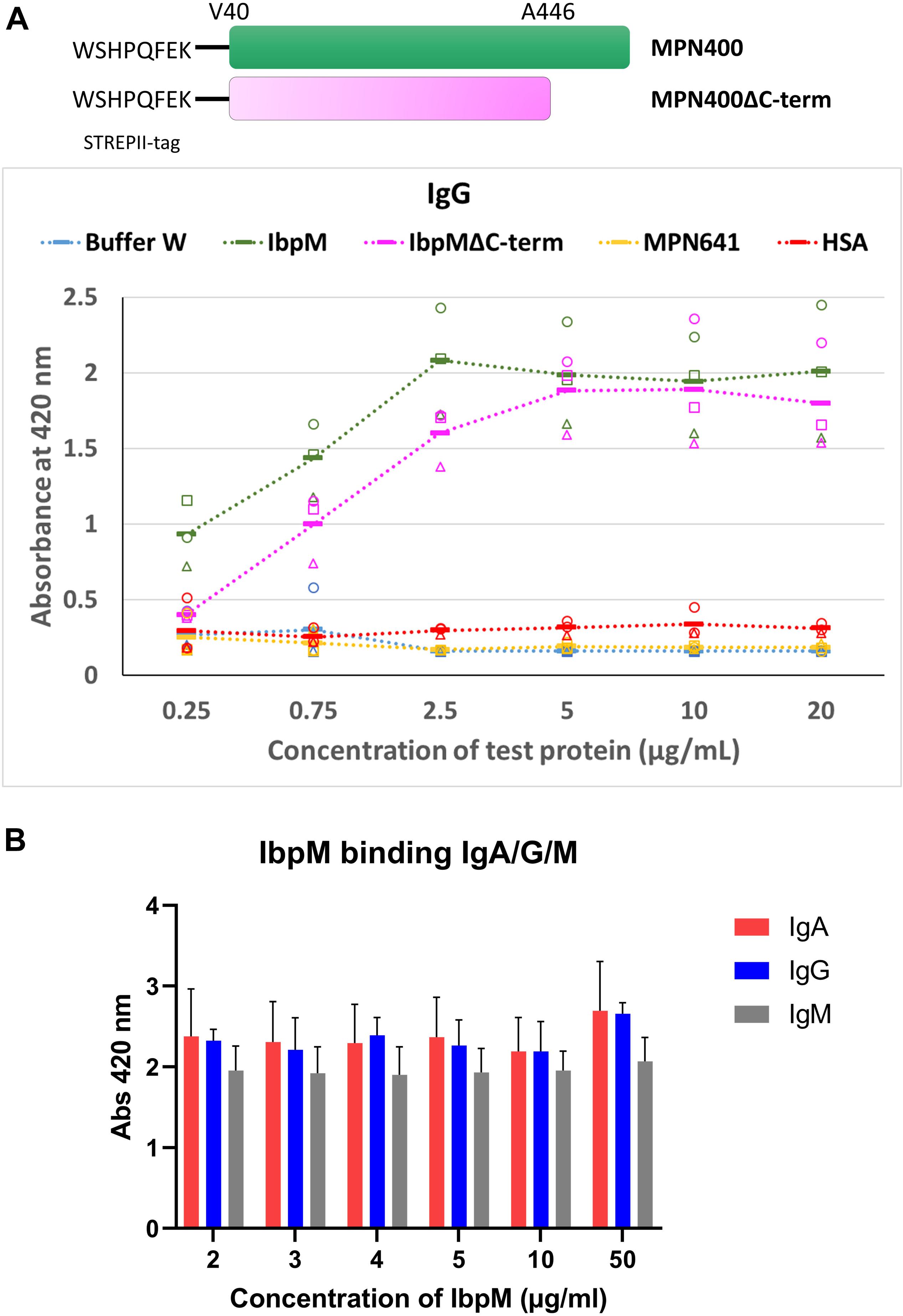
Figure 6. Binding of different Ig’s toward IbpM in an ELISA experiment. (A) Purified full-length IbpM, C-terminal truncated IbpM, and human serum albumin (HSA) were coated in different concentrations onto 96-well plates and incubated with IgG, IgA, or IgM. Binding of Ig’s was quantified by the detection with rabbit α-human Ig-AP antibody responsible for ABTS color formation and detection at 420 nm. Squares, circles, and triangles, each indicate a biological replicate mean, each biological replicate itself consisting of three technical replicates. The solid bar represents the average of all measurements. Full-length IbpM in green, truncated IbpM in pink, and HSA in orange. Values from IgG measurement in the graphic are representative as well for IgA and IgM (for graphs of IgA and IgM binding please refer Supplementary Figure S1). (B) Binding profiles of full length IbpM to IgA, IgG, and IgM.
Studies with the homologous Protein M of M. genitalium (Grover et al., 2014) indicated that the C-terminal region of the protein is probably disordered and flexible, which could influence the binding of antibodies. Due to the similarities in the predicted 3D structures for Protein M and MPN400, we also investigated the binding ability of a truncated MPN400 lacking the C-terminal region (residues 486–582) using the ELISA assay. We observed a reduced ability to bind IgG/A/M in the absence of the C-terminal region (see Figure 6). Specifically, at IbpM concentrations up to and including 5 μg/ml, the ability to bind all three antibodies was significantly compromised in recombinant proteins lacking the C-terminal region (Supplementary Figure S1). Thus, contrary to the suggestion that the C-terminal region sterically blocks access to the antigen-binding site, the C-terminal region instead promotes MPN400-antibody interaction or stabilizes it.
To address the question whether MPN400 binds also to other human proteins, we performed an ELISA assay with fibronectin and plasminogen. Bovine serum albumin (BSA) served as a negative control. As expected, MPN400 did not bind to BSA, whereas we observed binding to fibronectin and plasminogen (see Figure 7). We therefore named mpn400 the immunoglobulin binding protein gene of M. pneumoniae (ibpM) and the protein IbpM, respectively.
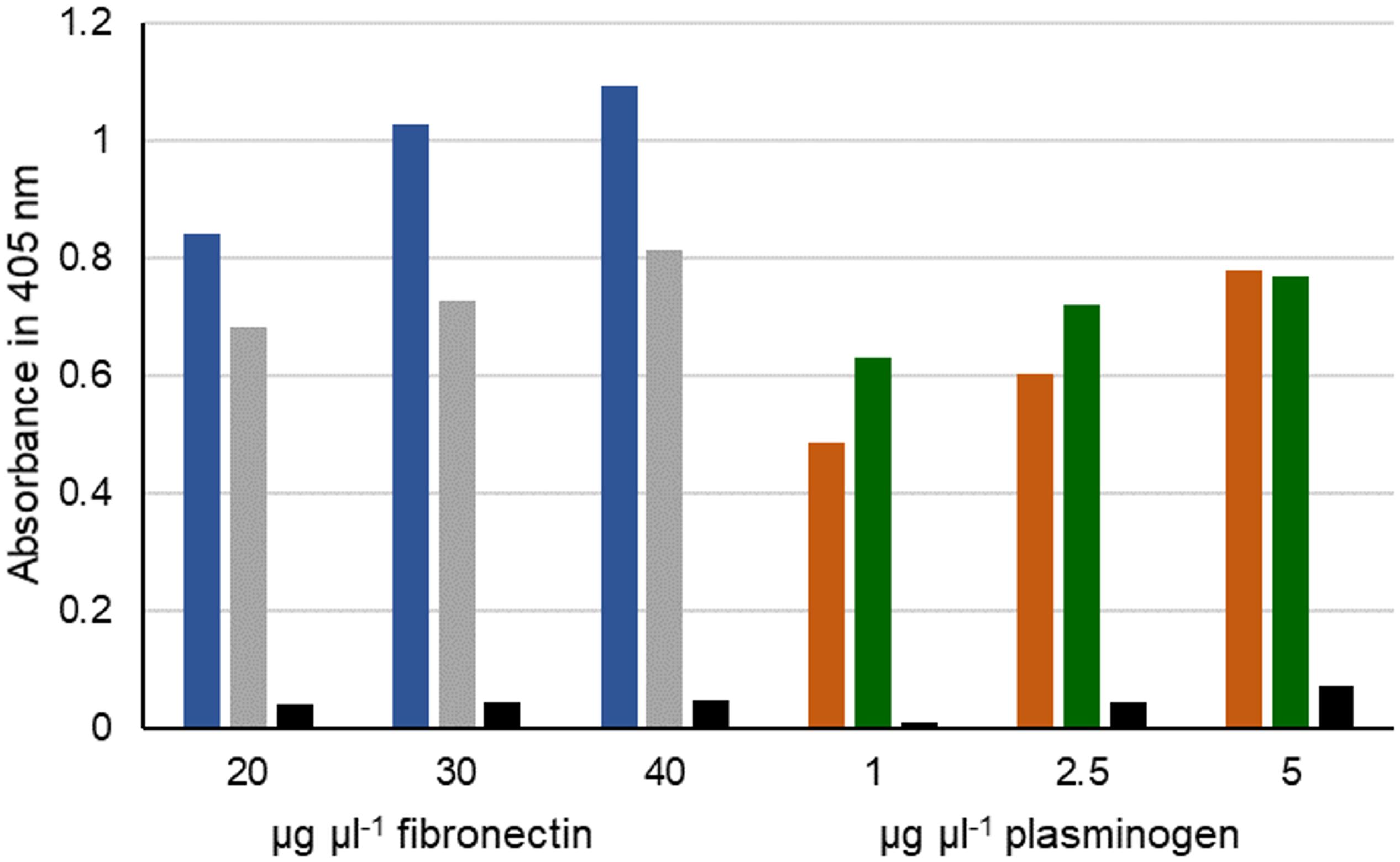
Figure 7. Interaction of fibronectin and plasminogen with IbpM. Purified IbpM (blue and brown) or whole cells of M. pneumoniae (gray and green) were coated onto 96-well plates and incubated with different concentrations of fibronectin (left) or plasminogen (right). Interaction of IbpM with the human proteins was quantified with peroxidase-conjugated antibodies detecting the corresponding human proteins. Absorbance was detected at 405 nm. BSA (black bars) served as negative control.
Analysis of Putative Immunoglobulin Proteases in M. pneumoniae
The protease of the MIB-MIP system (Arfi et al., 2016) was annotated as putative protease bearing a conserved domain of unknown function DUF31. We used the predicted domain DUF31 to scan the M. pneumoniae protein sequences for presence of similar domains. The function of this domain still remains elusive, but the amino acid sequence is similar to that of serine proteases. We found sixteen proteins containing a DUF31 domain. Interestingly, 14 of these genes encoding the DUF31 proteins localize in one single cluster of the M. pneumoniae genome (mpn577, mpn580 to mpn592, see Table 1). It is worth mentioning, that a similar comparison was done with many other Mollicutes and it seems, that only animal and human pathogenic Mycoplasmas encode such DUF31 containing proteins (Arfi et al., 2016). Furthermore, the presence of multiple copies of DUF31 proteins was only found in species lacking the MIB-MIP system (Arfi et al., 2016). This indicates that MIB-MIP and Protein M are mutually exclusive.
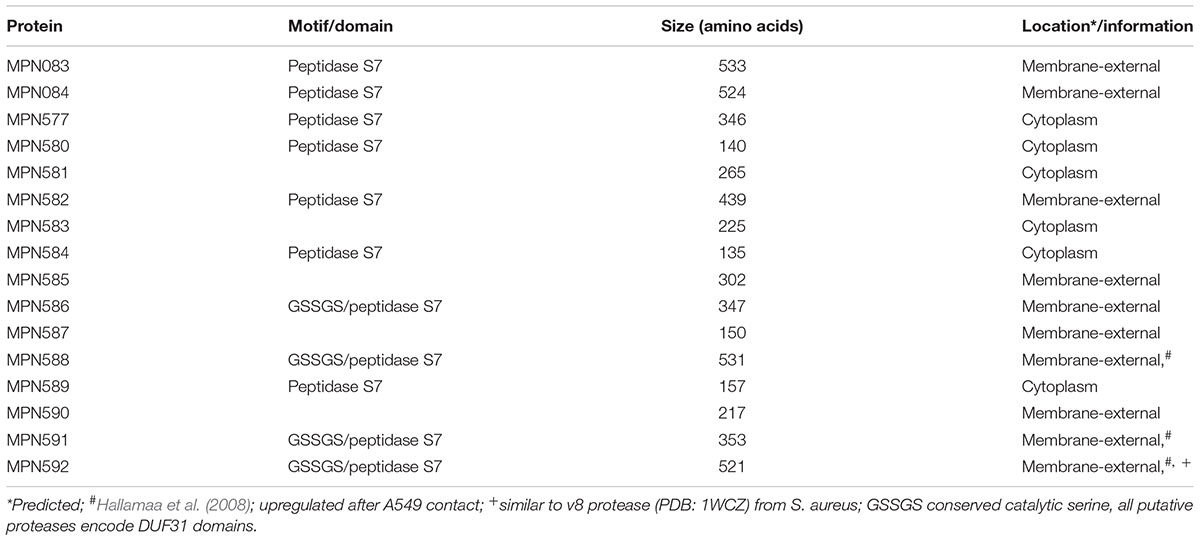
Table 1. M. pneumoniae proteins containing DUF31 domains.
As MPN592 is similar to the Staphylococcus aureus V8 protease, we selected this protein for further analysis. Using the purified protein, we assayed the potential protease activity of this enzyme on IgG, IgA, and IgM as the substrates. However, no activity was detectable, suggesting that MPN592 and probably also the other DUF31 proteases are not involved in immunoglobulin degradation in concert with IbpM.
Discussion
After successful adherence to epithelial cells, bacteria are exposed to the defense mechanisms of the human host, mainly cell-mediated and humoral immunity. The latter includes antibody-dependent cellular cytotoxicity (ADCC) and activation of complement proteins. The complement can detect antibody–antigen labeled complexes and consequently trigger a degradation cascade for bacterial clearance. To overcome opsonization, phagocytosis, cell lysis and successfully infect host tissue, bacteria must subvert or manipulate the immune system. There are two possibilities, on one hand they could modulate effectors required for detection and degradation of bacterial antigens while on the other hand they could evade the recognition by neutrophils and macrophages in a stealth-like manner (reviewed in Reddick and Alto, 2014; van Avondt et al., 2015).
In this study, we have demonstrated that the surface-localized M. pneumoniae IbpM protein binds to human immunoglobulins. Our data show the surface localization of IbpM by two different approaches, colony blot and trypsin digestion. Furthermore, we demonstrate binding of IgG, IgA, and IgM to recombinant IbpM with ELISA experiments. Additionally, we also tested and confirmed the binding capabilities to human plasminogen and fibronectin, which are required for innate immune response against bacterial infection. The C-terminal domain of the IbpM homolog in M. genitalium was thought to be not influencing the binding of Igs (Grover et al., 2014) but a truncated version of IbpM showed a reduced binding affinity. However, the precise function of the C-terminal domain remains elusive. As discussed earlier (Grover et al., 2014), after binding of IbpM the C-terminal domain could allow to cover the antigen-binding site and therefore interfere with antigen-antibody interaction. Such a masked Ig could not be detected by the complement or in turn an Ig-wrapped Mycoplasma cell could be barely detectable for the host defense mechanism. Lastly, strong binding of IgG by IbpM could invert the effect of opsonization and prevent ADCC (Wang et al., 2015) hindering natural killer cells to eliminate Mycoplasma cells. These ideas are in agreement with the chronicity of Mycoplasma infection and low mortality rates.
We evaluated proteases in M. pneumoniae, based on the described MIB-MIP system from M. mycoides (Arfi et al., 2016). Our results revealed the presence of several DUF31 containing proteases in the genome of M. pneumoniae. However, specific activity on immunoglobulins was not detectable for the selected protease MPN592, whereas other proteases could not be expressed in E. coli. We can thus not exclude the possibility that an Ig-specific protease is encoded in the genome of M. pneumoniae. However, it would be interesting to test if any of the DUF31 proteases contributes to the virulence of M. pneumoniae even if not involved in Ig degradation. Proteases are known to impact the pathogenicity of bacteria, e.g., the proteases InhA and ClpX of Bacillus anthracis contribute to invasion and survival of the pathogen in host tissue (McGillivray et al., 2009; Tonry et al., 2012).
Moreover, we show the binding of fibronectin and plasminogen to IbpM. For surface displayed proteins and lipoproteins in M. pneumoniae and other Mycoplasma species, the binding of plasminogen and fibronectin was already described (Yavlovich et al., 2004; Henderson et al., 2011; Gründel et al., 2016a, b; Hagemann et al., 2017; Chen et al., 2018; Qi et al., 2018). Neither fibronectin nor plasminogen binding was reported for components of the MIB-MIP system in M. mycoides. However, M. hyopneumoniae and M. gallisepticum MIB both bind plasminogen, but only MIB of M. hyopneumoniae also binds fibronectin (Sanderson-Smith et al., 2012). Both organisms possess MIB-MIP homologs. The binding of plasminogen could be mediated by interactions between the Kringle domains of human plasminogen and the leucine-rich repeat (LRR)-like structure in IbpM (see Figure 1B), as the affinity of these domains toward lysine-residues is well known (Bhattacharya et al., 2012; Sanderson-Smith et al., 2012). Furthermore, the binding of IbpM to host cell components could be important for cell invasion (Dallo and Baseman, 2000). Cell migration through tissue barriers involving LRR-containing proteins is already known. For example, Listeria monocytogenes uses InlB, a LRR containing protein that induces phagocytosis (Marino et al., 1999; Shen et al., 2000).
The observation of binding of plasminogen to IbpM suggests that M. pneumoniae already lost the MIP homolog during evolutionary genome minimization. The protease might be replaced by an Ig-specific protease from the human host, e.g., the plasmin (active form of plasminogen). Plasmin shares 48% amino acid similarity to serine proteases and is known to cleave IgG (Harpel et al., 1989; Chuba, 1994). In addition, the activation of plasminogen to plasmin, was already shown with different surface proteins of M. pneumoniae (Gründel et al., 2015; Hagemann et al., 2017). Moreover, binding of plasminogen and fibronectin for themselves could be important. After binding of fibronectin or plasminogen their activated forms can modulate the complement system which would allow Mycoplasmas to survive in host tissue (Barthel et al., 2012; Foley et al., 2016). The use of host derived enzymes to circumvent detection and elimination has been described and summarized for Gram-positive and Gram-negative bacteria (Bhattacharya et al., 2012).
Conclusion
In conclusion, IbpM was confirmed as a surface located multi-immunoglobulin binding protein with affinity for plasminogen and fibronectin. With this, IbpM exhibits all important abilities to subvert the host immune system. Beside other mycoplasmal lipoproteins that facilitate surveillance in the host (Citti and Rosengarten, 1997; Chambaud et al., 1999; Goret et al., 2016), IbpM is a new target for better understanding virulence of M. pneumoniae. The functions of IbpM in vivo and its implication in virulence, especially potential immunomodulatory effects need to be elucidated in the future. Testing the mpn400-deficent mutant in comparison with the wild type in animal models seem to be important.
Data Availability Statement
All datasets generated for this study are included in the article/Supplementary Material.
Ethics Statement
The animal study was reviewed and approved by the Ethical Board of Landesdirektion Sachsen, Dresden, Germany (permit no. 24-9168.25-1/2011/1).
Author Contributions
CB and JS designed the study. CB, NS, and RD performed the experiments and evaluated the data. CB, NS, and JS wrote the manuscript.
Funding
Results have been achieved within the framework of the ERASynBio 2nd Joint Call for Transnational Research Projects: “Building Synthetic Biology Capacity Through Innovative Transnational Projects” with funding from the corresponding ERASynBio National Funding Agencies.
Conflict of Interest
The authors declare that the research was conducted in the absence of any commercial or financial relationships that could be construed as a potential conflict of interest.
Acknowledgments
We are grateful to Kerstin Schmitt and Oliver Valerius for LCMS analyses which were done with the Service Unit LCMS Protein Analytics of the Göttingen Center for Molecular Biosciences (GZMB) at the Georg-August-University Göttingen (Grant ZUK 41/1 DFG-GZ A 630 (mass spectrometer; Prof. G. H. Braus). We wish to thank Johannes Gibhardt and Julia Busse for helpful discussions and technical assistance.
Supplementary Material
The Supplementary Material for this article can be found online at: https://www.frontiersin.org/articles/10.3389/fmicb.2020.00685/full#supplementary-material
FIGURE S1 | Ability of IbpM to bind different Ig’s and the effect of the C-terminal region on binding. Using ELISA, purified full-length IbpM was coated in different concentrations onto 96-well plates and incubated withIgG (A), IgA (B) or IgM (C). Binding of Igs was quantified by the detection with rabbit α-human Ig -AP antibody responsible for ABTS color formation and detection at 420 nm. The statistical significance of the influence of the C-terminalregion on the binding between IbpM and Igs is indicated by the following scheme: **** is P ≤ 0.0001, *** is P ≤ 0.001, ** is P ≤ 0.01, ns is P > 0.05.
TABLE S1 | MPN400 pulldown complete protein pulldown list.
DATA SHEET S1 | Original Blots Figures 2, 3, 5.
References
Akerström, B., and Björck, L. (1989). Protein L: an immunoglobulin light chain-binding bacterial protein. Characterization of binding and physicochemical properties. J. Biol. Chem. 264, 19740–19746.
Arfi, Y., Minder, L., di Primo, C., Le Roy, A., Ebel, C., Coquet, L., et al. (2016). MIB-MIP is a mycoplasma system that captures and cleaves immunoglobulin G. Proc. Natl. Acad. Sci. U.S.A. 113, 5406–5411. doi: 10.1073/pnas.1600546113
Atkinson, T. P., Balish, M. F., and Waites, K. B. (2008). Epidemiology, clinical manifestations, pathogenesis and laboratory detection of Mycoplasma pneumoniae infections. FEMS Microbiol. Rev. 32, 956–973. doi: 10.1111/j.1574-6976.2008.00129.x
Balish, M. F., and Krause, D. C. (2006). Mycoplasmas: a distinct cytoskeleton for wall-less bacteria. J. Mol. Microbiol. Biotechnol. 11, 244–255. doi: 10.1159/000094058
Barthel, D., Schindler, S., and Zipfel, P. F. (2012). Plasminogen is a complement inhibitor. J. Biol. Chem. 287, 18831–18842. doi: 10.1074/jbc.M111.323287
Becker, A., Kannan, T. R., Taylor, A. B., Pakhomova, O. N., Zhang, Y., Somarajan, S. R., et al. (2015). Structure of CARDS toxin, a unique ADP-ribosylating and vacuolating cytotoxin from Mycoplasma pneumoniae. Proc. Natl. Acad. Sci. U.S.A. 112, 5165–5170. doi: 10.1073/pnas.1420308112
Bhattacharya, S., Ploplis, V. A., and Castellino, F. J. (2012). Bacterial plasminogen receptors utilize host plasminogen system for effective invasion and dissemination. J. Biomed. Biotechnol. 2012:482096. doi: 10.1155/2012/482096
Bhavsar, A. P., Guttman, J. A., and Finlay, B. B. (2007). Manipulation of host-cell pathways by bacterial pathogens. Nature 449, 827–834. doi: 10.1038/nature06247
Blötz, C., and Stülke, J. (2017). Glycerol metabolism and its implication in virulence in Mycoplasma. FEMS Microbiol. Rev. 41, 640–652. doi: 10.1093/femsre/fux033
Boyle M. D. P. (ed.) (1990). “Complement activation and bacterial immunoglobulin binding proteins,” in Bacterial Immunoglobulin-Binding Proteins, Vol. 1, (San Diego, CA: Academic Press), 369–391. doi: 10.1016/b978-0-12-123012-8.50025-9
Burgos, R., Wood, G. E., Iverson-Cabral, S. L., and Tottena, P. A. (2018). Mycoplasma genitalium nonadherent phase variants arise by multiple mechanisms and escape antibody-dependent growth inhibition. Infect. Immun. 86:e00866-17. doi: 10.1128/IAI.00866-17
Campos, G. B., Marques, L. M., Rezende, I. S., Barbosa, M. S., Abrão, M. S., and Timenetsky, J. (2018). Mycoplasma genitalium can modulate the local immune response in patients with endometriosis. Fertil. Steril. 109, 549–560. doi: 10.1016/j.fertnstert.2017.11.009
Catrein, I., Dumke, R., Weiner, J., Jacobs, E., and Herrmann, R. (2004). Cross-complementation between the products of the genes P1 and ORF6 of Mycoplasma pneumoniae subtypes 1 and 2. Microbiology 150, 3989–4000. doi: 10.1099/mic.0.27506-0
Chambaud, I., Wróblewski, H., and Blanchard, A. (1999). Interactions between Mycoplasma lipoproteins and the host immune system. Trends Microbiol. 7, 493–499. doi: 10.1016/s0966-842x(99)01641-8
Chaudhry, R., Varshney, A. K., and Malhotra, P. (2007). Adhesion proteins of Mycoplasma pneumoniae. Front. Biosci. 12:690–699. doi: 10.2741/2093
Chen, S., Hao, H., Zhao, P., Ji, W., Li, M., Liu, Y., et al. (2018). Differential immunoreactivity to bovine convalescent serum between Mycoplasma bovis biofilms and planktonic cells revealed by comparative immunoproteomic analysis. Front. Microbiol. 9:379. doi: 10.3389/fmicb.2018.00379
Chuba, J. V. (1994). Susceptibility of monoclonal IgG paraproteins to plasmic cleavage using glycerin-stabilized human plasmin. Biochem. Biophys. Res. Commun. 202, 367–373. doi: 10.1006/bbrc.1994.1937
Citti, C., and Blanchard, A. (2013). Mycoplasmas and their host: emerging and re-emerging minimal pathogens. Trends Microbiol. 21, 196–203. doi: 10.1016/j.tim.2013.01.003
Citti, C., and Rosengarten, R. (1997). Mycoplasma genetic variation and its implication for pathogenesis. Wien. Klin. Wochenschr. 109, 562–568.
Cizelj, I., Bercic, R. L., Dusanic, D., Narat, M., Kos, J., Dovc, P., et al. (2011). Mycoplasma gallisepticum and Mycoplasma synoviae express a cysteine protease CysP, which can cleave chicken IgG into Fab and Fc. Microbiology 157, 362–372. doi: 10.1099/mic.0.045641-0
Dallo, S. F., and Baseman, J. B. (2000). Intracellular DNA replication and long-term survival of pathogenic Mycoplasmas. Microb. Pathog. 29, 301–309. doi: 10.1006/mpat.2000.0395
Dandekar, T., Huynen, M., Regula, J. T., Ueberle, B., Zimmermann, C. U., Andrade, M. A., et al. (2000). Re-annotating the Mycoplasma pneumoniae genome sequence: adding value, function and reading frames. Nucleic Acids Res. 28, 3278–3288. doi: 10.1093/nar/28.17.3278
Diethmaier, C., Pietack, N., Gunka, K., Wrede, C., Lehnik-Habrink, M., Herzberg, C., et al. (2011). A novel factor controlling bistability in Bacilllus subtilis: the YmdB protein affects flagellin expression and biofilm formation. J. Bacteriol. 193, 5997–6007. doi: 10.1128/JB.05360-11
Fischetti, V. A. (1989). Streptococcal M protein: molecular design and biological behavior. Clin. Microbiol. Rev. 2, 285–314. doi: 10.1128/cmr.2.3.285-314.1989
Foley, J. H., Walton, B. L., Aleman, M. M., O’Byrne, A. M., Lei, V., Harrasser, M., et al. (2016). Complement activation in arterial and venous thrombosis is mediated by plasmin. EBioMedicine 5, 175–182. doi: 10.1016/j.ebiom.2016.02.011
Frick, I. M., Wikström, M., Forsén, S., Drakenberg, T., Gomi, H., Sjöbring, U., et al. (1992). Convergent evolution among immunoglobulin G-binding bacterial proteins. Proc. Natl. Acad. Sci. U.S.A. 89, 8532–8536.
Goret, J., Le Roy, C., Touati, A., Mesureur, J., Renaudin, H., Claverol, S., et al. (2016). Surface lipoproteome of Mycoplasma hominis PG21 and differential expression after contact with human dendritic cells. Future Microbiol. 11, 179–194. doi: 10.2217/fmb.15.130
Graille, M., Harrison, S., Crump, M. P., Findlow, S. C., Housden, N. G., Muller, B. H., et al. (2002). Evidence for plasticity and structural mimicry at the immunoglobulin light chain-protein L interface. J. Biol. Chem. 277, 47500–47506. doi: 10.1074/jbc.m206105200
Graille, M., Stura, E. A., Corper, A. L., Sutton, B. J., Taussig, M. J., Charbonnier, J.-B., et al. (2000). Crystal structure of a Staphylococcus aureus protein A domain complexed with the Fab fragment of a human IgM antibody: structural basis for recognition of B-cell receptors and superantigen activity. Proc. Natl. Acad. Sci. U.S.A. 97, 5399–5404. doi: 10.1073/pnas.97.10.5399
Grover, R. K., Zhu, X., Nieusma, T., Jones, T., Boero, I., MacLeod, A. S., et al. (2014). A structurally distinct human Mycoplasma protein that generically blocks antigen-antibody union. Science 343, 656–661. doi: 10.1126/science.1246135
Großhennig, S., Ischebeck, T., Gibhardt, J., Busse, J., Feussner, I., and Stülke, J. (2016). Hydrogen sulfide is a novel potential virulence factor of Mycoplasma pneumoniae: characterization of the unusual cysteine desulfurase/desulfhydrase HapE. Mol. Microbiol. 100, 42–54. doi: 10.1111/mmi.13300
Gründel, A., Jacobs, E., and Dumke, R. (2016a). Interactions of surface-displayed glycolytic enzymes of Mycoplasma pneumoniae with components of the human extracellular matrix. Int. J. Med. Microbiol. 306, 675–685. doi: 10.1016/j.ijmm.2016.09.001
Gründel, A., Pfeiffer, M., Jacobs, E., and Dumke, R. (2016b). Network of surface-displayed glycolytic enzymes in Mycoplasma pneumoniae and their interactions with human plasminogen. Infect. Immun. 84, 666–676. doi: 10.1128/IAI.01071-15
Gründel, A., Pfeiffer, M., Jacobs, E., and Dumke, R. (2015). The network of surface-displayed glycolytic enzymes in Mycoplasma pneumoniae and their interactions with human plasminogen. Infect. Immun. 84, 666–676. doi: 10.1128/IAI.01071-15
Hagemann, L., Gründel, A., Jacobs, E., and Dumke, R. (2017). The surface-displayed chaperones GroEL and DnaK of Mycoplasma pneumoniae interact with human plasminogen and components of the extracellular matrix. Pathog. Dis. 75:ftx017. doi: 10.1093/femspd/ftx017
Halbedel, S., Busse, J., Schmidl, S., and Stülke, J. (2006). Regulatory protein phosphorylation in Mycoplasma pneumoniae: a PP2C-type phosphatase serves to dephosphorylate HPr(Ser-P). J. Biol. Chem. 281, 26253–26259. doi: 10.1074/jbc.m605010200
Halbedel, S., Hames, C., and Stülke, J. (2004). In vivo activity of enzymatic and regulatory components of the phosphoenolpyruvate:sugar phosphotransferase system in Mycoplasma pneumoniae. J. Bacteriol. 186, 7936–7943. doi: 10.1128/jb.186.23.7936-7943.2004
Halbedel, S., Hames, C., and Stülke, J. (2007). Regulation of carbon metabolism in the mollicutes and its relation to virulence. J. Mol. Microbiol. Biotechnol. 12, 147–154. doi: 10.1159/000096470
Hallamaa, K. M., Tang, S. L., Ficorilli, N., and Browning, G. F. (2008). Differential expression of lipoprotein genes in Mycoplasma pneumoniae after contact with human lung epithelial cells, and under oxidative and acidic stress. BMC Microbiol. 8:124. doi: 10.1186/1471-2180-8-124
Hames, C., Halbedel, S., Hoppert, M., Frey, J., and Stülke, J. (2009). Glycerol metabolism is important for cytotoxicity of Mycoplasma pneumoniae. J. Bacteriol. 191, 747–753. doi: 10.1128/JB.01103-08
Hames, C., Halbedel, S., Schilling, O., and Stülke, J. (2005). MMR: a method for the simultaneous introduction of multiple mutations into the glpK gene of Mycoplasma pneumoniae. Appl. Environ. Microbiol. 71, 4097–4100. doi: 10.1128/aem.71.7.4097-4100.2005
Harpel, P. C., Gordon, B. R., and Parker, T. S. (1989). Plasmin catalyzes binding of lipoprotein (a) to immobilized fibrinogen and fibrin. Proc. Natl. Acad. Sci. U.S.A. 86, 3847–3851. doi: 10.1073/pnas.86.10.3847
Henderson, B., Nair, S., Pallas, J., and Williams, M. A. (2011). Fibronectin: a multidomain host adhesin targeted by bacterial fibronectin-binding proteins. FEMS Microbiol. Rev. 35, 147–200. doi: 10.1111/j.1574-6976.2010.00243.x
Horstmann, R. D., Sievertsen, H. J., Knobloch, J., and Fischetti, V. A. (1988). Antiphagocytic activity of streptococcal M protein: selective binding of complement control protein factor H. Proc. Natl. Acad. Sci. U.S.A. 85, 1657–1661. doi: 10.1073/pnas.85.5.1657
Into, T., Dohkan, J. I., Inomata, M., Nakashima, M., Shibata, K. I., and Matsushita, K. (2007). Synthesis and characterization of a dipalmitoylated lipopeptide derived from paralogous lipoproteins of Mycoplasma pneumoniae. Infect. Immun. 75, 2253–2259. doi: 10.1128/iai.00141-07
Into, T., Kiura, K., Yasuda, M., Kataoka, H., Inoue, N., Hasebe, A., et al. (2004). Stimulation of human Toll-like receptor (TLR) 2 and TLR6 with membrane lipoproteins of Mycoplasma fermentans induces apoptotic cell death after NF-kappa B activation. Cell. Microbiol. 6, 187–199. doi: 10.1046/j.1462-5822.2003.00356.x
Johnson, C., Kannan, T. R., and Baseman, J. B. (2011). Cellular vacuoles induced by Mycoplasma pneumoniae CARDS toxin originate from Rab9-associated compartments. PLoS One 6:e22877. doi: 10.1371/journal.pone.0022877
Kannan, T. R., and Baseman, J. B. (2006). ADP-ribosylating and vacuolating cytotoxin of Mycoplasma pneumoniae represents unique virulence determinant among bacterial pathogens. Proc. Natl. Acad. Sci. U.S.A. 103, 6724–6729. doi: 10.1073/pnas.0510644103
Kannan, T. R., Musatovova, O., Balasubramanian, S., Cagle, M., Jordan, J. L., Krunkosky, T. M., et al. (2010). Mycoplasma pneumoniae community acquired respiratory distress syndrome toxin expression reveals growth phase and infection-dependent regulation. Mol. Microbiol. 76, 1127–1141. doi: 10.1111/j.1365-2958.2010.07092.x
Kenri, T., Kawakita, Y., Kudo, H., Matsumoto, U., Mori, S., Furukawa, Y., et al. (2018). Production and characterization of recombinant P1 adhesin essential for adhesion, gliding, and antigenic variation in the human pathogenic bacterium, Mycoplasma pneumoniae. Biochem. Biophys. Res. Commun. 508, 1050–1055. doi: 10.1016/j.bbrc.2018.11.132
Krause, D. C., Chen, S., Shi, J., Jensen, A. J., Sheppard, E. S., and Jensen, G. J. (2018). Electron cryotomography of Mycoplasma pneumoniae mutants correlates terminal organelle architectural features and function. Mol. Microbiol. 108, 306–318. doi: 10.1111/mmi.13937
Lin, C.-J., Sasse, C., Gerke, J., Valerius, O., Irmer, H., Frauendorf, H., et al. (2015). Transcription factor SomA is required for adhesion, development and virulence of the human pathogen Aspergillus fumigatus. PLoS Pathog. 11:e1005205. doi: 10.1371/journal.ppat.1005205
Lluch-Senar, M., Delgado, J., Chen, W. H., Lloréns-Rico, V., O’Reilly, F. J., Wodke, J. A., et al. (2015). Defining a minimal cell: essentiality of small ORFs and ncRNAs in a genome-reduced bacterium. Mol. Syst. Biol. 11:780. doi: 10.15252/msb.20145558
Marino, M., Braun, L., Cossart, P., and Ghosh, P. (1999). Structure of the lnlB leucine-rich repeats, a domain that triggers host cell invasion by the bacterial pathogen L. monocytogenes. Mol. Cell 4, 1063–1072. doi: 10.1016/s1097-2765(00)80234-8
McGillivray, S. M., Ebrahimi, C. M., Fisher, N., Sabet, M., Zhang, D. X., Chen, Y., et al. (2009). ClpX contributes to innate defense peptide resistance and virulence phenotypes of Bacillus anthracis. J. Innate Immun. 1, 494–506. doi: 10.1159/000225955
Merzbacher, M., Detsch, C., Hillen, W., and Stülke, J. (2004). Mycoplasma pneumoniae HPr kinase/phosphorylase. Assigning functional roles to the P-loop and the HPr kinase/phosphorylase signature sequence motif. Eur. J. Biochem. 271, 367–374. doi: 10.1046/j.1432-1033.2003.03935.x
Mühlradt, P. F., Kiess, M., Meyer, H., Süssmuth, R., and Jung, G. (1997). Isolation, structure elucidation, and synthesis of a macrophage stimulatory lipopeptide from Mycoplasma fermentans acting at picomolar concentration. J. Exp. Med. 185, 1951–1958. doi: 10.1084/jem.185.11.1951
Okusawa, T., Fujita, M., Nakamura, J.-I., Into, T., Yasuda, M., Yoshimura, A., et al. (2004). Relationship between structures and biological activities of mycoplasmal diacylated lipopeptides and their recognition by toll-like receptors 2 and 6. Infect. Immun. 72, 1657–1665. doi: 10.1128/iai.72.3.1657-1665.2004
Omasits, U., Ahrens, C. H., Müller, S., and Wollscheid, B. (2014). Protter: interactive protein feature visualization and integration with experimental proteomic data. Bioinformatics 30, 884–886. doi: 10.1093/bioinformatics/btt607
Parrott, G. L., Kinjo, T., and Fujita, J. (2016). A compendium for Mycoplasma pneumoniae. Front. Microbiol. 7:513. doi: 10.3389/fmicb.2016.00513
Pollack, J. D., Williams, M. V., and McElhaney, R. N. (1997). The comparative metabolism of the mollicutes (Mycoplasmas): the utility for taxonomic classification and the relationship of putative gene annotation and phylogeny to enzymatic function in the smallest free-living cells. Crit. Rev. Microbiol. 23, 269–354. doi: 10.3109/10408419709115140
Qi, J., Zhang, F., Wang, Y., Liu, T., Tan, L., Wang, S., et al. (2018). Characterization of Mycoplasma gallisepticum pyruvate dehydrogenase alpha and beta subunits and their roles in cytoadherence. PLoS One 13:e0208745. doi: 10.1371/journal.pone.0208745
Rappsilber, J., Mann, M., and Ishihama, Y. (2007). Protocol for micro-purification, enrichment, pre-fractionation and storage of peptides for proteomics using StageTips. Nat. Protoc. 2, 1896–1906. doi: 10.1038/nprot.2007.261
Reddick, L. E., and Alto, N. M. (2014). Bacteria fighting back: how pathogens target and subvert the host innate immune system. Mol. Cell 54, 321–328. doi: 10.1016/j.molcel.2014.03.010
Robertson, J. A., Stemler, M. E., and Stemke, G. W. (1984). Immunoglobulin A protease activity of Ureaplasma urealyticum. J. Clin. Microbiol. 19, 255–258. doi: 10.1128/jcm.19.2.255-258.1984
Rottem, S. (2003). Interaction of Mycoplasmas with host cells. Physiol. Rev. 83, 417–432. doi: 10.1152/physrev.00030.2002
Sambrook, J., Fritsch, E. F., and Maniatis, T. (1989). Molecular Cloning: A Laboratory Manual, 2nd Edn. Cold Spring Harbor, NY: Cold Spring Harbor Laboratory.
Sanderson-Smith, M. L., De Oliveira, D. M. P., Ranson, M., and McArthur, J. D. (2012). Bacterial plasminogen receptors: mediators of a multifaceted relationship. J. Biomed. Biotechnol. 2012:272148. doi: 10.1155/2012/272148
Schmidl, S. R., Gronau, K., Hames, C., Busse, J., Becher, D., Hecker, M., et al. (2010). The stability of cytadherence proteins in Mycoplasma pneumoniae requires activity of the protein kinase PrkC. Infect. Immun. 78, 184–192. doi: 10.1128/IAI.00958-09
Schmidl, S. R., Otto, A., Lluch-Senar, M., Piñol, J., Busse, J., Becher, D., et al. (2011). A trigger enzyme in Mycoplasma pneumoniae: impact of the glycerophosphodiesterase GlpQ on virulence and gene expression. PLoS Pathog. 7:e1002263. doi: 10.1371/journal.ppat.1002263
Schmitt, K., Smolinski, N., Neumann, P., Schmaul, S., Hofer-Pretz, V., Braus, G. H., et al. (2017). Asc1p/RACK1 connects ribosomes to eukaryotic phosphosignaling. Mol. Cell. Biol. 37:e00279-16. doi: 10.1128/MCB.00279-16
Schurwanz, N., Jacobs, E., and Dumke, R. (2009). Strategy to create chimeric proteins derived from functional adhesin regions of Mycoplasma pneumoniae for vaccine development. Infect. Immun. 77, 5007–5015. doi: 10.1128/IAI.00268-09
Seybert, A., Gonzalez-Gonzalez, L., Scheffer, M. P., Lluch-Senar, M., Mariscal, A. M., Querol, E., et al. (2018). Cryo-electron tomography analyses of terminal organelle mutants suggest the motility mechanism of Mycoplasma genitalium. Mol. Microbiol. 108, 319–329. doi: 10.1111/mmi.13938
Shen, Y., Naujokas, M., Park, M., and Ireton, K. (2000). InlB-dependent internalization of Listeria is mediated by the met receptor tyrosine kinase. Cell 103, 501–510. doi: 10.1016/s0092-8674(00)00141-0
Shevchenko, A., Wilm, M., Vorm, O., and Mann, M. (1996). Mass spectrometric sequencing of proteins silver-stained polyacrylamide gels. Anal. Chem. 68, 850–858. doi: 10.1021/ac950914h
Sidorin, E. V., and Solov’eva, T. F. (2011). IgG-binding proteins of bacteria. Biochemistry 76, 295–308. doi: 10.1134/s0006297911030023
Sjöbring, U., Björck, L., and Kastern, W. (1991). Streptococcal protein G. Gene structure and protein binding properties. J. Biol. Chem. 266, 399–405.
Spooner, R. K., Russell, W. C., and Thirkell, D. (1992). Characterization of the immunoglobulin A protease of Ureaplasma urealyticum. Infect. Immun. 60, 2544–2546. doi: 10.1128/iai.60.6.2544-2546.1992
Thomas, C., Jacobs, E., and Dumke, R. (2013). Characterization of pyruvate dehydrogenase subunit B and enolase as plasminogen-binding proteins in Mycoplasma pneumoniae. Microbiology 159, 352–365. doi: 10.1099/mic.0.061184-0
Tonry, J. H., Mcnichol, B. A., Ramarao, N., Chertow, D. S., Kim, K. S., Stibitz, S., et al. (2012). Bacillus anthracis protease InhA regulates BslA-mediated adhesion in human endothelial cells. Cell. Microbiol. 14, 1219–1230. doi: 10.1111/j.1462-5822.2012.01791.x
van Avondt, K., van Sorge, N. M., and Meyaard, L. (2015). Bacterial immune evasion through manipulation of host inhibitory immune signaling. PLoS Pathog. 11:e1004644. doi: 10.1371/journal.ppat.1004644
van der Woude, M. W., and Bäumler, A. J. (2004). Phase and antigenic variation in bacteria. Clin. Microbiol. Rev. 17, 581–611. doi: 10.1128/cmr.17.3.581-611.2004
Waites, K. B., and Talkington, D. F. (2004). Mycoplasma pneumoniae and its role as a human pathogen. Clin. Microbiol. Rev. 17, 697–728. doi: 10.1128/cmr.17.4.697-728.2004
Waldo, R. H., and Krause, D. C. (2006). Synthesis, stability, and function of cytadhesin P1 and accessory protein B/C complex of Mycoplasma pneumoniae. J. Bacteriol. 188, 569–575. doi: 10.1128/jb.188.2.569-575.2006
Wang, W., Erbe, A. K., Hank, J. A., Morris, Z. S., and Sondel, P. M. (2015). NK cell-mediated antibody-dependent cellular cytotoxicity in cancer immunotherapy. Front. Immunol. 6:368. doi: 10.3389/fimmu.2015.00368
Keywords: immune evasion, Mycoplasma pneumoniae, mollicutes, protein–protein interaction, host–pathogen interaction
Citation: Blötz C, Singh N, Dumke R and Stülke J (2020) Characterization of an Immunoglobulin Binding Protein (IbpM) From Mycoplasma pneumoniae. Front. Microbiol. 11:685. doi: 10.3389/fmicb.2020.00685
Received: 06 December 2019; Accepted: 24 March 2020;
Published: 16 April 2020.
Edited by:
Christine Citti, Institut National de la Recherche Agronomique (INRA), FranceReviewed by:
Joerg Jores, University of Bern, SwitzerlandSabine Pereyre, Université de Bordeaux, France
Copyright © 2020 Blötz, Singh, Dumke and Stülke. This is an open-access article distributed under the terms of the Creative Commons Attribution License (CC BY). The use, distribution or reproduction in other forums is permitted, provided the original author(s) and the copyright owner(s) are credited and that the original publication in this journal is cited, in accordance with accepted academic practice. No use, distribution or reproduction is permitted which does not comply with these terms.
*Correspondence: Jörg Stülke, anN0dWVsa0Bnd2RnLmRl
†These authors have contributed equally to this work