 Gang Lu
 Jiajun Ou
 Jinzhao Ji1,2,3
 Shoujun Li
94% of researchers rate our articles as excellent or good
Learn more about the work of our research integrity team to safeguard the quality of each article we publish.
Find out more
ORIGINAL RESEARCH article
Front. Microbiol. , 20 June 2019
Sec. Virology
Volume 10 - 2019 | https://doi.org/10.3389/fmicb.2019.01416
Getah virus (GETV) is a mosquito-borne virus that was first determined in Malaysia in 1955, and can infect humans and multiple other mammals. GETV infection in horses has been reported in Japan and India, and causes great economic losses. In China, GETV has been identified in mosquitoes, pigs, foxes, and cattle with a wide geographical distribution, but has not been detected in horses. In August 2018, a sudden onset of fever was observed in racehorse in an equestrian training center in Guangdong Province in southern China. Blood samples were collected from the sick horse, and PCR/RT-PCR analysis was performed to screen for equine viral pathogens associated with fever. The results indicated that the samples were GETV RNA positive. After RT-PCR, sequencing, and assembly, the genome of the first Chinese horse-derived GETV strain, GZ201808, was obtained. Compared with the genome sequences of other GETV strains, twelve unique nucleotide substitutions were observed in GZ201808. The genome of GZ201808 had the highest genetic identity (99.6%) with AH9192, which was detected in pigs in China in 2017. Phylogenetic analysis indicated that GZ201808 clustered in Group III, and was located in an independent branch distant from other horse-derived GETV strains, indicating a unique evolutionary pattern of GZ201808. This study first determined and described the disease course of horse infected with GETV in China, sequenced and characterized the genome of the field horse-derived GETV strain, and therefore presented an unequivocal report of GETV infection in horses in China.
The species Getah virus (GETV) belongs to the Alphavirus genus of the Togaviridae family. Its genome is linear, positive-sense ssRNA and contains a 5′ terminal cap, two large open reading frames (ORFs), and a 3′ terminal poly(A) tract. The two ORFs encode nonstructural (nsP1 to nsP4) and structural polyproteins [C (capsid), E3, E2, 6K, E1], which are flanked by a 44 nt 26S junction region associated with transcription of an intracellular subgenomic 26S RNA (Strauss and Strauss, 1994).
As a mosquito-borne virus, GETV was first isolated from Culex sp. mosquitoes in Malaysia in 1955 (Griffin, 2013). Subsequent studies indicated that this virus has a broad geographical distribution, including Asia (Ksiazek et al., 1981; Morita and Igarashi, 1984; Leake et al., 1986; Li et al., 1992; Brown and Timoney, 1998; Turell et al., 2003; Bryant et al., 2005), Europe (Lvov et al., 1990), and Australia (Spradbrow, 1972), and could infect humans, monkeys, cattle, birds, pigs, horses, and other mammals (Fukunaga et al., 2000).
In China, GETV was first identified in Hainan Province in southern China in 1964, and one GETV strain designated M-1 was isolated from wild-caught mosquitoes (Li et al., 1992). Since then, several investigations have demonstrated that GETV is present in mosquitoes in China with a wide distribution, ranging between latitudes 19°N and 40°N and longitudes 97°E and 122°E (Zhai et al., 2008). In 2011, the first GETV outbreak in pigs was confirmed on a farm with piglet death in Hebei Province in northern China (Zhou et al., 2018). In addition, two GETV strains were detected in cattle and foxes in China in 2017 and 2018. The GETV E2 protein is located on the surface of viral particles, and is a glycoprotein that mediates viral entry. Evolutionary analysis of the E2 gene of GETV strains characterized worldwide since 1955 indicated that the virus has evolved into four distinct groups: Groups I–IV (Li et al., 2017). All the Chinese GETV strains were clustered in Group III with the exception of a mosquito-derived GETV strain, YN12031, detected in Yunnan Province in southern China that clustered in Group IV (Li et al., 2017).
GETV is known to be pathogenic for horses and pigs, and has resulted in large economic losses. Horses are considered an important host for the amplification and circulation of GETV (Bannai et al., 2015). To date, six GETV outbreaks in horses have been reported worldwide, five outbreaks in Japan in 1978, 1979, 1983, 2014, and 2015 (Kamada et al., 1980; Fukunaga et al., 2000; Bannai et al., 2015, 2016; Nemoto et al., 2015), and one outbreak in India in 1990 (Brown and Timoney, 1998). The clinical signs of horses with GETV infection often include pyrexia, rash on the body, and edema in the legs. Nasal discharge was only found in experimentally inoculated horses, but not in natural infection cases (Kamada et al., 1991a, b).
To the best of our knowledge, until now, there is only one example of serological evidence supporting the presence of antibodies against GETV in horses in China, with a positive rate of 4/16, which was determined in serum samples collected in Hainan Province in 1979 (Li et al., 1992). There has been no direct evidence supporting the presence of GETV in horses in China thus far. In this study, we described the disease course of racehorse infected with GETV in Guangdong Province in southern China in 2018, characterized the genetic features of the GETV detected in the sick horse and therefore presented an unequivocal report of GETV infection in horses in China.
EDTA-treated blood samples were collected from racehorses in an equestrian training center in Guangdong Province in southern China, in which the racehorses were routinely immunized with equine influenza (Proteqflu Te, France) and Japanese encephalitis (Nisseiken, Japan) vaccines each year. After collection, the samples were immediately transported to our laboratory in an icebox for further use.
In the present study, a total of 16 serum samples were collected from racehorses in the equestrian training center. Among them, two samples were collected from the sick horse on day 1 and 13, respectively. The remaining samples were collected from 14 other healthy racehorses on day 13 (Figure 1).
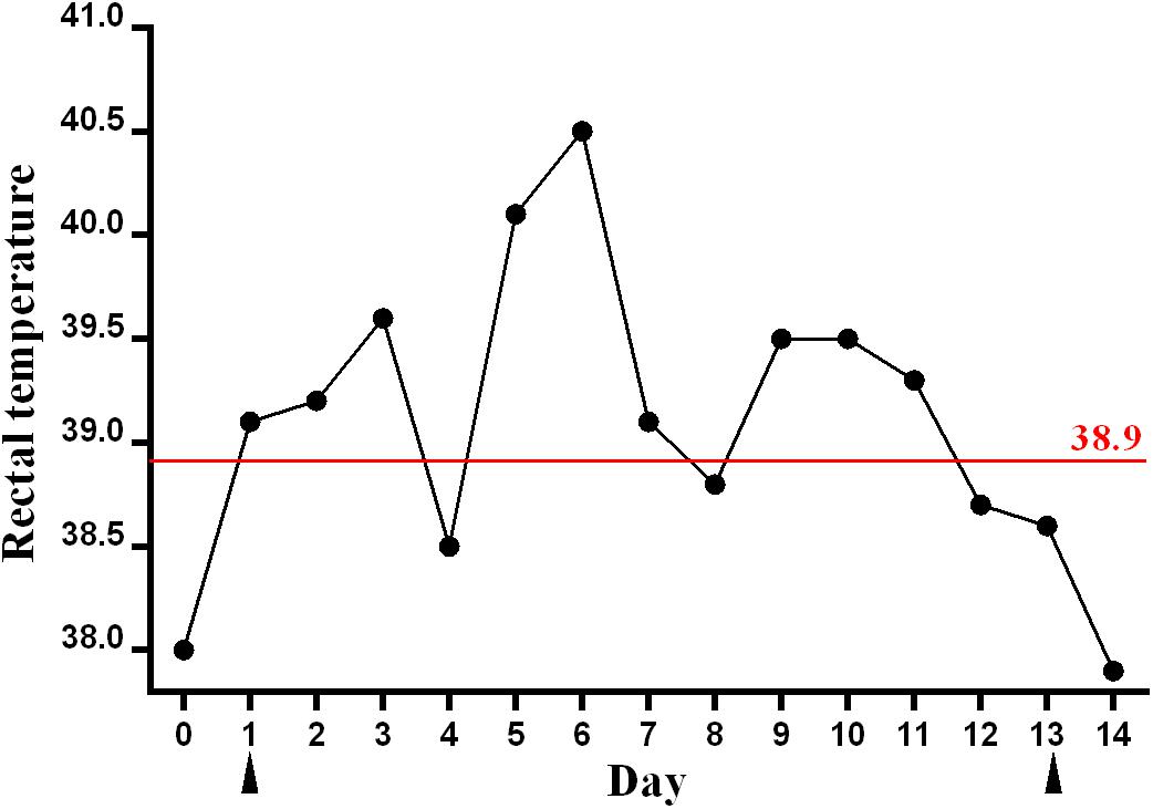
Figure 1. Rectal temperature of the sick racehorse infected with GETV. A rectal temperature of ≥38.9°C was considered as the reference for fever and is indicated by the red line. The blood samples were collected on days 1 and 13, which are indicated by black triangles.
Total RNA/DNA of 200 μl of the samples collected from the sick horse on day 1 was extracted using a MiniBEST Viral RNA/DNA Extraction Kit (Takara, Japan) following the manufacturer’s recommendations, and finally diluted in 30 μl of nuclease-free water. A total of 6 μl of RNA was then used for reverse transcription into cDNA using a HiScript 1st Strand cDNA Synthesis Kit (Vazyme, China) with random primers. Conventional or nested PCRs targeting the conserved regions of the viral genome were then performed to detect nucleic acids of equine arteritis virus, African horse sickness virus, equine infectious anemia virus, Japanese encephalitis virus, equid herpesviruses 1 and 4, and GETV using previously published PCR primers and procedures (Table 1), and GenStar Taq Polymerase Premix (Kangrun Chengye, China). One horse serum sample tested positive for both DNA (equine parvovirus) and RNA (equine hepacivirus) viruses in our previous study was used as positive control for PCR/RT-PCR in the present study (Lu et al., 2018), and whether these two viruses were present in the serum samples collected from the sick horse was also tested (Table 1).
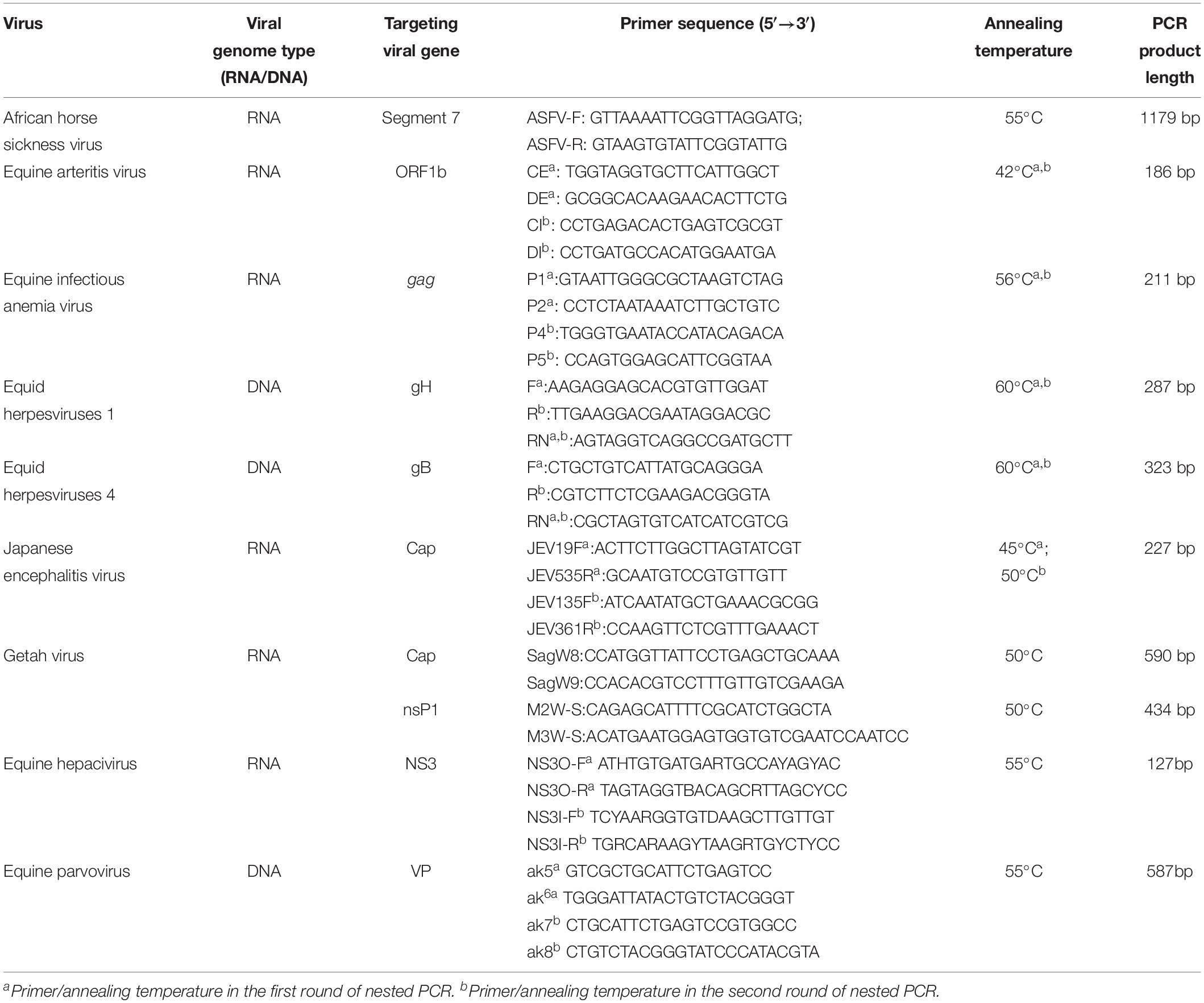
Table 1. Detailed information on the primers used for screening for the equine viral pathogens associated with fever.
After 1.5% agarose gel electrophoresis, the PCR products with the expected size were considered positive for the corresponding viruses (Table 1). The DNA fragments were then purified using a universal DNA purification kit (Tiangen, China), cloned into the pCloneEZ vector (Clone smarter, United States), and transformed into E. coli DH5α competent cells (Weidi, China). After culture for 10 h, the bacterial clones were picked and detected by PCR, and the positive samples were sent for Sanger sequencing from both ends (BGI, China).
The genome sequences of all of the GETV strains available in the NCBI database were obtained. The sequences were aligned using BioEdit 7.0.9.01 with the Clustal W method, and a total of 14 primer pairs covering the near complete genome of GETV were designed using Oligo 7.02 based on the conserved region among all of the GETV strains (Table 2). PCR was then performed to acquire the corresponding fragments using Phanta max superfidelity DNA polymerase (Vazyme, China). The annealing temperature for each PCR procedure was set to 55°C. After sequencing, the obtained raw data were processed using SeqMan 7.1.0 (DNAStar Lasergene package, Version 7.1.03) and the genome of GZ201808 was assembled.
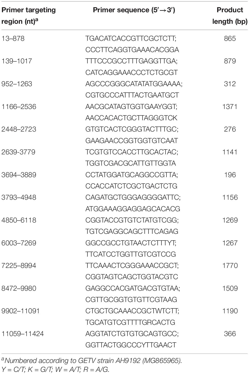
Table 2. Detailed information on the primers used for sequencing GZ201808.
The genome of the first GETV strain detected in horses in China, GZ201808, was aligned with 34 other GETV strains detected in mosquitoes, pigs, horses, foxes, and cattle from China, South Korea, Japan, Mongolia, and Russia (Table 3). The nucleotide and protein identity of the genome and the nonstructural and structural polyprotein coding sequences between GETV strains was calculated and displayed using MegAlign 7.1.0 (DNAStar Lasergene package, Version 7.1.03; Table 4).
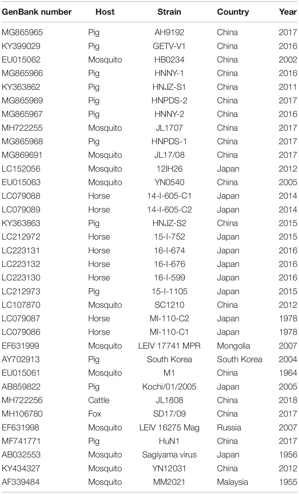
Table 3. Detailed information on GETV strains in China, Japan, Mongolia, South Korea, and Malaysia in this study.
Table 4. Nucleotide/amino acid identity of the genome and nonstructural and structural polyprotein coding sequences between GZ201808 and other GETV strains.
Maximum likelihood phylogenetic trees were constructed using MEGA 7.04 based on the GETV genome sequence, the nonstructural and structural polyprotein coding sequences, and the E2 gene. By “Find Best DNA Models,” the phylogenetic trees were inferred using general time reversible + gamma distributed or Tamura-Nei + gamma distributed models, based on a bootstrap value of 1,000 replicates.
EDTA-treated blood samples collected from the sick horse on day 1 were processed for virus isolation, as described previously (Kamada et al., 1980; Bannai et al., 2015). Briefly, the plasma and leukocytes were separated, frozen and thawed three times, and then centrifuged. The supernatant was inoculated onto confluent Vero, BHK-21, RK-13, and C6/36 cell monolayers, respectively. Cultures were inspected daily for a possible change in cytopathic effects (CPE). After 5 days, both the culture and supernatant were collected, and total RNA was extracted and tested for GETV RNA by RT-PCR using two primer pairs targeting the cap and nsp1 genes. Attempts for virus isolation were also performed through intracranial inoculation of suckling mice. The mice were observed for 14 days. The brains of mice were collected and tested for GETV RNA by RT-PCR.
To understand whether archived horse serum samples in our laboratory contained GETV RNA, a total of 320 samples collected during 2014–2018 in our previously studies were processed for GETV detection by RT-PCR (Lu et al., 2016, 2018).
In August 2018, a sudden onset of fever was observed in a racehorse (warmblood, 17 years old) in an equestrian training center in Guangdong Province in southern China. Blood sample from the sick animal was collected after the onset of illness (day 1) (Figure 1), and tested by PCR/RT-PCR to screen for equine viral pathogens associated with fever, including equine arteritis virus, African horse sickness virus, equine infectious anemia virus, Japanese encephalitis virus, equid herpesviruses, and GETV (Table 1). This animal had been routinely vaccinated with equine influenza vaccines. No respiratory symptoms were observed in this racehorse, and equine influenza virus was not included in the screening tests.
After agarose gel electrophoresis, only two PCR products testing for the Cap and nsP1 gene of GETV showed the expected band. The subsequent sequencing and BLAST results also indicated GETV, with the highest genetic identity of 99.6% with a Chinese swine-derived GETV strain (AH9192, GenBank no. MG865965) and 99.7% with three Chinese mosquito-derived GETV strains [SC1210 (LC107810), Z12 (FJ897146), YN0540 (EU015063)], a Korean mosquito-derived GETV strain [KorL915 (JN410944)], and a Korean swine-derived GETV strain [South Korea (AY702913)] when analyzing the Cap and nsP1 genes, respectively. This GETV strain detected in a racehorse in our study was designated GZ201808, and was used for further genomic sequencing and analysis.
To acquire the genome of GZ201808, a gap-filling PCR strategy was used based on 14 primer pairs covering the genome of all of the published GETV strains in the NCBI database (Table 2). After sequencing and assembly, the 11,421 nt near-complete genome of the first GETV strain detected in horses in China was obtained, including a 66 nt partial 5′UTR, 7404 nt nonstructural polyprotein coding region, 44 nt 26S junction region (ATGCAGGATTACACTACATCTAAAGACCACGTATTACAG ACACC), 3762 nt structural polyprotein coding region, and 145 nt partial 3′UTR. The genome of GZ201808 has been summited in the GenBank database with accession number MK487997. Consistent with a previous description, the nonstructural polyprotein coding region of GZ201808 covers the 5′-terminal two-thirds of the viral genome (Strauss and Strauss, 1994). As determined from alignment with other GETV strains, the genome of GZ201808 encodes putative nsP1, nsP2, nsP3, nsP4, C, E3, E2, 6K, and E1 viral proteins consisting of 534, 798, 523, 611, 267, 64, 422, 61, and 438 amino acids, respectively. Among them, nsP4 is translated via readthrough of a leaky opal stop codon, UGA (Firth et al., 2011). In addition, the nucleotide length and sequence of the 26S junction region of GZ201808 were identical to those in other horse-derived GETV strains, with the exception of nucleotide substitution C→T at the last nucleotide position in some strains. The G+C content of the genome of GZ201808 (52.50%) was similar to that of other GETV strains detected in mosquitoes, horses, pigs, cattle, and foxes worldwide (52.39–52.78%). Compared with other GETV strains (Table 3), a total of 12 unique nucleotide substitutions were observed in the genome of GZ201808: G1461A, C1632T, G3291A, T3585C, G3985C, C4530T, C5037T, A5703G, C6669T, C8375T, G8972A, and A/C9494T (numbered according to GETV strain M1), causing four unique amino acid substitutions in the nsP1 (M461I), nsP2 (A746S, D769H), and C (Q75K) viral proteins, respectively. Compared with other GETV strains, the genome sequence of GZ201808 had the most nucleotide substitutions (nos.: 423) compared to YN12031, which was isolated from mosquitoes in China in 2012, and the least nucleotide substitutions (nos.: 34) compared to AH9192, which was isolated from pigs in China in 2017.
The nucleotide identity of the genome between GZ201808 and other GETV strains was calculated (Table 4). The results indicated that GZ201808 had a nucleotide identity of 96.2–99.6% with other GETV strains at the genome level, with the highest homology (99.6%) with AH9192 and the lowest homology (96.2%) with YN12031. The first Chinese horse-derived GETV strain, GZ201808, identified in this study had a nucleotide identity of 98.5–98.8% with other horse-derived GETV strains at the genome level, with the highest homology (98.8%) with 14-I-605-C1, which was detected in Japan in 2014, and the lowest homology (98.5%) with both GETV strains MI-110-C1 and MI-110-C2, which were detected in Japan in 1978.
When analyzing the nonstructural polyprotein coding sequence, it was found that GZ201808 had the highest nucleotide identity (99.6%) with AH9192 and the highest amino acid identity (99.5%) with three GETV strains, GETV-V1, HNJZ-S2, and South Korea, detected in pigs in China or South Korea. When analyzing the structural polyprotein coding sequence, it was observed that GZ201808 had the highest nucleotide (99.7%) and amino acid (99.8%) identity with AH9192.
The genome sequence, E2 gene, and the nonstructural and structural polyprotein coding sequences of GZ201808 and other GETV strains were used to construct four maximum likelihood phylogenetic trees to understand their genetic relationship (Figure 2). The phylogenetic tree based on the E2 gene had an identical topology to that identified in a previous study (Li et al., 2017). All GETV strains were rooted by the prototype GETV MM2021 in Malaysia and were classified into four groups by the E2 gene. Group III contained all of the GETV strains found worldwide after 1964, except a Chinese strain (YN12031) and a Russian strain (LEIV/16275/Mag), and included all of the GETV strains that were pathogenic to domestic animals, i.e., horses and pigs. Three other phylogenetic trees inferred from the genome sequence and the nonstructural and structural polyprotein coding sequences also indicated that GZ201808 was clustered in Group III. It was noted that Group I contained a single strain (MM2021), and only the complete structural and partial nonstructural polyprotein coding sequences of MM2021 have been published (Morita and Igarashi, 1984). In the two phylogenetic analyses based on genome and nonstructural polyprotein coding sequence, GETV strains were clustered into three groups.
Figure 2. Phylogenetic analyses of GETV strains based on genome (A), nonstructural (B) and structural (C) polyprotein coding sequences, and the E2 gene (D). A phylogenetic tree based on the E2 gene was constructed using the TN93+G method, and three other phylogenetic trees were constructed using the GTR+G method. The GETV strain detected in this study is shown by a filled circle. The Japanese GETV strain is shown by filled triangle. The GETV strain name, country of origin, and isolation year are indicated in the phylogenetic tree.
In the four phylogenetic trees, it was observed that GZ201808 was located in Group III, always had the closest relationship with AH9192 and had a closer relationship with the recently determined Chinese GETV strains than with the first Chinese strain (M1).
Among the horse-derived GETV strains, it was observed that the Japanese strains detected during 2014-2016 (14-I-605-C1, 14-I-605-C2, 15-I-752, 16-I-674, 16-I-676, 16-I-599) and those in GETV outbreaks in 1978 (MI-110-C1, MI-110-C2) were located in two distant branches. The Chinese horse-derived GETV strain GZ201808 was located in an independent branch, clustered together with the pig-derived GETV strain AH9192 detected in China in 2017. In addition, the Japanese horse-derived GETV strains determined during 2014–2016 had a closer relationship with the pig-derived GETV strain 15-I-1105 detected in Japan in 2015. It indicated the horse derived GETV strains had a close relationship with the locally prevalent strains in other animals, exhibiting a distinct geographical pattern.
To date, there has been no reported effective antiviral treatment for horses infected with GETV. During the acute and recovery stages of the disease, routine training programs were terminated, and an increase in rest time was prescribed for the sick animal.
In this study, a rectal temperature of ≥38.9°C was considered as the reference for fever in horses, which was observed in the following 10 days after day 1, with transient drops below the fever threshold applied on days 4 and 8 (Figure 1). The highest rectal temperature of this animal reached 40.5°C. The rectal temperature returned to normal after day 12. No clinical signs of nasal discharge, depression, or anorexia were observed. Blood sample was collected from the sick animal again on day 13, and was determined as GETV RNA negative by RT-PCR.
No apparent clinical sign of GETV infection was observed in other racehorses in the equestrian training center. Blood samples from the other 14 racehorses were collected on days 1 and 13, and were confirmed to be GETV negative by RT-PCR.
After serial passages, no CPE were observed in cultured cells, and no clinical signs caused by GETV were found in suckling mice. In addition, no GETV RNA was detected in the cultured cells or mice by RT-PCR.
After RNA extraction, cDNA synthesis, and PCR, all the 320 archived horse serum samples were negative for GETV RNA.
Previously, GETV infection in horses was only recorded in Japan and India. In this study, GETV was detected for the first time in a racehorse with fever in China, which is the first description of disease caused by GETV infection in horses outside Japan and India. It has been reported that horses infected with GETV could recover spontaneously within 1 week without special treatment (Fukunaga et al., 2000), and the GETV viremia in affected horses usually lasted for only 3–5 days after onset (Kamada et al., 1991a). The short-term virus shedding period increases the difficulty of detecting GETV. In fact, before this study, considering the high detection frequency of GETV in mosquitoes and domestic animals recently in China, we analyzed 320 archived horse serum samples collected during 2014–2018 by RT-PCR (Lu et al., 2016, 2018), but found no evidence of GETV infection, although the clinical status of the tested animals was unknown. This study described GETV infection in a racehorse in China from multiple aspects, including clinical signs, pathogen confirmation, and genetic characterization, which provides a typical clinical GETV infection case in horses and is beneficial for future studies on this virus.
RNA-dependent RNA polymerase lacks proofreading/repair function and has a low fidelity (Holland et al., 1982). Accordingly, RNA viruses evolve rapidly, with a nucleotide substitution rate of close to 1 × 10⁻³ substitutions/site/year (Jenkins et al., 2002). However, a significantly lower nucleotide substitution rate was determined in vector-borne RNA viruses (Jenkins et al., 2002). The nucleotide substitution rate for the E2 gene of GETV was estimated to be 3.47 × 10⁻⁴ substitutions/site/year (Li et al., 2017). To sequence the genome of GZ201808, we designed 14 primer pairs based on online-published GETV genome sequences. Each primer pair matched well with the genome of GZ201808. In addition, genetic analysis between GZ201808 and other GETV strains showed a close genetic identity of >96%, which was consistent with a previous finding that GETV has been evolving slowly (Zhai et al., 2008). The reason for the low nucleotide substitution rate of GETV still needs further investigation.
A phylogenetic tree based on the E2 gene of GETV indicated that Group I rooted all GETV strains, and Group II rooted Group III and Group IV, which could be observed in the phylogenetic analysis based on the structural polyprotein coding sequence (Figure 2). Group I contained only one strain (MM2021), and its complete nonstructural polyprotein coding sequence has not been sequenced. In the phylogenetic analysis based on the nonstructural polyprotein coding sequence, Group II rooted Group IV, and Group II and Group III shared a same ancestor. In the phylogenetic analysis based on genome sequence, Group II rooted Group III, and Group III and Group IV shared the same ancestor. This analysis clearly demonstrated that three distant evolutionary patterns were involved in the genome and structural and nonstructural polyprotein coding sequence of GETV.
In this study, we attempted to isolate GETV GZ201808 in vivo and in vitro but failed. In a previously reported virus isolation experiment, a total of 209 blood samples were collected from sick horses with pyrexia in GETV outbreaks in 1978 in Japan, and only 62 virus strains were successfully isolated (Kamada et al., 1980). Of the tested cell types, Vero cells were most suitable for GETV isolation in vitro (Kamada et al., 1980), which were also used for virus isolation in this study. One explanation for the low successful isolation rate of GETV is strain-specific genome characteristics. For example, a total of 12 unique nucleotide substitutions were found in the genome of GZ201808, and these substitutions might influence GZ201808’s adaption and replication in Vero cells. However, the exact molecular mechanism remains to be determined. Further attempts are still needed to monitor and isolate GETV, and understand the prevalence of the virus in the horse population in China.
GETV is responsible for six major outbreaks among racehorses in Japan and India, and has caused huge economic losses. In Japan, an inactivated whole-virus vaccine (Nisseiken, Japan) was developed and used for preventing GETV infection in racehorses. In China, there is a large number of horses, and the racehorse industry is developing rapidly. To our knowledge, domestic horses in China have not been vaccinated against GETV infection. GETV is a potential threat to the racehorse industry in China. Developing a GETV vaccine using a field-prevalent strain as antigen components will help to prevent and control GETV in horses in China.
All datasets generated for this study are included in the manuscript and/or the supplementary files.
The blood sample collection method was conducted under the guidance of the South China Agricultural University Experimental Animal Welfare Ethics Committee. All procedures associated with the animal experiments were also approved by the South China Agricultural University Experimental Animal Welfare Ethics Committee.
SL: conceptualization, writing – review and editing, supervision, project administration, and funding acquisition. GL: methodology, software, formal analysis, resources, writing – original draft preparation, and visualization. JO, JJ, ZR, XH, and CW: validation. GL, JO, JJ, ZR, XH, and CW: investigation.
All sources of funding received for the research have been submitted. This work was supported by the Guangdong Provincial Natural Science Foundation (Grant No. 2017A030310367).
The authors declare that the research was conducted in the absence of any commercial or financial relationships that could be construed as a potential conflict of interest.
Bannai, H., Nemoto, M., Ochi, A., Kikuchi, T., Kobayashi, M., Tsujimura, K., et al. (2015). Epizootiological investigation of getah virus infection among racehorses in Japan in 2014. J. Clin. Microbiol. 53, 2286–2291. doi: 10.1128/JCM.00550-15
Bannai, H., Ochi, A., Nemoto, M., Tsujimura, K., Yamanaka, T., and Kondo, T. (2016). A 2015 outbreak of Getah virus infection occurring among Japanese racehorses sequentially to an outbreak in 2014 at the same site. BMC Vet. Res. 12:98. doi: 10.1186/s12917-016-0741-5
Brown, C. M., and Timoney, P. J. (1998). Getah virus infection of Indian horses. Trop. Anim. Health Prod. 30, 241–252.
Bryant, J. E., Crabtree, M. B., Nam, V. S., Yen, N. T., Duc, H. M., and Miller, B. R. (2005). Isolation of arboviruses from mosquitoes collected in northern Vietnam. Am. J. Trop. Med. Hyg. 73, 470–473. doi: 10.4269/ajtmh.2005.73.470
Firth, A. E., Wills, N. M., Gesteland, R. F., and Atkins, J. F. (2011). Stimulation of stop codon readthrough: frequent presence of an extended 3’ RNA structural element. Nucleic Acids Res. 39, 6679–6691. doi: 10.1093/nar/gkr224
Fukunaga, Y., Kumanomido, T., and Kamada, M. (2000). Getah virus as an equine pathogen. Vet. Clin. North Am. Equine Pract. 16, 605–617. doi: 10.1016/s0749-0739(17)30099-8
Griffin, D. E. (2013). “Alphaviruses,” in Fields Virology, ed. D. M. Knipe (Philadelphia, PA: Lippincott Williams and Wilkins), 651–686.
Holland, J., Spindler, K., Horodyski, F., Grabau, E., Nichol, S., and Vandepol, S. (1982). Rapid evolution of RNA genomes. Science 215, 1577–1585. doi: 10.1126/science.7041255
Jenkins, G. M., Rambaut, A., Pybus, O. G., and Holmes, E. C. (2002). Rates of molecular evolution in RNA viruses: a quantitative phylogenetic analysis. J. Mol. Evol. 54, 156–165. doi: 10.1007/s00239-001-0064-3
Kamada, M., Ando, Y., Fukunaga, Y., Kumanomido, T., Imagawa, H., Wada, R., et al. (1980). Equine Getah virus infection: isolation of the virus from racehorses during an enzootic in Japan. Am. J. Trop. Med. Hyg. 29, 984–988. doi: 10.4269/ajtmh.1980.29.984
Kamada, M., Kumanomido, T., Wada, R., Fukunaga, Y., Imagawa, H., and Sugiura, T. (1991a). Intranasal infection of Getah virus in experimental horses. J. Vet. Med. Sci. 53, 855–858. doi: 10.1292/jvms.53.855
Kamada, M., Wada, R., Kumanomido, T., Imagawa, H., Sugiura, T., and Fukunaga, Y. (1991b). Effect of viral inoculum size on appearance of clinical signs in equine Getah virus infection. J. Vet. Med. Sci. 53, 803–806. doi: 10.1292/jvms.53.803
Ksiazek, T. G., Trosper, J. H., Cross, J. H., and Basaca-Sevilla, V. (1981). Isolation of Getah virus from Nueva Ecija Province, Republic of the Philippines. Trans. R. Soc. Trop. Med. Hyg. 75, 312–313. doi: 10.1016/0035-9203(81)90346-1
Leake, C. J., Ussery, M. A., Nisalak, A., Hoke, C. H., Andre, R. G., and Burke, D. S. (1986). Virus isolations from mosquitoes collected during the 1982 Japanese encephalitis epidemic in northern Thailand. Trans. R. Soc. Trop. Med. Hyg. 80, 831–837. doi: 10.1016/0035-9203(86)90397-4
Li, Y. Y., Liu, H., Fu, S. H., Li, X. L., Guo, X. F., Li, M. H., et al. (2017). From discovery to spread: the evolution and phylogeny of Getah virus. Infect. Genet. Evol. 55, 48–55. doi: 10.1016/j.meegid.2017.08.016
Li Qiu, F. X., Yang, H., Rao, Y. N., and Calisher, C. H. (1992). Isolation of Getah Virus from mosquitos collected on Hainan Island, China, and results of a Serosurvey. Southeast Asian J. Trop. Med. Public Health 23, 730–734.
Lu, G., Sun, L., Ou, J., Xu, H., Wu, L., and Li, S. (2018). Identification and genetic characterization of a novel parvovirus associated with serum hepatitis in horses in China. Emerg. Microbes Infect. 7:170. doi: 10.1038/s41426-018-0174-2
Lu, G., Sun, L., Xu, T., He, D., Wang, Z., Ou, S., et al. (2016). First description of hepacivirus and pegivirus infection in domestic horses in China: a study in guangdong province, Heilongjiang Province and Hong Kong District. PLoS One 11:e0155662. doi: 10.1371/journal.pone.0155662
Lvov, S. D., Gromashevsky, V. L., Andreev, V. P., Skvortsova, T. M., Kondrashina, N. G., Morozova, T. N., et al. (1990). natural foci of arboviruses in far northern latitudes of eurasia. Arch. Virol. Suppl. 1, 267–275. doi: 10.1007/978-3-7091-9091-3_30
Morita, K., and Igarashi, A. (1984). Oligonucleotide fingerprint analysis of strains of Getah virus isolated in Japan and Malaysia. J. Gen. Virol. 65(Pt 11), 1899–1908. doi: 10.1099/0022-1317-65-11-1899
Nemoto, M., Bannai, H., Tsujimura, K., Kobayashi, M., Kikuchi, T., Yamanaka, T., et al. (2015). Getah virus infection among racehorses, Japan, 2014. Emerg. Infect. Dis. 21, 883–885. doi: 10.3201/eid2105.141975
Spradbrow, P. B. (1972). A survey for arbovirus antibodies in pigs and sheep in Queensland. Aust. Vet. J. 48, 402–407. doi: 10.1111/j.1751-0813.1972.tb05178.x
Strauss, J. H., and Strauss, E. G. (1994). The alphaviruses: gene expression, replication, and evolution. Microbiol. Rev. 58, 491–562.
Turell, M. J., O’Guinn, M. L., Wasieloski, L. P. Jr., Dohm, D. J., Lee, W. J., Cho, H. W., et al. (2003). Isolation of Japanese encephalitis and Getah viruses from mosquitoes (Diptera: Culicidae) collected near Camp Greaves, Gyonggi Province, Republic of Korea, 2000. J. Med. Entomol. 40, 580–584. doi: 10.1603/0022-2585-40.4.580
Zhai, Y. G., Wang, H. Y., Sun, X. H., Fu, S. H., Wang, H. Q., Attoui, H., et al. (2008). Complete sequence characterization of isolates of Getah virus (genus Alphavirus, family Togaviridae) from China. J. Gen. Virol. 89, 1446–1456. doi: 10.1099/vir.0.83607-0
Keywords: Getah virus, horses, China, phylogenetic analysis, genetic identity
Citation: Lu G, Ou J, Ji J, Ren Z, Hu X, Wang C and Li S (2019) Emergence of Getah Virus Infection in Horse With Fever in China, 2018. Front. Microbiol. 10:1416. doi: 10.3389/fmicb.2019.01416
Received: 02 March 2019; Accepted: 05 June 2019;
Published: 20 June 2019.
Edited by:
Juan-Carlos Saiz, Instituto Nacional de Investigación y Tecnología Agraria y Alimentaria (INIA), SpainReviewed by:
Victor Hugo Aquino, University of São Paulo, BrazilCopyright © 2019 Lu, Ou, Ji, Ren, Hu, Wang and Li. This is an open-access article distributed under the terms of the Creative Commons Attribution License (CC BY). The use, distribution or reproduction in other forums is permitted, provided the original author(s) and the copyright owner(s) are credited and that the original publication in this journal is cited, in accordance with accepted academic practice. No use, distribution or reproduction is permitted which does not comply with these terms.
*Correspondence: Shoujun Li, c2hvdWp1bmxpQHNjYXUuZWR1LmNu
Disclaimer: All claims expressed in this article are solely those of the authors and do not necessarily represent those of their affiliated organizations, or those of the publisher, the editors and the reviewers. Any product that may be evaluated in this article or claim that may be made by its manufacturer is not guaranteed or endorsed by the publisher.
Research integrity at Frontiers
Learn more about the work of our research integrity team to safeguard the quality of each article we publish.