 Aida Sarmiento-Vizcaíno1
 Julia Espadas1
 Jesús Martín2
 Alfredo F. Braña1
 Fernando Reyes2
 Luis A. García3
 Gloria Blanco1*
- 1Departamento de Biología Funcional, Área de Microbiología, e Instituto Universitario de Oncología del Principado de Asturias, Universidad de Oviedo, Oviedo, Spain
- 2Fundación MEDINA, Centro de Excelencia en Investigación de Medicamentos Innovadores en Andalucía, Parque Tecnológico de Ciencias de la Salud, Granada, Spain
- 3Departamento de Ingeniería Química y Tecnología del Medio Ambiente, Área de Ingeniería Química, Universidad de Oviedo, Oviedo, Spain
A cultivation-dependent approach revealed that highly diverse populations of Streptomyces were present in atmospheric precipitations from a hailstorm event sampled in February 2016 in the Cantabrian Sea coast, North of Spain. A total of 29 bioactive Streptomyces strains isolated from small samples of hailstone and rainwater, collected from this hailstorm event, were studied here. Taxonomic identification by 16S rRNA sequencing revealed more than 20 different Streptomyces species, with their closest homologs displaying mainly oceanic but also terrestrial origins. Backward trajectory analysis revealed that the air-mass sources of the hailstorm event, with North Western winds, were originated in the Arctic Ocean (West Greenland and North Iceland) and Canada (Labrador), depending on the altitude. After traveling across the North Atlantic Ocean during 4 days the air mass reached Europe and precipitated as hailstone and rain water at the sampling place in Spain. The finding of Streptomyces species able to survive and disperse through the atmosphere increases our knowledge of the biogeography of genus Streptomyces on Earth, and reinforces our previous dispersion model, suggesting a generalized feature for the genus which could have been essential in his evolution. This unique atmospheric-derived Streptomyces collection was screened for production of bioactive secondary metabolites. Analyses of isolates ethyl acetate extracts by LC-UV-MS and further database comparison revealed an extraordinary diversity of bioactive natural products. One hundred molecules were identified, mostly displaying contrasted antibiotic and antitumor/cytotoxic activities, but also antiparasitic, antiviral, anti-inflammatory, neuroprotector, and insecticide properties. More interestingly, 38 molecules not identified in natural products databases might represent new natural products. Our results revealed for the first time an extraordinary diversity of Streptomyces species in the atmosphere able to produce an extraordinary repertoire of bioactive molecules, thus providing a very promising source for the discovery of novel pharmaceutical natural products.
Introduction
Natural products are essential to human health and constitute a primary resource in biomedicine and biotechnology. Streptomyces species (Phylum Actinobacteria) are the most prolific source of bioactive natural products with pharmaceutical activities. New trends in the discovery of novel drugs, such antibiotics and antitumor compounds, are focused on the search of producing microorganisms from unexplored habitats (Subramani and Aalbersberg, 2013; Behie et al., 2016; Maciejewska et al., 2016; Law et al., 2017).
Although Streptomyces species have been traditionally considered as soil bacteria, in the last decades became evident their presence and wide distribution in oceanic ecosystems and associated to diverse marine organisms. Previous work in the North Atlantic region, Cantabrian Sea (Bay of Biscay), Northern Spain, revealed the presence of a great number of Streptomyces strains in intertidal seaweeds, and deep-sea coral reef invertebrates at the Aviles Canyon. New natural products with antimicrobial and cytotoxic activities against tumor cell lines were recently discovered in this submarine Canyon (Braña et al., 2015, 2017a,b; Sarmiento-Vizcaíno et al., 2015, 2016, 2017a,b).
Besides Earth and oceans, there is increasing evidence of the presence of Streptomyces strains in the atmosphere. In culture-dependent approaches, Streptomyces strains were isolated from cloud water at Puy de Dôme, Southern France (Amato et al., 2007) and repeatedly isolated from atmospheric precipitations such as rainwater, hailstone and snow in the Cantabrian region, during 2013–2014 (Braña et al., 2015; Sarmiento-Vizcaíno et al., 2016). These included three ubiquitous species, Streptomyces albidoflavus, Streptomyces cyaneofuscatus, and Streptomyces carnosus, previously isolated from terrestrial and oceanic environments (Braña et al., 2015; Sarmiento-Vizcaíno et al., 2016).
Following this line of evidence, we have previously proposed an atmospheric dispersion model, which follows the Earth hydrological cycle, to explain the biogeography and distribution among terrestrial, marine and atmospheric environments of Streptomyces species (Sarmiento-Vizcaíno et al., 2016). According to this hypothetical cycle, oceanic bioaerosols-forming clouds contribute to streptomycetes dissemination from marine ecosystems to the atmosphere, where they undergo long distances transport by winds and finally fall down to inland and oceanic ecosystems by precipitation.
Clouds have been defined as atmospheric air masses with water condensed in ice crystals or liquid state (Amato et al., 2007). They are considered as low-temperature “aquatic” environments which contribute to transport and aerial connection between Earth ecosystems (Amato et al., 2007), having been considered as possible atmospheric oases for microorganisms (Amato et al., 2017). Recent studies at the Puy de Dôme Mountain revealed that some microorganisms are even metabolically active in clouds (Amato et al., 2017). Rainwater droplets can coalesce into hailstones which circulate inside storm clouds following unpredictable pathways (Amato et al., 2017). Biogeochemical studies on hailstones indicate that storm clouds can be considered among the most extreme habitats for microbial life on Earth. (Šantl-Temkiv et al., 2013).
Here is reported the exploration of the phylogenetic and biosynthetic diversity of atmospheric-derived Streptomyces strains collected from a storm cloud in Northern Spain, using hailstone and rainwater precipitations as natural sampling sources. This work constitutes the first large insight into the Streptomyces diversity existing in hailstone and rainwater and the natural compounds produced. Meteorological analyses addressed to estimate the air mass sources and trajectories support our previous Streptomyces dispersion cycle model.
Materials and Methods
Sampling of Atmospheric Precipitations (Hailstone and Rain Water)
Cloud precipitations samples, such as hailstone and rainwater, were taken during a thunderstorm discharge over the coastal location of Gijón (Asturias) in the afternoon of 14th February 2016 at 16.00 h. Gijón (43°32′ N, 5°39′ W) is located in the North of Spain (Bay of Biscay, Figure 1). The prevailing wind direction during this storm event in this area was Northwestern.
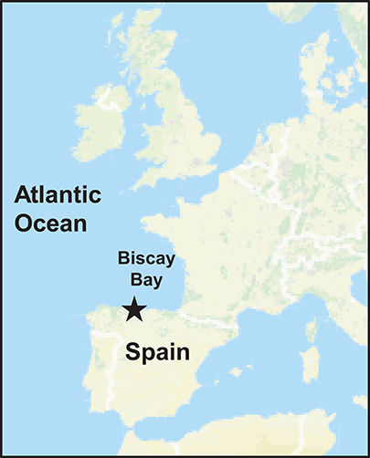
Figure 1. Sampling location. Overview of the Western European Seas (Atlantic Ocean). Star indicates the sampling location at Gijón, North of Spain (Iberian Peninsula).
Hailstone and rainwater samples from this storm event were collected in sterile recipients, while they were falling on the ground, in a terrace in front of the sea at about 30 m above sea level. Samples were stored at −20°C (hailstone) and 4°C (rain water) until immediately processing as has been reported (Braña et al., 2015).
Air Mass Backward Trajectories Analysis
In order to investigate the long-range transport journey of air masses that originated the precipitation event, backward trajectories were obtained using the HYSPLIT model (Hybrid Single Particle Lagrangian Integrated Trajectory) obtained from the Global Data Assimilation System of National Oceanic and Atmospheric Administration, USA (Stein et al., 2015). To track the transport pathways of air masses, 5-day backward trajectories (commonly used in bioaerosol studies) were generated using the NOAA model (http://ready.arl.noaa.gov/hypub-bin/trajtype.pl?runtype=archive) to determine the origin of a given air parcel. The sampling location for this study was used as the backward trajectory start point with altitudes of 30, 1,000, and 3,000 m, respectively above the ground level to estimate the accurate trajectories of atmospheric air masses.
Isolation of Streptomyces Strains and Culture Media
Atmospheric samples were inoculated on selective agar media prepared with cycloheximide (80 μg ml−1) as antifungal and nalidixic acid (20 μg ml−1) as anti-Gram negative bacteria, using MOPS BLEB 1/6 (Oxoid) basal medium as previously reported (Sarmiento-Vizcaíno et al., 2016). For selection, two different media either prepared with distilled water or with a supplement of 3.5% NaCl were used. After 2–3 weeks of incubation at 28°C, growing colonies were selected based on different morphological features and pigment production on R5A agar plates. Isolates obtained in pure cultures were conserved in 20% glycerol at −20°C, and at −70°C. For addressing halotolerance studies, MOPS BLEB 1/6 (Oxoid) was used as the basal medium, adding NaCl at of 0, 3.5, 7.0, and 10.5% (w/v) final concentrations. For secondary metabolite production streptomycetes isolates were cultured on R5A medium as previously described (Braña et al., 2015).
Antimicrobial Bioassays
Agar diffusion methods were used to determine antimicrobial activities. Antibiotic production was assessed using the following indicator microorganisms: the Gram-positive bacteria Micrococcus luteus ATCC 14452 and Streptomyces 85E ATCC 55824 (Shanbhag et al., 2015), the Gram-negative Escherichia coli ESS, and the yeast Saccharomyces cerevisiae var. carlsbergensis. Analyses were performed in TSA1/2 (Merck) against bacteria and in Sabouraud 1/2 (Pronadisa) against yeast. Bioassays were carried out both with agar plugs (7 mm diameter) and in parallel with different ethyl acetate extracts obtained from solid cultures of the isolates.
16S RNA Phylogenetic Analysis
For phylogenetic analysis of the strains based on 16S rRNA sequences, DNA was extracted with a microbial isolation kit (Ultra Clean, MoBio Laboratories, Inc.) and standard methods were used for checking the purity (Russell and Sambrook, 2001). Partial 16S rRNA gene sequences of the bacterial strains were obtained by using the 616V (forward) and 699R (reverse) primers (Arahal et al., 2008) in PCR amplification as previously described (Braña et al., 2015). Once obtained the nucleotide sequences were compared to sequences in databases using the BLAST program (Basic Local Alignment Search Tool) against the NCBI (National Centre for Biotechnology Information). The nucleotide sequences were submitted and deposited in the EMBL sequence database. Phylogenetic analysis of the strains based on 16S rRNA sequences was carried out as previously reported (Sarmiento-Vizcaíno et al., 2017a).
Chromatographic Analysis
Plugs of R5A plates (about 7 ml) were extracted using ethyl acetate in neutral and acidic (with 1% formic acid) conditions. After evaporation, the organic fraction residue was redissolved in 100 μL of a mixture of DMSO and methanol (50:50). The analysis of the samples were performed by reversed phase liquid chromatography as has been described (Braña et al., 2014; Sarmiento-Vizcaíno et al., 2016).
Identification of Compounds by LC-UV-Vis and LC-UV-HRMS Analyses
Samples were first analyzed and evaluated using an in-house HPLC-UV-Vis database. LC-UV-HRMS analyses were carried out as has been described (Pérez-Victoria et al., 2016) and major peaks in each chromatogram were searched against the MEDINA's internal database and also against the Dictionary of Natural Products (DNP) (Chapman and Hall/CRC, 2015).
Results
Backward Transport Trajectories
To estimate the sources of the air masses that caused the precipitation event in the Cantabrian Sea coast, 120 h backward trajectories were determined at three different arriving heights (30, 1,000, and 3,000 m). As shown in Figure 2, the results of the NOAA meteorological analysis indicated three different routes followed by the air layers depending on the altitude. The air-mass at 30 m altitude originate from Newfoundland and Labrador (Canada); the one at 1,000 m came from the Davies Strait in the Arctic Ocean, between North Canada and West Greenland; the air layer at 3,000 m originated at Northwest Iceland. All air-masses at different altitudes crossed the Atlantic Ocean, and after 4 days reached the Iberian Peninsula precipitating as hailstones and rainwater in the North of Spain, were samples were collected. In addition, possible mixing events were detected between different air layers at different altitudes meanwhile traveling across the Atlantic Ocean (Figure 2). Thus, the estimated backward trajectories mainly revealed an oceanic route, involving both the Arctic and Atlantic Oceans, but there was also a terrestrial route from continental America.
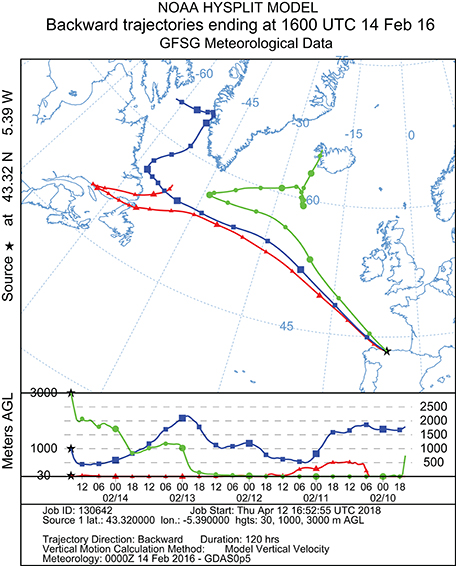
Figure 2. Five-day backward trajectories of air masses generating the storm that arrived at Gijón (North Spain) on February 14, 2016, at 16.00, calculated with the NOAA Hysplit Model with three different transects with different arriving height: red = 30 m; blue = 1,000 m; green = 3,000 m. Sampling time and place are indicated by the black asterisk.
Isolation of Bioactive Streptomyces From a Hailstorm Event
A cultivation-dependent approach revealed in atmospheric precipitations the presence of highly diverse Streptomyces populations by using a sample of hailstone (300 ml of unfrozen hailstones) and a sample of 25 ml of rainwater. Only strains cultivable at 28°C and atmospheric pressure were recovered on selective agar plates, prepared either with 3.5% NaCl (to simulate the salt content of the Cantabrian Sea water) or without salt. The percentage of streptomycete colonies recovered on selective medium without salt was higher, approximately 66% from hailstone and 56% from rainwater, than on saline medium.
Among a total of 136 streptomycete colonies isolated on selection plates (92 colonies from hailstone and 44 from rainwater), 45 morphologically different isolates from hailstone and 14 from rainwater samples were selected after the dereplication process. None of these isolates required NaCl for growth. An important feature observed in all isolates, with a single exception, was their halotolerance. Most isolates tolerate around 7% NaCl, which doubles the salt concentration in the Cantabrian Sea (3.5% average). This is in agreement with previous studies of NaCl tolerance for the genus Streptomyces in which it was estimated that around 50% of the species tolerate up to 7% NaCl (Tresner et al., 1968).
All isolates were initially tested for antimicrobial activity using agar diffusion assays. Further 29 different bioactive strains, displaying diverse antimicrobial activities, were selected for this study (Table 1). Most of the isolates displayed antibiotic activities against M. luteus and Streptomyces 85E (Gram-positive bacteria), and also against the yeast S. cerevisiae; whereas only three strains were active against the E. coli ESS (Gram-negative).
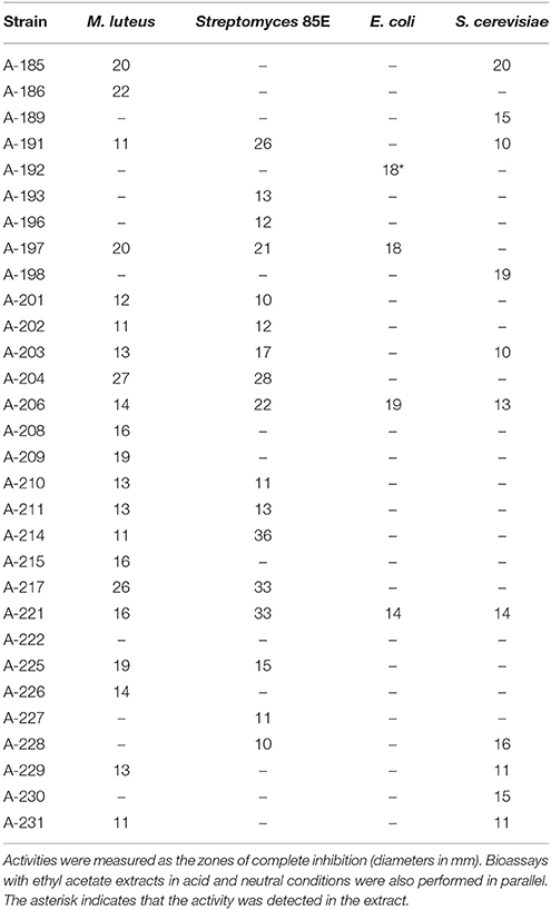
Table 1. Antibiotic activities of Streptomyces cultures (agar plugs) against Gram-positive, Gram-negative bacteria, and yeasts.
Taxonomic Identification of Isolates
For taxonomic identifications of the atmospheric-derived bioactive strains, we sequenced fragments of their 16S rDNA and deposited the nucleotide sequences in the EMBL database. Table 2 displays the accession numbers of the strains. Based on 16S rRNA gene alignments, phylogenetic analyses clearly demonstrated that all 29 isolates belonged to the Streptomyces genus, since all of them shared 99–100% identity with previously known Streptomyces species. The relationship between the atmospheric isolates and their closest phylogenetic relatives with indication of their isolation site is shown in Table 2. A phylogenetic tree was built to display the diversity among atmospheric isolates and assess their phylogenetic relationship (Figure 3).
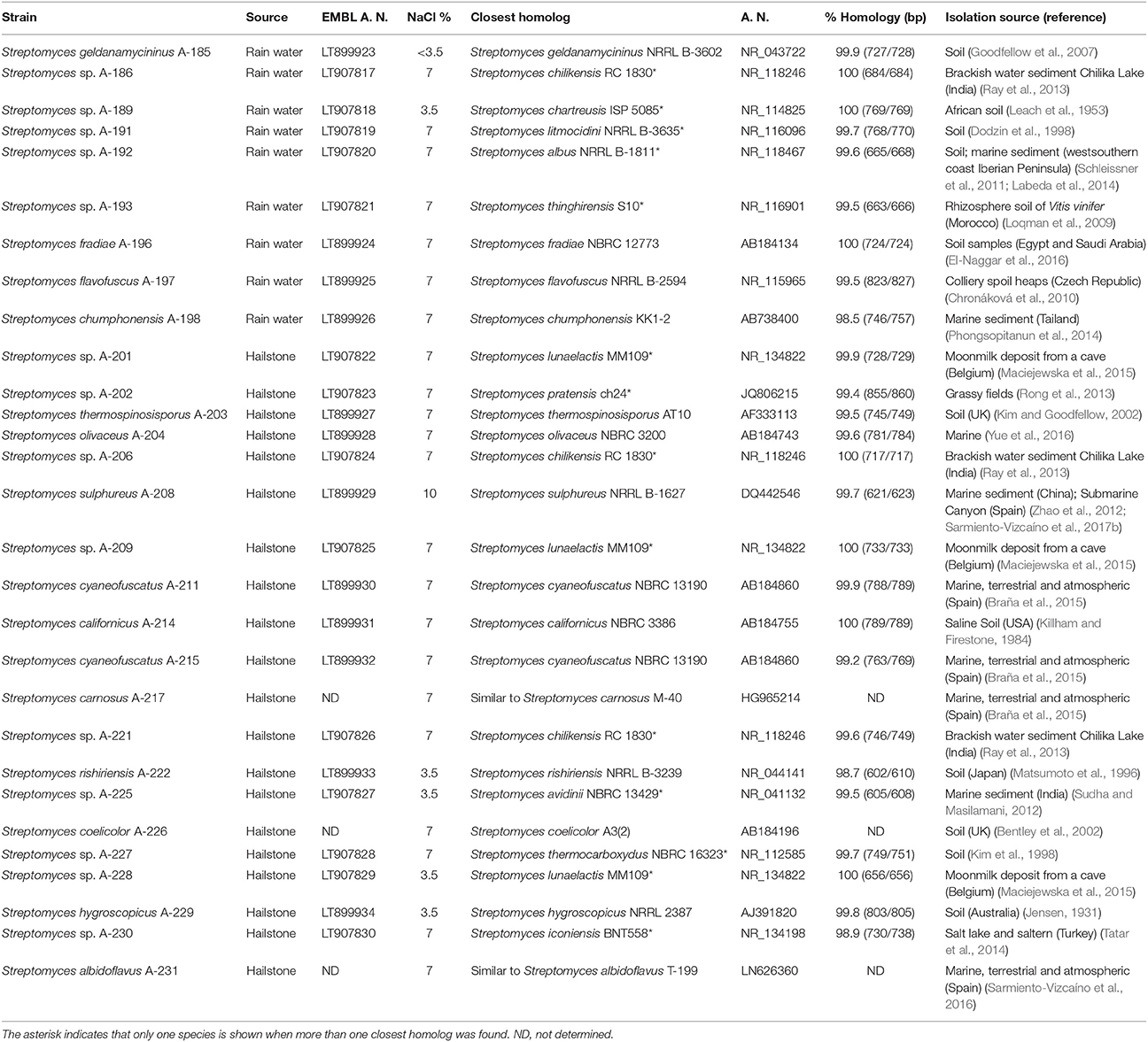
Table 2. Phylogenetic diversity of atmospheric-derived bioactive Streptomyces isolates.
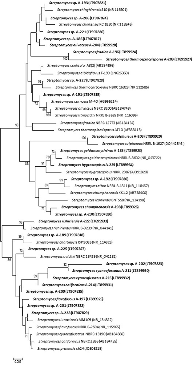
Figure 3. Neighbor-joining phylogenetic tree generated by distance matrix analysis of 16S rDNA sequences from atmospheric Streptomyces strains (highlighted) and nearest phylogenetic relatives. The numbers on branch nodes indicate bootstrap values (1,000 resamplings; only values >70% are shown). Bar represents 0.5% sequence divergence.
As shown by their identification, strains related to more than 20 different Streptomyces species have been isolated from a small sample of hailstone and rainwater (Table 2). This constitutes a significant proportion of the global number of Streptomyces species described in our planet so far, estimated in 550–823 (http://www.bacterio.net/streptomyces.html). Among 29 isolates, 15 were identified at species level and were designated with the species name (see Table 2), whereas the rest displayed 16S rDNA gene similarity to more than one species. Unfortunately, just based on these 16S rRNA sequences it is not enough to discriminate among closely related species and further assays are needed to complement the results of 16S rRNA sequence analysis for Streptomyces identification at specific level. Among identified isolates, S. albidoflavus, S. cyaneofuscatus, and S. carnosus, were repeatedly isolated in our geographical area from atmospheric precipitations, marine ecosystems (intertidal seaweeds and deep-sea corals) and terrestrial lichens. Interestingly the model species Streptomyces coelicolor, the genetically best known representative of the genus, was here isolated from hailstone; and Streptomyces albus, used as heterologous host in many laboratories, was isolated from rain water. Rare or infrequent Streptomyces species, here obtained from atmospheric precipitations, were previously isolated from highly diverse environments, mainly from the North Hemisphere (Table 2).
The habitats of known Streptomyces species closely related to the atmospheric-derived strains include soils and aquatic environments, such as oceans, lakes, and groundwater. This information was used together with the backward trajectory analysis (see above) to estimate the possible sources of airborne Streptomyces reported here.
Identification of Secondary Metabolites by Metabolite Profiling Analysis
To uncover the biosynthetic abilities of the airborne Streptomyces strains, ethyl acetate extracts were screened for secondary metabolites production by LC/UV and LC/HRMS analyses in combination with searches in UV and MS databases or the DNP after generation of a molecular formula of each peak based on HRMS results. Most of the strains displayed complex metabolic profiles revealing their high potential as a source of novel natural products (Supplementary Material 1). As an example, Figure 4 displays UV210nm chromatograms corresponding to samples A-185 and A-191 showing identified compounds.
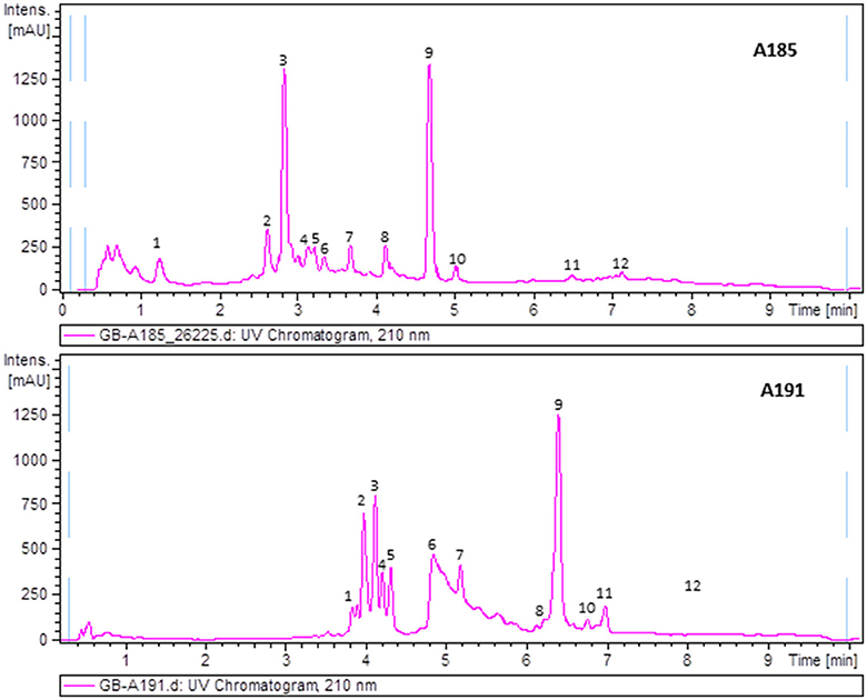
Figure 4. UV210nm chromatogram of samples A185 and A191 with peaks annotated showing components identified by HRMS. Dereplicated components in sample A185: (1) Ketomycin, (2) Thiazinogeldanamycin, (3) 4,5-Dihydrogeldanamycin, (4) Reblastatin, (5) Herbimycin E, (6) 15R-Hydroxygeldanamycin, (7) 6-Demethoxy-6-methylgeldanamycin, (8) 8-Demethylgeldanamycin or 17-O-Demethylgeldanamycin, (9) Geldanamycin, (10) C30H40N2O10 (geldanamycin related but with a molecular formula not found in the Dictionary of Natural Products), (11) Nigericin, and (12) 30-O-Acetylnigericin. Dereplicated components in sample A191: (1) Antibiotic RK 397, (2) Flavofungin I, (3 and 4) – C36H56O9 (related to flavofungin but with a molecular formula not included in the Dictionary of Natural Products), (5) Flavofungin II, (6) Prodigiosin 25b, (7) C18H30O (molecular formula not present in the Dictionary of Natural Products as produced by prokaryotes), (8) Undecylprodigiosin, (9) Spectinabilin or Arabilin, (10) Lyngbyamide D, (11) C28H33NO5 (molecular formula not found in the Dictionary of Natural Products as produced by prokaryotes), and (12) Pamamycins.
Among a total of 138 metabolites detected by HPLC/MS in the ethyl acetate extracts of the studied strains (Supplementary Material 1), 100 were identified after metabolite profiling analysis and comparison with natural products databases (some of them only at the family level). Among identified products, 45 were reported to display biological activities as antibiotics (17 antifungal and 13 antibacterial), 29 as cytotoxic/ antitumor agents, 5 as antiviral, 3 as antiparasitic, 3 as immunosupresors, 2 as anti-inflammatory, 1 as neuroprotector, 1 as insecticide, and other compounds with physiological roles in the Streptomyces life cycle (Table 3).
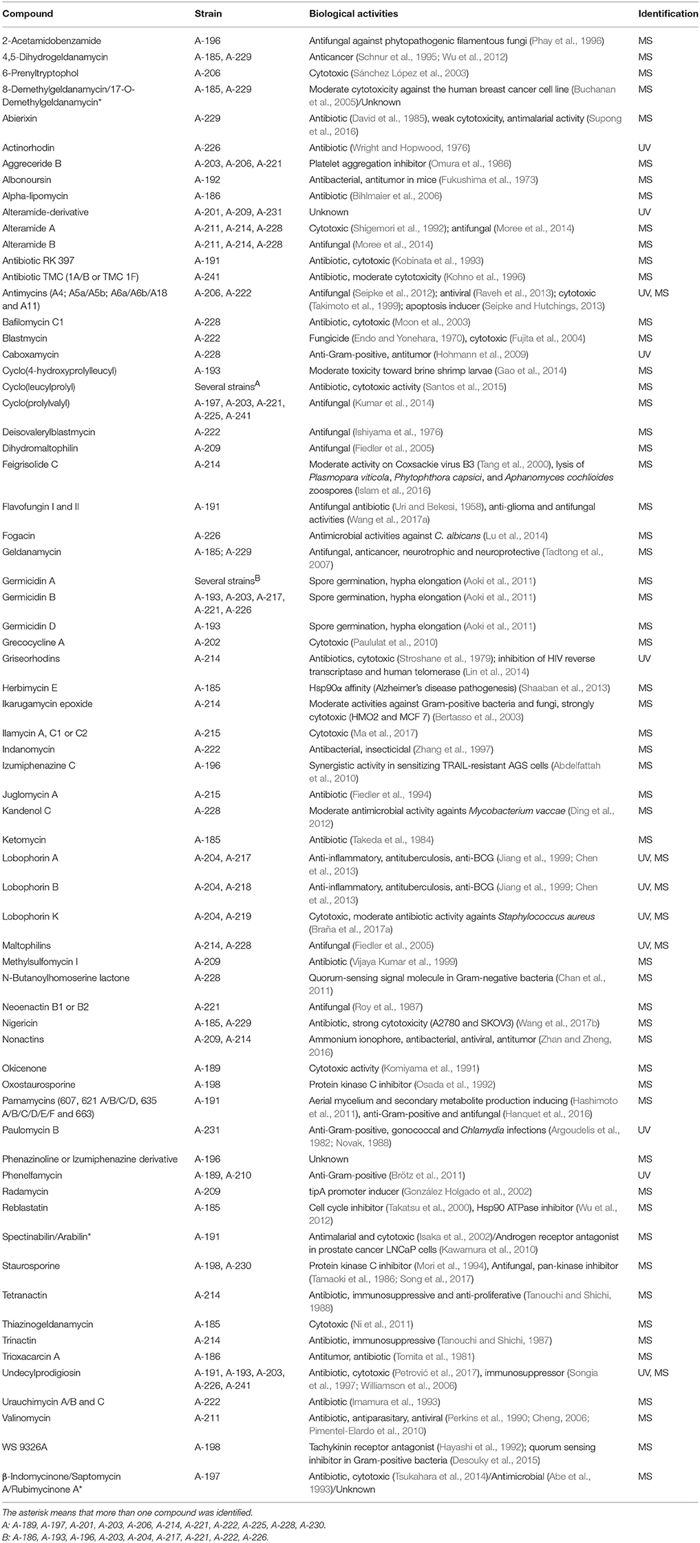
Table 3. Bioactive compounds produced by atmospheric-derived Streptomyces strains and their biological activities.
Remarkably, there are 38 metabolites whose molecular formulae determined by HRMS does not correspond to any compound included in Natural Products Databases and remained unidentified. These molecules might be new natural products, and therefore constitute an excellent starting point for the discovery of new bioactive molecules with pharmaceutical interest.
Discussion
We provide here the first insight ever made into the Streptomyces species diversity within a small storm cloud sample, which was collected after a hailstone and rainwater precipitation event during the afternoon of the 14th of February 2016 in the Cantabrian Sea coast (North of Spain). Our results revealed a striking richness of Streptomyces species present in the atmosphere. A generalized feature observed in the airborne strains here isolated (with a single exception) is their high halotolerance, since they mostly grew very well in culture media containing 7% NaCl, thus suggesting a marine origin. This has been previously proposed for Streptomyces species isolated from atmospheric precipitations in Northern Spain (Braña et al., 2015; Sarmiento-Vizcaíno et al., 2016).
Air mass backward trajectories analysis of this precipitation event revealed two main sources, oceanic (mainly from the Arctic Ocean, Greenland and Iceland) and continental (Canada) depending on the altitude. Mixing of the different air layers during the travel was observed. All air masses crossed the Atlantic Ocean and arrived to continental Europe after 4 days, reaching the North of Spain where samples were collected by atmospheric precipitation. Consistent with estimated air mass backward trajectories all isolated strains are similar to previously isolated species from highly diverse environments, either marine or terrestrial, mainly from the North hemisphere. The results of Blast search from 16S rRNA partial sequences herein provided revealed the presence of 29 strains belonging to 20–25 different Streptomyces species. Bearing in mind that the currently estimated number of Streptomyces species is of 550–823, the number of species isolated during this hail event represents a non-negligible 3–4% of all Streptomyces species known so far in our planet. This fact suggests that the presence in the atmosphere could be a generalized phenomenon within the Streptomyces genus and is in agreement with our previously stablished atmospheric dispersion model (Sarmiento-Vizcaíno et al., 2016). This model has recently received further support from culture-independent report from precipitations in Japan. That work also shows seasonal variations of microbial communities in the atmosphere in correlation with estimated air mass trajectories (Hiraoka et al., 2017).
Overall, the most relevant feature of the atmospheric-derived Streptomyces strains here studied is that they represent a striking great reservoir of structurally diverse bioactive compounds (Table 3). One hundred molecules have been identified, and for 60 of them different biological activities, mainly antimicrobial (antibacterial, antifungal, and antiviral) and antitumor properties, have been previously described. Interestingly 38 potentially bioactive natural products have not been identified and their possible novelty is the subject of current active research. During the time of writing this manuscript a new natural product was identified, after purification and NMR structure elucidation, in one of the strains here isolated (unpublished results). The number of produced secondary metabolites for these strains is estimated to be much higher than the one presented here, since only diffusible apolar molecules produced in a unique culture condition (R5A medium, 28°C) were analyzed so far, and possible diffusible polar or volatile products were not analyzed. Even more, most of the Streptomyces metabolic abilities are mainly hidden, not expressed under standard culture conditions. This silent (or cryptic) potential represents most of the metabolome (Reen et al., 2015).
Conclusion
Our findings highlight the relevance of the atmosphere as a novel source of highly diverse Streptomyces species able to produce an incredible reservoir of natural products, which has been overlooked so far. Atmospheric precipitations might represent a relevant unexplored environment for discovering bioactive natural products with pharmacological and biotechnological interest.
Author Contributions
GB isolated the strains and analyzed the air masses backward trajectories. AS-V performed the taxonomic identification and phylogenetic analyses of the strains. AS-V and JE conducted the bioactivity assays. AS-V and AB analyzed the compounds by LC-UV. JM and FR performed the metabolite profiling analysis and identified the compounds produced by LC-MS. GB wrote the manuscript which has been revised and approved by all the authors. LG and GB conceived and coordinated the project.
Conflict of Interest Statement
The authors declare that the research was conducted in the absence of any commercial or financial relationships that could be construed as a potential conflict of interest.
Acknowledgments
The authors want to thank the TBR group from the Universidad de Oviedo (GRUPIN14-140) for financial support and to Julian Davies for sending us the Streptomyces 85E strain. We also thank Victor González and Manuel Mora from the Oviedo Meteorological Observatory (AEMET), for their assistance with the NOAA analysis. We are also grateful to José L. Martínez and Daniel Serna (Servicios científico-técnicos, edificio Severo Ochoa, Universidad de Oviedo) for their great help in strains identification. This is a contribution of the Asturias Marine Observatory.
Supplementary Material
The Supplementary Material for this article can be found online at: https://www.frontiersin.org/articles/10.3389/fmicb.2018.00773/full#supplementary-material
Supplementary Material 1. UV 210 nm chromatograms corresponding to all samples.
References
Abdelfattah, M. S., Kazufumi, T., and Ishibashi, M. (2010). Izumiphenazines A-C: isolation and structure elucidation of phenazine derivatives from Streptomyces sp. IFM 11204. J. Nat. Prod. 73, 1999–2002. doi: 10.1021/np100400t
Abe, N., Nakakita, Y., Nakamura, T., Enoki, N., Uchida, H., and Munekata, M. (1993). Novel antitumor antibiotics, saptomycins. I. Taxonomy of the producing organism, fermentation, HPLC analysis and biological activities. J. Antibiot. 46, 1530–1535. doi: 10.7164/antibiotics.46.1530
Amato, P., Joly, M., Besaury, L., Oudart, A., Taib, N., Moné, A. I., et al. (2017). Active microorganisms thrive among extremely diverse communities in cloud water. PLoS ONE 12:e0182869. doi: 10.1371/journal.pone.0182869
Amato, P., Parazols, M., Sancelme, M., Laj, P., Mailhot, G., and Delort, A. M. (2007). Microorganisms isolated from the water phase of tropospheric clouds at the Puy de Dôme: major groups and growth abilities at low temperatures. FEMS Microbiol. Ecol. 59, 242–254. doi: 10.1111/j.1574-6941.2006.00199.x
Aoki, Y., Matsumoto, D., Kawaide, H., and Natsume, M. (2011). Physiological role of germicidins in spore germination and hyphal elongation in Streptomyces coelicolor A3(2). J. Antibiot. 64, 607–611. doi: 10.1038/ja.2011.59
Arahal, D. R., Sánchez, E., Macián, M. C., and Garay, E. (2008). Value of recN sequences for species identification and as a phylogenetic marker within the family “Leuconostocaceae.” Int. Microbiol. Off. J. Span. Soc. Microbiol. 11, 33–39. doi: 10.2436/20.1501.01.42
Argoudelis, A. D., Brinkley, T. A., Brodasky, T. F., Buege, J. A., Meyer, H. F., and Mizsak, S. A. (1982). Paulomycins A and B. Isolation and characterization. J. Antibiot. 35, 285–294. doi: 10.7164/antibiotics.35.285
Behie, S. W., Bonet, B., Zacharia, V. M., McClung, D. J., and Traxler, M. F. (2016). Molecules to ecosystems: actinomycete natural products In situ. Front. Microbiol. 7:2149. doi: 10.3389/fmicb.2016.02149
Bentley, S. D., Chater, K. F., Cerdeño-Tárraga, A. M., Challis, G. L., Thomson, N. R., James, K. D., et al. (2002). Complete genome sequence of the model actinomycete Streptomyces coelicolor A3(2). Nature 417, 141–147. doi: 10.1038/417141a
Bertasso, M., Holzenkämpfer, M., Zeeck, A., Stackebrandt, E., Beil, W., and Fiedler, H.-P. (2003). Ripromycin and other polycyclic macrolactams from Streptomyces sp. Tü 6239: taxonomy, fermentation, isolation and biological properties. J. Antibiot. 56, 364–371. doi: 10.7164/antibiotics.56.364
Bihlmaier, C., Welle, E., Hofmann, C., Welzel, K., Vente, A., Breitling, E., et al. (2006). Biosynthetic gene cluster for the polyenoyltetramic acid alpha-lipomycin. Antimicrob. Agents Chemother. 50, 2113–2121. doi: 10.1128/AAC.00007-06
Braña, A. F., Fiedler, H.-P., Nava, H., González, V., Sarmiento-Vizcaíno, A., et al. (2015). Two Streptomyces species producing antibiotic, antitumor, and anti-inflammatory compounds are widespread among intertidal macroalgae and deep-sea coral reef invertebrates from the central Cantabrian Sea. Microb. Ecol. 69, 512–524. doi: 10.1007/s00248-014-0508-0
Braña, A. F., Rodríguez, M., Pahari, P., Rohr, J., García, L. A., and Blanco, G. (2014). Activation and silencing of secondary metabolites in Streptomyces albus and Streptomyces lividans after transformation with cosmids containing the thienamycin gene cluster from Streptomyces cattleya. Arch. Microbiol. 196, 345–355. doi: 10.1007/s00203-014-0977-z
Braña, A. F., Sarmiento-Vizcaíno, A., Osset, M., Pérez-Victoria, I., Martín, J., de Pedro, N., et al. (2017a). Lobophorin K, a new natural product with cytotoxic activity produced by Streptomyces sp. M-207 associated with the deep-sea coral Lophelia pertusa. Mar. Drugs 15:144. doi: 10.3390/md15050144
Braña, A. F., Sarmiento-Vizcaíno, A., Pérez-Victoria, I., Otero, L., Fernández, J., Palacios, J. J., et al. (2017b). Branimycins B and C, antibiotics produced by the abyssal actinobacterium Pseudonocardia carboxydivorans M-227. J. Nat. Prod. 80, 569–573. doi: 10.1021/acs.jnatprod.6b01107
Brötz, E., Kulik, A., Vikineswary, S., Lim, C. T., Tan, G. Y. A., Zinecker, H., et al. (2011). Phenelfamycins G and H, new elfamycin-type antibiotics produced by Streptomyces albospinus Acta 3619. J. Antibiot. 64, 257–266. doi: 10.1038/ja.2010.170
Buchanan, G. O., Regentin, R., Piagentini, M., Rascher, A., McDaniel, R., Galazzo, J. L., et al. (2005). Production of 8-demethylgeldanamycin and 4,5-epoxy-8-demethylgeldanamycin from a recombinant strain of Streptomyces hygroscopicus. J. Nat. Prod. 68, 607–610. doi: 10.1021/np0496744
Chan, K.-G., Puthucheary, S. D., Chan, X. Y., Yin, W. F., Wong, C. S., Too, W. S. S., et al. (2011). Quorum sensing in Aeromonas species isolated from patients in Malaysia. Curr. Microbiol. 62, 167–172. doi: 10.1007/s00284-010-9689-z
Chen, C., Wang, J., Guo, H., Hou, W., Yang, N., Ren, B., et al. (2013). Three antimycobacterial metabolites identified from a marine-derived Streptomyces sp. MS100061. Appl. Microbiol. Biotechnol. 97, 3885–3892. doi: 10.1007/s00253-012-4681-0
Cheng, Y. Q. (2006). Deciphering the biosynthetic codes for the potent anti-SARS-CoV cyclodepsipeptide valinomycin in Streptomyces tsusimaensis ATCC 15141. Chembiochem Eur. J. Chem. Biol. 7, 471–477. doi: 10.1002/cbic.200500425
Chronáková, A., Kristufek, V., Tichý, M., and Elhottová, D. (2010). Biodiversity of streptomycetes isolated from a succession sequence at a post-mining site and their evidence in Miocene lacustrine sediment. Microbiol. Res. 165, 594–608. doi: 10.1016/j.micres.2009.10.002
David, L., Leal Ayala, H., and Tabet, J. C. (1985). Abierixin, a new polyether antibiotic. Production, structural determination and biological activities. J. Antibiot. 38, 1655–1663. doi: 10.7164/antibiotics.38.1655
Desouky, S. E., Shojima, A., Singh, R. P., Matsufuji, T., Igarashi, Y., Suzuki, T., et al. (2015). Cyclodepsipeptides produced by actinomycetes inhibit cyclic-peptide-mediated quorum sensing in Gram-positive bacteria. FEMS Microbiol. Lett. 362:fnv109. doi: 10.1093/femsle/fnv109
Ding, L., Maier, A., Fiebig, H.-H., Lin, W.-H., Peschel, G., and Hertweck, C. (2012). Kandenols A-E, eudesmenes from an endophytic Streptomyces sp. of the mangrove tree Kandelia candel. J. Nat. Prod. 75, 2223–2227. doi: 10.1021/np300387n
Chapman and Hall/CRC (2015). Dictionary of Marine Natural Products. Available online at: http://dnp.chemnetbase.com
Dodzin, M. E., Vinogradova, K. A., and Kotova, I. B. (1998). [Novel L-glutamate oxidase producing organisms: Streptomyces litmocidini and Streptomyces cremeus]. Antibiot. Khimioterapiia Antibiot. Chemoterapy Sic. 43, 7–13.
El-Naggar, Nel-A., Deraz, S. F., Soliman, H. M., El-Deeb, N. M., and El-Ewasy, S. M. (2016). Purification, characterization, cytotoxicity and anticancer activities of L-asparaginase, anti-colon cancer protein, from the newly isolated alkaliphilic Streptomyces fradiae NEAE-82. Sci. Rep. 6:32926. doi: 10.1038/srep32926
Endo, T., and Yonehara, H. (1970). Chemical studies on blastmycin. 3. Gas-liquid chromatography of antimycin A-blastmycin antibiotics. J. Antibiot. 23, 91–95. doi: 10.7164/antibiotics.23.91
Fiedler, H. P., Bruntner, C., Bull, A. T., Ward, A. C., Goodfellow, M., Potterat, O., et al. (2005). Marine actinomycetes as a source of novel secondary metabolites. Antonie Van Leeuwenhoek 87, 37–42. doi: 10.1007/s10482-004-6538-8
Fiedler, H. P., Kulik, A., Schüz, T. C., Volkmann, C., and Zeeck, A. (1994). Biosynthetic capacities of actinomycetes. 2. Juglomycin Z, a new naphthoquinone antibiotic from Streptomyces tendae. J. Antibiot. 47, 1116–1122. doi: 10.7164/antibiotics.47.1116
Fujita, K. I., Kiso, T., Usuki, Y., Tanaka, T., and Taniguchi, M. (2004). UK-2A, B, C and D, novel antifungal antibiotics from streptomyces sp. 517-02 VI (3). Role of substituents on dilactone ring of UK-2A and antimycin A3 against generation of reactive oxygen species in porcine renal proximal tubule LLC-PK1 cells. J. Antibiot. 57, 687–690. doi: 10.7164/antibiotics.57.687
Fukushima, K., Yazawa, K., and Arai, T. (1973). Biological activities of albonoursin. J. Antibiot. 26, 175–176. doi: 10.7164/antibiotics.26.175
Gao, C., Lin, L., Long, B., Chen, Y., He, B., Sun, H., et al. (2014). A new diketopiperazine from the gorgonian coral Menella kanisa. Nat. Prod. Res. 28, 473–476. doi: 10.1080/14786419.2013.879134
González Holgado, G., Castro Rodríguez, J., Cañedo Hernández, L. M., Díaz, M., Fernández-Abalos, J. M., Trujillano, I., et al. (2002). Radamycin, a novel thiopeptide produced by streptomyces sp. RSP9. I. Taxonomy, fermentation, isolation and biological activities. J. Antibiot. 55, 383–390. doi: 10.7164/antibiotics.55.383
Goodfellow, M., Kumar, Y., Labeda, D. P., and Sembiring, L. (2007). The Streptomyces violaceusniger clade: a home for Streptomycetes with rugose ornamented spores. Antonie Van Leeuwenhoek 92, 173–199. doi: 10.1007/s10482-007-9146-6
Hanquet, G., Salom-Roig, X., and Lanners, S. (2016). New insights into the synthesis and biological activity of the pamamycin macrodiolides. Chimia 70, 20–28. doi: 10.2533/chimia.2016.20
Hashimoto, M., Katsura, H., Kato, R., Kawaide, H., and Natsume, M. (2011). Effect of pamamycin-607 on secondary metabolite production by Streptomyces spp. Biosci. Biotechnol. Biochem. 75, 1722–1726. doi: 10.1271/bbb.110251
Hayashi, K., Hashimoto, M., Shigematsu, N., Nishikawa, M., Ezaki, M., Yamashita, M., et al. (1992). WS9326A, a novel tachykinin antagonist isolated from Streptomyces violaceusniger no. 9326. I. Taxonomy, fermentation, isolation, physico-chemical properties and biological activities. J. Antibiot. 45, 1055–1063. doi: 10.7164/antibiotics.45.1055
Hiraoka, S., Miyahara, M., Fujii, K., Machiyama, A., and Iwasaki, W. (2017). Seasonal analysis of microbial communities in precipitation in the greater Tokyo area, Japan. Front. Microbiol. 8:1506. doi: 10.3389/fmicb.2017.01506
Hohmann, C., Schneider, K., Bruntner, C., Irran, E., Nicholson, G., Bull, A. T., et al. (2009). Caboxamycin, a new antibiotic of the benzoxazole family produced by the deep-sea strain Streptomyces sp. NTK 937. J. Antibiot. 62, 99–104. doi: 10.1038/ja.2008.24
Imamura, N., Nishijima, M., Adachi, K., and Sano, H. (1993). Novel antimycin antibiotics, urauchimycins A and B, produced by marine actinomycete. J. Antibiot. 46, 241–246. doi: 10.7164/antibiotics.46.241
Isaka, M., Jaturapat, A., Kramyu, J., Tanticharoen, M., and Thebtaranonth, Y. (2002). Potent in vitro antimalarial activity of metacycloprodigiosin isolated from Streptomyces spectabilis BCC 4785. Antimicrob. Agents Chemother. 46, 1112–1113. doi: 10.1128/AAC.46.4.1112-1113.2002
Ishiyama, T., Endo, T., Otake, N., and Yonehara, H. (1976). Deisovalerylblastmycin produced by Streptomyces sp. J. Antibiot. 29, 804–808. doi: 10.7164/antibiotics.29.804
Islam, M. T., Laatsch, H., and von Tiedemann, A. (2016). Inhibitory effects of macrotetrolides from Streptomyces spp. on Zoosporogenesis and motility of Peronosporomycete zoospores are likely linked with enhanced ATPase activity in mitochondria. Front. Microbiol. 7:1824. doi: 10.3389/fmicb.2016.01824
Jensen, H. L. (1931). Contributions to our knowledge of the Actinomycetales. II. The definition and subdivision of the genus Actinomyces, with a preliminary account of Australian soil Actinomycetes. Proc. Linn. Soc. New South Wales 56, 345–370.
Jiang, Z. D., Jensen, P. R., and Fenical, W. (1999). Lobophorins A and B, new antiinflammatory macrolides produced by a tropical marine bacterium. Bioorg. Med. Chem. Lett. 9, 2003–2006. doi: 10.1016/S0960-894X(99)00337-6
Kawamura, T., Fujimaki, T., Hamanaka, N., Torii, K., Kobayashi, H., Takahashi, Y., et al. (2010). Isolation and structure elucidation of a novel androgen antagonist, arabilin, produced by Streptomyces sp. MK756-CF1. J. Antibiot. 63, 601–605. doi: 10.1038/ja.2010.98
Killham, K., and Firestone, M. K. (1984). Salt stress control of intracellular solutes in Streptomycetes indigenous to saline soils. Appl. Environ. Microbiol. 47, 301–306.
Kim, S. B., Falconer, C., Williams, E., and Goodfellow, M. (1998). Streptomyces thermocarboxydovorans sp. nov. and Streptomyces thermocarboxydus sp. nov., two moderately thermophilic carboxydotrophic species from soil. Int. J. Syst. Bacteriol. 48(Pt 1), 59–68. doi: 10.1099/00207713-48-1-59
Kim, S. B., and Goodfellow, M. (2002). Streptomyces thermospinisporus sp. nov., a moderately thermophilic carboxydotrophic streptomycete isolated from soil. Int. J. Syst. Evol. Microbiol. 52, 1225–1228. doi: 10.1099/00207713-52-4-1225
Kobinata, K., Koshino, H., Kudo, T., Isono, K., and Osada, H. (1993). RK-397, a new oxo pentaene antibiotic. J. Antibiot. 46, 1616–1618. doi: 10.7164/antibiotics.46.1616
Kohno, J., Nishio, M., Kawano, K., Nakanishi, N., Suzuki, S., Uchida, T., et al. (1996). TMC-1 A, B, C and D, new antibiotics of the manumycin group produced by Streptomyces sp. taxonomy, production, isolation, physico-chemical properties, structure elucidation and biological properties. J. Antibiot. 49, 1212–1220. doi: 10.7164/antibiotics.49.1212
Komiyama, K., Funayama, S., Anraku, Y., Ishibashi, M., Takahashi, Y., Kawakami, T., et al. (1991). A new antibiotic, okicenone. I. taxonomy, fermentation, isolation and biological characteristics. J. Antibiot. 44, 814–818. doi: 10.7164/antibiotics.44.814
Kumar, S. N., Nambisan, B., Sundaresan, A., Mohandas, C., and Anto, R. J. (2014). Isolation and identification of antimicrobial secondary metabolites from Bacillus cereus associated with a rhabditid entomopathogenic nematode. Ann. Microbiol. 64, 209–218. doi: 10.1007/s13213-013-0653-6
Labeda, D. P., Doroghazi, J. R., Ju, K. S., and Metcalf, W. W. (2014). Taxonomic evaluation of Streptomyces albus and related species using multilocus sequence analysis and proposals to emend the description of Streptomyces albus and describe Streptomyces pathocidini sp. nov. Int. J. Syst. Evol. Microbiol. 64, 894–900. doi: 10.1099/ijs.0.058107-0
Law, J. W. F., Ser, H. L., Duangjai, A., Saokaew, S., Bukhari, S. I., Khan, T. M., et al. (2017). Streptomyces colonosanans sp. nov., A novel Actinobacterium isolated from malaysia mangrove soil exhibiting antioxidative activity and cytotoxic potential against human colon cancer cell lines. Front. Microbiol. 8:877. doi: 10.3389/fmicb.2017.00877
Leach, B. E., Calhoun, K. M., Johnson, L. E., Teeters, C. M., and Jackson, W. G. (1953). Chartreusin, a new antibiotic produced by Streptomyces chartreusis, a new species. J. Am. Chem. Soc. 75, 4011–4012. doi: 10.1021/ja01112a040
Lin, Z., Zachariah, M. M., Marett, L., Hughen, R. W., Teichert, R. W., Concepcion, G. P., et al. (2014). Griseorhodins D-F, neuroactive intermediates and end products of post-PKS tailoring modification in Griseorhodin biosynthesis. J. Nat. Prod. 77, 1224–1230. doi: 10.1021/np500155d
Loqman, S., Bouizgarne, B., Barka, E. A., Clément, C., von Jan, M., Spröer, C., et al. (2009). Streptomyces thinghirensis sp. nov., isolated from rhizosphere soil of Vitis vinifera. Int. J. Syst. Evol. Microbiol. 59, 3063–3067. doi: 10.1099/ijs.0.008946-0
Lu, Y., Li, S., Zhou, D., and Zhang, Y. (2014). [Isolation and identification of termitarium antagonistic actinomycetes BYC 01 and its active metabolites]. Wei Sheng Wu Xue Bao 54, 754–759.
Ma, J., Huang, H., Xie, Y., Liu, Z., Zhao, J., Zhang, C., et al. (2017). Biosynthesis of ilamycins featuring unusual building blocks and engineered production of enhanced anti-tuberculosis agents. Nat. Commun. 8:391. doi: 10.1038/s41467-017-00419-5
Maciejewska, M., Adam, D., Martinet, L., Naômé, A., Całusinska, M., Delfosse, P., et al. (2016). A phenotypic and genotypic analysis of the antimicrobial potential of cultivable streptomyces isolated from cave moonmilk deposits. Front. Microbiol. 7:1455. doi: 10.3389/fmicb.2016.01455
Maciejewska, M., Pessi, I. S., Arguelles-Arias, A., Noirfalise, P., Luis, G., Ongena, M., et al. (2015). Streptomyces lunaelactis sp. nov., a novel ferroverdin A-producing Streptomyces species isolated from a moonmilk speleothem. Antonie Van Leeuwenhoek 107, 519–531. doi: 10.1007/s10482-014-0348-4
Matsumoto, N., Tsuchida, T., Maruyama, M., Sawa, R., Kinoshita, N., Homma, Y., et al. (1996). Lactonamycin, a new antimicrobial antibiotic produced by Streptomyces rishiriensis. J. Antibiot. 49, 953–954. doi: 10.7164/antibiotics.49.953
Moon, S. S., Hwang, W. H., Chung, Y. R., and Shin, J. (2003). New cytotoxic bafilomycin C1-amide produced by Kitasatospora cheerisanensis. J. Antibiot. 56, 856–861. doi: 10.7164/antibiotics.56.856
Moree, W. J., McConnell, O. J., Nguyen, D. D., Sanchez, L. M., Yang, Y. L., Zhao, X., et al. (2014). Microbiota of healthy corals are active against fungi in a light-dependent manner. ACS Chem. Biol. 9, 2300–2308. doi: 10.1021/cb500432j
Mori, T., Ando, M., and Takagi, K. (1994). Staurosporine and its derivatives enhance f-Met-Leu-Phe-induced superoxide production via phospholipase D activation in human polymorphonuclear leukocytes. Int. J. Clin. Pharmacol. Ther. 32, 422–428.
Ni, S., Wu, L., Wang, H., Gan, M., Wang, Y., He, W., et al. (2011). Thiazinogeldanamycin, a new geldanamycin derivative produced by Streptomyces hygroscopicus 17997. J. Microbiol. Biotechnol. 21, 599–603.
Novak, E. (1988). Treating Chlamydia Infections With Paulomycin. Patent PCT/US1987/002420: The Upjohn Company.
Omura, S., Nakagawa, A., Fukamachi, N., Otoguro, K., and Kobayashi, B. (1986). Aggreceride, a new platelet aggregation inhibitor from Streptomyces. J. Antibiot. 39, 1180–1181. doi: 10.7164/antibiotics.39.1180
Osada, H., Koshino, H., Kudo, T., Onose, R., and Isono, K. (1992). A new inhibitor of protein kinase C, RK-1409 (7-oxostaurosporine). I. Taxonomy and biological activity. J. Antibiot. 45, 189–194. doi: 10.7164/antibiotics.45.189
Paululat, T., Kulik, A., Hausmann, H., Karagouni, A. D., Zinecker, H., Imhoff, J. F., et al. (2010). Grecocyclines: new angucyclines from Streptomyces sp. Acta 1362. Eur. J. Org. Chem. 2010, 2344–2350. doi: 10.1002/ejoc.201000054
Pérez-Victoria, I., Martín, J., and Reyes, F. (2016). Combined LC/UV/MS and NMR strategies for the dereplication of marine natural products. Planta Med. 82, 857–871. doi: 10.1055/s-0042-101763
Perkins, J. B., Guterman, S. K., Howitt, C. L., Williams, V. E., and Pero, J. (1990). Streptomyces genes involved in biosynthesis of the peptide antibiotic valinomycin. J. Bacteriol. 172, 3108–3116. doi: 10.1128/jb.172.6.3108-3116.1990
Petrović, S., Vasić, V., Mitrović, T., Lazović, S., and Leskovac, A. (2017). The impact of concentration and administration time on the radiomodulating properties of undecylprodigiosin in vitro. Arh. Hig. Rada Toksikol. 68, 1–8. doi: 10.1515/aiht-2017-68-2897
Phay, N., Yada, H., Higashiyama, T., Yokota, A., Ichihara, A., and Tomita, F. (1996). NP-101A, antifungal antibiotic from Streptomyces aurantiogriseus NPO-101. J. Antibiot. 49, 703–705. doi: 10.7164/antibiotics.49.703
Phongsopitanun, W., Thawai, C., Suwanborirux, K., Kudo, T., Ohkuma, M., and Tanasupawat, S. (2014). Streptomyces chumphonensis sp. nov., isolated from marine sediments. Int. J. Syst. Evol. Microbiol. 64, 2605–2610. doi: 10.1099/ijs.0.062992-0
Pimentel-Elardo, S. M., Kozytska, S., Bugni, T. S., Ireland, C. M., Moll, H., and Hentschel, U. (2010). Anti-parasitic compounds from Streptomyces sp. strains isolated from Mediterranean sponges. Mar. Drugs 8, 373–380. doi: 10.3390/md8020373
Raveh, A., Delekta, P. C., Dobry, C. J., Peng, W., Schultz, P. J., Blakely, P. K., et al. (2013). Discovery of potent broad spectrum antivirals derived from marine actinobacteria. PLoS ONE 8:e82318. doi: 10.1371/journal.pone.0082318
Ray, L., Suar, M., Pattnaik, A. K., and Raina, V. (2013). Streptomyces chilikensis sp. nov., a halophilic streptomycete isolated from brackish water sediment. Int. J. Syst. Evol. Microbiol. 63, 2757–2764. doi: 10.1099/ijs.0.046284-0
Reen, F. J., Romano, S., Dobson, A. D. W., and O'Gara, F. (2015). The sound of silence: activating silent biosynthetic gene clusters in marine microorganisms. Mar. Drugs 13, 4754–4783. doi: 10.3390/md13084754
Rong, X., Doroghazi, J. R., Cheng, K., Zhang, L., Buckley, D. H., and Huang, Y. (2013). Classification of Streptomyces phylogroup pratensis (Doroghazi and Buckley, 2010) based on genetic and phenotypic evidence, and proposal of Streptomyces pratensis sp. nov. Syst. Appl. Microbiol. 36, 401–407. doi: 10.1016/j.syapm.2013.03.010
Roy, S. K., Inouye, Y., Nakamura, S., Furukawa, J., and Okuda, S. (1987). Isolation, structural elucidation and biological properties of neoenactins B1, B2, M1 and M2, neoenactin congeners. J. Antibiot. 40, 266–274. doi: 10.7164/antibiotics.40.266
Russell, D. W., and Sambrook, J. F. (2001). Molecular Cloning: A Laboratory Manual, 3rd Edn. New York, NY: Cold Spring Harbor Laboratory Press.
Sánchez López, J. M., Martínez Insua, M., Pérez Baz, J., Fernández Puentes, J. L., and Cañedo Hernández, L. M. (2003). New cytotoxic indolic metabolites from a marine Streptomyces. J. Nat. Prod. 66, 863–864. doi: 10.1021/np0204444
Santos, O. C., Soares, A. R., Machado, F. L. S., Romanos, M. T. V., Muricy, G., Giambiagi-deMarval, M., et al. (2015). Investigation of biotechnological potential of sponge-associated bacteria collected in Brazilian coast. Lett. Appl. Microbiol. 60, 140–147. doi: 10.1111/lam.12347
Sarmiento-Vizcaíno, A., Braña, A. F., González, V., Nava, H., Molina, A., Llera, E., et al. (2016). Atmospheric dispersal of bioactive Streptomyces albidoflavus strains among terrestrial and marine environments. Microb. Ecol. 71, 375–386. doi: 10.1007/s00248-015-0654-z
Sarmiento-Vizcaíno, A., Braña, A. F., Pérez-Victoria, I., Martín, J., de Pedro, N., de la Cruz, M., et al. (2017a). Paulomycin G, a new natural product with cytotoxic activity against tumor cell lines produced by deep-sea sediment derived Micromonospora matsumotoense M-412 from the Avilés Canyon in the Cantabrian Sea. Mar. Drugs 15:271. doi: 10.3390/md15090271
Sarmiento-Vizcaíno, A., González, V., Braña, A. F., Molina, A., Acuña, J. L., García, L. A., et al. (2015). Myceligenerans cantabricum sp. nov., a barotolerant actinobacterium isolated from a deep cold-water coral. Int. J. Syst. Evol. Microbiol. 65, 1328–1334. doi: 10.1099/ijs.0.000107
Sarmiento-Vizcaíno, A., González, V., Braña, A. F., Palacios, J. J., Otero, L., Fernández, J., et al. (2017b). Pharmacological potential of phylogenetically diverse actinobacteria isolated from deep-sea coral ecosystems of the submarine Avilés Canyon in the Cantabrian Sea. Microb. Ecol. 73, 338–352. doi: 10.1007/s00248-016-0845-2
Šantl-Temkiv, T., Finster, K., Dittmar, T., Hansen, B. M., Thyrhaug, R., Nielsen, N. W., et al. (2013). Hailstones: a window into the microbial and chemical inventory of a storm cloud. PLoS ONE 8:e53550. doi: 10.1371/journal.pone.0053550
Schleissner, C., Pérez, M., Losada, A., Rodríguez, P., Crespo, C., Zúñiga, P., et al. (2011). Antitumor actinopyranones produced by Streptomyces albus POR-04-15-053 isolated from a marine sediment. J. Nat. Prod. 74, 1590–1596. doi: 10.1021/np200196j
Schnur, R. C., Corman, M. L., Gallaschun, R. J., Cooper, B. A., Dee, M. F., Doty, J. L., et al. (1995). Inhibition of the oncogene product p185erbB-2 in vitro and in vivo by geldanamycin and dihydrogeldanamycin derivatives. J. Med. Chem. 38, 3806–3812. doi: 10.1021/jm00019a010
Seipke, R. F., and Hutchings, M. I. (2013). The regulation and biosynthesis of antimycins. Beilstein J. Org. Chem. 9, 2556–2563. doi: 10.3762/bjoc.9.290
Seipke, R. F., Kaltenpoth, M., and Hutchings, M. I. (2012). Streptomyces as symbionts: an emerging and widespread theme? FEMS Microbiol. Rev. 36, 862–876. doi: 10.1111/j.1574-6976.2011.00313.x
Shaaban, K. A., Wang, X., Elshahawi, S. I., Ponomareva, L. V., Sunkara, M., Copley, G. C., et al. (2013). Herbimycins D-F, ansamycin analogues from Streptomyces sp. RM-7-15. J. Nat. Prod. 76, 1619–1626. doi: 10.1021/np400308w
Shanbhag, P., Bhave, S., Vartak, A., Kulkarni-Almeida, A., Mahajan, G., Villanueva, I., et al. (2015). Screening of microbial extracts for anticancer compounds using Streptomyces kinase inhibitor assay. Nat. Prod. Commun. 10, 1287–1291.
Shigemori, H., Bae, M. A., Yazawa, K., Sasaki, T., and Kobayashi, J. (1992). Alteramide A, a new tetracyclic alkaloid from a bacterium Alteromonas sp. associated with the marine sponge Halichondria okadai. J. Org. Chem. 57, 4317–4320. doi: 10.1021/jo00041a053
Song, X., Liu, X., and Ding, X. (2017). Staurosporine scaffold-based rational discovery of the wild-type sparing reversible inhibitors of EGFR T790M gatekeeper mutant in lung cancer with analog-sensitive kinase technology. J. Mol. Recognit. 30:e2590. doi: 10.1002/jmr.2590
Songia, S., Mortellaro, A., Taverna, S., Fornasiero, C., Scheiber, E. A., Erba, E., et al. (1997). Characterization of the new immunosuppressive drug undecylprodigiosin in human lymphocytes: retinoblastoma protein, cyclin-dependent kinase-2, and cyclin-dependent kinase-4 as molecular targets. J. Immunol. Baltim. Md 158, 3987–3995.
Stein, A. F., Draxler, R. R., Rolph, G. D., Stunder, B. J. B., Cohen, M. D., and Ngan, F. (2015). NOAA's HYSPLIT atmospheric transport and dispersion modeling system. Bull. Am. Meteorol. Soc. 96, 2059–2077. doi: 10.1175/BAMS-D-14-00110.1
Stroshane, R. M., Chan, J. A., Rubalcaba, E. A., Garretson, A. L., Aszalos, A. A., and Roller, P. P. (1979). Isolation and structure elucidation of a novel griseorhodin. J. Antibiot. 32, 197–204. doi: 10.7164/antibiotics.32.197
Subramani, R., and Aalbersberg, W. (2013). Culturable rare Actinomycetes: diversity, isolation and marine natural product discovery. Appl. Microbiol. Biotechnol. 97, 9291–9321. doi: 10.1007/s00253-013-5229-7
Sudha, S., and Masilamani, S. M. (2012). Characterization of cytotoxic compound from marine sediment derived actinomycete Streptomyces avidinii strain SU4. Asian Pac. J. Trop. Biomed. 2, 770–773. doi: 10.1016/S2221-1691(12)60227-5
Supong, K., Thawai, C., Choowong, W., Kittiwongwattana, C., Thanaboripat, D., Laosinwattana, C., et al. (2016). Antimicrobial compounds from endophytic Streptomyces sp. BCC72023 isolated from rice (Oryza sativa L.). Res. Microbiol. 167, 290–298. doi: 10.1016/j.resmic.2016.01.004
Tadtong, S., Meksuriyen, D., Tanasupawat, S., Isobe, M., and Suwanborirux, K. (2007). Geldanamycin derivatives and neuroprotective effect on cultured P19-derived neurons. Bioorg. Med. Chem. Lett. 17, 2939–2943. doi: 10.1016/j.bmcl.2006.12.041
Takatsu, T., Ohtsuki, M., Muramatsu, A., Enokita, R., and Kurakata, S. I. (2000). Reblastatin, a novel benzenoid ansamycin-type cell cycle inhibitor. J. Antibiot. 53, 1310–1312. doi: 10.7164/antibiotics.53.1310
Takeda, Y., Mak, V., Chang, C. C., Chang, C. J., and Floss, H. G. (1984). Biosynthesis of ketomycin. J. Antibiot. 37, 868–875. doi: 10.7164/antibiotics.37.868
Takimoto, H., Machida, K., Ueki, M., Tanaka, T., and Taniguchi, M. (1999). UK-2A, B, C and D, novel antifungal antibiotics from Streptomyces sp. 517-02. IV. Comparative studies of UK-2A with antimycin A3 on cytotoxic activity and reactive oxygen species generation in LLC-PK1 cells. J. Antibiot. 52, 480–484. doi: 10.7164/antibiotics.52.480
Tamaoki, T., Nomoto, H., Takahashi, I., Kato, Y., Morimoto, M., and Tomita, F. (1986). Staurosporine, a potent inhibitor of phospholipid/Ca++dependent protein kinase. Biochem. Biophys. Res. Commun. 135, 397–402. doi: 10.1016/0006-291X(86)90008-2
Tang, Y. Q., Sattler, I., Thiericke, R., Grabley, S., and Feng, X. Z. (2000). Feigrisolides A, B, C and D, new lactones with antibacterial activities from Streptomyces griseus. J. Antibiot. 53, 934–943. doi: 10.7164/antibiotics.53.934
Tanouchi, Y., and Shichi, H. (1987). Immunosuppressive effects of polynactins (tetranactin, trinactin and dinactin) on experimental autoimmune uveoretinitis in rats. Jpn. J. Ophthalmol. 31, 218–229.
Tanouchi, Y., and Shichi, H. (1988). Immunosuppressive and anti-proliferative effects of a macrotetrolide antibiotic, tetranactin. Immunology 63, 471–475.
Tatar, D., Guven, K., Spröer, C., Klenk, H.-P., and Sahin, N. (2014). Streptomyces iconiensis sp. nov. and Streptomyces smyrnaeus sp. nov., two halotolerant actinomycetes isolated from a salt lake and saltern. Int. J. Syst. Evol. Microbiol. 64, 3126–3133. doi: 10.1099/ijs.0.062216-0
Tomita, F., Tamaoki, T., Morimoto, M., and Fujimoto, K. (1981). Trioxacarcins, novel antitumor antibiotics. I. Producing organism, fermentation and biological activities. J. Antibiot. 34, 1519–1524. doi: 10.7164/antibiotics.34.1519
Tresner, H. D., Hayes, J. A., and Backus, E. J. (1968). Differential tolerance of streptomycetes to sodium chloride as a taxonomic aid. Appl. Microbiol. 16, 1134–1136.
Tsukahara, K., Toume, K., Ito, H., Ishikawa, N., and Ishibashi, M. (2014). Isolation of β-indomycinone guided by cytotoxicity tests from Streptomyces sp. IFM11607 and revision of its double bond geometry. Nat. Prod. Commun. 9, 1327–1328.
Uri, J., and Bekesi, I. (1958). Flavofungin, a new crystalline antifungal antibiotic: origin and biological properties. Nature 181:908. doi: 10.1038/181908a0
Vijaya Kumar, E. K., Kenia, J., Mukhopadhyay, T., and Nadkarni, S. R. (1999). Methylsulfomycin I, a new cyclic peptide antibiotic from a Streptomyces sp. HIL Y-9420704. J. Nat. Prod. 62, 1562–1564. doi: 10.1021/np990088y
Wang, W., Song, T., Chai, W., Chen, L., Chen, L., Lian, X. Y., et al. (2017a). Rare polyene-polyol macrolides from mangrove-derived Streptomyces sp. ZQ4BG. Sci. Rep. 7:1703. doi: 10.1038/s41598-017-01912-z
Wang, W., Zhao, Y., Yao, S., Cui, X., Pan, W., Huang, W., et al. (2017b). Nigericin inhibits epithelial ovarian cancer metastasis by suppressing the cell cycle and epithelial-mesenchymal transition. Biochem. Biokhimiia 82, 933–941. doi: 10.1134/S0006297917080089
Williamson, N. R., Fineran, P. C., Leeper, F. J., and Salmond, G. P. (2006). The biosynthesis and regulation of bacterial prodiginines. Nat. Rev. Microbiol. 4, 887–899. doi: 10.1038/nrmicro1531
Wright, L. F., and Hopwood, D. A. (1976). Actinorhodin is a chromosomally-determined antibiotic in Streptomyces coelicolar A3(2). J. Gen. Microbiol. 96, 289–297. doi: 10.1099/00221287-96-2-289
Wu, C. Z., Jang, J. H., Ahn, J. S., and Hong, Y. S. (2012). New geldanamycin analogs from Streptomyces hygroscopicus. J. Microbiol. Biotechnol. 22, 1478–1481. doi: 10.4014/jmb.1206.06026
Yue, C., Niu, J., Liu, N., Lü, Y., Liu, M., and Li, Y. (2016). Cloning and identification of the lobophorin biosynthetic gene cluster from marine Streptomyces olivaceus strain FXJ7.023. Pak. J. Pharm. Sci. 29, 287–293.
Zhan, Y., and Zheng, S. (2016). Efficient production of nonactin by Streptomyces griseus subsp. griseus. Can. J. Microbiol. 62, 711–714. doi: 10.1139/cjm-2016-0248
Zhang, D., Nair, M. G., Murry, M., and Zhang, Z. (1997). Insecticidal activity of indanomycin. J. Antibiot. 50, 617–620. doi: 10.7164/antibiotics.50.617
Keywords: hailstorm, bioaerosols, Streptomyces, antibiotic, antimicrobial, antitumor
Citation: Sarmiento-Vizcaíno A, Espadas J, Martín J, Braña AF, Reyes F, García LA and Blanco G (2018) Atmospheric Precipitations, Hailstone and Rainwater, as a Novel Source of Streptomyces Producing Bioactive Natural Products. Front. Microbiol. 9:773. doi: 10.3389/fmicb.2018.00773
Received: 16 February 2018; Accepted: 05 April 2018;
Published: 23 April 2018.
Edited by:
Pierre Amato, UMR6296 Institut de Chimie de Clermont-Ferrand, FranceReviewed by:
Atsuko Matsumoto, Kitasato University, JapanLearn-Han Lee, Monash University Malaysia, Malaysia
Copyright © 2018 Sarmiento-Vizcaíno, Espadas, Martín, Braña, Reyes, García and Blanco. This is an open-access article distributed under the terms of the Creative Commons Attribution License (CC BY). The use, distribution or reproduction in other forums is permitted, provided the original author(s) and the copyright owner are credited and that the original publication in this journal is cited, in accordance with accepted academic practice. No use, distribution or reproduction is permitted which does not comply with these terms.
*Correspondence: Gloria Blanco, Z2JiQHVuaW92aS5lcw==