- 1Laboratory of Microbiology and Probiotics, Institute of Nutrition and Food Technology, University of Chile, Santiago, Chile
- 2Department of Nutrition, Faculty of Medicine, University of Chile, Santiago, Chile
- 3Centro de Genética y Genómica, Facultad de Medicina, Clínica Alemana Universidad del Desarrollo, Santiago, Chile
- 4Microbiology and Mycology Program, Institute of Biomedical Sciences, Faculty of Medicine, University of Chile, Santiago, Chile
The gut microbiota is currently recognized as an important factor regulating the homeostasis of the gastrointestinal tract and influencing the energetic metabolism of the host as well as its immune and central nervous systems. Determining the gut microbiota composition of healthy subjects is therefore necessary to establish a baseline allowing the detection of microbiota alterations in pathologic conditions. Accordingly, the aim of this study was to characterize the gut microbiota of healthy Chilean subjects using 16S rRNA gene sequencing. Fecal samples were collected from 41 young, asymptomatic, normal weight volunteers (age: 25 ± 4 years; ♀:48.8%; BMI: 22.5 ± 1.6 kg/m2) with low levels of plasma (IL6 and hsCRP) and colonic (fecal calprotectin) inflammatory markers. The V3-V4 region of the 16S rRNA gene of bacterial DNA was amplified and sequenced using MiSeq Illumina system. 109,180 ± 13,148 sequences/sample were obtained, with an α-diversity of 3.86 ± 0.37. The dominant phyla were Firmicutes (43.6 ± 9.2%) and Bacteroidetes (41.6 ± 13.1%), followed by Verrucomicrobia (8.5 ± 10.4%), Proteobacteria (2.8 ± 4.8%), Actinobacteria (1.8 ± 3.9%) and Euryarchaeota (1.4 ± 2.7%). The core microbiota representing the genera present in all the subjects included Bacteroides, Prevotella, Parabacteroides (phylum Bacteroidetes), Phascolarctobacterium, Faecalibacterium, Ruminococcus, Lachnospira, Oscillospira, Blautia, Dorea, Roseburia, Coprococcus, Clostridium, Streptococcus (phylum Firmicutes), Akkermansia (phylum Verrucomicrobia), and Collinsella (phylum Actinobacteria). Butyrate-producing genera including Faecalibacterium, Roseburia, Coprococcus, and Oscillospira were detected. The family Methanobacteriaceae was reported in 83% of the subjects and Desulfovibrio, the most representative sulfate-reducing genus, in 76%. The microbiota of the Chilean individuals significantly differed from those of Papua New Guinea and the Matses ethnic group and was closer to that of the Argentinians and sub-populations from the United States. Interestingly, the microbiota of the Chilean subjects stands out for its richness in Verrucomicrobia; the mucus-degrading bacterium Akkermansia muciniphila is the only identified member of this phylum. This is an important finding considering that this microorganism has been recently proposed as a hallmark of healthy gut due to its anti-inflammatory and immunostimulant properties and its ability to improve gut barrier function, insulin sensitivity and endotoxinemia. These results constitute an important baseline that will facilitate the characterization of dysbiosis in the main diseases affecting the Chilean population.
Introduction
The gastrointestinal tract is colonized by trillions of microorganisms known as the gut microbiota, which includes more than 1,000 different bacterial species. The two predominant phyla, Firmicutes and Bacteroidetes, represent about 90% of the microbial population (Qin et al., 2010). This microbial community has coevolved with humans for hundreds of thousands of years, establishing a symbiotic relationship with their host and performing essential activities considered as vital. It contributes to the extraction of energy and nutrients from foodstuffs, synthesis of vitamins, development of the immune system and protection against pathogens (Qin et al., 2010). Alterations of the microbial interactions with the host affect the gut barrier function as well as the local immune system, resulting in the disruption of the intestinal homeostasis and contributing to the development of several human diseases including gastrointestinal disorders (inflammatory bowel diseases (IBD), diarrheic syndrome, colorectal cancer, etc.), autoimmune diseases (multiple sclerosis, type-1 diabetes, rheumatoid arthritis), metabolic diseases (obesity, type 2 diabetes, non-alcoholic hepatic steatosis, atherosclerosis) and neurological disorders (autism, Parkinson’s disease) (Bravo et al., 2012; Julio-Pieper et al., 2014; Jandhyala et al., 2015). In fact, the gut microbiota composition of the individuals affected by these non-communicable diseases is altered and characterized by a decrease in microbial diversity (Konturek et al., 2015). For these reasons, the gut microbiota is currently considered as a new target to improve patient care through therapeutic (specific antibiotics, fecal transplant) or dietary intervention (newly designed probiotics or prebiotics) allowing the return to a “healthy” gut microbiota (González-Arancibia et al., 2016). Determining the composition of the gut microbiota of healthy subjects is therefore necessary to establish a baseline that can help us to understand the association between altered gut states and diseases.
Various factors are known to affect the taxonomic composition of the gut microbiota, making it difficult to define a shared core microbiota common to all individuals in a population. Microbiota can vary among individuals or populations according to host genetics, dietary habits, age, ethnic origin, geographic location and lifestyle (Zoetendal et al., 1998; Turnbaugh et al., 2008, 2009; De Filippo et al., 2010; Arumugam et al., 2011; Yatsunenko et al., 2012; Claesson et al., 2012; Cotillard et al., 2013; Schnorr et al., 2014; Suzuki and Worobey, 2014). For example, it has been hypothesized that variations in the gut microbiota may be observed in individuals or animals living in colder regions that need to extract more energy and to store more fat compared to those living in warmer regions (Suzuki and Worobey, 2014). Nevertheless, it is difficult to distinguish the effects of these multiple elements in the composition of the gut microbiota when different populations are compared. In addition, most of these studies did not consider that diet represents one of the most important factors affecting the gut microbiota. Collectively, it is more plausible that there is no core microbiome shared by all individuals, but rather multiple types that differ according to the geographic location and lifestyle of human populations.
Most of the studies on the human gut microbiota to date have focused on populations from North America and Europe. Rapid changes in lifestyle and socioeconomic conditions in association with an epidemiological transition have occurred in Latin-American countries in the last decades (Albala et al., 2001; Vio et al., 2008), which are probably accompanied by progressive changes in the composition of the gut microbiota of their inhabitants (Magne et al., 2016). New data on microbiome composition are becoming available from Latin American populations, but we are still orders of magnitude away from the knowledge available for other human groups from other continents (Escobar et al., 2014; Clemente et al., 2015; Carbonetto et al., 2016). In Chile, where the incidence of chronic metabolic diseases associated with gut dysbiosis is increasing, there is no available data about the microbiota composition of healthy or diseased subjects. For these reasons, this study describes the first characterization of the gut microbiota of healthy Chilean subjects using 16S rRNA gene sequencing.
Materials and Methods
Subject Enrollment Criteria
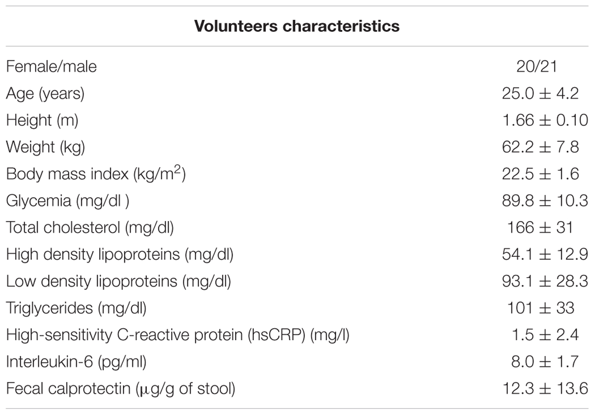
The study protocol was approved by the Ethics Committee for Research in Humans of the Institute of Nutrition and Food Technology (INTA), University of Chile, in compliance with the Helsinki Declaration. Asymptomatic, normal weight (BMI between 19.2 and 24.7 kg/m2) volunteers of both genders, aged 18–39 (Table 1), living in Santiago city and who had provided a signed, informed consent, were recruited in the study. Exclusion criteria included smoking, pregnancy and history of gastrointestinal, autoimmune, neurological and/or chronic metabolic diseases. Subjects with treatments interfering with intestinal permeability, motility or microbiota, such as dietary treatment for weight loss or drug intake (antibiotics, anti-inflammatory drugs, laxatives or prokinetics) during the month preceding the study were also excluded. Health status was confirmed through biochemical and lipid profiles and by the absence of colonic and systemic inflammation as suggested by fecal calprotectin and plasma Il-6 and high-sensitivity C-reactive protein (hsCRP), respectively (Table 1), as previously described (Morales et al., 2016). The subjects consumed their habitual diet and during the week before stool sampling, they were counseled by two registered dietitians to standardize their daily intake of macronutrients and avoid excess of dietary fats. The daily intake of energy (kcal) and macronutrients (% of caloric intake) of the subjects, as determined by a 48-h dietary recall, was as follows: (Means [CI95%]): energy 2078 [1946–2210]; carbohydrates: 58.9 [56.5–60.8]; proteins 15.2 [14.8–16.1] and lipids: 25.9 [23.9–27.9], according to the recommended dietary allowances. The subjects also consumed 19.9 g/day [18.3–21.6] of dietary fiber.
Sampling, DNA Extraction and Identification of the Fecal Microbiota
Fecal samples were collected by the 41 volunteers in sterile plastic containers and stored at -30°C until further processing. Genomic DNA was extracted from 220 mg of stool samples using the QIAmp DNA Stool Mini Kit (Qiagen, Hilden, Germany) according to manufacturer instructions. Library preparation and Illumina sequencing were performed at the Roy J. Carver Biotechnology Center, University of Illinois at Urbana-Champaign, Champaign, IL, United States. In brief, library preparation was performed using the Fluidigm Access Array (Fluidigm, South San Francisco, CA, United States), in which 2 ng of DNA measured with a Qubit 2.0 fluorometer (Thermo Fisher Scientific, Carlsbad, CA, United States) were amplified in a two-step process. In the first step, the V3–V4 region of the 16S rRNA gene was amplified using the primers 341F (5′-CCTACGGGNGGCWGCAG-3′) and 785R (5′-GACTACHVGGGTATCTAATCC-3′) (Klindworth et al., 2013). The index and sequencing adaptors required were added in a second PCR. The amplicons obtained were quantified through Qubit fluorometry and their sizes verified for 11 random samples using an Agilent 2100 Bioanalyzer (Agilent Technologies, Santa Clara, CA, United States) to determine their overall quality. Amplicons were then pooled in equimolar concentration and purified with a 2% agarose e-gel (EX 2% agarose, Invitrogen, Life Technologies, Grand Island, NY, United States). The cleaned pool was reanalyzed with the Bioanalyzer to verify the effectivity of the clean-up step and to determine the average size of the amplicons. Finally, the pooled libraries were quantified with qPCR performed using a CFX connect Real-Time PCR (Bio-Rad, Hercules, CA, United States) to achieve an accurate quantification, before loading the libraries to the sequencer. Sequencing was performed with MiSeq Illumina system (Illumina, San Diego, CA, United States), using the V3 kit, generating 2×300 nt paired end reads.
Sequence Processing, Comparison against Other Microbiome Datasets
De-multiplexing of the reads was performed using the software CASAVA v. 1.82. Paired reads were merged using FLASH (V1.2.11) with default parameters (Magoč and Salzberg, 2011), adaptors removed using Cutadapt (V1.9) (Martin, 2011), and quality trimmed using Trimmomatic (V0.36) (Bolger et al., 2014). To be able to compare this dataset with other studies that targeted different regions, we used a closed-reference strategy. First, 16S rRNA gene sequence data used in this study were obtained from: (1) Human Microbiome Project (HMP) data set (Human Microbiome Project Consortium, 2012)1, (2) Argentina (Carbonetto et al., 2016), (3) Colombia (Escobar et al., 2014), (4) Hadza population from Tanzania (Schnorr et al., 2014), (5) Italy (Schnorr et al., 2014), (6) South Korea (Nam et al., 2011), (7) Matses ethnic group, Peru (Obregon-Tito et al., 2015), and (8) Papua New Guinea (Martínez et al., 2015). These studies targeted different regions of 16S rRNA, but all the studies shared either the V3 and/or V4 regions, sequenced using either the 454 or Illumina platform. Paired-end reads were processed as previously mentioned (Masella et al., 2012). For all datasets, chimeric sequences were filtered using VSEARCH (Rognes et al., 2016) against the Greengenes database (DeSantis et al., 2006; 13_8 database). OTUs were built using the closed reference protocol in QIIME (V 1.8.0) (Caporaso et al., 2010) using the Greengenes 13.8 database, at 97% sequence similarity (Mcdonald et al., 2012). All abundance and diversity analyses were done using Phyloseq (McMurdie and Holmes, 2013). Rarefactions curves for the Chilean subjects were calculated using QIIME, subsampling in the range of 10–17,000 sequences, with a step of 1,000 sequences, and 10 re-samplings on each step. Values were obtained for the observed OTUs and Chao1 index.
To test for association of microbial abundance (at the genus level) with clinical metadata we performed a multivariate analysis using Multivariate Analysis by Linear Models (MaAsLin) version for R. MaAsLin2 performs boosted, additive general linear models between metadata and microbial abundance. Boosting of metadata and selection of a model was performed per taxon. All microbial data were arcsine-square root transformed. The metadata used were age, sex, body mass index (BMI), glycemia, triglycerides, total cholesterol, fecal calprotectin, and plasma interleukin-6 and hsCRP level. Associations were considered significant below a q-value threshold of 0.25 as recommended by the authors and as used in previous microbiome studies (Tong et al., 2014; Lim et al., 2016).
Data analysis was done using R (R Core Team, 2015). Calculation of alpha and beta diversities was done using Phyloseq (McMurdie and Holmes, 2013). Sample alpha diversity was calculated using the Shannon index. Differences in alpha diversity between genders for the Chilean subjects were determined using Kruskal–Wallis test. Comparisons between different countries were visualized using principal coordinate analysis (PCoA) of the weighted UniFrac distances between samples (Lozupone et al., 2006). Ellipses were drawn into the visualization using a 95% confidence interval. Evaluation of differences between countries, based on the beta diversity values, was done using ADONIS. A test for homogeneity of multivariate dispersions was applied to evaluate if these differences were due to group dispersions. Both analyses were done using the Vegan package in R. Linear discriminant analysis (LDA) effect, implemented in LEfSe (Segata et al., 2011), was used to compare the relative abundance of the different taxa between all the groups. An alpha value of 0.05 was used for the ANOVA and Wilcoxon tests to identify features with significantly different abundances between assigned taxa compared to other populations. A size-effect threshold of 4.0 on the logarithmic LDA score was used for discriminative microbial taxa. A one-against-all test was used to compare each different country of origin, against the rest of the dataset.
Results
The anthropometrical, biochemical and clinical characteristics of the volunteers are described in Table 1 and Supplementary Table S1 (Supplementary Data). The fecal microbiota of the volunteers was characterized through sequencing the V3–V4 region of the 16S RNA gene. A total of 4,476,399 high-quality filtered sequences were obtained, i.e., 109,180 ± 13,148 sequences per sample. The rarefaction curves reached an asymptote, which indicated that the sequence depth was sufficient to represent the majority of bacterial community diversity (Supplementary Figure S1). Overall, 13 phyla and 57 families were detected (Figures 1A,B) and an α-diversity (Shannon index) of 3.86 ± 0.37 was estimated. No differences in α-diversity were observed between genders (p = 0.1362, Supplementary Figure S2). The most abundant phyla were Firmicutes (43.6 ± 9.2%) and Bacteroidetes (41.6 ± 13.1%); followed by Verrucomicrobia (8.5 ± 10.4%), Proteobacteria (2.8 ± 4.8%), Actinobacteria (1.8 ± 3.9%) and Euryarchaeota (1.4 ± 2.7%). Euryarchaeota corresponds to the only identified phylum belonging to the Archaea domain. The other 7 phyla identified (Cyanobacteria, Tenericutes, Fusobacteria, Lentisphaerae, Spirochaetes, Synergistetes, and TM7) had relative abundance <0.2%. The phyla Firmicutes, Bacteroidetes, Verrucomicrobia, Proteobacteria, and Actinobacteria were detected in all subjects. High inter-individual variability was detected in the microbiota composition: the relative abundances ranged from 25.3 to 67.3% for Firmicutes, 4.1–63.5% for Bacteroidetes, 0.002–41.2% for Verrucomicrobia and 0.05–24.9% for Proteobacteria (Figure 1A).
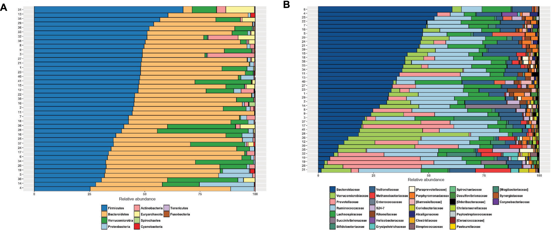
FIGURE 1. Fecal microbiota composition of healthy, normal weight Chilean subjects by sequencing the V3–V4 of 16S rRNA gene using the MiSeq Illumina system. Relative abundance (%) of phyla (A) and families (B) identified in healthy normal weight Chileans (n = 41).
Bacterial families were dominated by Bacteroidaceae (30.2 ± 16.1%), Ruminococcaceae (19.4 ± 6.7%), Lachnospiraceae (10.4 ± 5.6), Verrucomicrobiaceae (9.4 ± 11.7), Prevotellaceae (8.8 ± 13.1%) and Veillonellaceae (7.8 ± 4.5%) (Figure 1B). These families were detected in all subjects. Porphyromonadaceae, Methanobacteriaceae, Rikenellaceae, Bifidobacteriaceae and S24-7 showed relative abundances ranging from 1 to 2%. The other bacterial families had relative abundances <1%. Only small differences were detected between women and men in the abundance of bacterial families, affecting two families with abundances <0.5%: Streptococcaceae (♀: 0.27 ± 0.65% vs. ♂: 0.096 ± 0.22%; p = 0.015) and Pasteurellaceae (♀: 0.060 ± 0.080% vs. ♂: 0.018 ± 0.035%; p = 0.007).
The genera identified in all subjects, representing the core microbiota, includes Bacteroides, Prevotella, Parabacteroides (Bacteroidetes phylum), Phascolarctobacterium, Faecalibacterium, Ruminococcus, Lachnospira, Oscillospira, Blautia, Dorea, Roseburia, Coprococcus, Clostridium, Streptococcus (Firmicutes phylum), Akkermansia (Verrucomicrobia phylum), and Collinsella (Actinobacteria phylum). We observed a dominance of Bacteroides over Prevotella in 34/41 (83%) of the subjects. The Methanobacteriaceae family, belonging to the phylum Euryarchaeota of the Archaea domain was detected in 83% (34/41) of the subjects, and Desulfovibrio, the most representative sulfate-reducing genus, in 76% (31/41) of the subjects. Some genera including pathogens were also detected: Treponema in 5 individuals, Campylobacteraceae (with reads assigned to the genera Arcobacter and Campylobacter) in 15 subjects, and Helicobacter in 2 subjects. To identify specific microbial taxa significantly associated with clinical metadata, we performed a multivariate association with linear models (MaAsLin) analysis. Of all the variables, only glycemia showed a significant negative association with genus Haemophilus (Pasteurellaceae family) (p-value: 2.18E-4, q-value = 0.153, Supplementary Figure S3).
To compare the differences between microbial compositions among the different populations, we analyzed the beta diversity of populations, using weighted UniFrac distances (Figure 2). The microbiota of the United States and that of the Papua New Guinea subjects were clearly segregated while those from the other populations were distributed between them. The microbiota of the Chilean subjects significantly differed from that of Papua New Guinea (p = 0.001) and the Matses ethnic group (p = 0.001) and did not differ from the subjects of the other countries, being closer to the populations of Argentina and some sub-populations from the United States. We also evaluated the contribution of component PC3 showing only 4.3%. A visualization of the same dataset using a Bray–Curtis distance metric, showed a similar distribution of the different populations (Supplementary Figure S4). The difference between populations seems to be explained by differences in the abundance of phyla, as described in Figure 3, where the most dominant were Bacteroidetes and Firmicutes. More particularly, the Papua New Guinea microbiota showed a high dominance of Firmicutes over Bacteroidetes while the contrary was observed in the United States population. With a higher Firmicutes/Bacteroidetes ratio, the gut microbiota of Colombia, Italy, South Korea, Hadza and Matses ethnic groups were more similar to the Papua New Guinea microbiota. The Chilean and Argentinean gut microbiota harbored the same relative abundance of Firmicutes and Bacteroidetes phyla. However, the composition of the gut microbiota from Chilean subjects had greater abundance of members of the phylum Verrucomicrobia. We then evaluated significant differences in relative abundance of bacterial orders, families and genera of the Chilean subjects compared to the other populations through LDA Effect Size (LEfSe). We observed that the Chilean population was more enriched in Verrucomicrobiales, Verrucomicrobiae and Akkermansia, Bifidobacteriales and Bifidobacterium, Odoribacteraceae and Odoribacter, Paraprevotella, Prevotella, Alistipes, and Desulfovibrio (Figure 4).
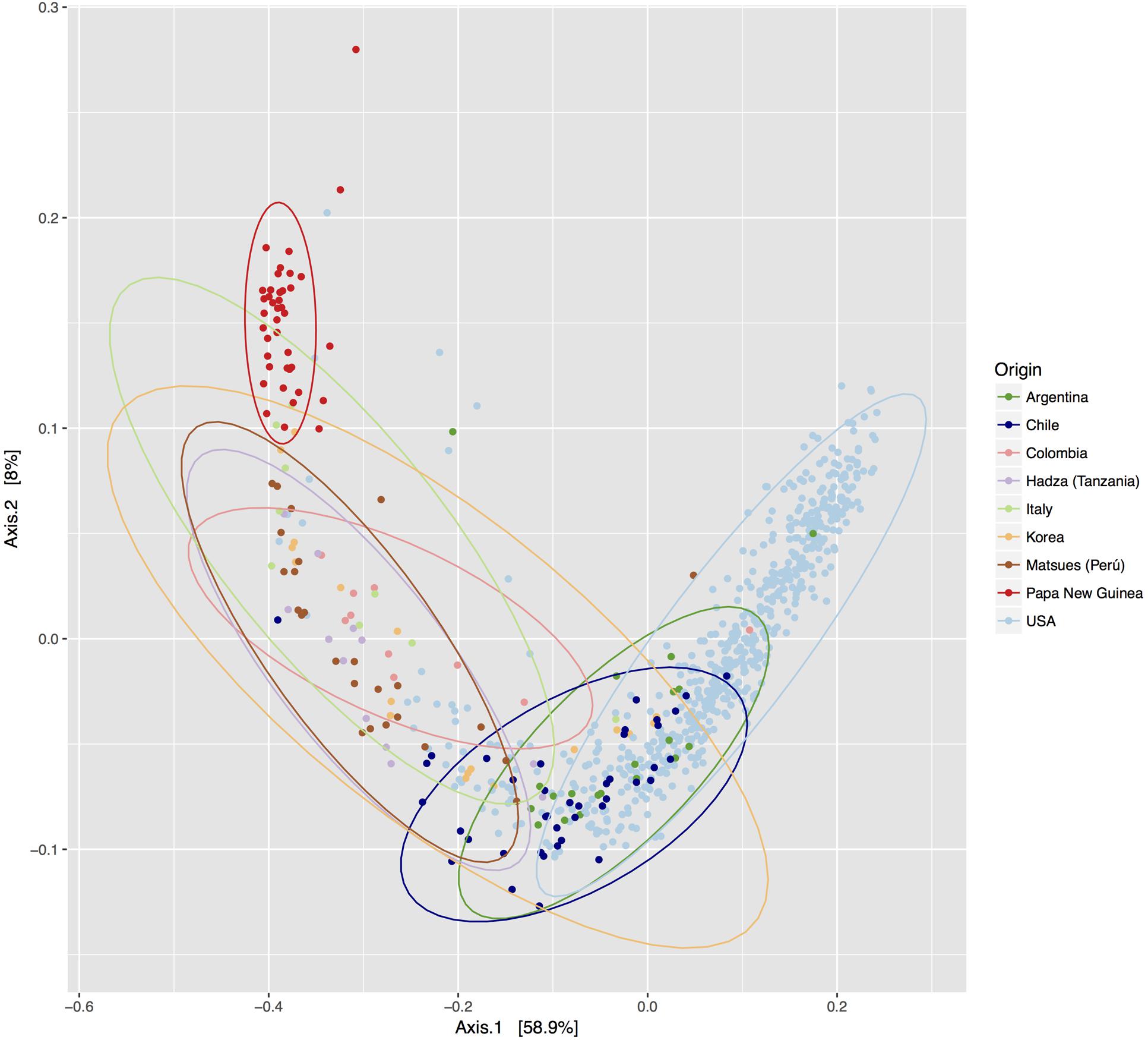
FIGURE 2. Comparison of the Chilean gut microbiota versus gut microbiota of individuals from other geographic locations. PCoA of the beta diversity values based on weighted Unifrac distances. Each color represents a population from a specific geographic area. Ellipses were drawn using a confidence interval of 95% for each group.
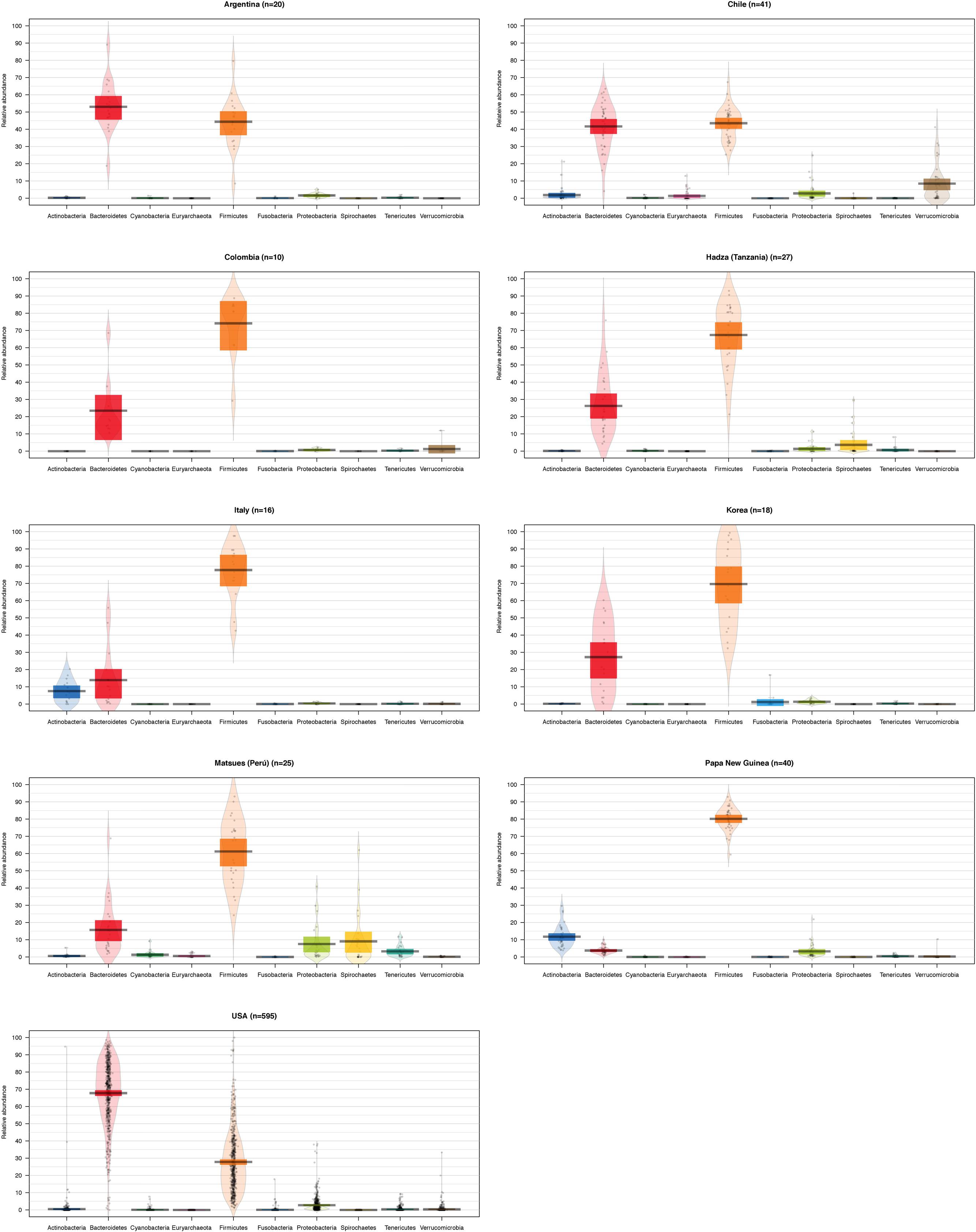
FIGURE 3. Abundance of the main bacterial phyla identified in the gut microbiota of individuals from different geographic locations. Violin plots showing the abundance of the most abundant bacterial phyla for the gut microbiota of different populations. The number of subjects for each population is shown on the title, and each bar represents a different bacterial phylum. The black line indicates the median of the values, the colored box the interquartile range and the area in light color corresponds to the distribution of the data.
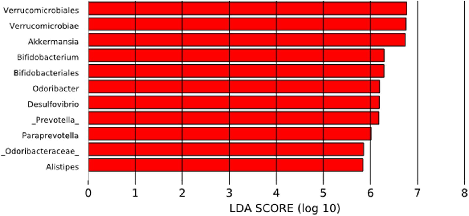
FIGURE 4. Linear discriminant analysis (LDA) Effect Size (LEfSe) analysis of bacterial taxa present in the Chilean subjects and those from other countries. The significantly enriched populations are shown only for the Chilean population.
Regarding the relative abundance of potentially pathogenic bacterial genera, no differences were observed between countries for Helicobacter. The abundance of Treponema in the Matses and Hadza populations was significantly higher (p < 0.002 and p < 0.006, respectively) than that reported for all the other countries, including Chile (Supplementary Figure S5A). For Campylobacter, no significant differences were observed between populations (Supplementary Figure S5B).
Discussion
This is the first report describing the taxonomic composition of the gut microbiota from young, healthy, normal weight, Chilean subjects. Although the great majority of the studies describing the composition of gut microbiota in different human populations are carried out from fecal samples, it is important to note that the human fecal microbiota is an unreliable reflection of the cecal or colonic microbiota. Marteau et al. (2001) showed, for example, that facultative anaerobes represent only 1% of total bacteria in the feces versus 25% in the cecum and that, more particularly, lower levels of Bifidobacterium, Bacteroides and members of the C. coccoides and C. leptum groups were detected in the cecum than in stool samples. This is obviously an important limitation that must be considered in the interpretation of the results.
Our results showed high inter-individual variability, despite the highly homogenous characteristics of the subjects (in terms of age, weight, biochemical and lipid profiles, absence of inflammation). Similar observations were also reported in other normal weight populations (Nam et al., 2011; Human Microbiome Project Consortium, 2012; Escobar et al., 2014; Schnorr et al., 2014; Obregon-Tito et al., 2015; Carbonetto et al., 2016). In most of our subjects we observed a dominance of Bacteroides over Prevotella. This enterotype was first shown by Wu et al. (2011) to correspond to individuals consuming western diets rich in animal proteins and fat, in contrast to fiber-rich diets that favor the presence of Prevotella. During the last 30 years Chile has experienced a rapid epidemiological and nutritional transition characterized by the gradual adoption of an energy-rich western-type diet accompanied by a shift from infectious to chronic diseases (Atalah et al., 2014). For example, the intake of calories from fat increased from 21 to 28% between 1980 and 1998; the daily intake of meat and meat-derived products is currently about 150 g (Albala et al., 2002; Ministerio de Salud, 2011). The modern urban Chilean diet has also been incorporating increasing amounts of processed foods, refined sugar and salt while decreasing the intake of fruits and vegetables. As a result, the prevalence of obesity in adults and children and type 2 diabetes is among the most elevated in the world. The volunteers of our study consumed this type of diet, rich in animal fat and proteins, but with a moderate daily caloric intake (∼2078 Kcal/day), explaining their normal weight. These findings could explain the dominance of Bacteroides over Prevotella in their microbiota. An appropriate characterization of the nutrient sources in studies of microbiota composition could improve the identification of specific nutrient-taxa associations.
In addition to Bacteroides, the bacterial core also includes taxa normally identified in the human gut microbiota such as the succinate-utilizing Phascolarctobacterium (Watanabe et al., 2012) and some beneficial butyrate-producing genera such as Faecalibacterium, Roseburia, Coprococcus, and Oscillospira (which have never been cultivated and are related to lean status and health) (Konikoff and Gophna, 2016). Collinsella, which belongs to the phylum Actinobacteria, has been found to be significantly increased in type 2 diabetes compared to prediabetic and non-diabetic American subjects (Lambeth et al., 2015). The presence and role of this microorganism in the Chilean gut microbiota should be explored in more detail in larger studies in the future. Interestingly, we detected a high proportion (>8%) of the mucus-degrading genus Akkermansia, belonging to the phylum Verrucomicrobia. This genus has been suggested to be a potential biomarker of a healthy gut status. It has been proposed that the growth of this bacterium is favored by low availability of enteral nutrients such as in long-term fasting and malnutrition (Belzer and de Vos, 2012).
Regarding the Archaea domain, several groups were detected in the microbiota of our subjects. The Methanobacteriaceae family of the phylum Euryarchaeota was observed in 83% (34/41) of the subjects. All of them harbored members of the genus Methanobrevibacter, in agreement with the high prevalence of this group in the adult population (>50%). Methanobrevibacter smithii, the most dominant methanogen in the human gut, is implicated in the production of methane through the reduction of CO2 using H2 (or formate) (Gaci et al., 2014). We identified sequences that matched the genus Methanosphaera in one individual. One of the most important representatives of this group is Methanosphaera stadtmaniae that produces methane from methanol (Gaci et al., 2014). Methanogens of the order Methanomassiliicoccales, which participate in the reduction of methyl-compounds [mono, di and trimethylamine (TMA)] to produce methane (Gaci et al., 2014) were not identified in our study. This could be important, considering that TMA, produced through the metabolism of choline and L-carnitine by gut microorganisms, is subsequently oxidized in the liver into the pro-atherogenic trimethylamine oxide (TMAO) (Gaci et al., 2014). Based on this consideration, it has been hypothesized that dietary supplementation with these “Archaebiotics” could prevent cardiovascular diseases in at-risk subjects (Gaci et al., 2014).
Desulfovibrio, the most representative sulfate-reducing genus, was detected in 76% (31/41) of the subjects. Although a high prevalence (>50%) of this bacterium has been reported in patients with IBD, it can exert pro- or anti-inflammatory effects depending on the concentration of hydrogen sulfide (H2S) produced in the gut lumen (Linden, 2014).
Another interesting taxon identified in our study was Treponema, which was detected in five individuals (12.5%). The genus Treponema belongs to the phylum Spirochaetes and includes human pathogenic and non-pathogenic species. Non-pathogenic Treponema has been described as part of the normal microbiota of the intestine, oral cavity and genital flora and was shown, for example, to be exclusively present in rural African children compared to European children (De Filippo et al., 2010). Recent studies using high-throughput sequencing of the 16S rRNA gene showed a high prevalence of Treponema in the gut of human populations consuming polysaccharide-rich diet, probably due to the fact that some Treponema species hydrolyze cellulose and xylans. Our results show that the relative abundance of this genus in the Chilean population was clearly lower than that described in the Matses and Hadza, hunter-gatherer populations consuming high amount of dietary fiber, especially from tubers (Schnorr et al., 2014).
Furthermore, we detected 15 (37.5%) subjects colonized with enteric pathogens belonging to the family Campylobacteraceae (Arcobacter and Campylobacter), and 2 subjects with Helicobacter. Campylobacter jejuni is the leading global cause of foodborne disease associated with the consumption of contaminated chicken in the developed world, and with contaminated water in the developing countries (Kaakoush et al., 2015; Johnson et al., 2017). Asymptomatic fecal carriage of the pathogen has been reported in humans (Dicksved et al., 2014; Kampmann et al., 2016). The high level of asymptomatic Campylobacter carriage observed in our study could be related to the high incidence (38–68%) of this microorganism in Chilean poultry meat (Figueroa et al., 2009), which is the main source of animal protein consumed in Chile. Other evidence reinforcing this hypothesis is the fact that some isolates from human campylobacteriosis and broiler meat are genetically indistinguishable by pulsed-field gel electrophoresis (PFGE) (González-Hein et al., 2013). Helicobacter pylori, a bacteria residing in the stomach, has been linked to several gastric diseases such as stomach cancer. It is estimated that more than 50% of the population is colonized with this microorganism worldwide, and over 80% of these infected humans remain asymptomatic (Peleteiro et al., 2014). H. pylori is highly prevalent in the Chilean population (Gotteland et al., 2006). Although the classical methods to detect this bacterium are culture of gastric biopsy or a urea breath test, H. pylori can also be detected in stool samples (Talarico et al., 2016).
We also explored possible correlation between the microbiota and metadata of the Chilean subjects. We only detected a significant association between the basal glycemia and Haemophilus: at higher relative abundance of Haemophilus, lower basal glycemia is observed. This data are in line with observations reported in other studies showing higher relative abundances of Haemophilus in healthy subjects compared to patients with glucose intolerance (Zhang et al., 2013) and with a type 2 diabetes mellitus group (Qin et al., 2012).
Finally, we compared our data to those obtained from public datasets generated from other populations. The results suggest that the diversity of the Chilean gut microbiota is similar to that of the Argentina and United States subpopulations, while it clearly differs from those from Papua New Guinea and the Matses ethnic groups. Further studies with greater coverage and power are needed to confirm these differences. It is interesting to note that, although the selection of our subjects was controlled as closely as possible in terms of age, health status, lifestyle and dietary intake, the dispersion of the abundances values was quite high. This could reflect differences in genetic background, particularly the Amerindian heritage whose importance may vary from one subject to another in the Chilean population. The composition of the microbiota of the Chileans and Argentinians appears to be closer because their lifestyle and diet are similar and the proportion of Amerindian heritage is lower than in other Latin American countries such as Colombia or Peru, where diet is also different. In the case of the United States subjects, it must be stated that the number of samples is clearly higher than those from the other countries, as well as their dispersion. This probably also reflects the heterogeneity of these subjects in terms of genetic background and the existence of other factors contributing to the microbiota composition.
An important difference in the Chilean subjects compared to populations from other countries was the higher abundance of phylum Verrucomicrobia. This phylum is a member of the PVC (Planctomycetes-Verrucomicrobia-Chlamydiae) superphylum which includes phylogenetically related bacteria with unusual characteristics such as the existence of a complex and dynamic endomembrane system that, in some aspects, makes them closer to eukaryotic cells. It includes a small number of genera isolated from fresh water, soil and animal feces; Akkermansia muciniphila is the main member of this phylum identified in humans (though the presence of Luteolibacter or Verrucomicrobium was also reported in three subjects from United States and Matses ethnic groups). A. muciniphila is a strict anaerobe, Gram-negative, mucus-inhabiting bacterium which is considered as a true symbiont due to its wide distribution in the animal kingdom. In humans, it is detected in the intestine of most healthy individuals where it represents 1–4% of the total microbiota (Collado et al., 2007); it is one of the bacteria on which the enterotype classification is based (Arumugam et al., 2011). It is a highly specialized bacterium capable of using mucins as a sole source of carbon and nitrogen (Derrien et al., 2004) and stimulating mucin expression and mucus secretion through a positive feedback (Derrien et al., 2010). This specialization would represent a competitive advantage in case of nutrient deprivation such as fasting, malnutrition or total parenteral nutrition. As a result of mucin degradation, A. muciniphila releases acetate and propionate which are easily available for the host (Derrien et al., 2004, 2011). A. muciniphila colonizes the human gut early in life (1 month) and increases rapidly with age, reaching 108/g of stool in adults (Collado et al., 2007). Its abundance is lower in patients with IBDs (Png et al., 2010) or appendicitis (Swidsinski et al., 2011), as well as in animal models of obesity and obese humans (Zhang et al., 2009; Santacruz et al., 2010). This is an interesting finding, considering that this agent also exhibits anti-inflammatory and immunostimulant properties. Increasing the intestinal counts of A. muciniphila in these subjects through the administration of polyphenols or prebiotics results in the improvement of barrier function, endotoxemia and insulin sensitivity (Anhê et al., 2015), the increased expression of intestinal Fiaf (a circulating inhibitor of lipoprotein lipase) and RegIII (a bactericidal C-type lectin targeting Gram-positive bacteria) (Anhê et al., 2015), and higher intestinal content of bioactive lipids participating in the endocannabinoid system. Interestingly, the abundance of A. muciniphila is higher after antibiotic administration (Dubourg et al., 2013) and in patients treated with metformin; this could represent one of the mechanisms of action by which this drug improves glucose homeostasis and insulin resistance in diabetic patients (Everard et al., 2013; Shin et al., 2014).
It is unclear why the Chilean subjects displayed higher abundance of Verrucomicrobia considering that none of them were consuming antibiotic or metformin (exclusion criterion) and their intake of dietary fiber (with eventual prebiotic properties) was not especially elevated. Accordingly, the presence of this taxon needs to be studied further to confirm the current results, to determine the factors subjacent to this high abundance and to evaluate its role in the gut microbiota of healthy subjects.
Conclusion
The Chilean gut microbiota shows high inter-individual variability despite the highly homogenous characteristics of the subjects. The differences between the Chilean gut microbiota and those of other populations seem to be associated with differences in the Firmicutes/Bacteroidetes ratio and the high abundance of Verrucomicrobia. These results constitute an important baseline to explore further the possible alterations of gut microbiota composition occurring in the most prevalent diseases in the Chilean population, and the host factors implicated in inter-individual variability.
Data Access
Raw datasets are available in the European Nucleotide Archive (ENA) under the accession number PRJEB16755.
Author Contributions
PM, PN, and MG designed the study. PM performed the recruitment of the volunteers, collection of stool samples and the extraction of fecal bacterial DNA. SF-V and JU processed the raw sequences. SF-V, YV, PV-W, and JU performed the bioinformatic and statistical analysis. SF-V, YV, PV-W, JU, FM, PN, and MG analyzed the results and wrote the manuscript.
Funding
The study was supported by grant FONDECYT 1120290 and 11140666 (CONICYT, Chile), CINUT (INTA, University of Chile), and ENLACE ENL010/15.
Conflict of Interest Statement
The authors declare that the research was conducted in the absence of any commercial or financial relationships that could be construed as a potential conflict of interest.
Supplementary Material
The Supplementary Material for this article can be found online at: http://journal.frontiersin.org/article/10.3389/fmicb.2017.01221/full#supplementary-material
Footnotes
References
Albala, C., Vio, F., Kain, J., and Uauy, R. (2001). Nutrition transition in Latin America: the case of Chile. Nutr. Rev. 59, 170–176. doi: 10.1111/j.1753-4887.2001.tb07008.x
Albala, C., Vio, F., Kain, J., and Uauy, R. (2002). Nutrition transition in Chile: determinants and consequences. Public Health Nutr. 5, 123–128. doi: 10.1079/PHN2001283
Anhê, F. F., Roy, D., Pilon, G., Dudonné, S., Matamoros, S., Varin, T. V., et al. (2015). A polyphenol-rich cranberry extract protects from diet-induced obesity, insulin resistance and intestinal inflammation in association with increased Akkermansia spp. population in the gut microbiota of mice. Gut 64, 872–883. doi: 10.1136/gutjnl-2014-307142
Arumugam, M., Raes, J., Pelletier, E., Le Paslier, D., Yamada, T., Mende, D. R., et al. (2011). Enterotypes of the human gut microbiome. Nature 473, 174–180. doi: 10.1038/nature09944
Atalah, E., Amigo, H., and Bustos, P. (2014). Does Chile’s nutritional situation constitute a double burden? Am. J. Clin. Nutr. 100, 1623S–1627S. doi: 10.3945/ajcn.114.083790
Belzer, C., and de Vos, W. M. (2012). Microbes inside–from diversity to function: the case of Akkermansia. ISME J. 6, 1449–1458. doi: 10.1038/ismej.2012.6
Bolger, A. M., Lohse, M., and Usadel, B. (2014). Trimmomatic: a flexible trimmer for Illumina sequence data. Bioinformatics 30, 2114–2120. doi: 10.1093/bioinformatics/btu170
Bravo, J. A., Julio-Pieper, M., Forsythe, P., Kunze, W., Dinan, T. G., Bienenstock, J., et al. (2012). Communication between gastrointestinal bacteria and the nervous system. Curr. Opin. Pharmacol. 12, 667–672. doi: 10.1016/j.coph.2012.09.010
Caporaso, J. G., Kuczynski, J., Stombaugh, J., Bittinger, K., Bushman, F. D., Costello, E. K., et al. (2010). QIIME allows analysis of high-throughput community sequencing data. Nat. Methods 7, 335–336. doi: 10.1038/nmeth.f.303
Carbonetto, B., Fabbro, M., Sciara, M., Seravalle, A., Méjico, G., Revale, S., et al. (2016). Human microbiota of Argentine population- a pilot study. Front. Microbiol. 7:51. doi: 10.3389/fmicb.2016.00051
Claesson, M. J., Jeffery, I. B., Conde, S., Power, S. E., O’Connor, E. M., Cusack, S., et al. (2012). Gut microbiota composition correlates with diet and health in the elderly. Nature 488, 178–184. doi: 10.1038/nature11319
Clemente, J., Pehrsson, E., Blaser, M., Sandhu, K., Gao, Z., Wang, B., et al. (2015). The microbiome of uncontacted Amerindians. Sci. Adv. 1:e1500183. doi: 10.1126/sciadv.1500183
Collado, M. C., Derrien, M., Isolauri, E., de Vos, W. M., and Salminen, S. (2007). Intestinal integrity and Akkermansia muciniphila, a mucin-degrading member of the intestinal microbiota present in infants, adults, and the elderly. Appl. Environ. Microbiol. 73, 7767–7770. doi: 10.1128/AEM.01477-07
Cotillard, A., Kennedy, S. P., Kong, L. C., Prifti, E., Pons, N., Le Chatelier, E., et al. (2013). Dietary intervention impact on gut microbial gene richness. Nature 500, 585–588. doi: 10.1038/nature12480
De Filippo, C., Cavalieri, D., Di Paola, M., Ramazzotti, M., Poullet, J. B., Massart, S., et al. (2010). Impact of diet in shaping gut microbiota revealed by a comparative study in children from Europe and rural Africa. Proc. Natl. Acad. Sci. U.S.A. 107, 14691–14696. doi: 10.1073/pnas.1005963107
Derrien, M., Van Baarlen, P., Hooiveld, G., Norin, E., Müller, M., and de Vos, W. M. (2011). Modulation of mucosal immune response, tolerance, and proliferation in mice colonized by the mucin-degrader Akkermansia muciniphila. Front. Microbiol. 1:166. doi: 10.3389/fmicb.2011.00166
Derrien, M., van Passel, M. W., van de Bovenkamp, J. H., Schipper, R. G., de Vos, W. M., and Dekker, J. (2010). Mucin-bacterial interactions in the human oral cavity and digestive tract. Gut Microbes 1, 254–268. doi: 10.4161/gmic.1.4.12778
Derrien, M., Vaughan, E. E., Plugge, C. M., and de Vos, W. M. (2004). Akkermansia muciniphila gen. nov., sp. nov., a human intestinal mucin-degrading bacterium. Int. J. Syst. Evol. Microbiol. 54, 1469–1476. doi: 10.1099/ijs.0.02873-0
DeSantis, T. Z., Hugenholtz, P., Larsen, N., Rojas, M., Brodie, E. L., Keller, K., et al. (2006). Greengenes, a chimera-checked 16S rRNA gene database and workbench compatible with ARB. Appl. Environ. Microbiol. 72, 5069–5072. doi: 10.1128/aem.03006-05
Dicksved, J., Ellström, P., Engstrand, L., and Rautelin, H. (2014). Susceptibility to Campylobacter infection is associated with the species composition of the human fecal microbiota. mBio 5:e01212-14. doi: 10.1128/mBio.01212-14
Dubourg, G., Lagiera, J. C., Armougoma, F., Roberta, C., Audolya, G., Papaziana, L., et al. (2013). High-level colonisation of the human gut by Verrucomicrobia following broad-spectrum antibiotic treatment. Int. J. Antimicrob. Agents 41, 149–155. doi: 10.1016/j.ijantimicag.2012.10.012
Escobar, J., Klotz, B., Valdes, B., and Agudelo, G. M. (2014). The gut microbiota of Colombians differs from that of Americans, Europeans and Asians. BMC Microbiol. 14:311. doi: 10.1186/s12866-014-0311-6
Everard, A., Belzer, C., Geurts, L., Ouwerkerk, J. P., Druart, C., Bindels, L. B., et al. (2013). Cross-talk between Akkermansia muciniphila and intestinal epithelium controls diet-induced obesity. Proc. Natl. Acad. Sci. U.S.A. 110, 9066–9071. doi: 10.1073/pnas.1219451110
Figueroa, G., Troncoso, M., López, C., Rivas, P., and Toro, M. (2009). Occurrence and enumeration of Campylobacter spp. during the processing of Chilean broilers. BMC Microbiol. 9:94. doi: 10.1186/1471-2180-9-94
Gaci, N., Borrel, G., Tottey, W., O’Toole, P. W., and Brugère, J. F. (2014). Archaea and the human gut: new beginning of an old story. World J. Gastroenterol. 20, 16062–16078. doi: 10.3748/wjg.v20.i43.16062
González-Arancibia, C., Escobar-Luna, J., Barrera-Bugueño, C., Díaz-Zepeda, C., González-Toro, M. P., Olavarría-Ramírez, L., et al. (2016). What goes around comes around: novel pharmacological targets in the gut-brain axis. Therap. Adv. Gastroenterol. 9, 339–353. doi: 10.1177/1756283X16630718
González-Hein, G., García, P., Foerster, C., Troncoso, M., and Figueroa, G. (2013). Campylobacter jejuni isolated from human cases in Chile showed indistinguishable PFGE profiles with strains isolated from poultry and bovine sources. Cyta J. Food 11, 185–189. doi: 10.1080/19476337.2012.722564
Gotteland, M., Brunser, O., and Cruchet, S. (2006). Systematic review: are probiotics useful in controlling gastric colonization by Helicobacter pylori? Aliment. Pharmacol. Ther. 23, 1077–1086. doi: 10.1111/j.1365-2036.2006.02868.x
Human Microbiome Project Consortium (2012). A framework for human microbiome research. Nature 486, 215–221. doi: 10.1038/nature11209
Jandhyala, S. M., Talukdar, R., Subramanyam, C., Vuyyuru, H., Sasikala, M., Reddy, N., et al. (2015). Role of the normal gut microbiota. World J. Gastroenterol. 21, 8787–8803. doi: 10.3748/wjg.v21.i29.8787
Johnson, T. J., Shank, J. M., and Johnson, J. G. (2017). Current and potential treatments for reducing Campylobacter colonization in animal hosts and disease in humans. Front. Microbiol. 8:487. doi: 10.3389/fmicb.2017.00487
Julio-Pieper, M., Bravo, J. A., Aliaga, E., and Gotteland, M. (2014). Review article: intestinal barrier dysfunction and central nervous system disorders–a controversial association. Aliment. Pharmacol. Ther. 40, 1187–1201. doi: 10.1111/apt.12950
Kaakoush, N. O., Castaño-Rodríguez, N., Mitchell, H. M., and Man, S. M. (2015). Global epidemiology of Campylobacter infection. Clin. Microbiol. Rev. 28, 687–720. doi: 10.1128/CMR.00006-15
Kampmann, C., Dicksved, J., Engstrand, L., and Rautelin, H. (2016). Composition of human faecal microbiota in resistance to Campylobacter infection. Clin. Microbiol. Infect. 22, 61.e1–61.e8. doi: 10.1016/j.cmi.2015.09.004
Klindworth, A., Pruesse, E., Schweer, T., Peplies, J., Quast, C., Horn, M., et al. (2013). Evaluation of general 16S ribosomal RNA gene PCR primers for classical and next-generation sequencing-based diversity studies. Nucleic Acids Res. 41:e1. doi: 10.1093/nar/gks808
Konikoff, T., and Gophna, U. (2016). Oscillospira: a central, enigmatic component of the human gut microbiota. Trends Microbiol. 24, 523–524. doi: 10.1016/j.tim.2016.02.015
Konturek, P. C., Haziri, D., Brzozowski, T., Hess, T., Heyman, S., Kwiecien, S., et al. (2015). Emerging role of fecal microbiota therapy in the treatment of gastrointestinal and extra-gastrointestinal diseases. J. Physiol. Pharmacol. 66, 483–491.
Lambeth, S. M., Carson, T., Lowe, J., Ramaraj, T., Leff, J. W., Luo, L., et al. (2015). Composition, diversity and abundance of gut microbiome in prediabetes and type 2 diabetes. J. Diabetes Obes. 2, 1–7.
Lim, M. Y., Yoon, H. S., Rho, M., Sung, J., Song, Y. M., Lee, K., et al. (2016). Analysis of the association between host genetics, smoking, and sputum microbiota in healthy humans. Sci. Rep. 6:23745. doi: 10.1038/srep23745
Linden, D. R. (2014). Hydrogen sulfide signaling in the gastrointestinal tract. Antioxid. Redox Signal. 20, 818–830. doi: 10.1089/ars.2013.5312
Lozupone, C., Hamady, M., and Knight, R. (2006). UniFrac–an online tool for comparing microbial community diversity in a phylogenetic context. BMC Bioinformatics 7:371. doi: 10.1186/1471-2105-7-371
Magne, F., O’Ryan, M. L., Vidal, R., and Farfan, M. (2016). The human gut microbiome of Latin America populations: a landscape to be discovered. Curr. Opin. Infect. Dis. 29, 528–537. doi: 10.1097/QCO.0000000000000300
Magoč, T., and Salzberg, S. L. (2011). FLASH: fast length adjustment of short reads to improve genome assemblies. Bioinformatics 27, 2957–2963. doi: 10.1093/bioinformatics/btr507
Marteau, P., Pochart, P., Doré, J., Béra-Maillet, C., Bernalier, A., and Corthier, G. (2001). Comparative study of bacterial groups within the human cecal and fecal microbiota. Appl. Environ. Microbiol. 67, 4939–4942. doi: 10.1128/AEM.67.10.4939-4942.2001
Martin, M. (2011). Cutadapt removes adapter sequences from high-throughput sequencing reads. EMBnet J. 17, 10–12. doi: 10.14806/ej.17.1.200
Martínez, I., Stegen, J. C., Maldonado-Gómez, M. X., Eren, A. M., Siba, P. M., Greenhill, A. R., et al. (2015). The gut microbiota of rural papua new guineans: composition, diversity patterns, and ecological processes. Cell Rep. 28, 527–538. doi: 10.1016/j.celrep.2015.03.049
Masella, A. P., Bartram, A. K., Truszkowski, J. M., Brown, D. G., and Neufeld, J. D. (2012). PANDAseq: paired-end assembler for illumina sequences. BMC Bioinformatics 13:31. doi: 10.1186/1471-2105-13-31
Mcdonald, D., Price, M. N., Goodrich, J., Nawrocki, E. P., DeSantis, T. Z., Probst, A., et al. (2012). An improved Greengenes taxonomy with explicit ranks for ecological and evolutionary analyses of bacteria and archaea. ISME J. 6, 610–618. doi: 10.1038/ismej.2011.139
McMurdie, P. J., and Holmes, S. (2013). phyloseq: a R package for reproducible interactive analysis and graphics of microbiome census data. PLoS ONE 84:e61217. doi: 10.1371/journal.pone.0061217
Ministerio de Salud (2011). Encuesta Nacional de Consumo Alimentario. [National Food Consumption Survey]. Available at: www.minsal.cl
Morales, P., Fujio, S., Navarrete, P., Ugalde, J. A., Magne, F., Carrasco-Pozo, C., et al. (2016). Impact of dietary lipids on colonic function and microbiota: an experimental approach involving orlistat-induced fat malabsorption in human volunteers. Clin. Transl. Gastroenterol. 7:e161. doi: 10.1038/ctg.2016.20
Nam, Y. D., Jung, M. J., Roh, S. W., Kim, M. S., and Bae, J. W. (2011). Comparative analysis of Korean human gut microbiota by barcoded pyrosequencing. PLoS ONE 6:e22109. doi: 10.1371/journal.pone.0022109
Obregon-Tito, A., Tito, R., Metcalf, J., Sankaranarayanan, K., Clemente, J., Ursell, L., et al. (2015). Subsistence strategies in traditional societies distinguish gut microbiomes. Nat. Commun. 6:6505. doi: 10.1038/ncomms7505
Peleteiro, B., Bastos, A., Ferro, A., and Lunet, N. (2014). Prevalence of Helicobacter pylori infection worldwide: a systematic review of studies with national coverage. Dig. Dis. Sci. 59, 1698–1709. doi: 10.1007/s10620-014-3063-0
Png, C. W., Lindén, S. K., Gilshenan, K. S., Zoetendal, E. G., McSweeney, C. S., Sly, L. I., et al. (2010). Mucolytic bacteria with increased prevalence in IBD mucosa augment in vitro utilization of mucin by other bacteria. Am. J. Gastroenterol. 105, 2420–2428. doi: 10.1038/ajg.2010.281
Qin, J., Li, R., Raes, J., Arumugam, M., Burgdorf, K. S., Manichanh, C., et al. (2010). A human gut microbial gene catalogue established by metagenomic sequencing. Nature 464, 59–65. doi: 10.1038/nature08821
Qin, J., Li, Y., Cai, Z., Li, S., Zhu, J., Zhang, F., et al. (2012). A metagenome-wide association study of gut microbiota in type 2 diabetes. Nature 490, 55–60. doi: 10.1038/nature11450
R Core Team (2015). R: A Language and Environment for Statistical Computing. Vienna: R Foundation for Statistical Computing.
Rognes, T., Flouri, T., Nichols, B., Quince, C., and Mahé, F. (2016). VSEARCH: a versatile open source tool for metagenomics. PeerJ 18:e2584. doi: 10.7717/peerj.2584
Santacruz, A., Collado, M. C., García-Valdés, L., Segura, M. T., Martín-Lagos, J. A., Anjos, T., et al. (2010). Gut microbiota composition is associated with body weight, weight gain and biochemical parameters in pregnant women. Br. J. Nutr. 104, 83–92. doi: 10.1017/S0007114510000176
Schnorr, S. L., Candela, M., Rampelli, S., Centanni, M., Consolandi, C., Basaglia, G., et al. (2014). Gut microbiome of the Hadza hunter-gatherers. Nat. Commun. 5:3654. doi: 10.1038/ncomms4654
Segata, N., Izard, J., Waldron, L., Gevers, D., Miropolsky, L., Garrett, W., et al. (2011). Metagenomic biomarker discovery and explanation. Genome Biol. 12:R60. doi: 10.1186/gb-2011-12-6-r60
Shin, N. R., Lee, J. C., Lee, H. Y., Kim, M. S., Whon, T. W., Lee, M. S., et al. (2014). An increase in the Akkermansia spp. population induced by metformin treatment improves glucose homeostasis in diet-induced obese mice. Gut 63, 727–735. doi: 10.1136/gutjnl-2012-303839
Suzuki, T. A., and Worobey, M. (2014). Geographical variation of human gut microbial composition. Biol. Lett. 10:20131037. doi: 10.1098/rsbl.2013.1037
Swidsinski, A., Dörffel, Y., Loening-Baucke, V., Theissig, F., Rückert, J. C., Ismail, M., et al. (2011). Acute appendicitis is characterised by local invasion with Fusobacterium nucleatum/necrophorum. Gut 60, 34–40. doi: 10.1136/gut.2009.191320
Talarico, S., Safaeian, M., Gonzalez, P., Hildesheim, A., Herrero, R., Porras, C., et al. (2016). Quantitative detection and genotyping of Helicobacter pylori from stool using droplet digital PCR reveals variation in bacterial loads that correlates with cagA virulence gene carriage. Helicobacter 21, 325–333. doi: 10.1111/hel.12289
Tong, M., McHardy, I., Ruegger, P., Goudarzi, M., Kashyap, P. C., Haritunians, T., et al. (2014). Reprograming of gut microbiome energy metabolism by the FUT2 Crohn’s disease risk polymorphism. ISME J. 8, 2193–2206. doi: 10.1038/ismej.2014.64
Turnbaugh, P. J., Bäckhed, F., Fulton, L., and Gordon, J. I. (2008). Diet-induced obesity is linked to marked but reversible alterations in the mouse distal gut microbiome. Cell Host Microbe 3, 213–223. doi: 10.1016/j.chom.2008.02.015
Turnbaugh, P. J., Hamady, M., Yatsunenko, T., Cantarel, B. L., Duncan, A., Ley, R. E., et al. (2009). A core gut microbiome in obese and lean twins. Nature 457, 480–484. doi: 10.1038/nature07540
Vio, F., Albala, C., and Kain, J. (2008). Nutrition transition in Chile revisited: mid-term evaluation of obesity goals for the period 2000-2010. Public Health Nutr. 11, 405–412. doi: 10.1017/S136898000700050X
Watanabe, Y., Nagai, F., and Morotomi, M. (2012). Characterization of Phascolarctobacterium succinatutens sp. nov., an asaccharolytic, succinate-utilizing bacterium isolated from human feces. Appl. Environ. Microbiol. 78, 511–518. doi: 10.1128/AEM.06035-11
Wu, G. D., Chen, J., Hoffmann, C., Bittinger, K., Chen, Y. Y., Keilbaugh, S. A., et al. (2011). Linking long-term dietary patterns with gut microbial enterotypes. Science 334, 105–108. doi: 10.1126/science.1208344
Yatsunenko, T., Rey, F. E., Manary, M. J., Trehan, I., Dominguez-Bello, M. G., Contreras, M., et al. (2012). Human gut microbiome viewed across age and geography. Nature 486, 222–227. doi: 10.1038/nature11053
Zhang, H., DiBaise, J. K., Zuccolo, A., Kudrna, D., Braidotti, M., Yu, Y., et al. (2009). Human gut microbiota in obesity and after gastric bypass. Proc. Natl. Acad. Sci. U.S.A. 106, 2365–2370. doi: 10.1073/pnas.0812600106
Zhang, X., Shen, D., Fang, Z., Jie, Z., Qiu, X., Zhang, C., et al. (2013). Human gut microbiota changes reveal the progression of glucose intolerance. PLoS ONE 8:e71108. doi: 10.1371/journal.pone.0071108
Keywords: Chilean gut microbiota, healthy normal weight, 16S rRNA gene sequencing, bacterial communities, fecal samples
Citation: Fujio-Vejar S, Vasquez Y, Morales P, Magne F, Vera-Wolf P, Ugalde JA, Navarrete P and Gotteland M (2017) The Gut Microbiota of Healthy Chilean Subjects Reveals a High Abundance of the Phylum Verrucomicrobia. Front. Microbiol. 8:1221. doi: 10.3389/fmicb.2017.01221
Received: 10 January 2017; Accepted: 16 June 2017;
Published: 30 June 2017.
Edited by:
M. Pilar Francino, Public Health (FISABIO), SpainReviewed by:
Naomi L. Ward, University of Wyoming, United StatesBoyang Ji, Chalmers University of Technology, Sweden
Ana E. Escalante, National Autonomous University of Mexico, Mexico
Copyright © 2017 Fujio-Vejar, Vasquez, Morales, Magne, Vera-Wolf, Ugalde, Navarrete and Gotteland. This is an open-access article distributed under the terms of the Creative Commons Attribution License (CC BY). The use, distribution or reproduction in other forums is permitted, provided the original author(s) or licensor are credited and that the original publication in this journal is cited, in accordance with accepted academic practice. No use, distribution or reproduction is permitted which does not comply with these terms.
*Correspondence: Martin Gotteland, bWdvdHRlbGFAbWVkLnVjaGlsZS5jbA== Paola Navarrete, cG5hdmFycmVAaW50YS51Y2hpbGUuY2w= Juan A. Ugalde, anVhbnV1QGdtYWlsLmNvbQ==
†These authors have contributed equally to this work.
 Sayaka Fujio-Vejar
 Yessenia Vasquez
 Pamela Morales2
 Fabien Magne
 Patricia Vera-Wolf
 Juan A. Ugalde
 Paola Navarrete
 Martin Gotteland