
94% of researchers rate our articles as excellent or good
Learn more about the work of our research integrity team to safeguard the quality of each article we publish.
Find out more
REVIEW article
Front. Med., 13 March 2025
Sec. Translational Medicine
Volume 12 - 2025 | https://doi.org/10.3389/fmed.2025.1539714
This article is part of the Research TopicThe Application of Multi-omics Analysis in Translational MedicineView all 5 articles
 Nikola Odehnalová1,2Viera Šandriková1Róbert Hromadka1,2
 Markéta Skaličková2,3Petr Dytrych4David Hoskovec4
 Zdeněk Kejík2,3,5Jan Hajduch2,3,6Frédéric Vellieux2,3Martina Koziar Vašáková7Pavel Martásek3*
 Milan Jakubek2,3,5*
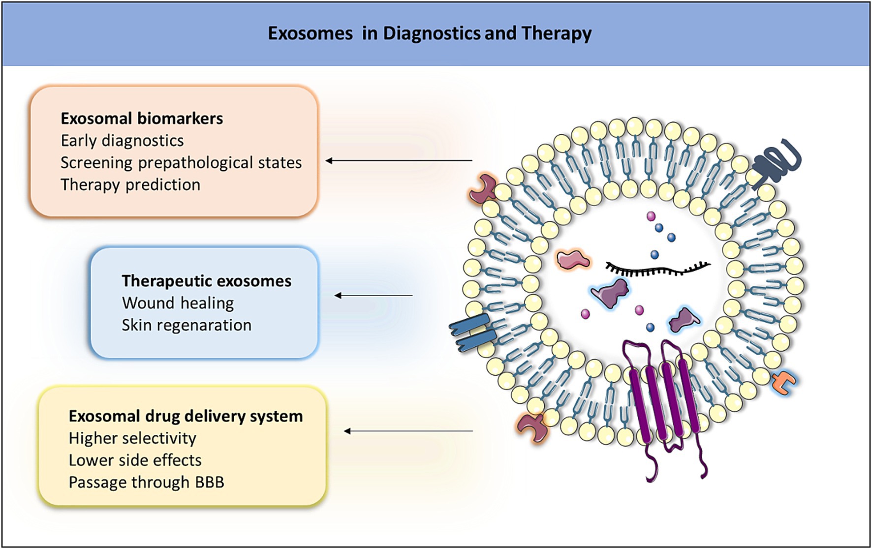
Exosomes, nanosized extracellular vesicles released by various cell types, are intensively studied for the diagnosis and treatment of cancer and neurodegenerative diseases, and they also display high usability in regenerative medicine. Emphasizing their diagnostic potential, exosomes serve as carriers of disease-specific biomarkers, enabling non-invasive early detection and personalized medicine. The cargo loading of exosomes with therapeutic agents presents an innovative strategy for targeted drug delivery, minimizing off-target effects and optimizing therapeutic interventions. In regenerative medicine, exosomes play a crucial role in intercellular communication, facilitating tissue regeneration through the transmission of bioactive molecules. While acknowledging existing challenges in standardization and scalability, ongoing research efforts aim to refine methodologies and address regulatory considerations. In summary, this review underscores the transformative potential of exosomes in reshaping the landscape of medical interventions, with a particular emphasis on cancer, neurodegenerative diseases, and regenerative medicine.
Graphical Abstract.
Extracellular vehicles (EVs) are cellular structures released by cells into the extracellular space and have recently become a focal point of research due to their multifunctional role in many biological processes (1, 2). According to the new classification, EVs are divided into several types based on their biogenesis (e.g., exosome, microvesicle, apoptosome, and autophagic EVs), concept (e.g., oncosome, matrix vesicle, stress EVs, and migrasome), and size (e.g., small EVs and large EVs) (3). In general, EVs were divided into three types including apoptotic bodies (apoptosomes), which are the largest EVs with a size range between 1 and 5 μm and are released during programmed cell death (apoptosis). They contain cellular organelles and fragmented DNA and are cleared by phagocytic cells. Microvesicles, typically 100–1,000 nm in diameter, are shed directly from the plasma membrane through outward budding, a process in which a portion of the cellular membrane protrudes outward from the cell surface. They contain proteins, lipids, polysaccharides, and nucleic acids and are involved in intercellular communication and signaling (2, 3). Exosomes (Figure 1), the smallest EVs ranging in size from 30 to 150 nm, are lipid bilayer vesicles and were discovered three decades ago by Pan and Johnstone during investigations of reticulocyte maturation (2–5). Recent studies discovered small exosomes (Exo-S) and large exosomes (Exo-L). Exo-S are in the size range 40–80 nm and contain exosomal tetraspanin marker CD63, while Exo-L (80–150 nm) contain CD9 (3). Initially perceived as cellular waste products responsible for eliminating unnecessary cellular components, our understanding of exosomes has undergone a paradigm shift over the years, revealing their multifaceted functions in cellular communication and signaling (1, 2).
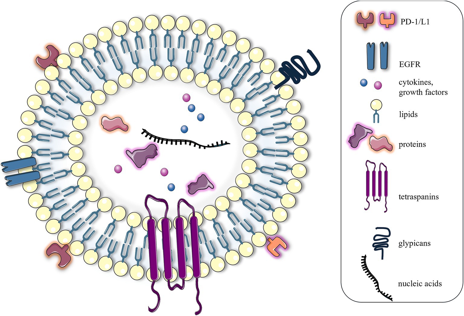
Figure 1. Structure of exosomes. From a structural perspective, exosomes can be defined as lipid nanoparticles characterized by a phospholipid bilayer membrane. The exosomal membrane is enriched with a diverse array of proteins and saccharide markers, including immunomodulatory molecules, such as PD-1 and PD-L1, and hormone receptors, such as EGFR, tetraspanins, and glypicans. The internal composition of exosomes comprises a variety of biomolecules, including intracellular and cytoskeletal proteins, nucleic acids, growth factors, and cytokines. The figure was partly generated using Servier Medical Art, provided by Servier, licensed under a Creative Commons Attribution 3.0 unported license.
Exosomes not only represent a promising material for the diagnosis of serious pathological states but can also be effectively utilized for medicinal applications and drug transport. Given their significance, this review presents the medicinal potential of exosomes, encompassing areas such as regenerative medicine, early diagnosis, and drug treatment. In addition, to provide a more comprehensive understanding, the review rigorously assesses exosome biogenesis, isolation, and characterization.
Understanding the biogenesis of exosomes is crucial for advancing the knowledge of their biological functions, their roles in diseases, and potential applications in therapeutics (1, 3). Their life cycle is a complex process involving three main steps: biogenesis, transport, and release (2, 3). The whole process is illustrated in Figure 2 and is initiated by an inward cell membrane budding (6). During this invagination of the plasma membrane, a portion of a cellular membrane undergoes inward folding, and a cup-shaped structure containing extracellular proteins, lipids, metabolites, and cell membrane proteins is formed. This leads to the formation of early endosomes (EEs), which subsequently mature and transform into late endosomes (LEs). Maturation involves the inward budding of the EEs membrane, leading to the sequestration of EE cytoplasmic contents and intraluminal vesicle (ILVs) formation within the endosomal lumen. During ILV formation, specific cargo molecules such as proteins, lipids, and nucleic acids are selectively sorted into the ILVs. LEs containing ILVs are called multi-vesicular bodies (MVBs). The fate of MVBs is determined by the specific proteins present on their surface, which in turn influence various intracellular pathways involved in cargo sorting and trafficking. The MVBs can either fuse with lysosomes or autophagosomes to be degraded or fuse with a plasma membrane to release the contained ILVs as exosomes (2, 6).
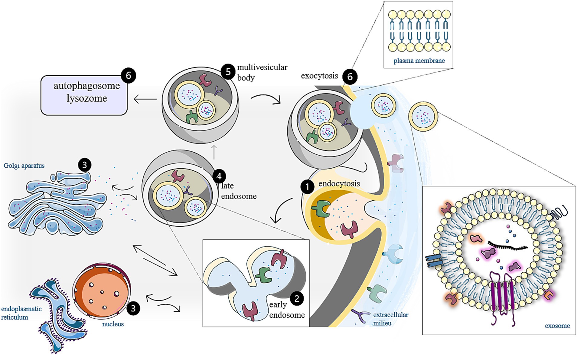
Figure 2. Biogenesis of exosomes. Extracellular membranes are characterized by the presence of numerous transmembrane proteins, including various receptors. (1) Upon ligand binding, receptor-mediated endocytosis is initiated, facilitated by actin filaments, which are integral components of the cytoskeleton. This process results in the invagination of the membrane surrounding the receptor, leading to the formation of an early endosome within the cell. (2) In this early endosome, the bilayer phospholipid membrane exhibits an orientation that is opposite to that of the cytoplasmic membrane, causing the extracellular domains of the transmembrane proteins to be directed inward, toward the lumen of the endosome. (3) Within the cellular context, endosomes are integral components of the complex endosomal-lysosomal system that interacts in a parallel manner (in both directions) with various organelles (e.g., Golgi apparatus and endoplasmic reticulum). This interaction allows endosomes and exosomes to encapsulate biomolecules derived from diverse cellular compartment. (4) During this process, early endosomes undergo maturation into late endosomes, which possess the normal orientation of transmembrane proteins and (5) generate intraluminal vesicles that will ultimately become exosomes. (6) Following the fusion of multi-vesicular bodies containing these intraluminal vesicles with the cytoplasmic membrane, exosomes are released into the extracellular environment. The figure was partly generated using Servier Medical Art, provided by Servier, licensed under a Creative Commons Attribution 3.0 unported license.
Isolation of exosomes is challenging due to the complexity of biological fluids (7, 8). The most common isolation methods include ultracentrifugation, ultrafiltration, size exclusion chromatography, polymer precipitation, tangential flow filtration, and immunoaffinity approaches. A comparison of these methods is provided in Table 1. The optimal isolation strategy should be selected based on the application field, as well as the volume and number of biological samples (7–9). Furthermore, the application of exosomes directly influences the use of subsequent characterization methods (9, 10). This chapter does not aim to provide a detailed explanation of the principles behind individual isolation and characterization methods of exosomes but rather to offer a comprehensive overview that assists researchers in selecting the most appropriate approaches based on considering the specific requirements of different experimental and application contexts.

Table 1. Advantages and disadvantages of the most common isolation techniques of exosomes.
For diagnostic purposes, exosomes from various biological fluids, including blood (serum and plasma), saliva, and urine, are used in non-invasive diagnostics due to their ability to carry specific molecular information (9, 11). Blood-derived exosomes provide systemic insights, reflecting the physiological and pathological state of the entire body, making them ideal for detecting various diseases such as cancer, neurodegenerative, cardiovascular, and autoimmune diseases (12–16). Salivary exosomes, primarily originating from the salivary glands and the oral cavity, are valuable for diagnosing oral diseases and gastrointestinal tract disorders (17–19). Urinary exosomes, secreted by epithelial cells of the urinary tract, are effective in diagnosing renal diseases, bladder cancer, and prostate conditions (20–22).
When utilizing exosomes for diagnostic purposes, the primary goal is to identify disease-specific exosomal markers. To determine disease-specific markers, it is essential to identify markers that differ in presence or expression level between samples from diseased and healthy patients. This requires the comparison and processing of a large number (tens to hundreds) of biological samples for statistical relevance (23, 24). This procedure thus involves processing large numbers of samples with small volume, typically in the maximum of a few milliliters. For limited numbers of samples, typically in the range of lower tens, differential ultracentrifugation (DUC) is considered the gold standard and is the most widely used method. It usually involves several consecutive rounds of centrifugation with increasing centrifugal force and centrifugation time to remove cells, cell debris, and larger microvesicles. The final step at 100,000 g or higher serves to precipitate the exosomes (12, 25–27).
Because DUC is time-consuming, for handling tens to hundreds of samples, the final ultracentrifugation step is often replaced with commercial kits based on precipitation. Several commercial kits use a polyethylene glycol precipitation technology, including the Total Exosome Isolation Kit (Invitrogen), ExoQuick-TC Exosome Precipitation Solution (System Biosciences), miRCURY Exosome Kits (QIAGEN), Exo-Prep (HansaBioMed), PureExo Exosome Isolation Kit (101Bio), ExoGAG (NasasBiotech), Exosome Precipitation Solutions (Immunostep), and the miRCURY Exosome Isolation Kit (Exiqon) (28–32). These methods result in the isolation of exosomes referred to as total exosomes. In neurodegenerative diseases (NDs), specific subpopulations of exosomes are isolated from the total exosome pool using immunoaffinity methods with specific antibodies, such as anti-L1CAM, anti-NCAM, anti-MOG, or anti-GLAST. These antibodies bind specifically neural, oligodendrocyte, or astrocyte exosomes, which are the most relevant for identifying markers of NDs as Alzheimer disease (AD), multiple sclerosis (MS), dementia, or schizophrenia (33–36).
After the isolation of total exosomes or specific exosomal subpopulations, the characterization of these exosomes and the identification of specific disease markers are performed (10). The most common markers include specific proteins or RNAs. For protein marker identification, techniques such as tandem of liquid chromatography and mass spectrometry (LC–MS) are used, while next-generation sequencing (NGS) is used for RNA. Once specific disease markers are identified, it is crucial to determine whether their exosomal expression differs significantly between diseased and healthy patients in a statistically relevant manner. To quantify markers, techniques such as enzyme-linked immunosorbent assay (ELISA) or quantitative reverse transcription polymerase chain reaction (qRT-PCR) are commonly used (37–42).
Currently, exosomes are being extensively investigated as drug delivery systems. Due to their nanoscale dimensions, they can deliver therapeutic agents specifically to tumor sites (43). Tumor tissues are characterized by a high absorption capacity and poor drainage (44), which facilitates the selective accumulation of exosomes with prolonged circulation times within these tissues. In addition, it is noteworthy that exosomes exhibit favorable permeability across BBB (45). Furthermore, the surface of exosomes can be effectively modified with various ligands that are specific to target cells, thereby significantly enhancing the selectivity of exosomes for these cells (46). As cell-derived products, exosomes are generally considered to be safer than conventional nanoparticles, particularly metal-based nanoparticles (47). Their unique structure, comprising a hydrophilic core and a lipid bilayer, allows them to transport a wide range of drug types (46). However, a significant limitation in the clinical application of exosomes is that cells produce only small quantities of various types of exosomes, which restricts their usability in therapeutic contexts (48).
In regenerative medicine and therapeutic applications, exosomes are isolated from cell culture media. The most commonly used cell sources are mesenchymal stem cells. Similar to diagnostic applications, where DUC is a preferred method for smaller sample quantities, DUC is also commonly employed for isolating exosomes from cell culture media for volumes ranging from tens to hundreds of milliliters. However, a drawback of the final ultracentrifugation step is that the high speeds lead to exosomal damage and the sedimentation of impurities, which diminishes exosomal therapeutic activity (12, 49, 50). Therefore, the final ultracentrifugation step is often replaced by density gradient ultracentrifugation (DGUC), utilizing sucrose or iodixanol gradients (OptiPrep™), where the exosomal fraction is not only less damaged but also better purified (49–51).
For larger volumes, in the range of hundreds of milliliters to liters, a combination of other techniques is employed due to the limited capacity of centrifuges. These primarily include ultrafiltration (UF), utilizing polymer filters of various pore sizes to remove cells, cell debris, and microvesicles, followed by tangential flow filtration (TFF) to remove contaminating proteins, to concentrate the sample, and to perform diafiltration of exosomes into the desired buffer (52–54). For final exosome purification, size exclusion chromatography (SEC) is employed, followed by a final TFF step to concentrate the sample and transfer it into suitable application buffers, most commonly PBS (55–57). The advantage of the combination of techniques such as UF, TFF, and SEC is that they are suitable for practical use on an industrial scale (55, 56). In addition, the isolated exosomes exhibit high purity and preserved therapeutic activity, which is usually subsequently confirmed by in vitro tests such as scratch or transwell assays (54, 58).
According to the recommendations of the International Society for Extracellular Vesicles (ISEV), exosomal samples should generally be characterized for size, concentration, and the presence of exosomes (59, 60). The presence of exosomes is typically confirmed by detecting at least one transmembrane protein (commonly tetraspanins: CD9, CD63, and CD81) or a GPI-anchored protein (e.g., integrins), along with one cytoplasmic lipid (e.g., sphingolipids, ceramides, and cholesterol) or cytoplasmic protein (e.g., ALIX, TSG101, and HSP70) (59, 60). These specific markers can be assayed using methods such as Western blot or ELISA (61, 62). In addition, electron microscopy is often employed to provide images of typical exosomal morphology, further confirming the presence of exosomes in the sample (63, 64).
The size and concentration of exosomes, expressed as particles per milliliter, are commonly determined using techniques such as dynamic light scattering (DLS), nanoparticle tracking analysis (NTA), and resistive pulse sensing (RPS). However, these techniques are not specific to exosomes and may overestimate their concentration (65, 66). Alternatively, exosomes can be labeled with specific dyes, where the measured concentration corresponds only to the positively stained population. This approach allows for more accurate quantification of exosomes and can be performed using nanoflow cytometry (nanoFCM). Nevertheless, underestimation of concentration may occur if the sample is not properly titrated (67, 68). In some cases, the concentration of exosomes is determined based on the total protein concentration per milliliter (μg/mL) using the Bradford assay (10). All these parameters must be thoroughly assessed to ensure sufficient sample purity and demonstrate that the observed therapeutic effect is primarily induced by exosomes rather than by potential contaminants.
As mentioned earlier, exosomes are natural intercellular communicators in normal biological processes but also in pathologies. They transport proteins, lipids, and nucleic acids specific for their parenteral pathogenic cells. From a clinical perspective, most applications use exosomes as biomarkers of diseases (69). The content of the exosome has been shown to be disease-specific, such as in NDs, prion diseases, viral infections, and cancer (69). Furthermore, exosomes play a transformative role in regenerative medicine, offering innovative therapeutic interventions. Their bioactive cargo, including growth factors and signaling molecules, has demonstrated significant potential in modulating immune responses and promoting tissue repair (70). In addition, exosomes serve as carriers for therapeutic cargo loading, holding promise for targeted drug delivery in disease treatment (69, 71, 72). Currently, 150 clinical trials registered on ClinicalTrials.gov are investigating exosome-based therapies for various diseases (73). The majority of applications utilizing exosomes for both therapy and diagnosis focus on their utilization in the fields of cancer and NDs, as depicted in Figure 3 (74). For this reason, this review will focus on the use of exosomes in these diseases.
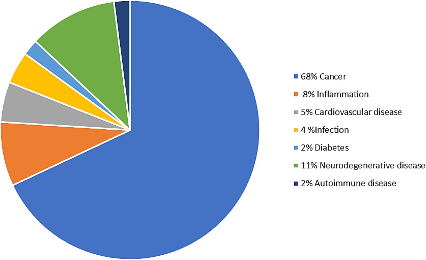
Figure 3. Distribution of exosome therapy and diagnostics concerning the target diseases (74). The figure was partly generated using Servier Medical Art, provided by Servier, licensed under a Creative Commons Attribution 3.0 unported license.
Nowadays, one of the main applications of exosomes is in regenerative medicine, a promising field dedicated to the regeneration and reconstruction of diseased or injured organs and tissues (72). Since the 1970s, mesenchymal stem cells (MSCs) have been under investigation in this field for their multipotent characteristics and their ability to migrate to injury sites. They are used in regenerative medicine due to their robust self-renewal capacity and ability to differentiate into adipogenic, chondrogenic, osteogenic, endothelial, neural, and epithelial cells, as proven in both in vivo and in vitro experiments (70, 73–75). MSCs, like every cell in the human body, release exosomes, which have started to be extensively researched due to their regenerative properties. Currently, at least 31 clinical trials are exploring the use of exosomes derived from MSCs (MSCs-EXOs) as an alternative to basic MSCs therapy (73). MSCs-EXOs have shown comparable or superior therapeutic efficacy compared to MSCs alone (72, 73). They exhibit lower immunogenicity, present an enhanced safety profile by avoiding concerns related to uncontrolled differentiation and tumorigenicity associated with live cells, and offer improved storage conditions, simplifying logistical challenges compared to live cell storage. In addition, there are no ethical issues, and their small size allows for sterilization by filtration (73–77). The comparison between exosomal and stem cell therapy is provided in Table 2.
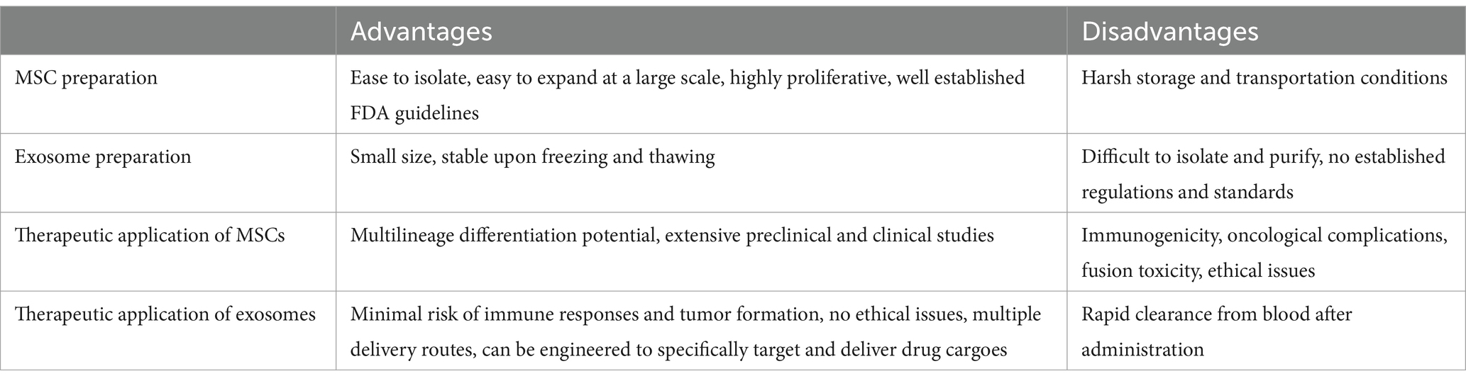
Table 2. Exosomes versus stem cells in preparation and therapy.
The greatest attention in regenerative medicine is focused on skin healing, a tissue which plays a crucial multifunctional role as a protective barrier, a temperature regulator, and facilitating tactile and pain sensations (78). However, the use of exosomes for wound treatment could be challenging due to their rapid clearance from the application site, which limited therapeutic effectiveness (79). To address this limitation, current research focuses on the combination of exosomes and biomaterials. This innovative approach extends the retention time of exosomes on the wound surface without compromising their biological activity, enabling the development of advanced exosome-based therapies (80, 81). Hydrogels as biomaterials exhibit a synergistic effect in exosome-induced wound healing and can serve as a versatile platform for the incorporation of therapeutic exosomes, enhancing their efficacy in tissue regeneration. To date, hyaluronic acid, gelatin, chitosan, and polypeptide-based hydrogels have been used for encapsulating exosomes from different cell sources (75, 80, 81). Chitosan is the often-used material for hydrogel preparation. Chitosan hydrogel enriched with MSCs-EXOs, specifically human endometrial stem cell-derived and human placenta-derived exosomes, demonstrated notable wound closure ability by promoting the formation of new epithelial cells, significant retention of MSCs-EXOs at injury sites, promotion of angiogenesis, and acceleration of the recovery of ischemic hind limbs (82). For the best contact of the hydrogel with the wounded skin and effective wound filling, thermosensitive hydrogels are used. Thermosensitive pluronic hydrogel combined with human umbilical cord MSCs-EXOs (HUCMSCs-EXOs) significantly accelerated wound closure, promoted angiogenesis, and improved skin healing of chronic diabetic wounds (83). For effective diabetic wound treatment, polyvinyl alcohol/alginate nanohydrogel with HUCMSCs-EXOs and an injectable antibacterial polypeptide-based hydrogel with adipose derived MSCs-EXOs were used. This type of hydrogels demonstrated the ability to promote proliferation, migration, and angiogenesis of human umbilical vein endothelial cells, expediting diabetic wound closure thus presenting a novel approach for complete skin regeneration (84, 85).
Further applications in regenerative medicine are focused on hard tissue regeneration, essential for bone and cartilage repair. Traditionally, MSCs, scaffolds, and growth factors are used in this field. While scaffolds have been proven beneficial for bone regeneration, the avascular nature of cartilage poses unique challenges (75). Osteoarthritis (OA), a prevalent joint disease extending beyond cartilage, demands innovative regenerative procedures (81). Promisingly, exosome-integrated scaffolds and MSCs-EXO therapy show potential in OA treatment. Articular cavity injection with HUCMSCs-EXOs in PBS demonstrated significant efficacy in preventing severe damage to knee articular cartilage in a rat OA model. These therapies not only promoted chondrocyte proliferation and migration but also exhibited anti-apoptotic effects and reversed cellular injuries. Moreover, HUCMSCs-EXOS played a crucial role in regulating the polarization of M2 macrophages, fostering chondrocyte survival by producing anti-inflammatory cytokines to suppress adverse inflammation (86). To enhance therapeutic efficiency and retention time in vivo, HUCMSCs-EXOs were engineered to specifically target chondrocytes and encapsulated within hyaluronic acid hydrogel, presenting a “two-phase” releasing system. This approach synergistically facilitated OA cartilage repair in a rat model and proved the rejuvenating effects of HUCMSCs-EXOs on aging chondrocytes in OA, offering a promising cell-free OA treatment strategy (87). Therapeutic potential of bone marrow MSCs-EXOs was explored in the context of mitochondrial dysfunction and oxidative stress in OA. 3D printed scaffolds, composed of extracellular matrix, gelatin methacrylate, and exosomes, effectively restored chondrocyte mitochondrial function, enhanced chondrocyte migration, and polarized the synovial macrophage response in vitro. Notably, a 3D printed scaffold significantly facilitated cartilage regeneration in a rabbit model, highlighting its potential as an early treatment strategy for OA (88).
In recent years, MSCs-EXOs have also been used increasingly in ophthalmology. MSCs-EXOs have shown promise in various applications, such as promoting ocular tissue regeneration and addressing vision-related disorders. MSCs-EXOs have explored the therapeutic potential of stem cell exosomes in treating ocular surface diseases, corneal injuries, and retinal degenerative conditions (89). Exosomes derived from bone marrow stem cells have demonstrated the ability to enhance corneal epithelialization and maintain corneal transparency in diabetic mice (90). MSCs-EXOs have been explored for their regenerative effects on corneal injuries (91). In retinal diseases, including age-related macular degeneration and retinitis pigmentosa, MSCs-EXOs have been investigated for their neuroprotective and regenerative properties. These exosomes may influence retinal cell survival, angiogenesis, and anti-inflammatory responses, offering a novel approach for treating degenerative conditions affecting the retina (92). As exosome research in regenerative medicine continues to progress, MSCs-EXOs are beginning to be applied into various fields, such as periodontitis (93, 94). Research on exosomes in regenerative medicine is expected to gain prominence, highlighting the growing need to integrate exosome-based regenerative therapies into clinical practice (95).
As already mentioned, exosomes are promising biomarkers for the diagnosis of several diseases. Most attention has been paid to cancer (Figure 4; Supplementary Table S1) (95–105) with less focus on NDs.
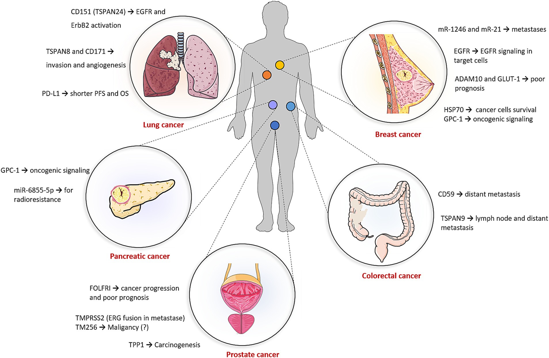
Figure 4. Potential of exosomes in cancer diagnostics. The applicability of exosomes in cancer diagnostics is detailed in Supplementary Table S1. The figure was partly generated using Servier Medical Art, provided by Servier, licensed under a Creative Commons Attribution 3.0 unported license.
Tumor-derived exosomes (TEXs) have emerged as critical players in cancer progression. Cancer cells release TEXs in large quantities, leading to a rapid increase in the concentration of total exosomes in the serum or plasma of cancer patients, which correlates with poor prognosis (106). TEXs provide immunosuppressive effects by inducing dysfunction in various immune cells, thereby suppressing anti-tumor immune responses (107). Initial interactions between TEXs and immune cells occur through ligand–receptor recognition, followed by either direct fusion with the plasma membrane or receptor-mediated uptake, resulting in the release of TEX cargo into the cytoplasm of immune cells. While T lymphocytes do not efficiently internalize TEXs, they interact with surface molecules to trigger sustained Ca2+ flux and downstream signaling cascades, ultimately altering the transcriptome of recipient cells. In contrast, phagocytic cells, such as dendritic cells and macrophages, rapidly internalize TEXs (108).
Macrophages are a prominent component of the tumor microenvironment (TME) and can make up more than half of the tumor mass in some cases. Moreover, tumor progression is associated mainly with macrophages. Macrophages can be induced to either tumor-suppressive immunological type M1 or tumor-promoting inflammatory M2 macrophages (109, 110). Tumor-associated macrophages predominantly exhibit an M2 phenotype, characterized by the secretion of pro-angiogenic factors and cytokines that promote angiogenesis, tumor growth, and metastasis (111, 112). Many studies have shown evidence on the role of TEXs in macrophage M2 polarization to promote tumor progression. For instance, exposure of macrophages to TEXs decreased the expression of M1 markers, such as IFNγ, while upregulating IL-1β, a marker of inflammation, suggesting that TEXs may help maintain a macrophage phenotype supportive of tumor survival and proliferation (113). In triple-negative breast cancer (TNBC), TEXs have been reported to promote M2 macrophage polarization, facilitating lymph node metastasis. Co-culture of TNBC-TEXs with macrophages led to significant morphological changes and an increased expression of M2 markers, including Fizz1, CD206, and Arg-1. An orthotopic TNBC model further confirmed the role of TEXs in driving M2 polarization, with enhanced tumor growth and axillary lymph node metastasis observed in vivo (114).
In cancer metastasis, epithelial–mesenchymal transition (EMT) is the key process during which cancer cells lose their epithelial properties and adopt a more mesenchymal and invasive phenotype. Generally, EMT initiation is characterized by loss of cell–cell adhesions and apicobasal polarity, leading to the formation of cells with increased migratory and invasion capabilities that are able to invade the extracellular matrix (115, 116). At a molecular level, EMT involves the downregulation of epithelial-type proteins, such as E-cadherin, and the acquisition of mesenchymal markers, such as vimentin (117). TEXs from bladder cancer cells have been shown to induce EMT-like changes in urothelial cells, enhancing their invasive potential. This effect was mediated by TEX-induced upregulation of vimentin and downregulation of E-cadherin through the TGF-β1 signaling pathway (118). Similar EMT-inducing effects of TEXs have been observed in glioblastoma, lung carcinoma, and gastric cancer models (119–121). TEXs can also influence EMT through their miRNA cargo. For example, miR-23a within TEXs promotes EMT by inhibiting E-cadherin synthesis in lung carcinoma and melanoma cells, while miR-191 and let-7a, present in TEXs from patients with melanoma, gastric, and colorectal cancers, have also been implicated in EMT regulation (122–126). In addition, TEX-derived miR-105 has been shown to promote vascular invasion by downregulating ZO-1 in endothelial cells. Notably, elevated miR-105 levels in the serum of breast cancer patients have been correlated with metastatic progression and poor prognosis (127). Given their critical role in modulating the TME, promoting EMT, and facilitating immune evasion, profiling TEXs in blood and other body fluids holds significant promise as a non-invasive method for cancer diagnosis and prognosis (128).
The WHO Global Cancer Observatory (GLOBOCAN) 2022 registry provides a list of the most common types of cancers. The three most common types are lung (12.4%), breast (11.5%), and colorectal (9.6%) cancers. Lung cancer is the leading cause of cancer death worldwide (129). Surface-enhanced Raman spectroscopy (SERS) of exosomes, combined with AI deep learning software, allows for accurate diagnosis of early-stage lung cancer. The deep learning model was trained with SERS signals of exosomes derived from normal and lung cancer cell lines and subsequently its ability to detect cancer was verified using exosome samples from patients’ blood. The model identified the lung cancer patients and even detected stage I patients with an accuracy of 90.7% (130). Regarding TEX protein biomarkers, CD151, TSPAN8, and CD171 were overexpressed in lung cancer samples. Of these, CD171 has been associated with EMT, metastases, and poor prognosis (96, 131, 132). Determination of PD-L1 can also provide important diagnostic information. Akbar et al. reported that exoPD-L1 from non-small cell lung cancer (NSCLC) patients and healthy controls showed a significantly higher difference than corresponding serum and tissue PD-L1 (97). For example, exoPD-L1 was found in all the patients (100%), while tissue PD-L1 was observed only in 71% patients. Furthermore, exoPD-L1 can also be used for the prediction of immune inhibitors efficiency. ROC curve analysis of change from baseline in exoPD-L1 levels between responders and non-responders showed 87% sensitivity and 100% specificity (p = 0.0015), indicating strong discriminatory power. An increasing number of studies have demonstrated that non-coding RNAs are closely correlated with the initiation and development of lung cancer as well (133). Detection of exosomal long non-coding RNA as potential lung cancer diagnosis is also performed in a current clinical study (NCT03830619) (134). Breast cancer is the most common type of cancer and the second leading cause of cancer-related deaths in women. In this type of cancer, media from breast cancer cell lines were analyzed to identify specific exosomal proteins such as glucose transporter-1, glypican-1, and the metalloproteinase domain-containing protein 10 (98). Epidermal growth factor receptor (EGFR) is a transmembrane protein that plays a key role in cell signaling pathways involved in cell growth, proliferation, and survival and is often overexpressed or mutated in various cancers. It was reported that triple-negative breast cancer cells (MDA-MB-468) can produce exosomes with encapsulated EGFR (protected from EGFR inhibitor), which can induce EGFR signaling in target cells, thereby promoting cancer progression or resistance to therapy (99). Typical non-coding RNA cancer biomarkers are miR-1246 and miR-21, which were also overexpressed in breast cancer cell lines (100). The plasma level of miR-1246 was measured in breast cancer patients and healthy controls using an Au nanoflare probe. This biomarker-based probe distinguished breast cancer patients from healthy individuals with 100% sensitivity and 92.9% specificity (95). Currently, a clinical trial (NCT02662621) is underway, focusing on an exosomal detection protocol for diagnosing various cancers, including breast cancer. The study indicates that exosomes displaying the stress protein HSP70 on their membrane may serve as cancer-specific exosomes (134). In colorectal cancer, tetraspanin-1 was found to be upregulated in plasma exosomes from patients compared to healthy controls, showing 75.7% sensitivity (135). Pancreatic cancer, one of the deadliest cancer types, involves TEXs that carry the glypican-1 biomarker. This co-receptor for various signaling molecules regulates key processes such as cell growth, motility, and differentiation (102). Currently, two clinical trials are underway to assess the efficacy of diagnostic exosomes in colon and liver cancers (NCT03432806) and in pancreatic cancer (NCT03334708). These studies aim to identify biomarkers circulating in blood and analyze the corresponding tissues (134). While previous studies have focused on TEXs from blood, urinary exosomes play a role in urological tumors (136). In prostate cancer, biomarkers such as TM256 and LAMTOR proteins found in urinary exosomes exhibited very high sensitivity (105). In addition, potential biomarkers such as TPP1, TMPRSS2, and FOLR1 were highly upregulated in urinary exosomes derived from the bladder. Notably, despite being histologically tumor-free at cystectomy, patients’ urinary exosomes displayed a carcinogenic metabolic profile, likely originating from undetected or partially transformed cancer cells (137).
Current diagnosis of NDs relies on clinical assessments, medical history, imaging techniques, and diagnostic tests based on observed symptoms (138, 139). However, predicting these diseases remains challenging due to the typically late-stage diagnosis. Emerging research highlights the potential of exosomes as biomarkers for early detection, offering a promising approach for more effective and timely diagnoses in the future (Figure 5) (140). Early diagnosis of NDs is crucial for enabling timely interventions that can significantly improve patient outcomes (141).
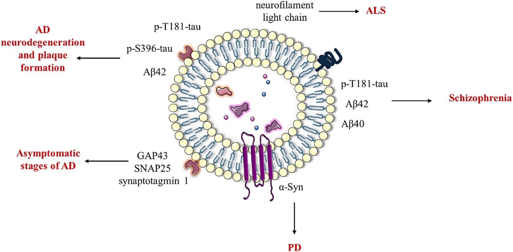
Figure 5. Potential of exosomes in the diagnosis of neurological diseases. In AD, protein markers such as total tau, phosphorylated T181-tau (p-T181-tau), phosphorylated S396-tau (p-S396-tau), and amyloid-beta 42 (Aβ42) derived from neural-derived blood exosomes are indicative of neurodegeneration and plaque formation. Notably, GAP43, SNAP25, and synaptotagmin 1 can be detected even in asymptomatic stages of AD. Furthermore, exosomal Aβ42, Aβ40, and P-T181-tau have been implicated in schizophrenia. In the context of PD and ALS, exosomal α-synuclein and neurofilament light chain exhibit promising potential as biomarkers for disease monitoring and progression. The figure was partly generated using Servier Medical Art, provided by Servier, licensed under a Creative Commons Attribution 3.0 unported license.
In Alzheimer’s disease (AD), neural-derived blood exosomes have emerged as a valuable source of AD-related overexpressed protein markers, including total tau, P-T181-tau, P-S396-tau, and Aβ42 (33). Exosomal synaptic proteins, such as growth-associated GAP43, neurogranin, SNAP25, and synaptotagmin 1, show promise in predicting AD at asymptomatic stages, potentially detecting the disease 5 to 7 years before cognitive impairment occurs (34). In addition, miRNA markers, including miR-137, miR-181c, miR-9, and miR-29a/b, are downregulated in blood serum and offer additional potential for early AD detection (142, 143). Among other types of biomarkers, lipids such as LDL-C, TG, and HDL-C are associated with AD (144). Furthermore, it was also shown that exosomal protein markers offer comparable diagnostic capacity to cerebrospinal markers, further enhancing the potential of blood-based diagnostics for AD (145). Exosomal levels of Aβ42, Aβ40, and P-T181-tau were also measured in the blood plasma of patients diagnosed with schizophrenia (36). Specifically, these markers were determined in neural and astrocytic exosomes. While Aβ42 levels were higher in astrocytic exosomes than in neural exosomes, other markers were similar between these two groups. It was also found that higher astrocytic P-T181-tau levels were associated with worse executive functioning, and astrocytic Aβ42 levels were more sensitive and specific in differentiating diagnostic groups. Other types of neural cells explored in the context of NDs are oligodendrocytes. These cells are associated with MS. Oligodendrocyte-derived extracellular vesicles showed higher concentrations of myelin basic protein, which could serve as potential biomarkers across diverse MS phenotypes (35). For Parkinson disease (PD), it was found that the most reliable biomarker of blood neural exosomes is α-synuclein (146). In amyotrophic lateral sclerosis (ALS), exosomal biomarkers, such as neurofilament light chain, have been identified in both cerebrospinal fluid and blood. This marker can serve for early diagnosis and monitoring disease progression (147).
The two main areas in which exosome applications are greatly investigated and hold great potential are cancer and NDs (74, 76). In cancer treatment, significant attention is paid to nanocarriers which can entrap chemotherapeutic drugs and deliver them to the diseased site, reducing the side effects associated with the systemic administration of conventional anticancer drugs (148, 149). In recent years, exosomes started to be explored as promising nanocarriers (48) that can affect tumor growth, metastasis, and even sensitize cancer cells to conventional therapies (Figure 6).
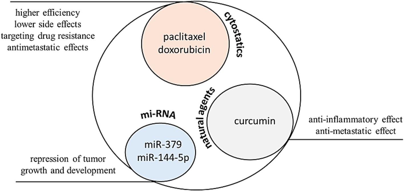
Figure 6. Exosomes represent a promising delivery system for the transport of anticancer agents. In murine cancer models, specifically in breast and ovarian carcinoma, exosomal formulations exhibit lower cardiotoxicity and higher efficacy compared to free doxorubicin. In the case of paclitaxel, exosomal formulations provide significantly improved intracellular delivery into 3LL-M27 cells (Lewis carcinoma expressing P-glycoprotein, which is associated with drug resistance) when compared to liposomal formulations and polystyrene nanoparticles. Furthermore, in these murine models, exosomes have been shown to suppress the development of metastases. Regarding natural agents, curcumin-loaded exosomes display promising efficacy against colon and pancreatic carcinomas, exhibiting antimetastatic effects and reduced inflammation. Exosomes are also suitable for the transport of biological agents such as microRNAs. For instance, exosomes loaded with miR-379 and miR-144-5p demonstrate potent effects against breast cancer and pancreatic ductal adenocarcinoma. The figure was partly generated using Servier Medical Art, provided by Servier, licensed under a Creative Commons Attribution 3.0 unported license.
Their ability to target specific cell types and deliver therapeutic cargos makes them a valuable asset in the fight against malignancies (150–153). For example, exosomes loaded with doxorubicin (ExoDOX) were used in a mouse model of breast and ovarian cancers. It was found that ExoDOX are less cardiotoxic than free DOX, enabling the use of higher concentrations of ExoDOX, thus increasing the efficacy of DOX (154). ExoDOX conjugated with gold nanoparticles (ExoDOX-GNPs) were used in vitro with lung cancer cell lines and normal lung fibroblasts. The pH sensitive conjugation bond enables the enhanced rate of drug release under acidic conditions and successful uptake of the ExoDOX-GNPs by the recipient cells. Cell viability assays indicated that ExoDOX-GNPs exhibit preferential cytotoxicity toward cancer cells and have minimal activity on non-cancerous cells (155). Exosomes loaded with paclitaxel (ExoPTX) were developed. The incorporation of paclitaxel into exosomes enhanced drug cytotoxicity more than 50-fold in drug-resistant MDCKMDR1 (Pgp+) cells (156). Furthermore, ExoPTX displayed a potent anticancer effect in a mouse model of murine Lewis Lung Carcinoma pulmonary metastases. In addition to separate therapeutics, other compounds are also used as exosomal cargo. Exosomes loaded with gemcitabine in combination with the survivin protein with mutation T34A induce apoptosis and enhance gemcitabine-killing effects in pancreatic adenocarcinoma cells (157). For cancer treatment, exosomes can also deliver nucleic acids, including miRNAs, siRNAs, and mRNAs (158). For instance, exosomes containing miR-379, a potential tumor suppressor, showed a significant reduction in the rate of tumor formation and growth for the in vitro and in vivo therapies of breast cancer (159). Exosomes loaded with miR-145-5p, which inhibits pancreatic ductal adenocarcinoma (PDAC) cell proliferation, invasion, and increase apoptosis, significantly reduced the growth of xenograft tumors in a PDAC mouse model (160). Natural products are also used in exosomal cancer therapies, including the use of curcumin. This molecule can mitigate cancer initiation and metastasis (161, 162). Exosomes loaded with curcumin induced the apoptosis of pancreatic cancer cells and significantly delayed brain tumor growth with reduced inflammation when delivered to a GL26 brain tumor model via an intranasal route (163, 164). Plant exosomes with curcumin are used also in Phase I clinical trial investigating the ability of plant exosomes to deliver curcumin to normal and malignant colon tissue (NCT01294072). This study is now recruiting patients (165). Grape exosomes are investigated in preliminary active clinical trial to abrogate oral mucositis induced by combined chemotherapy and radiation in head and neck cancer patients (NCT01668849) (165). Vaccination with tumor antigen-loaded dendritic cell-derived exosomes on patients with unresectable NSCLC lung cancer responding to induction chemotherapy was explored in another clinical study (NCT01159288), where the first phase of this study has now been completed. The primary endpoint was progression-free survival at 4 months after chemotherapy cessation, with a target of at least 50% of patients achieving this endpoint. However, this target was not met as only 32% of patients experienced disease stabilization at 4 months (165, 166). A study of mesenchymal stromal cell-derived exosomes with KrasG12D siRNA for metastatic pancreas cancer patients harboring the KrasG12D mutation is now recruiting patients for the NCT03608631 clinical trial (166). G12D is the most common KRAS mutation detected in carcinomas and confers a unique structural conformation that influences downstream signaling and may lead to its potent oncogenic activity (167).
It is noteworthy that anti-tumor exosomes could be produced by activated T cells themselves (Figure 7). It is well established that tumor cells often express programmed death-ligand 1 (PD-L1) to deactivate T cells through the activation of programmed death-1 (PD-1) signaling pathways (168). Furthermore, research has shown that these tumor cells also release exosomes containing PD-L1, which exhibit similar immunosuppressive functionality. However, Qiu et al. have reported that activated T cells produce exosomes containing PD-1, which serve to neutralize exosomes carrying PD-L1 (169) and induce the degradation of PD-L1 on the surface of TNBC cells. This dynamic interplay highlights the potential of T-cell-derived exosomes in counteracting tumor-mediated immune evasion.
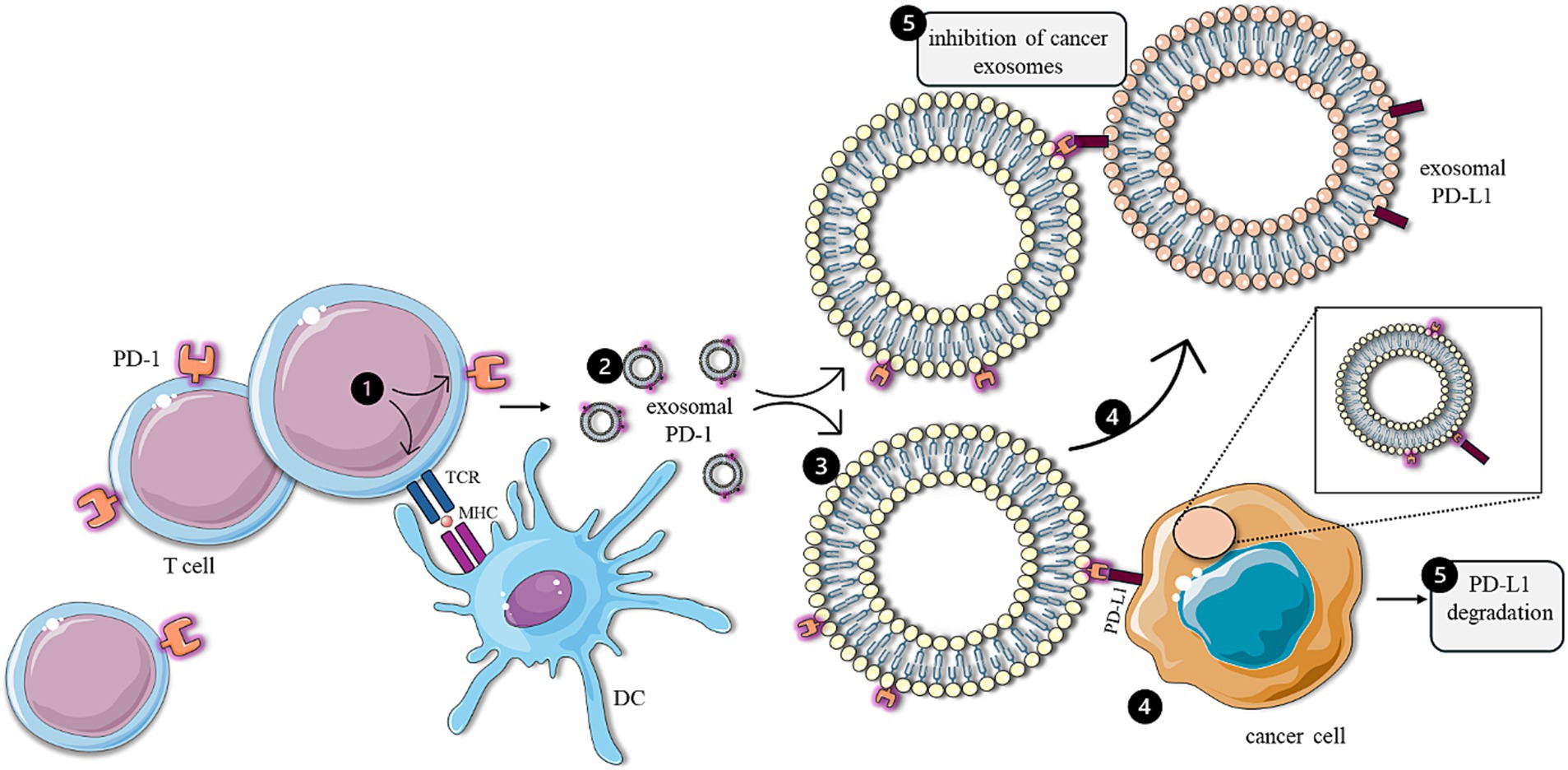
Figure 7. Anti-tumor effects of T-cell-derived exosomes. Following activation by antigen-presenting cells (1), T cells generate exosomes containing PD-1 (2), which directly attenuate the immunosuppressive effects of cancer cells (4) by facilitating the degradation of PD-L1 (5) present on the surface of tumor cells. In addition, T-cell-derived exosomes (3) inhibit the functionality of immunosuppressive exosomes expressing PD-L1 (5), produced by cancer cells. The figure was partly generated using Servier Medical Art, provided by Servier, licensed under a Creative Commons Attribution 3.0 unported license.
The ability of exosomes to cross the blood–brain barrier (BBB) provides the opportunity for their use in the treatment of NDs (45, 170). Many publications highlight the application of MSCs-EXOs in the treatment of NDs (Figure 8) (171, 172).
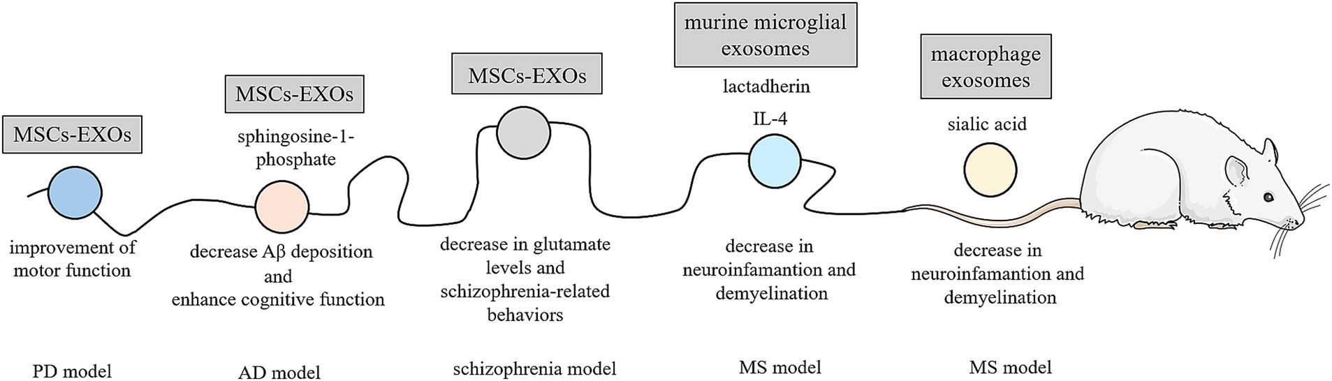
Figure 8. Exosomes in the treatment of neurodegenerative diseases. Due to their ability to cross the BBB, exosomes are being investigated for the treatment of neurodegenerative diseases. In a rat model of PD, bone marrow-derived mesenchymal stem cell exosomes (MSCs-EXOs) have been shown to improve motor function. Similarly, MSCs-EXOs with a high content of sphingosine-1-phosphate have been found to decrease Aβ deposition and enhance cognitive function in a mouse model of AD. In a mouse model of schizophrenia, MSCs-EXOs have been reported to reduce cerebrospinal fluid glutamate levels and alleviate schizophrenia-related behaviors. Exosomes derived from murine microglial cells contain IL-4, an anti-inflammatory cytokine, and lactadherin, a phagocytic “eat me” signal, which reduce neuroinflammation in a mouse model of experimental autoimmune encephalomyelitis (a MS model). Similarly, effects were observed with macrophage-derived exosomes that were modified with derivatives of sialic acid, containing resveratrol. The figure was partly generated using Servier Medical Art, provided by Servier, licensed under a Creative Commons Attribution 3.0 unported license.
For example, the effect of bone marrow-derived MSCs-EXOs was examined in rats with induced Parkinson’s disease (PD). Rats treated with these exosomes showed significant improvements in motor function and histopathological outcomes, demonstrating a greater suppression of PD symptoms compared to L-DOPA treatment, which is a medication commonly used to treat PD (173). In AD treatment, bone marrow MSCS-EXOs with high content of sphingosine-1-phosphate (second messenger downregulated in the AD tissue) were injected into double transgenic AD mice (174). Their application led to reduce Aβ deposition and promote cognitive function recovery. According to the observed results, sphingosine kinase inhibitor (SKI-II) or sphingosine-1-phosphate 1 receptor blocker (VPC23019) repress the therapeutic effects of exosomes. Intracerebral injection of bone marrow MSCS-EXOs in a preclinical mouse model of early stage of AD suggests the possibility of intervening before overt clinical manifestations. The study indicated that bone marrow MSC-EXOs are effective at reducing the Aβ plaque burden and the number of dystrophic neurites in both cortex and hippocampus (175). Adipose-MSCs-EXOs administered intranasally and HUCMSCs-EXOs administered intravenously have also shown promise in AD treatment. These treatments effectively improved neurological damage in entire brain regions, increased newborn neurons, powerfully rescued memory deficits, and reduced Aβ expression in transgenic AD mice (176–179). MSCS-EXOs were also used in a mouse model of schizophrenia, where they improved the core schizophrenia-like behavior and biochemical markers of schizophrenia (e.g., cerebrospinal fluid glutamate level) (180). In addition to MSCs-EXOs, macrophage-derived EXOs loaded with resveratrol, nature antioxidant, or exosomes from murine microglia cell line containing anti-inflammatory cytokine IL-4 were also used in mice with MS (181, 182). The efficacy of this approach can be increased by suitable derivatization of the exosome membrane with compounds such as lactadherin (“eat me” signal for the phagocytes) or sialic acid derivatives (BBB transport). Both exosome agents significantly inhibited inflammatory responses in the CNS and the peripheral system in a mouse model and effectively improved the clinical evolution of MS in vivo (182, 183). In clinical studies, there is just a single trial registered in CLinicalTrials.gov. This study evaluates the safety and the efficacy of adipose MSC-EXOs in AD (NCT04388982). It was found that the intranasal administration of MSCs-EXOs was safe and well tolerated for a semi-weekly treatment frequency. A dose of at least 4 × 10⁸ particles was selected for a randomized phase II and phase III clinical trial in further steps (181).
Exosomes are increasingly recognized for their pivotal role in the diagnostic realm of cancer and NDs. Nonetheless, a multitude of high-impact clinical trials have unveiled their promising potential in diagnosing a spectrum of additional serious conditions. Hyperuricemia (HUA) is acknowledged as a significant risk factor for chronic heart failure (CHF), a disease frequently linked to elevated morbidity and mortality rates (184, 185). Notably, fluctuations in miRNA expression have been correlated with cardiovascular diseases, including CHF and HUA (186, 187). Analysis of miRNA patterns in serum exosomes revealed that miR-27a-5p was upregulated (p < 0.01), while miR-139-3p was downregulated (p < 0.01) in patient groups (CHF with HUA) (188). When used in combination, these markers exhibited an AUC of 0.899 (95%) with 79.2% sensitivity and 91.7% specificity. Moreover, exosomes are integral to the pathogenesis of osteoarthritis, with those isolated from synovial fluids emerging as promising diagnostic instruments for affected patients (189, 190). However, exosome isolation methods can be invasive. In contrast, urine biomarkers present a non-invasive alternative for osteoarthritis diagnostics (191), positioning urine-derived exosomes as potential diagnostic assets. Cao et al. introduced a nanopolymer modified with an exosome-affinity component (CD63 aptamer and distearoyl phosphoethanolamine) (192). This innovative approach leads to aggregation of exosomes upon binding, facilitating easy centrifugation (4,000 g for 3 min at pH 6). Notably, these precipitated exosomes can be re-dissolved in a basic pH environment. Metabolomic analyses of urine exosomes have identified 30 biomarkers, including catechol (AUC = 0.917, p < 0.001), which effectively differentiate between healthy individuals and those with early osteoarthritis.
In the future, exosomes such as natural nanocarriers should emerge as a novel therapeutic alternative in the fields of oncology, immunology, and regenerative medicine. Future research may focus on refining loading techniques, optimizing targeting strategies, and exploring novel applications in diverse disease contexts (193). For example, in ND treatment to enhance targeted drug delivery efficiency, exosomes can be surface-modified with RVG protein/peptides to specifically bind to the acetylcholine receptor expressed on neuronal cells (194). Comparing RVG-tagged MSC-EXOs to MSCS-EXOs, EXOs tagged with RVG exhibit improved targeting to the cortex and hippocampus after being administered intravenously, plaque deposition and Aβ levels are decreased sharply, and the activation of astrocytes is obviously reduced compared to the observations made in the group of AD mice treated only with MSCs-EXOs. In the group of AD mice injected with RVG-EXOs, there is a significant improvement in learning and memory capabilities with reduced plaque deposition and Aβ levels (195).
Exosomes can also be combined with other biomaterials or inorganic materials for biomedical uses. These hybrid nanoparticles can be further loaded with specific cargo or drug and surface engineered to increase the local concentration of the particles at the diseased site, thereby reducing toxicity and side effects and maximizing therapeutic efficacy (196). The surface engineered exosomal hybrid nanoparticles with specific cargo loading, and modifications conferring desired properties such as pH sensitivity or photosensitivity can be called “ExoBots.” This term reflects the hybrid nature of these structures, which incorporate elements from both biological (exosomes) and technological (robotics) realms to form advanced nanoparticle system. ExoBots combine the advantageous properties of exosomes and other nanoparticles, holding great promise for advancing therapeutic interventions across various biomedical applications. For instance, an engineered core-shell hybrid system was prepared for the in vivo treatment of PD mice. This hybrid system consisted of a curcumin-loaded polymer nanoparticle core and an RVG-modified exosome shell. This hybrid was able to clear α-synuclein aggregates, reduce their cytotoxicity in neurons, and improve the motor behavior of PD mice (197).
Another type of hybrid is composed from exosomes and liposomes. Long circulating and pH sensitive hybrids loaded with DOX were investigated for anti-tumor effect on a mouse model of breast cancer. The results indicated that this hybrid system may be a promising nanocarrier for the treatment of breast cancer, reducing toxicity and inhibiting metastasis mainly in the lungs (198). This hybrid approach was also used to overcome chemotherapy resistance in OC. The hybrid system was developed by fusing cRGD-modified liposomes loaded with miR-497 and triptolide with CD47-expressing tumor-derived exosomes. RGD peptides specifically bind to integrin receptors overexpressed on the surface of many cancer cells. Overexpression of miR-497 may overcome OC chemotherapy resistance, and triptolide was confirmed to possess a superior killing effect on cisplatin-resistant cell lines. The in vitro results indicated that these hybrids were efficiently taken up by tumor cells, thus significantly enhancing tumor cell apoptosis and exerting significant anticancer activity without any negative effects observed in vivo. These hybrids may provide a translational strategy to overcome cisplatin-resistant OC (199). In addition, paclitaxel was used as the cargo of hybrids for in vivo colon cancer treatment. The study revealed that hybrids significantly suppressed tumor growth in a colon tumor-bearing mouse model, reduced the expression of M2 type tumor-associated macrophages, and decreased regulatory T cells (200). Many other types of hybrids were explored, such as thermosensitive hybrids for improved treatment of metastatic peritoneal cancer, pH sensitive macrophage hybrids loaded with DOX for tumor targeted drug delivery, or long circulated pH sensitive hybrids loaded with dasatinib for pancreatic cancer treatment. All these ExoBots show positive therapeutic results and can thus serve as potential therapeutics for cancer treatment (201–203).
Recently, alongside the extensive research on exosomes derived from eukaryotic cells, there has been a growing interest in exploring plant-derived exosomes (P-ELNs; puerarin) as a novel source of exosomes with potential applications in various biotechnological and therapeutic fields. Polyphenolic compounds found in various plant exosomes show great promise in treating serious health disturbances. High-impact studies (204, 205) suggest that their therapeutic effects may be partially attributed to their ability to target ferroptosis, a process linked to numerous pathological states (206, 207).
For instance, exosome-like nanovesicles derived from P. lobata roots have been shown to alleviate alcoholic intoxication, enhance alcohol metabolism, and reduce alcohol levels in the liver and serum of mouse models (204). These effects are associated with the induction of acetaldehyde dehydrogenase activity and a decrease in glutathione peroxidase 4 and glutathione levels, as well as with the suppression of acyl-CoA synthetase long-chain family member 4, likely contributing to the repression of ferroptosis. In addition, Robinia pseudoacacia L. flower-derived P-ELNs, when administered orally, significantly reduce hypoxia-induced ferroptosis and mucosal injury in the gastrointestinal tract of mouse models (205). Their effects are mediated through the modulation of HIF-1α and HIF-2α expression, subsequently influencing ROS production and lipid peroxidation via NOX4 and ALOX5 pathways.
Another promising application of P-ELNs is their potential to mitigate obesity, a well-known risk factor for cancer, metabolic disorders, cardiovascular diseases, and inflammation (208–210). The global prevalence of obesity has been on the rise, making this research particularly relevant. Wang et al. reported that turmeric-derived ELNs exhibit potent anti-obesity effects, achieving weight reductions of 8.68 and 14.56% through intragastric and subcutaneous delivery, respectively (211). This effect is linked to the stimulation of adipocyte apoptosis, the induction of lipolysis, and the inhibition of lipogenesis, highlighting the therapeutic potential of these natural compounds. However, the biological functions of P-ELNs are not fully understood, standard isolation protocols are lacking, and P-ELNs are a promising new frontier in precision medicine. Their plant origin offers advantages in terms of biocompatibility, scalability, and reduced immunogenicity. Moreover, bioactive molecules from plants are associated with disease-preventive effects making P-ELNs an attractive alternative to mammalian EVs in future biomedical innovations (212–214).
Although further fundamental research, especially regarding the biogenesis of exosomes and the optimization of their isolation techniques and characterization methods, is necessary, their significant potential has been demonstrated in several biomedical areas, particularly in regenerative medicine, disease diagnosis, and treatment. The continued growth of the exosome market is also evidenced by the recent announcement of four collaborations between pharmaceutical companies, including two potentially worth close to $1 billion each. One of the largest is Lilly’s partnership with Evox Therapeutics of Oxford, UK. In this deal, which could bring in up to $1.2 billion in milestone payments, CNS-targeting exosomes developed by Evox will be loaded with RNA interference and antisense oligonucleotide therapies from Lilly, targeting up to five undisclosed targets. In another major deal, potentially worth over $900 million, Carmine Therapeutics has partnered with Takeda to develop gene therapies for two undisclosed rare diseases targets (215). Exosomes may thus be the future of medicine, used as ExoBots programmed to deliver specific drugs to specific locations within the organism with minimal side effects and high therapeutic efficacies.
NO: Writing – original draft. VŠ: Writing – original draft. RH: Formal analysis, Writing – review & editing. MS: Writing – original draft. PD: Writing – original draft. DH: Writing – review & editing. ZK: Writing – review & editing. JH: Writing – original draft. FV: Writing – review & editing. MV: Writing – original draft. PM: Writing – review & editing, Formal analysis. MJ: Supervision, Writing – review & editing.
The author(s) declare that financial support was received for the research and/or publication of this article. This study was supported by the project of C2P (NEXARS IP 073-23-04). This study was supported by the projects of Charles University in Prague (SVV260637; SVV260521; UNCE 204064; Cooperatio DIAG). This study was also supported by the Ministry of Education, Youth, and Sports grant no. LM2023053 (EATRIS-CZ), the Technology Agency of the Czech Republic within project TN02000122. The study was also supported by the Ministry of Health grant no. RVO-VFN 64165. We are also grateful for the support from the project National Institute for Cancer Research (Program EXCELES, ID Project No. LX22NPO5102) funded by the European Union—Next Generation. The study was also supported by the National Institute for Neurological Research (Programme EXCELES, ID Project No. LX22NPO5107) funded by the European Union—Next Generation EU. The study was supported from the project MULTIOMICS_CZ (Programme Johannes Amos Comenius, Ministry of Education, Youth and Sports of the Czech Republic,__ ID Project CZ.02.01.01/00/23_020/0008540) co-funded by the European Union. We also acknowledge the Operational Programme Research, Development, and Education within the project Center for Tumor Ecology—Research of the Cancer Microenvironment Supporting Cancer Growth and Spread (reg. no. CZ.02.1.01/0.0/0.0/16_019/0000785).
The authors declare that the research was conducted in the absence of any commercial or financial relationships that could be construed as a potential conflict of interest.
The author(s) declared that they were an editorial board member of Frontiers, at the time of submission. This had no impact on the peer review process and the final decision.
The authors declare that no Gen AI was used in the creation of this manuscript.
All claims expressed in this article are solely those of the authors and do not necessarily represent those of their affiliated organizations, or those of the publisher, the editors and the reviewers. Any product that may be evaluated in this article, or claim that may be made by its manufacturer, is not guaranteed or endorsed by the publisher.
The Supplementary material for this article can be found online at: https://www.frontiersin.org/articles/10.3389/fmed.2025.1539714/full#supplementary-material
1. Bayraktar, HRME, Abd-Ellah, KHGMF, Amero, P, Chavez-Reyes, A, and Rodriguez-Aguayo, C. Exosomes: from garbage bins to promising therapeutic targets. Int J Mol Sci. (2017) 18:538. doi: 10.3390/ijms18030538
2. Burtenshaw, D, Regan, B, Owen, K, Collins, D, McEneaney, D, Megson, IL, et al. Exosomal composition, biogenesis and profiling using point-of-care diagnostics-implications for cardiovascular disease. Front Cell Dev Biol. (2022) 10:853451. doi: 10.3389/fcell.2022.853451
3. Sheta, M, Taha, EA, Lu, Y, and Eguchi, T. Extracellular vesicles: new classification and tumor immunosuppression. Biology. (2023) 12:110. doi: 10.3390/biology12010110
4. Pan, BT, Teng, K, Wu, C, Adam, M, and Johnstone, RM. Electron microscopic evidence for externalization of the transferrin receptor in vesicular form in sheep reticulocytes. J Cell Biol. (1985) 101:942–8. doi: 10.1083/jcb.101.3.942
5. Johnstone, RM, Adam, M, Hammond, JR, Orr, L, and Turbide, C. Vesicle formation during reticulocyte maturation. Association of plasma membrane activities with released vesicles (exosomes). J Biol Chem. (1987) 262:9412–20. doi: 10.1016/S0021-9258(18)48095-7
6. Kalluri, R, and LeBleu, VS. The biology, function, and biomedical applications of exosomes. Science. (2020) 367:eaau6977. doi: 10.1126/science.aau6977
7. Chen, J, Li, P, Zhang, T, Xu, Z, Huang, X, Wang, R, et al. Review on strategies and Technologies for Exosome Isolation and Purification. Front Bioeng Biotechnol. (2021) 9:811971. doi: 10.3389/fbioe.2021.811971
8. Yakubovich, EI, Polischouk, AG, and Evtushenko, VI. Principles and problems of exosome isolation from biological fluids. Biochem Suppl Ser A Membr Cell Biol. (2022) 16:115–26. doi: 10.1134/S1990747822030096
9. Dilsiz, N. A comprehensive review on recent advances in exosome isolation and characterization: toward clinical applications. Transl Oncol. (2024) 50:102121. doi: 10.1016/j.tranon.2024.102121
10. Kumari, S, Lausted, C, Scherler, K, Ng, AHC, Lu, Y, Lee, I, et al. Approaches and challenges in characterizing the molecular content of extracellular vesicles for biomarker discovery. Biomol Ther. (2024) 14:1599. doi: 10.3390/biom14121599
11. Chen, YF, Luh, F, Ho, YS, and Yen, Y. Exosomes: a review of biologic function, diagnostic and targeted therapy applications, and clinical trials. J Biomed Sci. (2024) 31:67. doi: 10.1186/s12929-024-01055-0
12. Zhang, X, Borg, EGF, Liaci, AM, Vos, HR, and Stoorvogel, W. A novel three step protocol to isolate extracellular vesicles from plasma or cell culture medium with both high yield and purity. J Extracell Vesicles. (2020) 9:1791450. doi: 10.1080/20013078.2020.1791450
13. Jafari, N, Llevenes, P, and Denis, GV. Exosomes as novel biomarkers in metabolic disease and obesity-related cancers. Nat Rev Endocrinol. (2022) 18:327–8. doi: 10.1038/s41574-022-00666-7
14. Ghosh, S, Rajendran, RL, Mahajan, AA, Chowdhury, A, Bera, A, Guha, S, et al. Harnessing exosomes as cancer biomarkers in clinical oncology. Cancer Cell Int. (2024) 24:278. doi: 10.1186/s12935-024-03464-5
15. Zhang, Z, Zou, Y, Song, C, Cao, K, Cai, K, Chen, S, et al. Advances in the study of exosomes in cardiovascular diseases. J Adv Res. (2024) 66:133–53. doi: 10.1016/j.jare.2023.12.014
16. Park, C, Weerakkody, JS, Schneider, R, Miao, S, and Pitt, D. CNS cell-derived exosome signatures as blood-based biomarkers of neurodegenerative diseases. Front Neurosci. (2024) 18:1426700. doi: 10.3389/fnins.2024.1426700
17. Bano, A, Vats, R, Verma, D, Yadav, P, Kamboj, M, and Bhardwaj, R. Exploring salivary exosomes as early predictors of oral cancer in susceptible tobacco consumers: noninvasive diagnostic and prognostic applications. J Cancer Res Clin Oncol. (2023) 149:15781–93. doi: 10.1007/s00432-023-05343-4
18. Bozyk, N, Tang, KD, Zhang, X, Batstone, M, Kenny, L, Vasani, S, et al. Salivary exosomes as biomarkers for early diagnosis of oral squamous cell carcinoma. Oral Oncol Rep. (2023) 6:100017. doi: 10.1016/j.oor.2023.100017
19. Nijakowski, K, and Surdacka, A. Salivary biomarkers for diagnosis of inflammatory bowel diseases: a systematic review. Int J Mol Sci. (2020) 21:7477. doi: 10.3390/ijms21207477
20. Sun, IO, and Lerman, LO. Urinary extracellular vesicles as biomarkers of kidney disease: from diagnostics to therapeutics. Diagnostics. (2020) 10:311. doi: 10.3390/diagnostics10050311
21. Lee, N, Canagasingham, A, Bajaj, M, Shanmugasundaram, R, Hutton, A, Bucci, J, et al. Urine exosomes as biomarkers in bladder cancer diagnosis and prognosis: from functional roles to clinical significance. Front Oncol. (2022) 12:1019391. doi: 10.3389/fonc.2022.1019391
22. Nilsson, J, Skog, J, Nordstrand, A, Baranov, V, Mincheva-Nilsson, L, Breakefield, XO, et al. Prostate cancer-derived urine exosomes: a novel approach to biomarkers for prostate cancer. Br J Cancer. (2009) 100:1603–7. doi: 10.1038/sj.bjc.6605058
23. Zhou, B, Xu, K, Zheng, X, Chen, T, Wang, J, Song, Y, et al. Application of exosomes as liquid biopsy in clinical diagnosis. Signal Transduct Target Ther. (2020) 5:144. doi: 10.1038/s41392-020-00258-9
24. Mosquera-Heredia, MI, Morales, LC, Vidal, OM, Barcelo, E, Silvera-Redondo, C, Velez, JI, et al. Exosomes: potential disease biomarkers and new therapeutic targets. Biomedicines. (2021) 9:1061. doi: 10.3390/biomedicines9081061
25. Baranyai, T, Herczeg, K, Onodi, Z, Voszka, I, Modos, K, Marton, N, et al. Isolation of exosomes from blood plasma: qualitative and quantitative comparison of ultracentrifugation and size exclusion chromatography methods. PLoS One. (2015) 10:e0145686. doi: 10.1371/journal.pone.0145686
26. Chhoy, P, Brown, CW, Amante, JJ, and Mercurio, AM. Protocol for the separation of extracellular vesicles by ultracentrifugation from in vitro cell culture models. STAR Protoc. (2021) 2:100303. doi: 10.1016/j.xpro.2021.100303
27. Plaschke, K, Brenner, T, Fiedler, MO, Holle, T, von der Forst, M, Wolf, RC, et al. Extracellular vesicles as possible plasma markers and mediators in patients with Sepsis-associated delirium-a pilot study. Int J Mol Sci. (2023) 24:15781. doi: 10.3390/ijms242115781
28. Soares Martins, T, Catita, J, Martins Rosa, I, Silva, OAB, and Henriques, AG. Exosome isolation from distinct biofluids using precipitation and column-based approaches. PLoS One. (2018) 13:e0198820. doi: 10.1371/journal.pone.0198820
29. Tangwattanachuleeporn, M, Muanwien, P, Teethaisong, Y, and Somparn, P. Optimizing concentration of Polyethelene glycol for exosome isolation from plasma for downstream application. Medicina (Kaunas). (2022) 58:1600. doi: 10.3390/medicina58111600
30. Coughlan, C, Bruce, KD, Burgy, O, Boyd, TD, Michel, CR, Garcia-Perez, JE, et al. Exosome isolation by ultracentrifugation and precipitation and techniques for downstream analyses. Curr Protoc Cell Biol. (2020) 88:e110. doi: 10.1002/cpcb.110
31. Liu, WZ, Ma, ZJ, and Kang, XW. Current status and outlook of advances in exosome isolation. Anal Bioanal Chem. (2022) 414:7123–41. doi: 10.1007/s00216-022-04253-7
32. Abdouh, M, Hamam, D, Gao, ZH, Arena, V, Arena, M, and Arena, GO. Exosomes isolated from cancer patients' sera transfer malignant traits and confer the same phenotype of primary tumors to oncosuppressor-mutated cells. J Exp Clin Cancer Res. (2017) 36:113. doi: 10.1186/s13046-017-0587-0
33. Fiandaca, MS, Kapogiannis, D, Mapstone, M, Boxer, A, Eitan, E, Schwartz, JB, et al. Identification of preclinical Alzheimer's disease by a profile of pathogenic proteins in neurally derived blood exosomes: a case-control study. Alzheimers Dement. (2015) 11:600–7.e1. doi: 10.1016/j.jalz.2014.06.008
34. Jia, L, Zhu, M, Kong, C, Pang, Y, Zhang, H, Qiu, Q, et al. Blood neuro-exosomal synaptic proteins predict Alzheimer's disease at the asymptomatic stage. Alzheimers Dement. (2021) 17:49–60. doi: 10.1002/alz.12166
35. Agliardi, C, Guerini, FR, Zanzottera, M, Bolognesi, E, Picciolini, S, Caputo, D, et al. Myelin basic protein in oligodendrocyte-derived extracellular vesicles as a diagnostic and prognostic biomarker in multiple sclerosis: a pilot study. Int J Mol Sci. (2023) 24:894. doi: 10.3390/ijms24010894
36. Lee, EE, Winston-Gray, C, Barlow, JW, Rissman, RA, and Jeste, DV. Plasma levels of neuron- and astrocyte-derived Exosomal amyloid Beta1-42, amyloid Beta1-40, and phosphorylated tau levels in schizophrenia patients and non-psychiatric comparison subjects: relationships with cognitive functioning and psychopathology. Front Psych. (2020) 11:532624. doi: 10.3389/fpsyt.2020.532624
37. Wang, YT, Shi, T, Srivastava, S, Kagan, J, Liu, T, and Rodland, KD. Proteomic analysis of exosomes for discovery of protein biomarkers for prostate and bladder Cancer. Cancers (Basel). (2020) 12:2335. doi: 10.3390/cancers12092335
38. Sun, Y, Liu, S, Qiao, Z, Shang, Z, Xia, Z, Niu, X, et al. Systematic comparison of exosomal proteomes from human saliva and serum for the detection of lung cancer. Anal Chim Acta. (2017) 982:84–95. doi: 10.1016/j.aca.2017.06.005
39. Logozzi, M, Di Raimo, R, Mizzoni, D, and Fais, S. Immunocapture-based ELISA to characterize and quantify exosomes in both cell culture supernatants and body fluids. Methods Enzymol. (2020) 645:155–80. doi: 10.1016/bs.mie.2020.06.011
40. Elkommos-Zakhary, M, Rajesh, N, and Beljanski, V. Exosome RNA sequencing as a tool in the search for cancer biomarkers. Noncoding RNA. (2022) 8:75. doi: 10.3390/ncrna8060075
41. Tang, S, Cheng, J, Yao, Y, Lou, C, Wang, L, Huang, X, et al. Combination of four serum exosomal MiRNAs as novel diagnostic biomarkers for early-stage gastric cancer. Front Genet. (2020) 11:237. doi: 10.3389/fgene.2020.00237
42. Gu, Z, Yin, H, Zhang, H, Zhang, H, Liu, X, Zeng, X, et al. Optimization of a method for the clinical detection of serum exosomal miR-940 as a potential biomarker of breast cancer. Front Oncol. (2022) 12:956167. doi: 10.3389/fonc.2022.956167
43. Shi, J, Kantoff, PW, Wooster, R, and Farokhzad, OC. Cancer nanomedicine: progress, challenges and opportunities. Nat Rev Cancer. (2017) 17:20–37. doi: 10.1038/nrc.2016.108
44. Azzi, S, Hebda, JK, and Gavard, J. Vascular permeability and drug delivery in cancers. Front Oncol. (2013) 3:211. doi: 10.3389/fonc.2013.00211
45. Abdelsalam, M, Ahmed, M, Osaid, Z, Hamoudi, R, and Harati, R. Insights into exosome transport through the blood-brain barrier and the potential therapeutical applications in brain diseases. Pharmaceuticals (Basel). (2023) 16:571. doi: 10.3390/ph16040571
46. Fu, S, Wang, Y, Xia, X, and Zheng, JC. Exosome engineering: current progress in cargo loading and targeted delivery. Nanoimpact. (2020) 20:100261. doi: 10.1016/j.impact.2020.100261
47. Cecchin, R, Troyer, Z, Witwer, K, and Morris, KV. Extracellular vesicles: the next generation in gene therapy delivery. Mol Ther. (2023) 31:1225–30. doi: 10.1016/j.ymthe.2023.01.021
48. Lee, J. Trends in developing extracellular vesicle-based therapeutics. Brain Tumor Res Treat. (2024) 12:153–61. doi: 10.14791/btrt.2024.0027
49. Zhang, Z, Wang, C, Li, T, Liu, Z, and Li, L. Comparison of ultracentrifugation and density gradient separation methods for isolating Tca8113 human tongue cancer cell line-derived exosomes. Oncol Lett. (2014) 8:1701–6. doi: 10.3892/ol.2014.2373
50. Lobb, RJ, Becker, M, Wen, SW, Wong, CS, Wiegmans, AP, Leimgruber, A, et al. Optimized exosome isolation protocol for cell culture supernatant and human plasma. J Extracell Vesicles. (2015) 4:27031. doi: 10.3402/jev.v4.27031
51. Collino, F, Pomatto, M, Bruno, S, Lindoso, RS, Tapparo, M, Sicheng, W, et al. Exosome and microvesicle-enriched fractions isolated from mesenchymal stem cells by gradient separation showed different molecular signatures and functions on renal tubular epithelial cells. Stem Cell Rev Rep. (2017) 13:226–43. doi: 10.1007/s12015-016-9713-1
52. Busatto, S, Vilanilam, G, Ticer, T, Lin, WL, Dickson, DW, Shapiro, S, et al. Tangential flow filtration for highly efficient concentration of extracellular vesicles from large volumes of fluid. Cells. (2018) 7:273. doi: 10.3390/cells7120273
53. Luo, H, Zhang, J, Yang, A, Ouyang, W, Long, S, Lin, X, et al. Large-scale isolation of exosomes derived from NK cells for anti-tumor therapy. Bio Protoc. (2023) 13:e4693. doi: 10.3389/fimmu.2022.1087689
54. Kim, JY, Rhim, WK, Seo, HJ, Lee, JY, Park, CG, and Han, DK. Comparative analysis of MSC-derived exosomes depending on cell culture Media for Regenerative Bioactivity. Tissue Eng Regen Med. (2021) 18:355–67. doi: 10.1007/s13770-021-00352-1
55. Kawai-Harada, Y, Nimmagadda, V, and Harada, M. Scalable isolation of surface-engineered extracellular vesicles and separation of free proteins via tangential flow filtration and size exclusion chromatography (TFF-SEC). BMC Methods. (2024) 1:9. doi: 10.1186/s44330-024-00009-0
56. Ahn, SH, Ryu, SW, Choi, H, You, S, Park, J, and Choi, C. Manufacturing therapeutic exosomes: from bench to industry. Mol Cells. (2022) 45:284–90. doi: 10.14348/molcells.2022.2033
57. Guerreiro, EM, Vestad, B, Steffensen, LA, Aass, HCD, Saeed, M, Ovstebo, R, et al. Efficient extracellular vesicle isolation by combining cell media modifications, ultrafiltration, and size-exclusion chromatography. PLoS One. (2018) 13:e0204276. doi: 10.1371/journal.pone.0204276
58. Nguyen, VVT, Witwer, KW, Verhaar, MC, Strunk, D, and van Balkom, BWM. Functional assays to assess the therapeutic potential of extracellular vesicles. J Extracell Vesicles. (2020) 10:e12033. doi: 10.1002/jev2.12033
59. Thery, C, Witwer, KW, Aikawa, E, Alcaraz, MJ, Anderson, JD, Andriantsitohaina, R, et al. Minimal information for studies of extracellular vesicles 2018 (MISEV2018): a position statement of the International Society for Extracellular Vesicles and update of the MISEV2014 guidelines. J Extracell Vesicles. (2018) 7:1535750. doi: 10.1080/20013078.2018.1535750
60. Welsh, JA, Goberdhan, DCI, O'Driscoll, L, Buzas, EI, Blenkiron, C, Bussolati, B, et al. Minimal information for studies of extracellular vesicles (MISEV2023): from basic to advanced approaches. J Extracell Vesicles. (2024) 13:e12404. doi: 10.1002/jev2.12404
61. Kowal, EJK, Ter-Ovanesyan, D, Regev, A, and Church, GM. Extracellular vesicle isolation and analysis by Western blotting. Methods Mol Biol. (2017) 1660:143–52. doi: 10.1007/978-1-4939-7253-1_12
62. Takizawa, K, Nishimura, T, and Harita, Y. Enzyme-linked immunosorbent assay to detect surface marker proteins of extracellular vesicles purified from human urine. STAR Protoc. (2023) 4:102415. doi: 10.1016/j.xpro.2023.102415
63. Noble, JM, Roberts, LM, Vidavsky, N, Chiou, AE, Fischbach, C, Paszek, MJ, et al. Direct comparison of optical and electron microscopy methods for structural characterization of extracellular vesicles. J Struct Biol. (2020) 210:107474. doi: 10.1016/j.jsb.2020.107474
64. van der Pol, E, Coumans, FA, Grootemaat, AE, Gardiner, C, Sargent, IL, Harrison, P, et al. Particle size distribution of exosomes and microvesicles determined by transmission electron microscopy, flow cytometry, nanoparticle tracking analysis, and resistive pulse sensing. J Thromb Haemost. (2014) 12:1182–92. doi: 10.1111/jth.12602
65. Koritzinsky, EH, Street, JM, Star, RA, and Yuen, PS. Quantification of exosomes. J Cell Physiol. (2017) 232:1587–90. doi: 10.1002/jcp.25387
66. Rupert, DLM, Claudio, V, Lasser, C, and Bally, M. Methods for the physical characterization and quantification of extracellular vesicles in biological samples. Biochim Biophys Acta Gen Subj. (1861) 2017:3164–79. doi: 10.1016/j.bbagen.2016.07.028
67. Fortunato, D, Mladenovic, D, Criscuoli, M, Loria, F, Veiman, KL, Zocco, D, et al. Opportunities and pitfalls of fluorescent labeling methodologies for extracellular vesicle profiling on high-resolution single-particle platforms. Int J Mol Sci. (2021) 22:10510. doi: 10.3390/ijms221910510
68. Dehghani, M, Montange, RK, Olszowy, MW, and Pollard, D. An emerging fluorescence-based technique for quantification and protein profiling of extracellular vesicles. SLAS Technol. (2021) 26:189–99. doi: 10.1177/2472630320970458
69. Chung, IM, Rajakumar, G, Venkidasamy, B, Subramanian, U, and Thiruvengadam, M. Exosomes: current use and future applications. Clin Chim Acta. (2020) 500:226–32. doi: 10.1016/j.cca.2019.10.022
70. Mager, E.L.A. S I., X.O. Breakefield, and Wood, M.J., Extracellular vesicles: biology and emerging therapeutic opportunities. Nat Rev Drug Discov 12 (2013) 347–357. doi: 10.1038/nrd3978
71. Aheget, H, Tristan-Manzano, M, Mazini, L, Cortijo-Gutierrez, M, Galindo-Moreno, P, Herrera, C, et al. Exosome: a new player in translational nanomedicine. J Clin Med. (2020) 9:2380. doi: 10.3390/jcm9082380
72. Tienda-Vazquez, MA, Hanel, JM, Marquez-Arteaga, EM, Salgado-Alvarez, AP, Scheckhuber, CQ, Alanis-Gomez, JR, et al. Exosomes: a promising strategy for repair, regeneration and treatment of skin disorders. Cells. (2023) 12:1625. doi: 10.3390/cells12121625
73. Zhang, K, and Cheng, K. Stem cell-derived exosome versus stem cell therapy. Nat Rev Bioeng. (2023) 1:608–9. doi: 10.1038/s44222-023-00064-2
74. Wang, X. Exosome therapy and diagnostics: The path to becoming clinical giants. Washington, USA: CAS (2022).
75. Nooshabadi, VT, Khanmohamadi, M, Valipour, E, Mahdipour, S, Salati, A, Malekshahi, ZV, et al. Impact of exosome-loaded chitosan hydrogel in wound repair and layered dermal reconstitution in mice animal model. J Biomed Mater Res A. (2020) 108:2138–49. doi: 10.1002/jbm.a.36959
76. Tan, F, Li, X, Wang, Z, Li, J, Shahzad, K, and Zheng, J. Clinical applications of stem cell-derived exosomes. Signal Transduct Target Ther. (2024) 9:17. doi: 10.1038/s41392-023-01704-0
77. Prasai, A, Jay, JW, Jupiter, D, Wolf, SE, and El Ayadi, A. Role of exosomes in dermal wound healing: a systematic review. J Invest Dermatol. (2022) 142:662–678.e8. doi: 10.1016/j.jid.2021.07.167
78. Shi, L, Song, D, Meng, C, Cheng, Y, Wang, B, and Yang, Z. Opportunities and challenges of engineered exosomes for diabetic wound healing. Giant. (2024) 18:100251. doi: 10.1016/j.giant.2024.100251
79. Fan, MH, Pi, JK, Zou, CY, Jiang, YL, Li, QJ, Zhang, XZ, et al. Hydrogel-exosome system in tissue engineering: a promising therapeutic strategy. Bioact Mater. (2024) 38:1–30. doi: 10.1016/j.bioactmat.2024.04.007
80. Xie, Y, Guan, Q, Guo, J, Chen, Y, Yin, Y, and Han, X. Hydrogels for exosome delivery in biomedical applications. Gels. (2022) 8:328. doi: 10.3390/gels8060328
81. Khayambashi, P, Iyer, J, Pillai, S, Upadhyay, A, Zhang, Y, and Tran, SD. Hydrogel encapsulation of mesenchymal stem cells and their derived exosomes for tissue engineering. Int J Mol Sci. (2021) 22:684. doi: 10.3390/ijms22020684
82. Zhang, K, Zhao, X, Chen, X, Wei, Y, Du, W, Wang, Y, et al. Enhanced therapeutic effects of mesenchymal stem cell-derived exosomes with an injectable hydrogel for Hindlimb ischemia treatment. ACS Appl Mater Interfaces. (2018) 10:30081–91. doi: 10.1021/acsami.8b08449
83. Yang, J, Chen, Z, Pan, D, Li, H, and Shen, J. Umbilical cord-derived mesenchymal stem cell-derived exosomes combined Pluronic F127 hydrogel promote chronic diabetic wound healing and complete skin regeneration. Int J Nanomedicine. (2020) 15:5911–26. doi: 10.2147/IJN.S249129
84. Zhang, Y, Zhang, P, Gao, X, Chang, L, Chen, Z, and Mei, X. Preparation of exosomes encapsulated nanohydrogel for accelerating wound healing of diabetic rats by promoting angiogenesis. Mater Sci Eng C. (2021) 120:111671. doi: 10.1016/j.msec.2020.111671
85. Wang, C, Wang, M, Xu, T, Zhang, X, Lin, C, Gao, W, et al. Engineering bioactive self-healing antibacterial exosomes hydrogel for promoting chronic diabetic wound healing and complete skin regeneration. Theranostics. (2019) 9:65–76. doi: 10.7150/thno.29766
86. Li, P, Lv, S, Jiang, W, Si, L, Liao, B, Zhao, G, et al. Exosomes derived from umbilical cord mesenchymal stem cells protect cartilage and regulate the polarization of macrophages in osteoarthritis. Ann Transl Med. (2022) 10:976. doi: 10.21037/atm-22-3912
87. Cao, H, Chen, M, Cui, X, Liu, Y, Liu, Y, Deng, S, et al. Cell-free osteoarthritis treatment with sustained-release of chondrocyte-targeting exosomes from umbilical cord-derived mesenchymal stem cells to rejuvenate aging chondrocytes. ACS Nano. (2023) 17:13358–76. doi: 10.1021/acsnano.3c01612
88. Chen, P, Zheng, L, Wang, Y, Tao, M, Xie, Z, Xia, C, et al. Desktop-stereolithography 3D printing of a radially oriented extracellular matrix/mesenchymal stem cell exosome bioink for osteochondral defect regeneration. Theranostics. (2019) 9:2439–59. doi: 10.7150/thno.31017
89. Wu, KY, Ahmad, H, Lin, G, Carbonneau, M, and Tran, SD. Mesenchymal stem cell-derived exosomes in ophthalmology: a comprehensive review. Pharmaceutics. (2023) 15:1167. doi: 10.3390/pharmaceutics15041167
90. Di, G, Du, X, Qi, X, Zhao, X, Duan, H, Li, S, et al. Mesenchymal stem cells promote diabetic corneal epithelial wound healing through TSG-6-dependent stem cell activation and macrophage switch. Invest Ophthalmol Vis Sci. (2017) 58:4344–54. doi: 10.1167/iovs.17-21506
91. Samaeekia, R, Rabiee, B, Putra, I, Shen, X, Park, YJ, Hematti, P, et al. Effect of human corneal mesenchymal stromal cell-derived exosomes on corneal epithelial wound healing. Invest Ophthalmol Vis Sci. (2018) 59:5194–200. doi: 10.1167/iovs.18-24803
92. Mead, B, and Tomarev, S. Bone marrow-derived mesenchymal stem cells-derived exosomes promote survival of retinal ganglion cells through miRNA-dependent mechanisms. Stem Cells Transl Med. (2017) 6:1273–85. doi: 10.1002/sctm.16-0428
93. Wang, T, Zhou, Y, Zhang, W, Xue, Y, Xiao, Z, Zhou, Y, et al. Exosomes and exosome composite scaffolds in periodontal tissue engineering. Front Bioeng Biotechnol. (2023) 11:1287714. doi: 10.3389/fbioe.2023.1287714
94. Lin, H, Chen, H, Zhao, X, Ding, T, Wang, Y, Chen, Z, et al. Advances of exosomes in periodontitis treatment. J Transl Med. (2022) 20:279. doi: 10.1186/s12967-022-03487-4
95. Zhai, LY, Li, MX, Pan, WL, Chen, Y, Li, MM, Pang, JX, et al. In situ detection of plasma Exosomal MicroRNA-1246 for breast Cancer diagnostics by a au Nanoflare probe. ACS Appl Mater Interfaces. (2018) 10:39478–86. doi: 10.1021/acsami.8b12725
96. Sandfeld-Paulsen, B, Jakobsen, KR, Baek, R, Folkersen, BH, Rasmussen, TR, Meldgaard, P, et al. Exosomal proteins as diagnostic biomarkers in lung Cancer. J Thorac Oncol. (2016) 11:1701–10. doi: 10.1016/j.jtho.2016.05.034
97. Akbar, S, Raza, A, Mohsin, R, Kanbour, A, Qadri, S, Parray, A, et al. Circulating exosomal immuno-oncological checkpoints and cytokines are potential biomarkers to monitor tumor response to anti-PD-1/PD-L1 therapy in non-small cell lung cancer patients. Front Immunol. (2022) 13:1097117. doi: 10.3389/fimmu.2022.1097117
98. Risha, Y, Minic, Z, Ghobadloo, SM, and Berezovski, MV. The proteomic analysis of breast cell line exosomes reveals disease patterns and potential biomarkers. Sci Rep. (2020) 10:13572. doi: 10.1038/s41598-020-70393-4
99. Hung, Y, Wang, Y-L, Lin, Y-Z, Chiang, S-F, Wu, W-R, and Wang, S-C. The exosomal compartment protects epidermal growth factor receptor from small molecule inhibitors. Biochem Biophys Res Commun. (2019) 510:42–7. doi: 10.1016/j.bbrc.2018.12.187
100. Hannafon, BN, Trigoso, YD, Calloway, CL, Zhao, YD, Lum, DH, Welm, AL, et al. Plasma exosome microRNAs are indicative of breast cancer. Breast Cancer Res. (2016) 18:90. doi: 10.1186/s13058-016-0753-x
101. Gobbo, J, Marcion, G, Cordonnier, M, Dias, AMM, Pernet, N, Hammann, A, et al. Restoring anticancer immune response by targeting tumor-derived exosomes with a HSP70 peptide Aptamer. JNCI J Natl Cancer Inst. (2015) 108:djv330. doi: 10.1093/jnci/djv330
102. Melo, SA, Luecke, LB, Kahlert, C, Fernandez, AF, Gammon, ST, Kaye, J, et al. Glypican-1 identifies cancer exosomes and detects early pancreatic cancer. Nature. (2015) 523:177–82. doi: 10.1038/nature14581
103. Ueda, H, Takahashi, H, Kobayashi, S, Kubo, M, Sasaki, K, Iwagami, Y, et al. miR-6855-5p enhances Radioresistance and promotes migration of pancreatic Cancer by inducing epithelial-mesenchymal transition via suppressing FOXA1: potential of plasma Exosomal miR-6855-5p as an indicator of radiosensitivity in patients with pancreatic cancer. Ann Surg Oncol. (2024) 32:720–35. doi: 10.1245/s10434-024-16115-w
104. Dash, S, Wu, CC, Wu, CC, Chiang, SF, Lu, YT, Yeh, CY, et al. Extracellular vesicle membrane protein profiling and targeted mass spectrometry unveil CD59 and Tetraspanin 9 as novel plasma biomarkers for detection of colorectal cancer. Cancers. (2022) 15:177. doi: 10.3390/cancers15010177
105. Overbye, A, Skotland, T, Koehler, CJ, Thiede, B, Seierstad, T, Berge, V, et al. Identification of prostate cancer biomarkers in urinary exosomes. Oncotarget. (2015) 6:30357–76. doi: 10.18632/oncotarget.4851
106. Jang, JY, Lee, JK, Jeon, YK, and Kim, CW. Exosome derived from epigallocatechin gallate treated breast cancer cells suppresses tumor growth by inhibiting tumor-associated macrophage infiltration and M2 polarization. BMC Cancer. (2013) 13:421. doi: 10.1186/1471-2407-13-421
107. Whiteside, TL. Exosomes and tumor-mediated immune suppression. J Clin Invest. (2016) 126:1216–23. doi: 10.1172/JCI81136
108. Hao, Q, Wu, Y, Wu, Y, Wang, P, and Vadgama, JV. Tumor-derived exosomes in tumor-induced immune suppression. Int J Mol Sci. (2022) 23:1416. doi: 10.3390/ijms23031461
109. Saccani, A, Schioppa, T, Porta, C, Biswas, SK, Nebuloni, M, Vago, L, et al. p50 nuclear factor-kappaB overexpression in tumor-associated macrophages inhibits M1 inflammatory responses and antitumor resistance. Cancer Res. (2006) 66:11432–40. doi: 10.1158/0008-5472.CAN-06-1867
110. Mantovani, A, Marchesi, F, Porta, C, Sica, A, and Allavena, P. Inflammation and cancer: breast cancer as a prototype. Breast. (2007) 16:27–33. doi: 10.1016/j.breast.2007.07.013
111. Chanmee, T, Ontong, P, Konno, K, and Itano, N. Tumor-associated macrophages as major players in the tumor microenvironment. Cancers (Basel). (2014) 6:1670–90. doi: 10.3390/cancers6031670
112. Sica, A, Schioppa, T, Mantovani, A, and Allavena, P. Tumour-associated macrophages are a distinct M2 polarised population promoting tumour progression: potential targets of anti-cancer therapy. Eur J Cancer. (2006) 42:717–27. doi: 10.1016/j.ejca.2006.01.003
113. Ham, S, Lima, LG, Chai, EPZ, Muller, A, Lobb, RJ, Krumeich, S, et al. Breast Cancer-derived exosomes Alter macrophage polarization via gp130/STAT3 signaling. Front Immunol. (2018) 9:871. doi: 10.3389/fimmu.2018.00871
114. Piao, YJ, Kim, HS, Hwang, EH, Woo, J, Zhang, M, and Moon, WK. Breast cancer cell-derived exosomes and macrophage polarization are associated with lymph node metastasis. Oncotarget. (2018) 9:7398–410. doi: 10.18632/oncotarget.23238
115. Nieto, MA. The ins and outs of the epithelial to mesenchymal transition in health and disease. Annu Rev Cell Dev Biol. (2011) 27:347–76. doi: 10.1146/annurev-cellbio-092910-154036
116. Thiery, JP, Acloque, H, Huang, RY, and Nieto, MA. Epithelial-mesenchymal transitions in development and disease. Cell. (2009) 139:871–90. doi: 10.1016/j.cell.2009.11.007
117. Shen, Y, and TanTai, J. Exosomes secreted by metastatic cancer cells promotes epithelial mesenchymal transition in small cell lung carcinoma: the key role of Src/TGF-beta1 axis. Gene. (2024) 892:147873. doi: 10.1016/j.gene.2023.147873
118. Franzen, CA, Blackwell, RH, Todorovic, V, Greco, KA, Foreman, KE, Flanigan, RC, et al. Urothelial cells undergo epithelial-to-mesenchymal transition after exposure to muscle invasive bladder cancer exosomes. Oncogenesis. (2015) 4:e163. doi: 10.1038/oncsis.2015.21
119. Christianson, HC, Svensson, KJ, van Kuppevelt, TH, Li, JP, and Belting, M. Cancer cell exosomes depend on cell-surface heparan sulfate proteoglycans for their internalization and functional activity. Proc Natl Acad Sci USA. (2013) 110:17380–5. doi: 10.1073/pnas.1304266110
120. Liu, D, Li, C, Trojanowicz, B, Li, X, Shi, D, Zhan, C, et al. CD97 promotion of gastric carcinoma lymphatic metastasis is exosome dependent. Gastric Cancer. (2016) 19:754–66. doi: 10.1007/s10120-015-0523-y
121. Rahman, MA, Barger, JF, Lovat, F, Gao, M, Otterson, GA, and Nana-Sinkam, P. Lung cancer exosomes as drivers of epithelial mesenchymal transition. Oncotarget. (2016) 7:54852–66. doi: 10.18632/oncotarget.10243
122. Cao, M, Seike, M, Soeno, C, Mizutani, H, Kitamura, K, Minegishi, Y, et al. MiR-23a regulates TGF-beta-induced epithelial-mesenchymal transition by targeting E-cadherin in lung cancer cells. Int J Oncol. (2012) 41:869–75. doi: 10.3892/ijo.2012.1535
123. Kim, J, Kim, TY, Lee, MS, Mun, JY, Ihm, C, and Kim, SA. Exosome cargo reflects TGF-beta1-mediated epithelial-to-mesenchymal transition (EMT) status in A549 human lung adenocarcinoma cells. Biochem Biophys Res Commun. (2016) 478:643–8. doi: 10.1016/j.bbrc.2016.07.124
124. Ohshima, K, Inoue, K, Fujiwara, A, Hatakeyama, K, Kanto, K, Watanabe, Y, et al. Let-7 microRNA family is selectively secreted into the extracellular environment via exosomes in a metastatic gastric cancer cell line. PLoS One. (2010) 5:e13247. doi: 10.1371/journal.pone.0013247
125. Tanaka, S, Hosokawa, M, Ueda, K, and Iwakawa, S. Effects of Decitabine on invasion and Exosomal expression of miR-200c and miR-141 in Oxaliplatin-resistant colorectal Cancer cells. Biol Pharm Bull. (2015) 38:1272–9. doi: 10.1248/bpb.b15-00129
126. Xiao, D, Barry, S, Kmetz, D, Egger, M, Pan, J, Rai, SN, et al. Melanoma cell-derived exosomes promote epithelial-mesenchymal transition in primary melanocytes through paracrine/autocrine signaling in the tumor microenvironment. Cancer Lett. (2016) 376:318–27. doi: 10.1016/j.canlet.2016.03.050
127. Zhou, W, Fong, MY, Min, Y, Somlo, G, Liu, L, Palomares, MR, et al. Cancer-secreted miR-105 destroys vascular endothelial barriers to promote metastasis. Cancer Cell. (2014) 25:501–15. doi: 10.1016/j.ccr.2014.03.007
128. Kumar, S, Dhar, R, Kumar, L, Shivji, GG, Jayaraj, R, and Devi, A. Theranostic signature of tumor-derived exosomes in cancer. Med Oncol. (2023) 40:321. doi: 10.1007/s12032-023-02176-6
129. Bray, F, Laversanne, M, Sung, H, Ferlay, J, Siegel, RL, Soerjomataram, I, et al. GLOBOCAN estimates of incidence and mortality worldwide for 36 cancers in 185 countries. CA Cancer J Clin. (2022) 74:229–63. doi: 10.3322/caac.21660
130. Shin, H, Oh, S, Hong, S, Kang, M, Kang, D, Ji, YG, et al. Early-stage lung Cancer diagnosis by deep learning-based spectroscopic analysis of circulating exosomes. ACS Nano. (2020) 14:5435–44. doi: 10.1021/acsnano.9b09119
131. Hai, J, Zhu, CQ, Bandarchi, B, Wang, YH, Navab, R, Shepherd, FA, et al. L1 cell adhesion molecule promotes tumorigenicity and metastatic potential in non-small cell lung cancer. Clin Cancer Res. (2012) 18:1914–24. doi: 10.1158/1078-0432.CCR-11-2893
132. Tischler, V, Pfeifer, M, Hausladen, S, Schirmer, U, Bonde, AK, Kristiansen, G, et al. L1CAM protein expression is associated with poor prognosis in non-small cell lung cancer. Mol Cancer. (2011) 10:127. doi: 10.1186/1476-4598-10-127
133. Liu, Y, Ding, W, Wang, J, Ao, X, and Xue, J. Non-coding RNAs in lung cancer: molecular mechanisms and clinical applications. Front Oncol. (2023) 13:1256537. doi: 10.3389/fonc.2023.1256537
134. Makler, A, and Asghar, W. Exosomal biomarkers for cancer diagnosis and patient monitoring. Expert Rev Mol Diagn. (2020) 20:387–400. doi: 10.1080/14737159.2020.1731308
135. Lee, CH, Im, EJ, Moon, PG, and Baek, MC. Discovery of a diagnostic biomarker for colon cancer through proteomic profiling of small extracellular vesicles. BMC Cancer. (2018) 18:1058. doi: 10.1186/s12885-018-4952-y
136. Xu, Y, Lou, J, Yu, M, Jiang, Y, Xu, H, Huang, Y, et al. Urinary exosomes diagnosis of urological tumors: a systematic review and meta-analysis. Front Oncol. (2021) 11:734587. doi: 10.3389/fonc.2021.734587
137. Hiltbrunner, S, Mints, M, Eldh, M, Rosenblatt, R, Holmstrom, B, Alamdari, F, et al. Urinary exosomes from bladder cancer patients show a residual cancer phenotype despite complete pathological downstaging. Sci Rep. (2020) 10:5960. doi: 10.1038/s41598-020-62753-x
138. Shusharina, N, Yukhnenko, D, Botman, S, Sapunov, V, Savinov, V, Kamyshov, G, et al. Modern methods of diagnostics and treatment of neurodegenerative diseases and depression. Diagnostics. (2023) 13:573. doi: 10.3390/diagnostics13030573
139. Konickova, D, Mensikova, K, Tuckova, L, Henykova, E, Strnad, M, Friedecky, D, et al. Biomarkers of neurodegenerative diseases: biology, taxonomy, clinical relevance, and current research status. Biomedicines. (2022) 10:1760. doi: 10.3390/biomedicines10071760
140. Gao, P, Li, X, Du, X, Liu, S, and Xu, Y. Diagnostic and therapeutic potential of exosomes in neurodegenerative diseases. Front Aging Neurosci. (2021) 13:790863. doi: 10.3389/fnagi.2021.790863
141. Rastogi, S, Sharma, V, Bharti, PS, Rani, K, Modi, GP, Nikolajeff, F, et al. The evolving landscape of exosomes in neurodegenerative diseases: exosomes characteristics and a promising role in early diagnosis. Int J Mol Sci. (2021) 22:440. doi: 10.3390/ijms22010440
142. Geekiyanage, H, Jicha, GA, Nelson, PT, and Chan, C. Blood serum miRNA: non-invasive biomarkers for Alzheimer's disease. Exp Neurol. (2012) 235:491–6. doi: 10.1016/j.expneurol.2011.11.026
143. Manna, I, De Benedittis, S, Quattrone, A, Maisano, D, Iaccino, E, and Quattrone, A. Exosomal miRNAs as potential diagnostic biomarkers in Alzheimer's disease. Pharmaceuticals. (2020) 13:243. doi: 10.3390/ph13090243
144. Agarwal, M, and Khan, S. Plasma lipids as biomarkers for Alzheimer's disease: a systematic review. Cureus. (2020) 12:e12008. doi: 10.7759/cureus.12008
145. Jia, L, Qiu, Q, Zhang, H, Chu, L, Du, Y, Zhang, J, et al. Concordance between the assessment of Abeta42, T-tau, and P-T181-tau in peripheral blood neuronal-derived exosomes and cerebrospinal fluid. Alzheimers Dement. (2019) 15:1071–80. doi: 10.1016/j.jalz.2019.05.002
146. Nila, IS, Sumsuzzman, DM, Khan, ZA, Jung, JH, Kazema, AS, Kim, SJ, et al. Identification of exosomal biomarkers and its optimal isolation and detection method for the diagnosis of Parkinson's disease: a systematic review and meta-analysis. Ageing Res Rev. (2022) 82:101764. doi: 10.1016/j.arr.2022.101764
147. Barbo, M, and Ravnik-Glavac, M. Extracellular vesicles as potential biomarkers in amyotrophic lateral sclerosis. Genes. (2023) 14:325. doi: 10.3390/genes14020325
148. Zafar, MN, Abuwatfa, WH, and Husseini, GA. Acoustically-activated liposomal Nanocarriers to mitigate the side effects of conventional chemotherapy with a focus on emulsion-liposomes. Pharmaceutics. (2023) 15:421. doi: 10.3390/pharmaceutics15020421
149. Srivastava, A, Amreddy, N, Razaq, M, Towner, R, Zhao, YD, Ahmed, RA, et al. Exosomes as Theranostics for lung Cancer. Adv Cancer Res. (2018) 139:1–33. doi: 10.1016/bs.acr.2018.04.001
150. Paskeh, MDA, Entezari, M, Mirzaei, S, Zabolian, A, Saleki, H, Naghdi, MJ, et al. Emerging role of exosomes in cancer progression and tumor microenvironment remodeling. J Hematol Oncol. (2022) 15:83. doi: 10.1186/s13045-022-01305-4
151. Maqsood, Q, Sumrin, A, Saleem, Y, Wajid, A, and Mahnoor, M. Exosomes in Cancer: diagnostic and therapeutic applications. Clin Med Insights Oncol. (2024) 18:11795549231215966. doi: 10.1177/11795549231215966
152. Cao, Y, Xu, P, Shen, Y, Wu, W, Chen, M, Wang, F, et al. Exosomes and cancer immunotherapy: a review of recent cancer research. Front Oncol. (2022) 12:1118101. doi: 10.3389/fonc.2022.1118101
153. Araldi, RP, Delvalle, DA, da Costa, VR, Alievi, AL, Teixeira, MR, Dias Pinto, JR, et al. Exosomes as a Nano-Carrier for chemotherapeutics: a new era of oncology. Cells. (2023) 12:2411. doi: 10.3390/cells12172144
154. Hadla, M, Palazzolo, S, Corona, G, Caligiuri, I, Canzonieri, V, Toffoli, G, et al. Exosomes increase the therapeutic index of doxorubicin in breast and ovarian cancer mouse models. Nanomedicine. (2016) 11:2431–41. doi: 10.2217/nnm-2016-0154
155. Srivastava, A, Amreddy, N, Babu, A, Panneerselvam, J, Mehta, M, Muralidharan, R, et al. Nanosomes carrying doxorubicin exhibit potent anticancer activity against human lung cancer cells. Sci Rep. (2016) 6:38541. doi: 10.1038/srep38541
156. Kim, MS, Haney, MJ, Zhao, Y, Mahajan, V, Deygen, I, Klyachko, NL, et al. Development of exosome-encapsulated paclitaxel to overcome MDR in cancer cells. Nanomedicine. (2016) 12:655–64. doi: 10.1016/j.nano.2015.10.012
157. Aspe, JR, Diaz Osterman, CJ, Jutzy, JM, Deshields, S, Whang, S, and Wall, NR. Enhancement of gemcitabine sensitivity in pancreatic adenocarcinoma by novel exosome-mediated delivery of the Survivin-T34A mutant. J Extracell Vesicles. (2014) 3:1–9. doi: 10.3402/jev.v3.23244
158. Geis-Asteggiante, L, Belew, AT, Clements, VK, Edwards, NJ, Ostrand-Rosenberg, S, El-Sayed, NM, et al. Differential content of proteins, mRNAs, and miRNAs suggests that MDSC and their exosomes may mediate distinct immune suppressive functions. J Proteome Res. (2018) 17:486–98. doi: 10.1021/acs.jproteome.7b00646
159. O'Brien, KP, Khan, S, Gilligan, KE, Zafar, H, Lalor, P, Glynn, C, et al. Employing mesenchymal stem cells to support tumor-targeted delivery of extracellular vesicle (EV)-encapsulated microRNA-379. Oncogene. (2018) 37:2137–49. doi: 10.1038/s41388-017-0116-9
160. Ding, Y, Cao, F, Sun, H, Wang, Y, Liu, S, Wu, Y, et al. Exosomes derived from human umbilical cord mesenchymal stromal cells deliver exogenous miR-145-5p to inhibit pancreatic ductal adenocarcinoma progression. Cancer Lett. (2019) 442:351–61. doi: 10.1016/j.canlet.2018.10.039
161. Song, H, Liu, B, Dong, B, Xu, J, Zhou, H, Na, S, et al. Exosome-based delivery of natural products in Cancer therapy. Front Cell Dev Biol. (2021) 9:650426. doi: 10.3389/fcell.2021.650426
162. Mansouri, K, Rasoulpoor, S, Daneshkhah, A, Abolfathi, S, Salari, N, Mohammadi, M, et al. Clinical effects of curcumin in enhancing cancer therapy: a systematic review. BMC Cancer. (2020) 20:791. doi: 10.1186/s12885-020-07256-8
163. Osterman, CJ, Lynch, JC, Leaf, P, Gonda, A, Ferguson Bennit, HR, Griffiths, D, et al. Curcumin Modulates Pancreatic Adenocarcinoma Cell-Derived Exosomal Function. PLoS One. (2015) 10:e0132845. doi: 10.1371/journal.pone.0132845
164. Zhuang, X, Xiang, X, Grizzle, W, Sun, D, Zhang, S, Axtell, RC, et al. Treatment of brain inflammatory diseases by delivering exosome encapsulated anti-inflammatory drugs from the nasal region to the brain. Mol Ther. (2011) 19:1769–79. doi: 10.1038/mt.2011.164
165. Shahraki, K, Boroumand, PG, Lotfi, H, Radnia, F, Shahriari, H, Sargazi, S, et al. An update in the applications of exosomes in cancer theranostics: from research to clinical trials. J Cancer Res Clin Oncol. (2023) 149:8087–116. doi: 10.1007/s00432-023-04701-6
166. Besse, B, Charrier, M, Lapierre, V, Dansin, E, Lantz, O, Planchard, D, et al. Dendritic cell-derived exosomes as maintenance immunotherapy after first line chemotherapy in NSCLC. Onco Targets Ther. (2016) 5:e1071008. doi: 10.1080/2162402X.2015.1071008
167. Zeissig, MN, Ashwood, LM, Kondrashova, O, and Sutherland, KD. Next batter up! Targeting cancers with KRAS-G12D mutations. Trends Cancer. (2023) 9:955–67. doi: 10.1016/j.trecan.2023.07.010
168. Han, Y, Liu, D, and Li, L. PD-1/PD-L1 pathway: current researches in cancer. Am J Cancer Res. (2020) 10:727–42.
169. Qiu, Y, Yang, Y, Yang, R, Liu, C, Hsu, J-M, Jiang, Z, et al. Activated T cell-derived exosomal PD-1 attenuates PD-L1-induced immune dysfunction in triple-negative breast cancer. Oncogene. (2021) 40:4992–5001. doi: 10.1038/s41388-021-01896-1
170. Banks, WA, Sharma, P, Bullock, KM, Hansen, KM, Ludwig, N, and Whiteside, TL. Transport of extracellular vesicles across the blood-brain barrier: brain pharmacokinetics and effects of inflammation. Int J Mol Sci. (2020) 21:4407. doi: 10.3390/ijms21124407
171. Fayazi, N, Sheykhhasan, M, Soleimani Asl, S, and Najafi, R. Stem cell-derived exosomes: a new strategy of neurodegenerative disease treatment. Mol Neurobiol. (2021) 58:3494–514. doi: 10.1007/s12035-021-02324-x
172. Luarte, A, Batiz, LF, Wyneken, U, and Lafourcade, C. Potential therapies by stem cell-derived exosomes in CNS diseases: focusing on the neurogenic niche. Stem Cells Int. (2016) 2016:5736059. doi: 10.1155/2016/5736059
173. Mohamed, AS, Abdel-Fattah, DS, Abdel-Aleem, GA, El-Sheikh, TF, and Elbatch, MM. Biochemical study of the effect of mesenchymal stem cells-derived exosome versus L-Dopa in experimentally induced Parkinson's disease in rats. Mol Cell Biochem. (2023) 478:2795–811. doi: 10.1007/s11010-023-04700-8
174. Wang, X, and Yang, G. Bone marrow mesenchymal stem cells-derived exosomes reduce Abeta deposition and improve cognitive function recovery in mice with Alzheimer's disease by activating sphingosine kinase/sphingosine-1-phosphate signaling pathway. Cell Biol Int. (2021) 45:775–84. doi: 10.1002/cbin.11522
175. Elia, CA, Tamborini, M, Rasile, M, Desiato, G, Marchetti, S, Swuec, P, et al. Intracerebral injection of extracellular vesicles from mesenchymal stem cells exerts reduced Abeta plaque burden in early stages of a preclinical model of Alzheimer's disease. Cells. (2019) 8:1059. doi: 10.3390/cells8091059
176. Ma, X, Huang, M, Zheng, M, Dai, C, Song, Q, Zhang, Q, et al. ADSCs-derived extracellular vesicles alleviate neuronal damage, promote neurogenesis and rescue memory loss in mice with Alzheimer's disease. J Control Release. (2020) 327:688–702. doi: 10.1016/j.jconrel.2020.09.019
177. Chen, YA, Lu, CH, Ke, CC, Chiu, SJ, Jeng, FS, Chang, CW, et al. Mesenchymal stem cell-derived exosomes ameliorate Alzheimer's disease pathology and improve cognitive deficits. Biomedicines. (2021) 9:594. doi: 10.3390/biomedicines9060594
178. Zhai, L, Shen, H, Sheng, Y, and Guan, Q. ADMSC Exo-MicroRNA-22 improve neurological function and neuroinflammation in mice with Alzheimer's disease. J Cell Mol Med. (2021) 25:7513–23. doi: 10.1111/jcmm.16787
179. Cone, AS, Yuan, X, Sun, L, Duke, LC, Vreones, MP, Carrier, AN, et al. Mesenchymal stem cell-derived extracellular vesicles ameliorate Alzheimer's disease-like phenotypes in a preclinical mouse model. Theranostics. (2021) 11:8129–42. doi: 10.7150/thno.62069
180. Tsivion-Visbord, H, Perets, N, Sofer, T, Bikovski, L, Goldshmit, Y, Ruban, A, et al. Mesenchymal stem cells derived extracellular vesicles improve behavioral and biochemical deficits in a phencyclidine model of schizophrenia. Transl Psychiatry. (2020) 10:305. doi: 10.1038/s41398-020-00988-y
181. Xie, X, Song, Q, Dai, C, Cui, S, Tang, R, Li, S, et al. Clinical safety and efficacy of allogenic human adipose mesenchymal stromal cells-derived exosomes in patients with mild to moderate Alzheimer's disease: a phase I/II clinical trial. Gen Psychiatr. (2023) 36:e101143. doi: 10.1136/gpsych-2023-101143
182. Zheng, X, Sun, K, Liu, Y, Yin, X, Zhu, H, Yu, F, et al. Resveratrol-loaded macrophage exosomes alleviate multiple sclerosis through targeting microglia. J Control Release. (2023) 353:675–84. doi: 10.1016/j.jconrel.2022.12.026
183. Casella, G, Colombo, F, Finardi, A, Descamps, H, Ill-Raga, G, Spinelli, A, et al. Extracellular vesicles containing IL-4 modulate neuroinflammation in a mouse model of multiple sclerosis. Mol Ther. (2018) 26:2107–18. doi: 10.1016/j.ymthe.2018.06.024
184. Borghi, C, Agabiti-Rosei, E, Johnson, RJ, Kielstein, JT, Lurbe, E, Mancia, G, et al. Hyperuricaemia and gout in cardiovascular, metabolic and kidney disease. Eur J Intern Med. (2020) 80:1–11. doi: 10.1016/j.ejim.2020.07.006
185. Tedeschi, A, Agostoni, P, Pezzuto, B, Corra, U, Scrutinio, D, La Gioia, R, et al. Role of comorbidities in heart failure prognosis part 2: chronic kidney disease, elevated serum uric acid. Eur J Prev Cardiol. (2020) 27:35–45. doi: 10.1177/2047487320957793
186. Wang, L, Liu, J, Xu, B, Liu, YL, and Liu, Z. Reduced exosome miR-425 and miR-744 in the plasma represents the progression of fibrosis and heart failure. Kaohsiung J Med Sci. (2018) 34:626–33. doi: 10.1016/j.kjms.2018.05.008
187. Bohatá, J, Horváthová, V, Pavlíková, M, and Stibůrková, B. Circulating microRNA alternations in primary hyperuricemia and gout. Arthritis Res Ther. (2021) 23:186. doi: 10.1186/s13075-021-02569-w
188. Chen, Z, Shi, J, Huang, X, Yang, Y, Cheng, Y, Qu, Y, et al. Exosomal miRNAs in patients with chronic heart failure and hyperuricemia and the underlying mechanisms. Gene. (2025) 933:148920. doi: 10.1016/j.gene.2024.148920
189. Zhao, Y, and Xu, J. Synovial fluid-derived exosomal lncRNA PCGEM1 as biomarker for the different stages of osteoarthritis. Int Orthop. (2018) 42:2865–72. doi: 10.1007/s00264-018-4093-6
190. Kolhe, R, Hunter, M, Liu, S, Jadeja, RN, Pundkar, C, Mondal, AK, et al. Gender-specific differential expression of exosomal miRNA in synovial fluid of patients with osteoarthritis. Sci Rep. (2017) 7:2029. doi: 10.1038/s41598-017-01905-y
191. Kraus, VB, Collins, JE, Hargrove, D, Losina, E, Nevitt, M, Katz, JN, et al. Predictive validity of biochemical biomarkers in knee osteoarthritis: data from the FNIH OA biomarkers consortium. Ann Rheum Dis. (2017) 76:186–95. doi: 10.1136/annrheumdis-2016-209252
192. Cao, Y, Liao, S, Deng, C, Qin, H, and Li, Y. A pH-responsive phase-transition bi-affinity nanopolymer-assisted exosome metabolomics for early screening of osteoarthritis. Talanta. (2025) 283:127144. doi: 10.1016/j.talanta.2024.127144
193. Zou, Z, Li, H, Xu, G, Hu, Y, Zhang, W, and Tian, K. Current knowledge and future perspectives of exosomes as Nanocarriers in diagnosis and treatment of diseases. Int J Nanomedicine. (2023) 18:4751–78. doi: 10.2147/IJN.S417422
194. Wang, Q, Cheng, S, Qin, F, Fu, A, and Fu, C. Application progress of RVG peptides to facilitate the delivery of therapeutic agents into the central nervous system. RSC Adv. (2021) 11:8505–15. doi: 10.1039/D1RA00550B
195. Cui, GH, Guo, HD, Li, H, Zhai, Y, Gong, ZB, Wu, J, et al. RVG-modified exosomes derived from mesenchymal stem cells rescue memory deficits by regulating inflammatory responses in a mouse model of Alzheimer's disease. Immun Ageing. (2019) 16:10. doi: 10.1186/s12979-019-0150-2
196. Liang, Y, Duan, L, Lu, J, and Xia, J. Engineering exosomes for targeted drug delivery. Theranostics. (2021) 11:3183–95. doi: 10.7150/thno.52570
197. Liu, L, Li, Y, Peng, H, Liu, R, Ji, W, Shi, Z, et al. Targeted exosome coating gene-chem nanocomplex as "nanoscavenger" for clearing α-synuclein and immune activation of Parkinson's disease. Sci Adv. (2020) 6:eaba3967. doi: 10.1126/sciadv.aba3967
198. Gomes, ER, Souza, FR, Cassali, GD, Sabino, AP, Barros, ALB, and Oliveira, MC. Investigation of the antitumor activity and toxicity of tumor-derived exosomes fused with Long-circulating and pH-sensitive liposomes containing doxorubicin. Pharmaceutics. (2022) 14:2256. doi: 10.3390/pharmaceutics14112256
199. Li, L, He, D, Guo, Q, Zhang, Z, Ru, D, Wang, L, et al. Exosome-liposome hybrid nanoparticle codelivery of TP and miR497 conspicuously overcomes chemoresistant ovarian cancer. J Nanobiotechnol. (2022) 20:50. doi: 10.1186/s12951-022-01264-5
200. Wang, X, Li, D, Li, G, Chen, J, Yang, Y, Bian, L, et al. Enhanced therapeutic potential of hybrid exosomes loaded with paclitaxel for Cancer therapy. Int J Mol Sci. (2024) 25:3645. doi: 10.3390/ijms25073645
201. Lv, Q, Cheng, L, Lu, Y, Zhang, X, Wang, Y, Deng, J, et al. Thermosensitive exosome-liposome hybrid nanoparticle-mediated chemoimmunotherapy for improved treatment of metastatic peritoneal cancer. Adv Sci. (2020) 7:2000515. doi: 10.1002/advs.202000515
202. Rayamajhi, S, Nguyen, TDT, Marasini, R, and Aryal, S. Macrophage-derived exosome-mimetic hybrid vesicles for tumor targeted drug delivery. Acta Biomater. (2019) 94:482–94. doi: 10.1016/j.actbio.2019.05.054
203. Zhou, X, Zhuang, Y, Liu, X, Gu, Y, Wang, J, Shi, Y, et al. Study on tumour cell-derived hybrid exosomes as dasatinib nanocarriers for pancreatic cancer therapy. Artif Cells Nanomed Biotechnol. (2023) 51:532–46. doi: 10.1080/21691401.2023.2264358
204. Zhang, W, Song, Q, Bi, X, Cui, W, Fang, C, Gao, J, et al. Preparation of Pueraria lobata root-derived exosome-like Nanovesicles and evaluation of their effects on mitigating alcoholic intoxication and promoting alcohol metabolism in mice. Int J Nanomedicine. (2024) 19:4907–21. doi: 10.2147/IJN.S462602
205. Wang, D, Zhang, H, Liao, X, Li, J, Zeng, J, Wang, Y, et al. Oral administration of Robinia pseudoacacia L. flower exosome-like nanoparticles attenuates gastric and small intestinal mucosal ferroptosis caused by hypoxia through inhibiting HIF-1α- and HIF-2α-mediated lipid peroxidation. J Nanobiotechnol. (2024) 22:479. doi: 10.1186/s12951-024-02663-6
206. Brogyanyi, T, Kejík, Z, Veselá, K, Dytrych, P, Hoskovec, D, Masařik, M, et al. Iron chelators as mitophagy agents: potential and limitations. Biomed Pharmacother. (2024) 179:117407. doi: 10.1016/j.biopha.2024.117407
207. Li, J, Cao, F, Yin, H, Huang, Z, Lin, Z, Mao, N, et al. Ferroptosis: past, present and future. Cell Death Dis. (2020) 11:88. doi: 10.1038/s41419-020-2298-2
208. Ahima, RS, and Lazar, MA. The health risk of obesity—better metrics imperative. Science. (2013) 341:856–8. doi: 10.1126/science.1241244
209. Matarese, G. The link between obesity and autoimmunity. Science. (2023) 379:1298–300. doi: 10.1126/science.ade0113
210. Blüher, M. Obesity: global epidemiology and pathogenesis. Nat Rev Endocrinol. (2019) 15:288–98. doi: 10.1038/s41574-019-0176-8
211. Wang, J, Zhang, T, Gu, R, Ke, Y, Zhang, S, Su, X, et al. Development and evaluation of reconstructed Nanovesicles from turmeric for multifaceted obesity intervention. ACS Nano. (2024) 18:23117–35. doi: 10.1021/acsnano.4c05309
212. Zheng, M, Chavda, VP, Vaghela, DA, Bezbaruah, R, Gogoi, NR, Patel, K, et al. Plant-derived exosomes in therapeutic nanomedicine, paving the path toward precision medicine. Phytomedicine. (2024) 135:156087. doi: 10.1016/j.phymed.2024.156087
213. Hazas, MCLDL, Tome-Carneiro, J, Del Pozo-Acebo, L, Del Saz-Lara, A, Chapado, LA, Balaguer, L, et al. Therapeutic potential of plant-derived extracellular vesicles as nanocarriers for exogenous miRNAs. Pharmacol Res. (2023) 198:106999. doi: 10.1016/j.phrs.2023.106999
214. Gao, C, Zhou, Y, Chen, Z, Li, H, Xiao, Y, Hao, W, et al. Turmeric-derived nanovesicles as novel nanobiologics for targeted therapy of ulcerative colitis. Theranostics. (2022) 12:5596–614. doi: 10.7150/thno.73650
215. Zipkin, M. Big pharma buys into exosomes for drug delivery. Nat Biotechnol. (2020) 38:1226–8. doi: 10.1038/s41587-020-0725-7
216. Greening, DW, Xu, R, Ji, H, Tauro, BJ, and Simpson, RJ. A protocol for exosome isolation and characterization: evaluation of ultracentrifugation, density-gradient separation, and immunoaffinity capture methods. Methods Mol Biol. (2015) 1295:179–209. doi: 10.1007/978-1-4939-2550-6_15
217. Onodi, Z, Pelyhe, C, Terezia Nagy, C, Brenner, GB, Almasi, L, Kittel, A, et al. Isolation of high-purity extracellular vesicles by the combination of Iodixanol density gradient ultracentrifugation and bind-elute chromatography from blood plasma. Front Physiol. (2018) 9:1479. doi: 10.3389/fphys.2018.01479
218. Shu, S, Allen, CL, Benjamin-Davalos, S, Koroleva, M, MacFarland, D, Minderman, H, et al. A rapid exosome isolation using ultrafiltration and size exclusion chromatography (REIUS) method for exosome isolation from melanoma cell lines. Methods Mol Biol. (2021) 2265:289–304. doi: 10.1007/978-1-0716-1205-7_22
219. Cheruvanky, A, Zhou, H, Pisitkun, T, Kopp, JB, Knepper, MA, Yuen, PS, et al. Rapid isolation of urinary exosomal biomarkers using a nanomembrane ultrafiltration concentrator. Am J Physiol Renal Physiol. (2007) 292:F1657–61. doi: 10.1152/ajprenal.00434.2006
220. Nordin, JZ, Lee, Y, Vader, P, Mager, I, Johansson, HJ, Heusermann, W, et al. Ultrafiltration with size-exclusion liquid chromatography for high yield isolation of extracellular vesicles preserving intact biophysical and functional properties. Nanomedicine. (2015) 11:879–83. doi: 10.1016/j.nano.2015.01.003
221. Ansari, FJ, Tafti, HA, Amanzadeh, A, Rabbani, S, Shokrgozar, MA, Heidari, R, et al. Comparison of the efficiency of ultrafiltration, precipitation, and ultracentrifugation methods for exosome isolation. Biochem Biophys Rep. (2024) 38:101668. doi: 10.1016/j.bbrep.2024.101668
222. Contreras, H, Alarcón-Zapata, P, Nova-Lamperti, E, Ormazabal, V, Varas-Godoy, M, Salomon, C, et al. Comparative study of size exclusion chromatography for isolation of small extracellular vesicle from cell-conditioned media, plasma, urine, and saliva. Front Nanotechnol. (2023) 5:1146772. doi: 10.3389/fnano.2023.1146772
223. Navajas, R, Corrales, FJ, and Paradela, A. Serum exosome isolation by size-exclusion chromatography for the discovery and validation of preeclampsia-associated biomarkers. Methods Mol Biol. (2019) 1959:39–50. doi: 10.1007/978-1-4939-9164-8_3
224. Jiao, R, Sun, S, Gao, X, Cui, R, Cao, G, Wei, H, et al. A polyethylene glycol-based method for enrichment of extracellular vesicles from culture supernatant of human ovarian Cancer cell line A2780 and body fluids of high-grade serous carcinoma patients. Cancer Manag Res. (2020) 12:6291–301. doi: 10.2147/CMAR.S228288
225. Rider, MA, Hurwitz, SN, and Meckes, DG Jr. ExtraPEG: a polyethylene glycol-based method for enrichment of extracellular vesicles. Sci Rep. (2016) 6:23978. doi: 10.1038/srep23978
226. Sharma, P, Ludwig, S, Muller, L, Hong, CS, Kirkwood, JM, Ferrone, S, et al. Immunoaffinity-based isolation of melanoma cell-derived exosomes from plasma of patients with melanoma. J Extracell Vesicles. (2018) 7:1435138. doi: 10.1080/20013078.2018.1435138
AD – Alzheimer’s disease
ALS – amyotrophic lateral sclerosis
BBB – blood–brain barrier
CHF – chronic heart failure
DGUC – density gradient ultracentrifugation
DLS – dynamic light scattering
DUC – differential ultracentrifugation
EEs – early endosomes
EGFR – epidermal growth factor receptor
ELISA – enzyme-linked immunosorbent assay
EMT – epithelial–mesenchymal transition
EVs – extracellular vehicles
ExoDOX – exosomes loaded with doxorubicin
ExoDOX-GNPs – exosomes loaded with doxorubicin conjugated with gold nanoparticles
Exo-L – large exosomes
ExoPTX – exosomes loaded with paclitaxel
Exo-S – small exosomes
HUA – hyperuricemia
HUCMSCs-EXOs – mesenchymal stem cell exosomes derived from human umbilical cord
ILVs – intraluminal vesicles
ISEV – International Society for Extracellular Vesicles
LC–MS – liquid chromatography–mass spectrometry tandem technique
LEs – late endosomes
MS – multiple sclerosis
MSCs – mesenchymal stem cells
MSCs-EXOs – mesenchymal stem cell-derived exosomes
MVBs – multi-vesicular bodies
nanoFCM – nanoflow cytometry
NDs – neurodegenerative diseases
NGS – next-generation sequencing
NSCLC – non-small cell lung cancer
NTA – nanoparticle tracking analysis
PD – Parkinson disease
PDAC – pancreaticductaladenocarcinoma
PD-1 – programmeddeath-1
PD-L1 – programmeddeath-ligand1
P-ELNs – plant-derivedexosomes
qRT-PCR – quantitativereversetranscriptionpolymerasechainreaction
RPS – resistivepulsesensing
SEC – sizeexclusionchromatography
SERS – surface-enhancedRamanspectroscopy
TEXs – tumor-derivedexosomes
TFF – tangentialflowfiltration
TME – tumormicroenvironment
TNBC – triple-negativebreastcancer
OA – osteoarthritis
UF – ultrafiltration
Keywords: exosomes, regenerative medicine, biomarker, cancer, neurodegenerative diseases
Citation: Odehnalová N, Šandriková V, Hromadka R, Skaličková M, Dytrych P, Hoskovec D, Kejík Z, Hajduch J, Vellieux F, Vašáková MK, Martásek P and Jakubek M (2025) The potential of exosomes in regenerative medicine and in the diagnosis and therapies of neurodegenerative diseases and cancer. Front. Med. 12:1539714. doi: 10.3389/fmed.2025.1539714
Edited by:
HaiHui Huang, Shaoguan University, ChinaReviewed by:
Medicharla Venkata Jagannadham, University of Hyderabad, IndiaCopyright © 2025 Odehnalová, Šandriková, Hromadka, Skaličková, Dytrych, Hoskovec, Kejík, Hajduch, Vellieux, Vašáková, Martásek and Jakubek. This is an open-access article distributed under the terms of the Creative Commons Attribution License (CC BY). The use, distribution or reproduction in other forums is permitted, provided the original author(s) and the copyright owner(s) are credited and that the original publication in this journal is cited, in accordance with accepted academic practice. No use, distribution or reproduction is permitted which does not comply with these terms.
*Correspondence: Milan Jakubek, TWlsYW4uSmFrdWJla0BsZjEuY3VuaS5jeg==; Pavel Martásek, UGF2ZWwuTWFydGFzZWtAbGYxLmN1bmkuY3o=
Disclaimer: All claims expressed in this article are solely those of the authors and do not necessarily represent those of their affiliated organizations, or those of the publisher, the editors and the reviewers. Any product that may be evaluated in this article or claim that may be made by its manufacturer is not guaranteed or endorsed by the publisher.
Research integrity at Frontiers
Learn more about the work of our research integrity team to safeguard the quality of each article we publish.