- 1Department of Clinical Medicine, Chongqing Medical University, Chongqing, China
- 2Chongqing Key Laboratory of Ophthalmology, Department of Ophthalmology, Chongqing Eye Institute, The First Affiliated Hospital of Chongqing Medical University, Chongqing, China
Purpose: To describe a case of acute macular neuroretinopathy (AMN) associated with COVID-19 infection and a related literature review.
Methods: A case from the First Affiliated Hospital of Chongqing Medical University was reported that could be linked to COVID-19 or SARS-CoV-2 infection. We performed a comprehensive search on PubMed, retrieving articles containing information on AMN after COVID-19 or SARS-CoV-2 infection. The key words used were ‘COVID-19’, ‘SARS-CoV-2’, ‘ophthalmic manifestations’, ‘acute macular neuroretinopathy’, and ‘paracentral scotomas’. The relevant data were extracted, charted, consolidated, and evaluated. Moreover, manual exploration of the reference lists of pertinent articles was carried out.
Results: We describe the case of a 30-year-old young woman who developed bilateral AMN one day after being infected with COVID-19 or SARS-CoV-2. She had severe visual impairment (20/2000 OD and 20/32 OS), and her vision recovered after taking oral corticosteroids. After reviewing the literature, we summarized 16 relevant reports and found that symptoms of AMN tend to arise 1 day to 1 month after COVID-19 or SARS-CoV-2 infection. Contraceptive pills and other risk factors should be avoided to reduce the risk of adverse outcomes. Oral prednisone may be an effective treatment for those experiencing important vision loss.
Conclusion: Symptoms of AMN can arise 1 day to 1 month after COVID-19 or SARS-CoV-2 infection. Ophthalmologists should remain vigilant about this disease, notably because patient characteristics may deviate from the norm.
Introduction
The 2019 coronavirus disease (COVID-19) pandemic has been a substantial public health concern (1). With the continuous mutation of the virus (2) and the expansion of the scope of infection, an increasing number of eye lesions are caused. The clinical manifestations of novel coronavirus eye disease are diverse and lack specificity. Symptoms include many aspects, such as ocular inflammatory reaction disease (3–6), vascular disease (7, 8), and neurological disease (9, 10). Acute neuroretinopathy following COVID-19 or SARS-CoV-2 infection, including acute macular neuroretinopathy (AMN), optic neuritis (ON), neuroretinitis, retinal vascular occlusion, Purtschner like retinopathy, central serous retinopathy, papillophlebitis, optic neuritis, panuveitis, multifocal retinitis, and necrotizing retinitis, is rare (11). Here, we describe the case of a young woman with new-onset AMN after experiencing symptoms of COVID-19 infection. In addition, we reviewed and pooled available data from AMN patients following COVID-19 or SARS-CoV-2 infection.
Methods
Patient signed informed consent forms. This study was conducted in accordance with the Declaration of Helsinki and approved by the Institutional Review Board of Chongqing Medical University, the First Affiliated Hospital of Chongqing Medical University (Approval No. 2023-181). A PubMed database search was performed for ‘COVID-19’, ‘SARS-CoV-2’, ‘ophthalmic manifestations’, ‘acute macular neuroretinopathy’, and ‘paracentral scotomas’. The reference lists of the obtained records were manually searched for additional reports. We included articles in the English language published between January 1, 2020, and March 31, 2023. There were no restrictions on study design, but duplicate reports were removed. The extracted data included patient demographic information, drug history, background conditions, COVID-19 or SARS-CoV-2 infection symptoms, infection-to-ocular symptom time intervals, symptom presentations, findings from imaging studies, treatment processes, and outcomes. While the search was not exhaustive, we tried to include all the articles.
Results
Case presentation
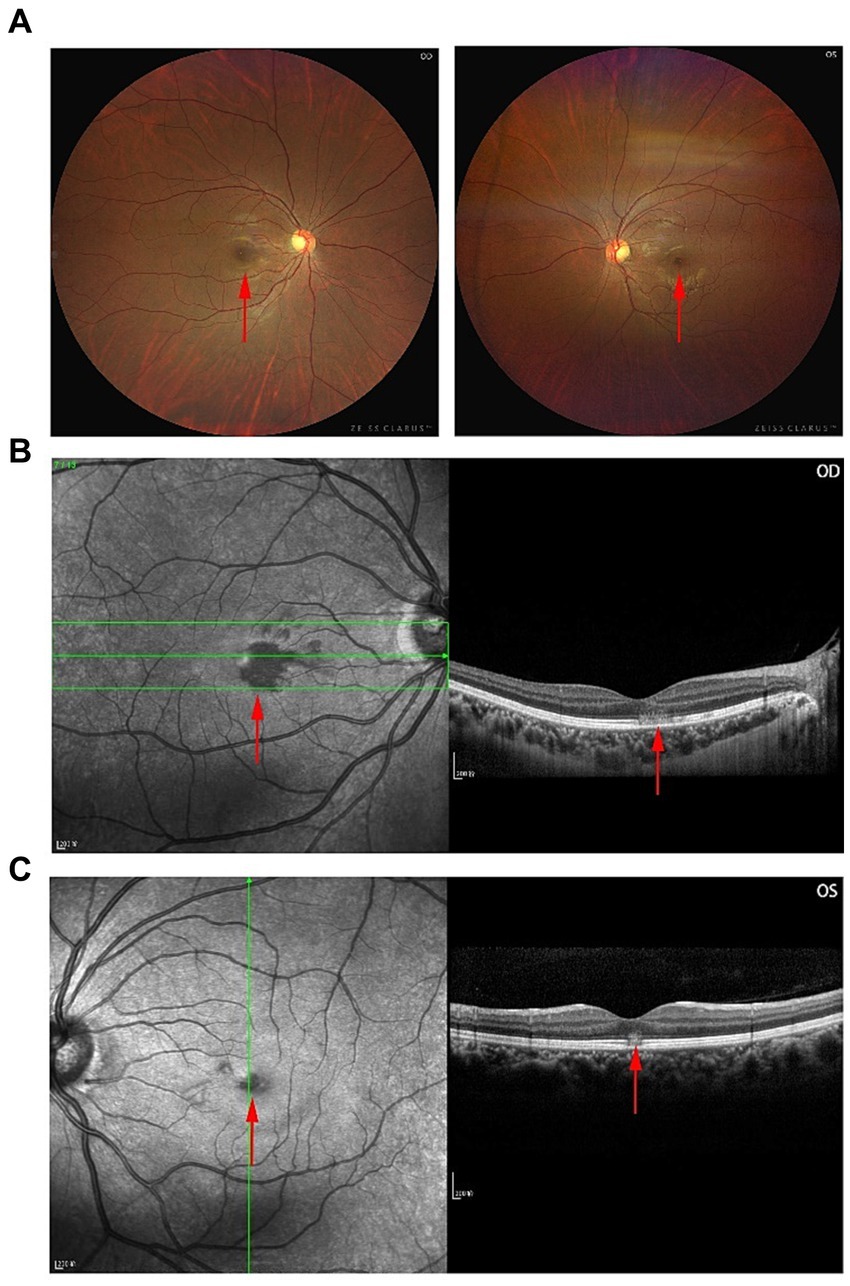
A 30-year-old Han woman complained of blurred vision in both eyes (more evident in the right eye) one day after the symptoms of COVID-19 or SARS-CoV-2 infection, i.e., fever (39.1°C), first appeared. The infection was diagnosed by reverse transcriptase polymerase chain reaction (PCR). Her visual acuity was 20/2000 OD and 20/32 OS. No relative afferent pupillary defect (RAPD) was found. No anterior segment abnormalities were detected. Color fundus imaging demonstrated perifoveal reddish-brown lesions OUs (Figure 1A). Near-infrared reflectance (NIR) imaging in both eyes revealed a well-demarcated, hyporeflective, oval-shaped macular lesion involving the fovea and that extended nasally, with the lesion area in the right eye being approximately three times that in the left eye (Figures 1B,C). Cross-sectional spectral-domain OCT (SD-OCT) revealed outer plexiform layer (OPL) thickening, outer nuclear layer (ONL) thinning, and disruption of the ellipsoid zone (EZ) in areas corresponding to the lesions (OU) (Figures 1B,C). She had no known ocular history, or systemic condition, and had not sought treatment prior to this presentation.

Figure 1. Multimodal images that display a partial reconstitution of the outer retinal architecture. (A) Fundus photographs of the right (OD) and left (OS) eyes at the time of presentation. (B,C) SD-OCT images of both eyes.
Given the acute development of these characteristic findings along with her clinical history, the patient was diagnosed with AMN. She was started on oral prednisolone 30 mg/day for 7 days. Afterward, the dose was reduced to 10 mg per week until 5 mg/day, after which the treatment was stopped. Notably, by one month, her visual acuity was 20/20 OD and 20/20 OS. During the four-month follow-up period, the patient’s visual acuity stabilized at 20/20, and no further discomfort was reported in either eye.
Literature search results
In the literature, we found 19 articles reporting cases of AMN in people with recent COVID-19 or SARS-CoV-2 infection (see Table 1).
Discussion
At present, COVID-19 or SARS-CoV-2 infection can be asymptomatic or cause mild influenza-like symptoms, and severe cases can present with respiratory distress and multiple organ failure. COVID-19 or SARS-CoV-2 seems to employ mechanisms for receptor recognition. It can bind with angiotensin-converting enzyme 2 (ACE-2) with the assistance of transmembrane serine protein 2 (TMPRSS2) or enter host cells by binding with the CD147 spike protein, thereby triggering a series of symptoms (29). ACE-2 receptors are present in the retinal ganglion cell layer, inner plexiform layer, inner nuclear layer, and outer photoreceptor segments of the eye. Moreover, TMPRSS2 is expressed in multiple retinal neuronal cells, vascular and perivascular cells, and retinal Müller glial cells. SARS-CoV-2 RNA was found in the retinas of patients who died from COVID-19, suggesting viral entry into retinal cells (30). Endothelial damage and microthrombi are the main pathological changes that lead to ocular disease.
Ophthalmologists worldwide have reported various manifestations of infection in the eye. Ophthalmic images vary in terms of presentation, severity, and timing (31). COVID-19 or SARS-CoV-2 can directly cause damage via keratoconjunctivitis, epiphora, or chemosis. Hyperinflammation with cytokine storms, stasis with hypoxia, and stasis with hypoxia that activate coagulation mechanisms can cause retinal disease (7, 8, 31, 32). Elevated D-dimer, serum ferritin, and lactate dehydrogenase levels and increased ESR/CRP inflammatory marker levels are observed in patients with ocular manifestations even after recovering from COVID-19 (33).
AMN was first reported by BOS in 1975 (34). Since the outbreak of COVID-19 or SARS-CoV-2, the incidence of AMN has increased from 0.66/100,000 in 2019 to 8.97/100,000 in 2020 (p = 0.001) at Rothschild Foundation Hospital, Paris, France. It is more common in young people (aged 12–65, median age 26), with a male-to-female ratio of approximately 1: 4–6. It can affect both eyes and is characterized by photophobia, paracentral scotoma (72–100%), floaters (3%), and visual distortions (35). Possible risk factors for AMN include infection or febrile illness (47.5%), oral contraceptives (35.6%), the use of adrenaline (7.9%), severe nonocular trauma (5.9%), shock (5%), dehydration, preeclampsia, postpartum hypotension, ulcerative colitis, Behcet’s disease, systemic lupus erythematosus, leukemia, and vaccine-related complications. Microvascular ischemia of the choriocapillaris after COVID-19 or SARS-CoV-2 infection may lead to hypoxic insult to the middle and outer retinal layers.
According to the available literature, symptoms of AMN can arise 1 day to 1 month after COVID-19 or SARS-CoV-2 infection. Risk factors such as contraceptive pills should be avoided. Oral prednisone may be an effective treatment for those experiencing marked vision loss. It is crucial to conduct additional research to uncover a potential cause-and-effect relationship between AMN and COVID-19 or SARS-CoV-2. However, whether a genetic susceptibility exists is unknown. To reinforce this hypothesis, further investigations with a larger sample size, including individuals with and without ocular symptoms and incorporating prolonged follow-up times are needed. As the pandemic continues and vaccination programs are rolled out extensively, the number of AMN cases may increase. Ophthalmologists should remain vigilant about this disease, notably because patient characteristics may deviate from the norm.
Data availability statement
The raw data supporting the conclusions of this article will be made available by the authors, without undue reservation.
Ethics statement
The studies involving humans were approved by the Institutional Review Board of Chongqing Medical University, the First Affiliated Hospital of Chongqing Medical University. The studies were conducted in accordance with the local legislation and institutional requirements. The participants provided their written informed consent to participate in this study. Written informed consent was obtained from the individual(s) for the publication of any potentially identifiable images or data included in this article (Approval No. 2023-181).
Author contributions
XW: Conceptualization, Data curation, Investigation, Methodology, Writing – original draft, Writing – review & editing. PW: Conceptualization, Investigation, Project administration, Writing – review & editing. JL: Data curation, Formal analysis, Writing – original draft. HJ: Data curation, Methodology, Validation, Writing – original draft. HX: Conceptualization, Formal analysis, Resources, Writing – original draft. HP: Investigation, Project administration, Supervision, Validation, Visualization, Writing – review & editing.
Funding
The author(s) declare that no financial support was received for the research, authorship, and/or publication of this article.
Acknowledgments
We would like to express our appreciation to the doctors and nurses in our department for their help.
Conflict of interest
The authors declare that the research was conducted in the absence of any commercial or financial relationships that could be construed as a potential conflict of interest.
Publisher’s note
All claims expressed in this article are solely those of the authors and do not necessarily represent those of their affiliated organizations, or those of the publisher, the editors and the reviewers. Any product that may be evaluated in this article, or claim that may be made by its manufacturer, is not guaranteed or endorsed by the publisher.
References
1. Hu, B, Guo, H, Zhou, P, and Shi, ZL. Characteristics of SARS-CoV-2 and COVID-19. Nat Rev Microbiol. (2021) 19:141–54. doi: 10.1038/s41579-020-00459-7
2. Carabelli, AM, Peacock, TP, Thorne, LG, Harvey, WT, Hughes, J, Consortium, C-GU, et al. SARS-CoV-2 variant biology: immune escape, transmission and fitness. Nat Rev Microbiol. (2023) 21:162–77. doi: 10.1038/s41579-022-00841-7
3. Francois, J, Collery, AS, Hayek, G, Sot, M, Zaidi, M, Lhuillier, L, et al. Coronavirus disease 2019-associated ocular neuropathy with Panuveitis: a case report. JAMA Ophthalmol. (2021) 139:247–9. doi: 10.1001/jamaophthalmol.2020.5695
4. Hutama, SA, Alkaff, FF, Intan, RE, Maharani, CD, Indriaswati, L, and Zuhria, I. Recurrent keratoconjunctivitis as the sole manifestation of COVID-19 infection: a case report. Eur J Ophthalmol. (2022) 32:NP17–21. doi: 10.1177/11206721211006583
5. Otaif, W, Al Somali, AI, and Al Habash, A. Episcleritis as a possible presenting sign of the novel coronavirus disease: a case report. Am J Ophthalmol Case Rep. (2020) 20:100917. doi: 10.1016/j.ajoc.2020.100917
6. Wu, P, Duan, F, Luo, C, Liu, Q, Qu, X, Liang, L, et al. Characteristics of ocular findings of patients with coronavirus disease 2019 (COVID-19) in Hubei Province, China. JAMA Ophthalmol. (2020) 138:575–8. doi: 10.1001/jamaophthalmol.2020.1291
7. Uzun, A, Keles Sahin, A, and Bektas, O. A unique case of branch retinal artery occlusion associated with a relatively mild coronavirus disease 2019. Ocul Immunol Inflamm. (2021) 29:715–8. doi: 10.1080/09273948.2021.1933071
8. Yeo, S, Kim, H, Lee, J, Yi, J, and Chung, YR. Retinal vascular occlusions in COVID-19 infection and vaccination: a literature review. Graefes Arch Clin Exp Ophthalmol. (2023) 261:1793–808. doi: 10.1007/s00417-022-05953-7
9. Tisdale, AK, and Chwalisz, BK. Neuro-ophthalmic manifestations of coronavirus disease 19. Curr Opin Ophthalmol. (2020) 31:489–94. doi: 10.1097/ICU.0000000000000707
10. Tisdale, AK, Dinkin, M, and Chwalisz, BK. Afferent and efferent neuro-ophthalmic complications of coronavirus disease 19. J Neuroophthalmol. (2021) 41:154–65. doi: 10.1097/WNO.0000000000001276
11. Sanjay, S, Agrawal, S, Jayadev, C, Kawali, A, Gowda, PB, Shetty, R, et al. Posterior segment manifestations and imaging features post-COVID-19. Med Hypothesis Discov Innov Ophthalmol. (2021) 10:95–106. doi: 10.51329/mehdiophthal1427
12. Virgo, J, and Mohamed, M. Paracentral acute middle maculopathy and acute macular neuroretinopathy following SARS-CoV-2 infection. Eye (Lond). (2020) 34:2352–3. doi: 10.1038/s41433-020-1069-8
13. Gascon, P, Briantais, A, Bertrand, E, Ramtohul, P, Comet, A, Beylerian, M, et al. Covid-19-associated retinopathy: a case report. Ocul Immunol Inflamm. (2020) 28:1293–7. doi: 10.1080/09273948.2020.1825751
14. Zamani, G, Ataei Azimi, S, Aminizadeh, A, Shams Abadi, E, Kamandi, M, Mortazi, H, et al. Acute macular neuroretinopathy in a patient with acute myeloid leukemia and deceased by COVID-19: a case report. J Ophthalmic Inflamm Infect. (2021) 10:39. doi: 10.1186/s12348-020-00231-1
15. Aidar, MN, Gomes, TM, de Almeida, MZH, de Andrade, EP, and Serracarbassa, PD. Low visual acuity due to acute macular neuroretinopathy associated with COVID-19: a case report. Am J Case Rep. (2021) 22:e931169. doi: 10.12659/AJCR.931169
16. David, JA, and Fivgas, GD. Acute macular neuroretinopathy associated with COVID-19 infection. Am J Ophthalmol Case Rep. (2021) 24:101232. doi: 10.1016/j.ajoc.2021.101232
17. El Matri, K, Werda, S, Chebil, A, Falfoul, Y, Hassairi, A, Bouraoui, R, et al. Acute macular outer retinopathy as a presumed manifestation of COVID-19. J Fr Ophtalmol. (2021) 44:1274–7. doi: 10.1016/j.jfo.2021.06.002
18. Masjedi, M, Pourazizi, M, and Hosseini, NS. Acute macular neuroretinopathy as a manifestation of coronavirus disease 2019: a case report. Clin Case Rep. (2021) 9:e04976. doi: 10.1002/ccr3.4976
19. Mace, T, and Pipelart, V. Acute macular neuroretinopathy and SARS-CoV-2 infection: case report. J Fr Ophtalmol. (2021) 44:e519–21. doi: 10.1016/j.jfo.2021.07.004
20. Capuano, V, Forte, P, Sacconi, R, Miere, A, Mehanna, CJ, Barone, C, et al. Querques G: acute macular neuroretinopathy as the first stage of SARS-CoV-2 infection. Eur J Ophthalmol. (2022) 33:NP105–11. doi: 10.1177/11206721221090697
21. Preti, RC, Zacharias, LC, Cunha, LP, and Monteiro, MLR. Acute macular Neuroretinopathy as the presenting manifestation of Covid-19 infection. Retin Cases Brief Rep. (2022) 16:12–5. doi: 10.1097/ICB.0000000000001050
22. Strzalkowski, P, Steinberg, JS, and Dithmar, S. COVID-19-associated acute macular neuroretinopathy. Fortschr Ophthalmol. (2022) 120:767–70. doi: 10.1007/s00347-022-01704-5
23. Kovalchuk, B, Kessler, LJ, Auffarth, GU, and Mayer, CS. Paracentral scotomas associated with COVID-19 infection. Fortschr Ophthalmol. (2022) 120:323–7. doi: 10.1007/s00347-022-01726-z
24. Hawley, L, and Han, LS. Acute macular neuroretinopathy following COVID-19 infection. N Z Med J. (2022) 135:105–7.
25. Bellur, S, Zeleny, A, Patronas, M, Jiramongkolchai, K, and Kodati, S. Bilateral acute macular neuroretinopathy after COVID-19 vaccination and infection. Ocul Immunol Inflamm. (2022) 31:1222–5. doi: 10.1080/09273948.2022.2093753
26. Giacuzzo, C, Eandi, CM, and Kawasaki, A. Bilateral acute macular neuroretinopathy following COVID-19 infection. Acta Ophthalmol. (2022) 100:e611–2. doi: 10.1111/aos.14913
27. Jalink, MB, and Bronkhorst, IHG. A sudden rise of patients with acute macular neuroretinopathy during the COVID-19 pandemic. Case Rep Ophthalmol. (2022) 13:96–103. doi: 10.1159/000522080
28. Sanjay, S, Gadde, SGK, Kumar Yadav, N, Kawali, A, Gupta, A, Shetty, R, et al. Bilateral sequential acute macular Neuroretinopathy in an Asian Indian female with beta thalassemia trait following (Corona virus disease) COVID-19 vaccination and probable recent COVID infection – multimodal imaging study. Ocul Immunol Inflamm. (2022) 30:1222–7. doi: 10.1080/09273948.2022.2026978
29. Gupta, A, Madhavan, MV, Sehgal, K, Nair, N, Mahajan, S, Sehrawat, TS, et al. Extrapulmonary manifestations of COVID-19. Nat Med. (2020) 26:1017–32. doi: 10.1038/s41591-020-0968-3
30. Reinhold, A, Tzankov, A, Matter, MS, Mihic-Probst, D, Scholl, HPN, and Meyer, P. Ocular pathology and occasionally detectable intraocular severe acute respiratory syndrome Coronavirus-2 RNA in five fatal coronavirus Disease-19 cases. Ophthalmic Res. (2021) 64:785–92. doi: 10.1159/000514573
31. Sen, M, Honavar, SG, Sharma, N, and Sachdev, MS. COVID-19 and eye: a review of ophthalmic manifestations of COVID-19. Indian J Ophthalmol. (2021) 69:488–509. doi: 10.4103/ijo.IJO_297_21
32. Mahjoub, A, Dlensi, A, Romdhane, A, Ben Abdesslem, N, Mahjoub, A, Bachraoui, C, et al. Bilateral central serous chorioretinopathy post-COVID-19. J Fr Ophtalmol. (2021) 44:1484–90. doi: 10.1016/j.jfo.2021.10.001
33. Sanjay, S, Bhakti Mistra, S, Patro, SK, Kawali, A, Shetty, R, and Mahendradas, P. Systemic markers in ophthalmic manifestations of post Corona virus Disease-19 (COVID-19). Ocul Immunol Inflamm. (2023) 31:410–5. doi: 10.1080/09273948.2021.2025253
34. Bos, PJ, and Deutman, AF. Acute macular neuroretinopathy. Am J Ophthalmol. (1975) 80:573–84. doi: 10.1016/0002-9394(75)90387-6
Keywords: COVID-19, SARS-CoV-2, acute macular neuroretinopathy, paracentral scotomas, corticosteroids
Citation: Wang X, Wang P, Lu J, Ju H, Xie H and Peng H (2024) Acute macular neuroretinopathy and COVID-19 or SARS-CoV-2 infection: case report and literature review. Front. Med. 11:1267392. doi: 10.3389/fmed.2024.1267392
Edited by:
Alan G. Palestine, University of Colorado Anschutz Medical Campus, United StatesReviewed by:
Srinivasan Sanjay, Singapore National Eye Center, SingaporeSeong Joon Ahn, Hanyang University Seoul Hospital, Republic of Korea
Copyright © 2024 Wang, Wang, Lu, Ju, Xie and Peng. This is an open-access article distributed under the terms of the Creative Commons Attribution License (CC BY). The use, distribution or reproduction in other forums is permitted, provided the original author(s) and the copyright owner(s) are credited and that the original publication in this journal is cited, in accordance with accepted academic practice. No use, distribution or reproduction is permitted which does not comply with these terms.
*Correspondence: Hui Peng, cGVuZ2g5QHNpbmEuY29t
 Xing Wang
 Peng Wang
 Jing Lu1
 Huan Ju
 Hui Peng