 Tinatin Tkemaladze1,2*
 Kakha Bregvadze
 Eka Kvaratskhelia
95% of researchers rate our articles as excellent or good
Learn more about the work of our research integrity team to safeguard the quality of each article we publish.
Find out more
CASE REPORT article
Front. Med. , 21 December 2023
Sec. Nephrology
Volume 10 - 2023 | https://doi.org/10.3389/fmed.2023.1281049
Alport syndrome is a rare genetic condition characterized by kidney disease, hearing impairment, and ocular abnormalities. It exhibits various inheritance patterns involving pathogenic variants in COL4A3, COL4A4, and COL4A5 genes. The phenotypes can range from isolated hematuria with a non-progressive or very slowly progressive course to progressive kidney disease with extrarenal abnormalities. Timely diagnosis of Alport syndrome facilitates the early and effective implementation of treatment, as well as genetic counseling. Here, we report the COL4A3 c.765G > A, p.((=)) mutation in three ethnically Azerbaijani, apparently unrelated, consanguineous families from the village of Algeti in the Marneuli region of Georgia. We speculate that this variant could represent a founder mutation within this population and recommend offering genetic testing to Algeti village residents with persistent hematuria.
Alport syndrome is one of the most commonly inherited kidney diseases, with population prevalence varying from 1 in 5,000 to 1 in 53,000 (1). Inheritance of Alport syndrome can be X-linked (OMIM 301050) due to pathogenic variants in COL4A5, autosomal recessive (OMIM 203780) caused by biallelic pathogenic variants in COL4A3 or COL4A4, autosomal dominant (OMIM 104200) caused by heterozygous COL4A3 or COL4A4 variants, or digenic, with variants in two of the COL4A3–COL4A5 genes (2). Individuals with heterozygous pathogenic variants in COL4A3 or COL4A4 genes are considered carriers of autosomal recessive Alport syndrome and are often diagnosed with thin basement membrane nephropathy (TBMN; also called thin basement membrane disease). However, there is no universal consensus on the terminology and some guidelines use the term “COL4A3 and COL4A4 heterozygotes” (2, 3).
Alport syndrome is a clinically heterogenous condition (4). Typically, recurrent microscopic or gross hematuria is the first sign of Alport syndrome, and it is usually detected in childhood. Progressive nephropathy leads to end-stage kidney disease (ESKD) either in early or middle adulthood (5). Sensorineural hearing loss and non-nephrotic range proteinuria are also common findings (6). Ocular abnormalities affecting the cornea, lens, and retina may be present as well (7). The clinical features of nephropathy are usually more severe and progressive in male than female patients. Heterozygous individuals with pathogenic variants in COL4A3 or COL4A4 genes have TBMN and present with persistent hematuria, occasional proteinuria, hypertension, and kidney dysfunction. A systematic review conducted by Matthaiou and colleagues demonstrated that approximately 29% of heterozygous patients developed chronic kidney disease and 15.1% reached ESKD (mean age of 52.8). Hearing loss or ocular abnormalities typical of X-linked Alport syndrome have low prevalence (8). It should be noted that many women with X-linked Alport syndrome have isolated microscopic hematuria (9). Patients with digenic COL4A3 and COL4A4 variants have increased risk of proteinuria, and the phenotype is intermediate between autosomal dominant and autosomal recessive Alport syndrome (10).
COL4A3–COL4A5 genes are characterized by significant allelic heterogeneity, with missense variants being the most common type of mutations, and nonsense variants resulting in nonsense-mediated mRNA decay. Rarely, large deletions and synonymous splice-site variants resulting in abnormal splicing have also been reported (1). Potential splicing effects can be examined with prediction tools such as MaxEntScan or SpliceSiteFinder, and eventually require confirmation with in vitro or in vivo functional studies or RNA sequencing.
Timely diagnosis of Alport syndrome is crucial as effective inexpensive treatment with the renin-angiotensin-aldosterone system (RAAS) blockade delays the development of kidney failure (11). However, diagnosis is often difficult on the basis of clinical features, family history, and even kidney biopsy. Genetic testing is sensitive and accurate, and provides information on the mode of inheritance.
Here, we describe a COL4A3 c.765G > A, p.((=)) variant in the homozygous and heterozygous state in three consanguineous families from Algeti village, Marneuli region, Georgia (population of 4,253), where 99.6% of the village residents are ethnic Azerbaijani. We speculate that this variant could be a founder mutation in this population.
The proband is a 5-year-old girl with asymptomatic microscopic hematuria, proteinuria, and leukocyturia detected by routine urine analysis. She was born at term as the second child of healthy, consanguineous ethnic Azerbaijani parents living in Algeti village, Marneuli region, Georgia (Figure 1A). Her developmental milestones were normal. The physical examination was unremarkable, with no edema. Her heart rate was normal (80 beats/min) and regular. She also had a normal blood pressure (90/60 mm Hg). The slit-lamp examination was normal.

Figure 1. (A) Pedigree of family 1 showing the high degree of consanguinity. (B) Pedigree of family 2. (C) Pedigree of family 3. “+” indicates a wild type allele and “−” corresponds to the COL4A3 c.765G > A variant.
Urine analysis (UA) was remarkable for protein + and erythrocytes +++. Urine microscopy revealed >1,600 red blood cells (RBC) and 8 white blood cells (WBC) per high-powered field. Phase-contrast microscopy of the urine revealed dysmorphic erythrocytes >40% and acanthocytes <5%. The spot urine protein/creatinine ratio (UPCR) was 719 mg/g (<200 mg/g). Her serum electrolyte, urea, and creatinine values were normal. Further serological tests (C3, C4, antinuclear antibodies (ANA), antineutrophilic cytoplasmic antibodies (ANCA), and circulating IgA) were within normal ranges. Abdominal and kidney ultrasound examinations were unremarkable. UA performed in both parents and her 13-year-old brother demonstrated mild hematuria (+).
Considering a positive family history of hematuria, whole exome sequencing (WES) was performed, which revealed the homozygous, likely pathogenic variant c.765G > A p.((=)) in the COL4A3 gene (NM_000091.4). The variant segregated in the heterozygous state in the parents and the brother.
Treatment with the angiotensin-converting enzyme inhibitor (ACEI) ramipril 1.5 mg/m2/once daily/orally was initiated. The patient had good adherence to the medication, without any side effects. There was a reduction in hematuria, and no significant rebound proteinuria was present after 2 years of treatment. Currently, her UPCR is 324 mg/g. Her pure-tone audiometry was normal.
The proband is a 12-year-old boy, in whom routine UA revealed microscopic hematuria at the age of 10. He was born at term as the third child of healthy, consanguineous Azerbaijani parents from Algeti village (Figure 1B). He had age-appropriate psychomotor development. The physical examination did not show any signs of edema. His heart rate (80 beats/min) and blood pressure (90/60 mm Hg) were normal. Abdominal and kidney ultrasound were unremarkable. His first UA revealed hematuria +++ and no proteinuria. Urine microscopy was remarkable for 238 RBC per high-powered field. Phase-contrast microscopy of the urine revealed dysmorphic erythrocytes <40% and acanthocytes 18%; spot UPCR was 180 mg/g. Abdominal and kidney ultrasound examinations were unremarkable.
UA and urinary microscopy were performed in both parents and his 16-year-old and 17-year-old sisters. Both sisters and the mother had hematuria +++, whereas the father had mild hematuria +. Additionally, both sisters revealed proteinuria +++, but not the parents.
WES was performed in the proband, and the homozygous variant c.765G > A, p.((=)) in the COL4A3 (NM_000091.4) gene was identified. The variant segregated in the heterozygous state in both parents and in the homozygous state in both sisters.
Treatment with ACEI (ramipril 1.5 mg/m2/once daily/orally) was initiated in all siblings. The patients had good adherence to the medication, without any side effects. After 2 years of treatment, proteinuria and hematuria persisted. Recent pure-tone audiometry revealed unilateral moderate sensorineural hearing loss in the proband and bilateral sensorineural hearing loss in both sisters.
The proband is an 8-year-old girl who presented with dark urine at 4 years of age. She was born from consanguineous Azerbaijani parents from Algeti village (Figure 1C). UA revealed hematuria (+++) and proteinuria (+), and urine microscopy was remarkable for 374 RBC per high-powered field. Phase-contrast microscopy of the urine revealed dysmorphic erythrocytes >40% and acanthocytes >5%. The spot UPCR was 183 mg/g. The physical examination was unremarkable and the heart rate, blood pressure, CBC, and slit-lamp examination were all normal.
UA was also performed in both parents; the father revealed hematuria +++ and the mother had normal UA. WES was performed in the proband, which detected the heterozygous variant c.765G > A, p. ((=)) in the COL4A3 (NM_000091.4) gene. The variant segregated in the father in the heterozygous state.
Treatment with ACEI (ramipril 1.5 mg/m2/once daily/orally) was initiated. The patient had good adherence to the medication, without any side effects. Her second UA 1 month later was negative for proteinuria but positive for hematuria +++. Urine microscopy showed 30 RBC per high-powered field. Her pure-tone audiometry was normal.
The COL4A5, COL4A3, and COL4A4 genes involved in Alport syndrome code for the collagen IV α5-, α3-, and α4-chains. Type IV collagen is a major structural component of the glomerular basement membrane (GBM). Mutations in α5-, α3-, and α4-chains may result in disrupted folding and assembly of monomers, which are then rapidly degraded in the cell. As a result, there is a switch, with persistence of α1:α1:α2 networks in the mature GBM. The α1:α1:α2 network is less resistant to higher intraglomerular pressure and proteolytic attacks compared to the α3:α4:α5 network (12).
Heterozygous mutations in COL4A3/COL4A4 are associated with a spectrum of phenotypes, ranging from complete absence of detectable symptomatology through isolated, asymptomatic hematuria to progressive kidney disease, sensorineural hearing loss, and ocular abnormalities (2). Recent data from the 100,000 Genomes project provide evidence that heterozygous variants in COL4A3/COL4A4 genes are present in approximately 1% of Europeans, which indicates that monoallelic variants may not significantly reduce fitness by producing a milder phenotype without ESKD (13). However, even within families, the phenotype associated with monoallelic mutations in COL4A3/COL4A4 genes can vary significantly. The underlying causes for clinical heterogeneity are likely diverse, including modifier genes that disrupt the effects of mutations on the synthesis, assembly, or function (or a combination of these) of α345(IV)21 and non-genetic factors such as smoking, hypertension, and dietary habits involving the consumption of salt and animal protein. The phenotype resulting from heterozygous pathogenic variants in COL4A3/COL4A4 is transmitted in an autosomal dominant manner, and the exact genotype–phenotype correlations have yet to be established.
Interpretation of DNA sequence variants and assessment of their pathogenicity are not always straightforward and simple, often requiring functional studies and computational tools. Generally, the interpretation of frameshift and nonsense variants poses less of a challenge compared to missense variants, whereas synonymous changes are the most challenging to interpret. As previously thought, synonymous variants are not necessarily silent, and evidence suggests that codon usage determines gene expression and splicing as well as protein function or conformation, especially if the variant is located close to the regulatory, donor, or acceptor splice site (14, 15). Moreover, several studies have shown that 15–60% of human genetic diseases are caused by splicing variants (16, 17).
The COL4A3 synonymous variant c.765G > A described in our series of patients is predicted not to change the amino acid sequence and is found at a very low frequency in the gnomAD v2.1.1 dataset (total allele frequency: <0.001%). It is located at the last nucleotide of exon 13. In silico predictions from MutationTaster suggest it to be deleterious, while Human Splicing Finder, NNSPLICE, NetGene2, and MaxEntScan indicate that it may affect splicing. A recent study employed an in vitro minigene assay, demonstrating that the COL4A3 variant c.765G > A affects pre-mRNA splicing by skipping exon 13 and consequently leading to an in-frame deletion of exon 13. Based on the data, the variant c.765G > A was reclassified as “pathogenic” (PVS1, PS3, PM2, PP3) according to the ACMG guidelines and the minigene assay results (18).
It is noteworthy to mention that the three families described in our study are from the same small village of Algeti (population of 4,253), Marneuli region, South Georgia (Figure 2). The Marneuli region shares a border with Azerbaijan and has a population of approximately 108,000, with 83.8% of its residents being ethnic Azerbaijanis. Azerbaijanis constitute 6.5% of Georgia’s population and form the country’s largest ethnic minority, primarily residing in the rural areas of Southern Georgia. Cousin marriage is a prevalent practice among Marneuli residents. Historically, beginning in the 16th century, several villages in the southern part of Georgia were settled by Qizilbash tribes, which gradually expanded into nearby eastern and western lands throughout the 18th century (19, 20).
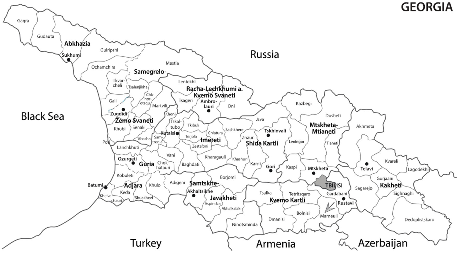
Figure 2. A map of Georgia with the Marneuli region indicated by the arrow.
The identification of a COL4A3 c.765G > A variant in carriers and homozygotes from three apparently unrelated families in the same village, along with the community’s historical background, suggest a possible founder effect. A recent demonstration of a founder effect was described in an isolated Czech Romani population, where two variants in the COL4A4 and COL4A3 genes contributed to a high prevalence of kidney failure (21). Currently, there is almost no literature describing genetic diseases from the Marneuli region of South Georgia, except for a single case report involving siblings with ultra-rare SC4MOL deficiency and a novel mutation in the MSMO1 gene (22).
Studies conducted in consanguineous families from isolated populations have enabled the discovery of new disease-causing alleles and disease genes, sometimes leading to the identification of more than one genetic disease in affected individuals (23, 24). A future large-scale study of the Marneuli population could significantly contribute to advancing such discoveries. The identification of a specific mutation in several families from the same area of Algeti village in Georgia, coupled with information about the community’s history, suggest that the c.765G > A variant in the COL4A3 gene likely results from a combination of a founder effect and consanguinity. Residents of Algeti village with persistent hematuria should be offered genetic testing to facilitate the early and effective implementation of treatment, as well as genetic counseling. Investigating the presence of founder mutations in isolated populations could potentially enhance the understanding of disease mechanisms, progression, and therapeutic strategies.
This study has several limitations. The lack of kidney biopsy data limits our ability to provide direct histological correlations with the observed clinical presentations and genetic findings. It would be valuable to conduct a large-scale study among Algeti village residents to identify individuals with hematuria and to make a detailed characterization of the phenotype and genotype to definitively establish the founder effect. Our study provides insights into the initial diagnosis and management of individuals with the COL4A3 c.765G > A variant. However, long-term follow-up data are lacking. The natural history of the disease, including the progression of kidney dysfunction and extrarenal manifestations, remains to be determined in this population. The focus of our study was on a specific COL4A3 c.765G > A variant. It is important to note that the phenotypic expression of Alport syndrome can vary significantly depending on the specific variant involved. Therefore, our findings may not entirely apply to individuals with different COL4A3 variants.
The original contributions presented in the study are included in the article/supplementary material, further inquiries can be directed to the corresponding author.
The studies involving humans were approved by Tbilisi State Medical University Ethics Committee. The studies were conducted in accordance with the local legislation and institutional requirements. Written informed consent for participation in this study was provided by the participants’ legal guardians/next of kin. Written informed consent was obtained from the individual(s) for the publication of any potentially identifiable images or data included in this article. Written informed consent was obtained from the participant/patient(s) for the publication of this case report.
TT: Conceptualization, Investigation, Writing – original draft, Writing – review & editing, Supervision. KB: Conceptualization, Investigation, Writing – original draft, Writing – review & editing, Methodology. EK: Visualization, Writing – review & editing. EA: Writing – review & editing. TD: Investigation, Supervision, Writing – review & editing.
The author(s) declare that no financial support was received for the research, authorship, and/or publication of this article.
We extend our gratitude to Aleksandre Kvakhadze for providing invaluable information about the historical aspects of Algeti village and the Marneuli region in general.
The authors declare that the research was conducted in the absence of any commercial or financial relationships that could be construed as a potential conflict of interest.
All claims expressed in this article are solely those of the authors and do not necessarily represent those of their affiliated organizations, or those of the publisher, the editors and the reviewers. Any product that may be evaluated in this article, or claim that may be made by its manufacturer, is not guaranteed or endorsed by the publisher.
1. Savige, J, Storey, H, Watson, E, Hertz, JM, Deltas, C, Renieri, A, et al. Consensus statement on standards and guidelines for the molecular diagnostics of Alport syndrome: refining the ACMG criteria. Eur J Hum Genet. (2021) 29:1186–97. doi: 10.1038/s41431-021-00858-1
2. Savige, J, Lipska-Zietkiewicz, BS, Watson, E, Hertz, JM, Deltas, C, Mari, F, et al. Guidelines for genetic testing and management of Alport syndrome. Clin J Am Soc Nephrol. (2022) 17:143–54. doi: 10.2215/CJN.04230321
3. Kashtan, CE, Ding, J, Garosi, G, Heidet, L, Massella, L, Nakanishi, K, et al. Alport syndrome: a unified classification of genetic disorders of collagen IV α345: a position paper of the Alport syndrome classification working group. Kidney Int. (2018) 93:1045–51. doi: 10.1016/j.kint.2017.12.018
4. Jais, JP, Knebelmann, B, Giatras, I, De Marchi, M, Rizzoni, G, Renieri, A, et al. X-linked Alport syndrome: natural history and genotype-phenotype correlations in girls and women belonging to 195 families: a “European Community Alport syndrome concerted action” study. J Am Soc Nephrol. (2003) 14:2603–10. doi: 10.1097/01.ASN.0000090034.71205.74
5. Hudson, BG, Tryggvason, K, Sundaramoorthy, M, and Neilson, EG. Alport's syndrome, Goodpasture's syndrome, and type IV collagen. N Engl J Med. (2003) 348:2543–56. doi: 10.1056/NEJMra022296
6. Alves, FR, and Ribeiro, FD. Revision about hearing loss in the Alport's syndrome, analyzing the clinical, genetic and bio-molecular aspects. Rev Bras Otorrinolaringol. (2005) 71:813–9. doi: 10.1590/S0034-72992005000600020
7. Savige, J, Sheth, S, Leys, A, Nicholson, A, Mack, HG, and Colville, D. Ocular features in Alport syndrome: pathogenesis and clinical significance. Clin J Am Soc Nephrol. (2015) 10:703–9. doi: 10.2215/CJN.10581014
8. Matthaiou, A, Poulli, T, and Deltas, C. Prevalence of clinical, pathological and molecular features of glomerular basement membrane nephropathy caused by COL4A3 or COL4A4 mutations: a systematic review. Clin Kidney J. (2020) 13:1025–36. doi: 10.1093/ckj/sfz176
9. Mencarelli, MA, Heidet, L, Storey, H, van Geel, M, Knebelmann, B, Fallerini, C, et al. Evidence of digenic inheritance in Alport syndrome. J Med Genet. (2015) 52:163–74. doi: 10.1136/jmedgenet-2014-102822
10. Savige, J, Renieri, A, Ars, E, Daga, S, Pinto, AM, Rothe, H, et al. Correction: digenic Alport syndrome. Clin J Am Soc Nephrol. (2023) 17:1697–706. doi: 10.2215/CJN.03120322
11. Savige, J, Gregory, M, Gross, O, Kashtan, C, Ding, J, and Flinter, F. Expert guidelines for the management of Alport syndrome and thin basement membrane nephropathy. J Am Soc Nephrol. (2013) 24:364–75. doi: 10.1681/ASN.2012020148
12. Khoshnoodi, J, Cartailler, JP, Alvares, K, Veis, A, and Hudson, BG. Molecular recognition in the assembly of collagens: terminal noncollagenous domains are key recognition modules in the formation of triple helical protomers. J Biol Chem. (2006) 281:38117–21. doi: 10.1074/jbc.R600025200
13. Gibson, J, Fieldhouse, R, Chan, MM, Sadeghi-Alavijeh, O, Burnett, L, Izzi, V, et al. Prevalence estimates of predicted pathogenic COL4A3–COL4A5 variants in a population sequencing database and their implications for Alport syndrome. J Am Soc Nephrol. (2021) 32:2273–90. doi: 10.1681/ASN.2020071065
14. Liu, Y, Yang, Q, and Zhao, F. Synonymous but not silent: the codon usage code for gene expression and protein folding. Annu Rev Biochem. (2021) 90:375–401. doi: 10.1146/annurev-biochem-071320-112701
15. Cartegni, L, Chew, SL, and Krainer, AR. Listening to silence and understanding nonsense: exonic mutations that affect splicing. Nat Rev Genet. (2002) 3:285–98. doi: 10.1038/nrg775
16. Wang, GS, and Cooper, TA. Splicing in disease: disruption of the splicing code and the decoding machinery. Nat Rev Genet. (2007) 8:749–61. doi: 10.1038/nrg2164
17. Sauna, ZE, and Kimchi-Sarfaty, C. Understanding the contribution of synonymous mutations to human disease. Nat Rev Genet. (2011) 12:683–91. doi: 10.1038/nrg3051
18. Deng, H, Zhang, Y, Ding, J, and Wang, F. Presumed COL4A3/COL4A4 missense/synonymous variants induce aberrant splicing. Front Med. (2022) 9:838983. doi: 10.3389/fmed.2022.838983
19. Nanji, A, and Daftary, F. What is shiite islam? In: VJ Cornell, editor. Voices of islam, vol. 1. Westport, CT, London: Praeger Publishers (2007). 223–7.
20. Parker, CH. Asian states and territorial empires In: D Critchlow, editor. Global interactions in the early modern age, 1400–1800. New York: Cambridge University Press (2010). 39–67.
21. Plevova, P, Indrakova, J, Savige, J, Kuhnova, P, Tvrda, P, Cerna, D, et al. A founder COL4A4 pathogenic variant resulting in autosomal recessive Alport syndrome accounts for most genetic kidney failure in Romani people. Front Med. (2023) 10:1096869. doi: 10.3389/fmed.2023.1096869
22. Tkemaladze, T, Bratland, E, Bregvadze, K, Shatirishvili, T, Tatishvili, N, Abzianidze, E, et al. MSMO1 deficiency: a potentially partially treatable, ultrarare neurodevelopmental disorder with psoriasiform dermatitis, alopecia and polydactyly. Clin Dysmorphol. (2023) 32:97–105. doi: 10.1097/MCD.0000000000000461
23. Monies, D, Abouelhoda, M, Assoum, M, Moghrabi, N, Rafiullah, R, Almontashiri, N, et al. Lessons learned from large-scale, first-tier clinical exome sequencing in a highly consanguineous population. Am J Hum Genet. (2019) 104:1182–201. doi: 10.1016/j.ajhg.2019.04.011
Keywords: Alport syndrome, thin basement membrane disease, COL4A3 , consanguinity, founder effect
Citation: Tkemaladze T, Bregvadze K, Kvaratskhelia E, Abzianidze E and Davitaia T (2023) A founder COL4A3 pathogenic variant resulting in Alport syndrome and thin basement membrane disease: a case report series. Front. Med. 10:1281049. doi: 10.3389/fmed.2023.1281049
Edited by:
Hitoshi Suzuki, Juntendo University Urayasu Hospital, JapanReviewed by:
Andrew Mallett, Townsville University Hospital, AustraliaCopyright © 2023 Tkemaladze, Bregvadze, Kvaratskhelia, Abzianidze and Davitaia. This is an open-access article distributed under the terms of the Creative Commons Attribution License (CC BY). The use, distribution or reproduction in other forums is permitted, provided the original author(s) and the copyright owner(s) are credited and that the original publication in this journal is cited, in accordance with accepted academic practice. No use, distribution or reproduction is permitted which does not comply with these terms.
*Correspondence: Tinatin Tkemaladze, dC50a2VtYWxhZHplQHRzbXUuZWR1
Disclaimer: All claims expressed in this article are solely those of the authors and do not necessarily represent those of their affiliated organizations, or those of the publisher, the editors and the reviewers. Any product that may be evaluated in this article or claim that may be made by its manufacturer is not guaranteed or endorsed by the publisher.
Research integrity at Frontiers
Learn more about the work of our research integrity team to safeguard the quality of each article we publish.