- Division of Pulmonary, Critical Care, and Allergy, Department of Internal Medicine, Faculty of Medicine, Chiang Mai University, Chiang Mai, Thailand
Background: Small airways play a major role in the pathogenesis and prognosis of chronic obstructive pulmonary disease (COPD) and asthma. More data on small airway dysfunction (SAD) using spirometry and impulse oscillometry (IOS) in these populations are required. The objective of this study was to compare the two methods, spirometry and IOS, for SAD detection and its prevalence defined by spirometry and IOS in subjects with COPD and asthma with and without fixed airflow obstruction (FAO).
Design: This is a cross-sectional study.
Methods: Spirometric and IOS parameters were compared across four groups (COPD, asthma with FAO, asthma without FAO, and healthy subjects). SAD defined by spirometry and IOS criteria were compared.
Results: A total of 262 subjects (67 COPD, 55 asthma with FAO, 101 asthma without FAO, and 39 healthy controls) were included. The prevalence of SAD defined by using IOS and spirometry criteria was significantly higher in patients with COPD (62.7 and 95.5%), asthma with FAO (63.6 and 98.2%), and asthma without FAO (38.6 and 19.8%) in comparison with healthy control (7.7 and 2.6%). IOS is more sensitive than spirometry in the detection of SAD in asthma without FAO (38.6% vs. 19.8%, p = 0.003) However, in subjects with FAO (COPD and asthma with FAO), spirometry is more sensitive than IOS to detect SAD (95.5% vs. 62.7%, p < 0.001 and 98.2% vs. 63.6%, p < 0.001, respectively).
Conclusion: Small airway dysfunction was significantly detected in COPD and asthma with and without FAO. Although IOS shows more sensitivity than spirometry in the detection of SAD in asthma without FAO, spirometry is more sensitive than IOS in patients with FAO including COPD and asthma with FAO.
Introduction
Small airways have a diameter of fewer than 2 mm (1). Small airways contribute a major role in both chronic obstructive pulmonary disease (COPD) and asthma (2, 3) caused by inflammation, hypersecretion of mucus, and airway remodeling (4). Small airway dysfunction (SAD) has been investigated for more than 60 years (5). No gold standard method currently exists for clinically assessing SAD. Spirometry is the most widely used method due to its relatively easy performance and simple measurement device (6).
The mid-maximal expiratory flow rate (MMEF) widely known as the average expired flow over the middle half (25–75%) of the forced vital capacity (FVC) maneuver (FEF25–75%) was proposed as the best parameter to identify SAD by spirometry (7). Its use is based on the hypothesis that the mid-late portion of the FVC reflects the airflow through the small airways, which are prone to expiratory collapse due to their lack of cartilaginous support (8). The American Thoracic Society (ATS) and European Respiratory Society (ERS) guidelines do not support using the FEF25–75% to identify SAD but suggest that oscillometry may provide evidence of airflow obstruction (9–11). Moreover, Quanjer et al. also suggested that the FEF25–75% does not contribute usefully to clinical decision-making (12). However, a recent large cohort analysis supports %predicted of FEF25–75% which helps link the anatomic pathology and deranged physiology of COPD independent of the %predicted forced expiratory measurement in the first second (FEV1) (13).
Impulse oscillometry (IOS) is a simple, non-invasive method, requiring only tidal breathing that allows for the evaluation of airway resistance and airway reactance in the airways and lungs (14). The IOS can be used for the diagnosis of COPD (15). It has been valuable for the evaluation of asthma control (16–18). Moreover, it detects SAD in COPD and asthma (19–22). Additionally, the IOS helped in the differentiation of COPD, asthma, and healthy subjects (15, 21, 23). All of the IOS parameters including resistance at 5 Hz (R5), resistance at 20 Hz (R20), heterogeneity of resistance (R5-R20), reactance at 5 Hz (X5), resonant frequency (Fres), and area under reactance curve between 5 Hz and Fres (AX) were significantly higher in COPD compared to healthy subjects (15, 23). The R20, R5–R20, and Fres were significantly different between COPD and asthma (23). Pornsuriyasak et al. also found significantly different IOS parameters across COPD, asthma with fixed airflow obstruction (FAO), and asthma without FAO (21).
In COPD and asthma, the small airway plays a major role in their pathogenesis (23). Air trapping and small airway wall thickening are associated with the progression of COPD (23). In asthma, inflammation and alterations of the small airways are associated with the severity of asthma and the level of asthma control (18, 24). Many methods have been introduced for measuring SAD including spirometry, plethysmography, IOS, inert gas washout, exhaled nitric oxide, and imaging, e.g., high-resolution computed tomography hyperpolarized magnetic resonance imaging and nuclear medicine (25). Of these, IOS is non-invasive, easy to perform, effort independent, and reproducible (25). By using IOS, more than half of subjects with COPD (60.0–74.0%) had SAD (21, 26–28). Additionally, Crisafulli et al. found the presence of SAD to progressively increase according to severity classifications of COPD (26). In asthma, a systematic review showed that the prevalence of SAD in asthma ranged from 33.0 to 69.9% when using IOS (22). In one study, the prevalence of SAD by using fixed R5–R20 criteria ≥0.075 kPa/L/s in COPD, asthma with FAO, and asthma without FAO were 68, 95, and 77%, respectively (21). By using spirometry, Manoharan et al. found that 54% of asthma had SAD defined using FEF25–75% of <60% predicted (29). The prevalence of SAD in airway diseases utilizing different methods varies. The study of SAD in COPD and asthma prevalence using IOS and spirometry still requires more clarification. Therefore, the objective of this study was to compare the IOS parameters and prevalence of SAD using IOS and spirometry in subjects with COPD, asthma with and without FAO, and healthy controls.
Materials and methods
Study procedures
This is a secondary analysis of cross-sectional studies in patients with COPD and asthma which were previously published (15, 18). The study was conducted at the Lung Health Center, Division of Pulmonary, Critical Care and Allergy, Department of Internal Medicine, Faculty of Medicine, Chiang Mai University, Chiang Mai, Thailand. All subjects were enrolled between July 2019 and June 2020. All tests were performed by a well-trained technician. Demographic data including age, sex, body mass index (BMI), comorbidities, smoking status, and inhaled medication used were recorded. The study was approved by the Research Ethics Committee of the Faculty of Medicine, Chiang Mai University [Institutional Review Board (IRB) approval number: MED-2562-06282, date of approval: 28 June 2019 and filed under Clinical Trials Registry (Study ID: TCTR20190709004, date of approval: 5 July 2019)]. Written informed consent was obtained from all subjects before enrollment.
Subjects
The inclusion criteria were subjects with ages greater than 40 years. The exclusion criteria were the subjects who were unable to perform acceptable spirometry according to the ATS/ERS standard (11) and who were unable to perform the IOS according to the standard recommended by the ERS standard (14). Diagnosis of COPD in this study was based on the post-bronchodilator (post-BD) ratio of FEV1/FVC below the lower limit of normal (LLN) (9) with a history of smoking ≥10 pack-years, age of onset of symptoms >40 years, and no history of asthma in first-degree family members. Asthma subjects in this study were based on a history of clinically diagnosed asthma based on episodic breathlessness, wheezing, cough, and tightness and/or bronchodilator responsiveness and had a history of non-smoking or ex-smoking of <5 pack-years. Asthma with FAO was defined by the on-treatment ratio of FEV1/FVC persistently below LLN for at least three measurements during stable visits over a year. Asthma without FAO was characterized by the on-treatment ratio of FEV1/FVC ≥ LLN measured at the stable visit. Healthy control subjects were classified as subjects with normal spirometry (%FEV1/FVC value > LLN and FVC > LLN), had no chronic respiratory symptoms, no previous diagnosis of any chronic respiratory diseases, and were non-smokers (30).
Definition
Due to the lack of consensus over which spirometry parameter is the best to identify SAD. FEF25–75% is a more sensitive parameter than FEV1 for assessing changes in peripheral airway function (7). FEF25–75% has been described as less effort-dependent than FEV1 and is a measurement of SAD (7, 8). Thus, this study used FEF25–75% from spirometry to identify SAD. However, there is no guideline regarding normal values for FEF25–75%. The most popular arbitrary cutoffs of FEF25-75% were between 60 and 75% (8). The previous study found that the normal 95th percentile for FEF25-75% is actually closer to 56% predicted in subjects with ages over 36 years (31). Therefore, the FEF25–75% < 60% predicted (8) was defined as SAD in our study. For the IOS test, SAD was defined when R5–R20 > ULN [predictive value +1.645 × Root Mean Square Error (RMSE)] (32).
Spirometry
Spirometry was performed using the Vmax 22 spirometer, Care Fusion, Hoechberg, Germany. Pre-BD spirometry was performed according to the standards of ATS/ERS (10). Spirometric parameters were collected including FVC, FEV1, the ratio of FEV1/FVC, and FEF25–75%. The predicted values of FVC, FEV1, and FEF25-75% were calculated using the Global Lung Initiative (GLI) 2012 (Southeast Asian sub-group) reference equations (33). The z-scores of FVC, FEV1, ratio of FEV1/FVC, and FEF25–75% of each subject were also calculated from the GLI 2012 (Southeast Asian sub-group) reference equations (33).
Impulse oscillometry
Pre-BD IOS was performed in all subjects before spirometry. The respiratory resistance and reactance were measured using IOS (Master Screen IOS, Viasys GmbH, Hoechberg, Germany). Each subject was asked to perform tidal breathing for 30–40 s via a mouthpiece that was connected to the IOS machine. A minimum of three tests following the recommendation by ERS standard were required (34). The average values from three IOS measurements were recorded. We collected the following IOS parameters: R5, R20, R5–R20, X5, Fres, and AX. The predicted values of all parameters in IOS were calculated using the Thai predictive value published by Deesomchok et al. (35).
Study size calculation
The size of this study was calculated based on data from the previous study (21) using G*Power Version 3.1.9.2. The proportions of SAD in patients with COPD and asthma with FAO were 68 and 95%, respectively (21). Therefore, at least 140 subjects (35 subjects for each group) needed to be included in this study (power = 0.8 with statistical significance <0.05).
Statistical analysis
Results for continuous data were expressed as mean ± standard deviation (SD) or median, interquartile range (IQR) as appropriate. Results for categorical data were expressed as frequencies and percentages. For baseline characteristics, IOS, and spirometry parameters, one-way analysis of variance (ANOVA) with the Bonferroni adjustment method was used to analyze differences across the four groups for parametric data. Kruskall–Wallis test was used to analyze the differences in baseline characteristics, IOS, and spirometry parameters across the four groups for non-parametric data. The Mann–Whitney U-Test was used to compare differences between the two groups for non-parametric data. The chi-square and Fisher’s exact test were used to compare the categorical data across the four groups and between groups, respectively. The McNemar test was used to compare the categorical data within the group. The correlations between the IOS parameters and FEF25–75% were determined using Spearman’s correlation coefficient analysis. The following cutoff parameters: 0 < |r| < 0.3 = weak correlation; 0.3 < |r| < 0.7 = moderate correlation; and |r| > 0.7 = strong correlation were used in this study (36). A value of p of <0.05 was considered statistical significance. In multiple comparisons, the adjusted level of significance was estimated by dividing the level of significance by several comparisons of four groups. Therefore, the value of p for multiple comparisons (6 and 3) was set as 0.008 (0.05/6) and 0.017 (0.05/3), respectively. All statistical analyses were performed using STATA version 16 (StataCorp, College Station, TX, United States).
Results
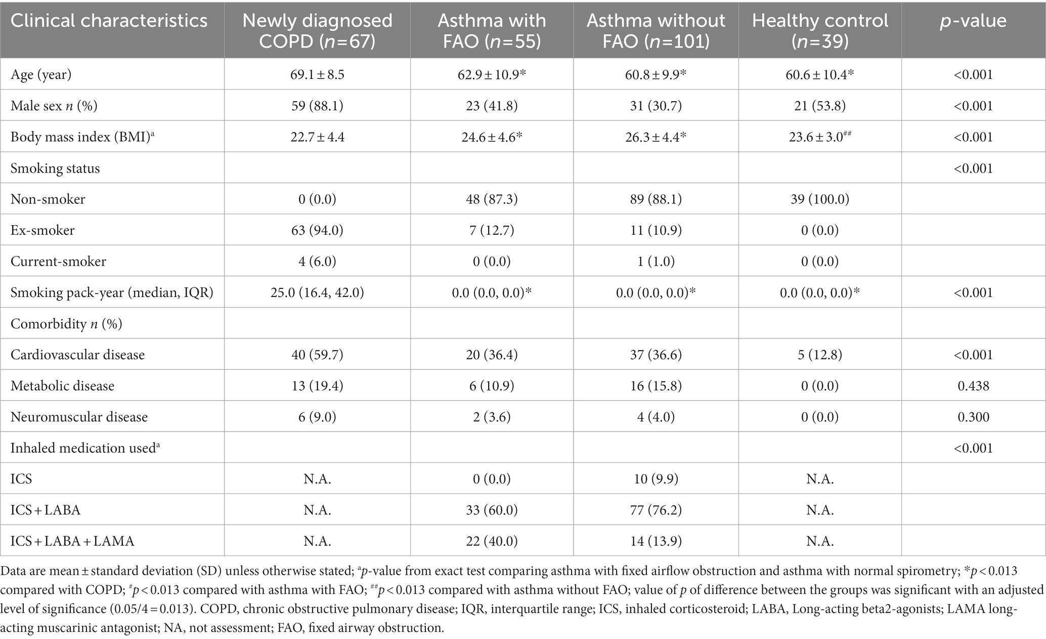
A total of 262 subjects (67 COPD, 55 asthma with FAO, 101 asthma without FAO, and 39 healthy controls) were included in the analysis. The baseline demographic data of all subjects in the four groups are shown in Table 1. There were significant differences in age, proportion of male sex, BMI, smoking status, and cardiovascular comorbidity across the four groups. Inhaled medications used were significantly different between asthma with FAO and asthma without FAO groups. Age and male sex were significantly higher in the COPD group in comparison with the other groups. More data are shown in Table 1.
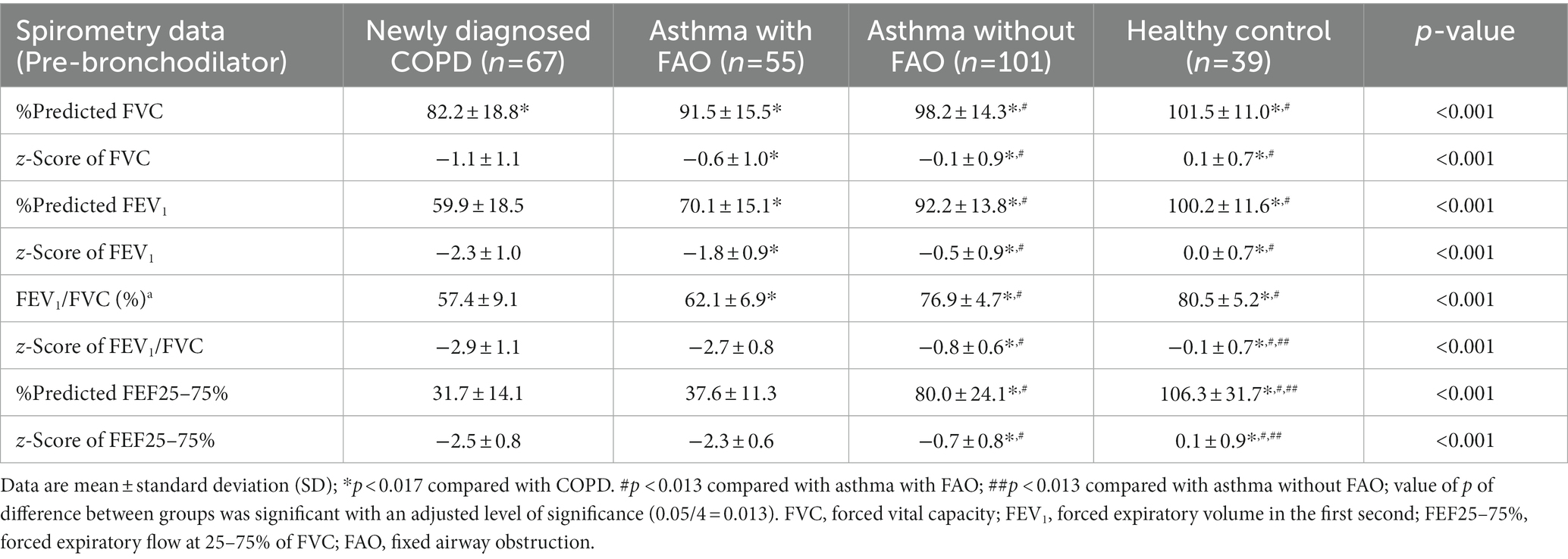
There were significantly lower %predicted and z-score of all parameters of spirometry in the COPD and asthma with FAO groups in contrast to asthma without FAO and healthy control groups. The %predicted and z-score of all parameters of spirometry in the COPD group were also significantly lower than asthma with FAO group, except for the z-score of FEV1/FVC and %predicted and z-score of FEF25–75%. In asthma without FAO, the z-score of FEV1/FVC and %predicted and z-score of FEF25–75% were significantly lower against the healthy controls. More details are shown in Table 2.
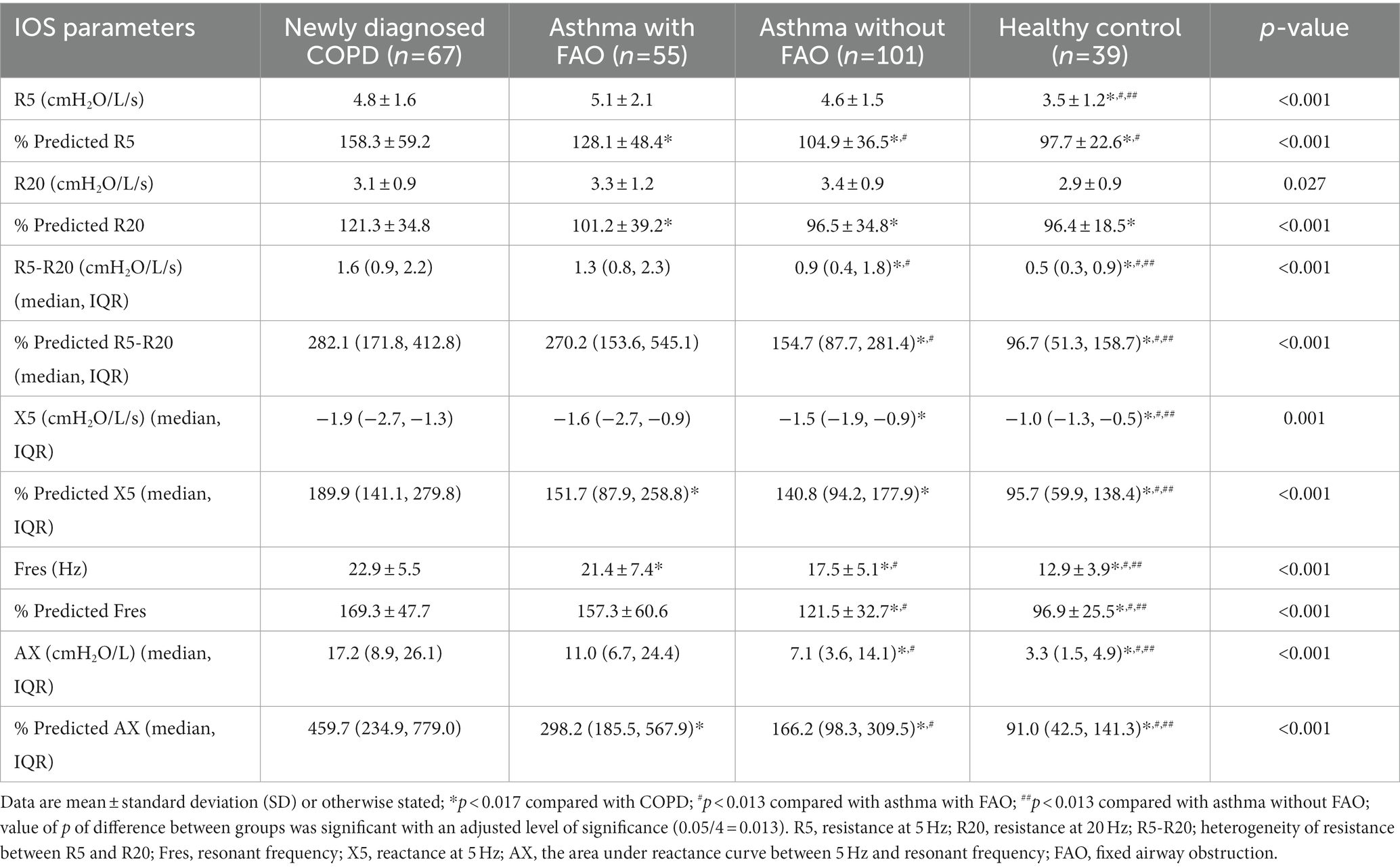
Impulse oscillometry parameters of all groups are shown in Table 3. The %predicted and the absolute values of IOS parameters including R5–R20, Fres, and AX were significantly lower in healthy controls in correlation with the other groups. The less negative reactance of X5 was also observed in the healthy control group. In the COPD and asthma with FAO groups, a significant increase in %predicted and the absolute value of IOS parameters, including R5–R20, Fres, and AX, was observed in comparison with asthma without FAO group. A significant decrease in %predicted of X5 was observed in COPD and asthma with FAO when contrasted with asthma without FAO group. The %predicted of AX was significantly higher in the group with COPD when compared with asthma in the FAO group. The %predicted and absolute values of R5-R20 were indifferent between COPD and asthma in the FAO group. More data are shown in Table 3.
Correlations between FEF25–75% and IOS parameters in subjects with chronic respiratory diseases and healthy controls are shown in Table 4. There was only a low-to-moderate correlation between some parameters of IOS and FEF25–75%, in which Fres in asthma with FAO showed the highest correlation. Correlations between FEF25–75% and IOS parameters in subjects with COPD, asthma with FAO, and asthma without FAO and healthy control with preserved FVC are also shown in Table 5. Most of the IOS parameters did not correlate with FEF25–75%. The highest correlation was shown in FEF25–75% and Fres in asthma with FAO, which was only a moderate correlation.
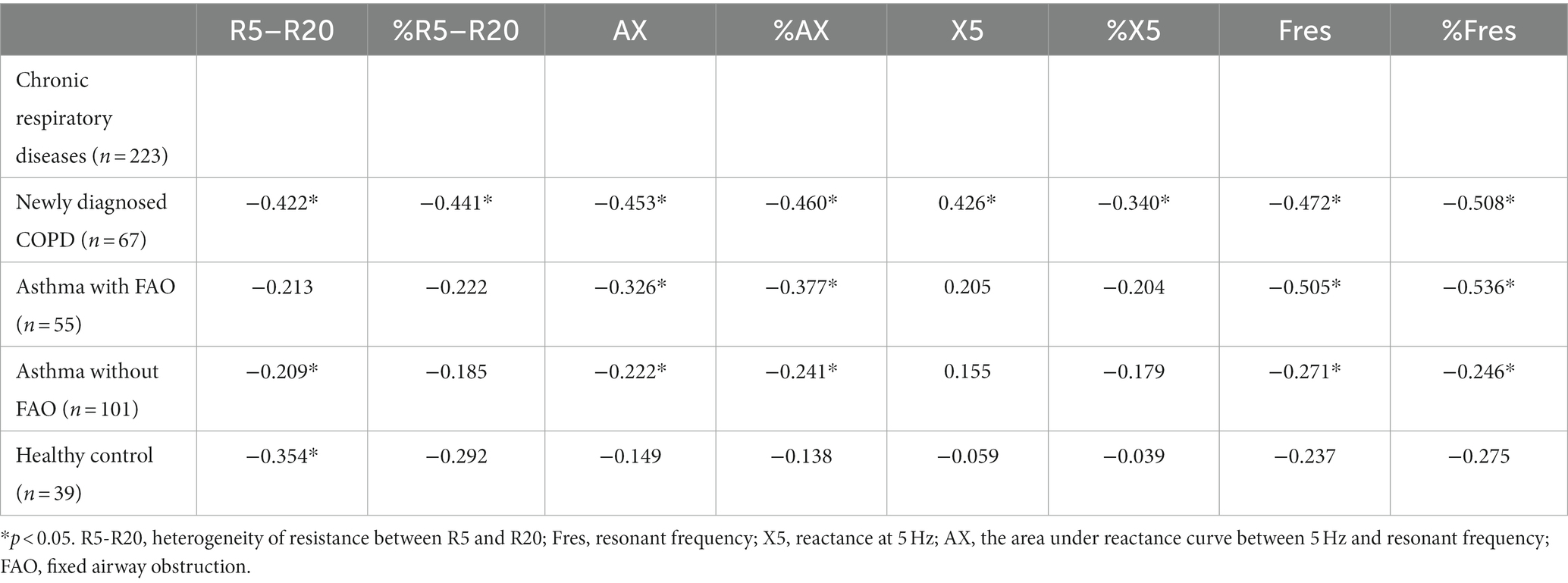
Table 4. Spearman correlation coefficients of FEF25–75% and IOS parameters in subjects with chronic respiratory diseases and healthy control.
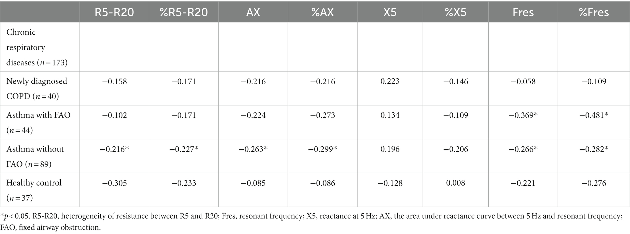
Table 5. Spearman correlation coefficients of FEF25–75% and IOS parameters in subjects with chronic respiratory diseases and healthy control with FVC ≥ 80% predicted.
The prevalence of SAD was significantly higher in COPD, asthma with FAO, and asthma without FAO in comparison with healthy control groups both in the whole and subgroups with preserved FVC (Tables 6 7). In COPD and asthma with FAO groups, the prevalence of SAD was also significantly higher than in asthma without FAO group. But there was no difference in the prevalence of SAD between COPD and asthma with FAO groups (Tables 6 7). In the case of airway obstruction including COPD and asthma with FAO, the FEF25–75% was more sensitive than R5–R20 for SAD diagnosis. But, R5–R20 was more sensitive than FEF25–75% for SAD detection in asthma without FAO. Additionally, the overestimation of SAD was observed when using the fixed criteria (R5-R20 ≥ 0.76 cmH2O) in asthma without FAO and healthy controls. More data are shown in Tables 6 7.

Table 6. Subjects with small airway dysfunction defined by R5–R20 ≥ upper limit of normal, R5–R20 ≥ 0.76 cmH2O, and %predicted FEF25–75% < 60.

Table 7. Subjects with small airway dysfunction defined by R5–R20 ≥ upper limit of normal, R5–R20 ≥ 0.76 cmH2O, and %predicted FEF25-75% < 60 in subjects with chronic respiratory diseases and healthy control with preserved FVC (FVC ≥ 80% predicted).
Discussion
In this study, we found that the prevalence of SAD classified by an increase in small airway resistance (R5–R20 ≥ ULN) was significantly higher in COPD, asthma with FAO, and asthma without FAO compared to healthy controls. The spirometry-derived FEF25–75% was more sensitive than R5–R20 for SAD diagnosis in patients with COPD and asthma with FAO. But, the IOS-derived R5–R20 was more sensitive than FEF25–75% for SAD diagnosis in asthma without FAO. In addition, SAD was overdiagnosed when using the fixed criteria of R5–R20 ≥ 0.76 cmH2O in asthma without FAO and healthy controls. We also found that the IOS parameters, especially for R5–R20, X5, Fres, and AX, were significantly lower in healthy subjects compared to COPD and asthma with and without FAO.
Respiratory resistance is largely affected by airway caliber (34). The narrower and longer airways have higher airway resistance (34). Our study showed an increase in R5 and R5–R20 in COPD, asthma with FAO, and asthma without FAO compared to healthy controls. Additionally, the R5–R20 was significantly higher in COPD and asthma with FAO compared to asthma without FAO groups. Our results were supported by the previous studies showing that airway resistances were increased in COPD and asthma (with and without FAO) compared to healthy subjects (15, 21, 23). Moreover, our results were comparable to the previous finding indicating that R5–R20 was increased in asthma with FAO compared to asthma without FAO (21).
Respiratory reactance is comprised of both inertance and elastance (34). King et al. suggested that more negative reactance indicated greater elastance or stiffness (34) and this typically occurred in subjects with obstructive airway diseases (34). The previous studies showed a decrease in X5 and an increase in AX in subjects with COPD and asthma compared with healthy controls (15, 23). In asthma with FAO, a decrease in X5 and an increase in AX were also observed in contrast with asthma without FAO (21). Our study also demonstrated that the X5 and AX were significantly decreased and increased, respectively, in COPD, asthma with FAO, and asthma without FAO when compared to healthy subjects.
From the previous studies, the diagnosis of SAD could be made by using IOS parameters, especially the R5–R20 (21, 22, 26, 27, 37). They reported that the ranges of the prevalence of SAD in COPD and asthma varied from 60.0 to 73.8% and 33.0 to 95.0%, respectively. In this study, we used the ULN of R5–R20 value for the diagnosis of SAD. We found the prevalence of SAD in COPD, asthma with FAO, and asthma without FAO to be 62.7, 63.6, and 38.6%, respectively. Our results were comparable with the previous findings (21, 22, 26, 27, 37). However, the prevalence of SAD in asthma with FAO and without FAO in our study was lower than in the previous study published by Pornsuriyasak et al. (21). They found that the prevalence of SAD was 95 and 77% in asthma with FAO and asthma without FAO, respectively. These discrepancies were due to the difference in criteria used for the diagnosis of SAD. The average of ULN of R5–R20 in our study was 1.182 cmH2O/L/s (0.116 kPa/L/s) (data not shown) which was much higher than the fixed cutoff R5–R20 level of >0.075 kPa/L/s used by the previous study (21). This could lead to overestimating SAD when using the fixed cutoff R5–R20 criteria.
This study found FEF25–75% to be more sensitive than R5–R20 in the case of FAO (COPD and asthma with FAO). R5–R20 was more sensitive than FEF25–75% for SAD detection in groups with normal FEV1/FVC (asthma without FAO) and in subgroups with preserved FVC. Additionally, the overestimation of SAD was observed when using the fixed criteria (R5-R20 ≥ 0.76 cmH2O) in asthma without FAO and healthy controls. Our results were supported by a previous study indicating that IOS shows more sensitivity for evaluating SAD than spirometry in patients with normal lung function and spirometry showed more sensitivity than IOS to detect SAD using spirometry in subjects with abnormal lung function (32). The difference in techniques of both tests might explain these discrepant results. The sound waves were applied to measure airway resistance and airway reactance during tidal breathing in IOS, whereas the forced expiratory maneuvers might induce airway collapse which resulted in decreasing of FEF25–75% in spirometry (8, 38). Therefore, the ULN of R5-R20 from IOS is more sensitive than FEF25–75% from spirometry for the detection of SAD in subjects with normal FEV1/FVC and patients with obstructive airway disease with normal FVC.
Impulse oscillometry is currently the most specific and sensitive test for the detection of SAD (37). We found that subjects with chronic respiratory disease including COPD and asthma were in high-risk groups for SAD because small airways play a major role in the pathogenesis and prognosis of both COPD and asthma (18, 23, 24). For example, the SAD classified by value of R5–R20 ≥ 1 cmH2O/L/s was an accurate tool for the detection of uncontrolled asthma in our previous study (18). SAD diagnosed by IOS in patients with intermittent asthma not treated with controller medications, mostly had normal FEV1/FVC, was shown to predict the future of more symptoms, more use of rescue medication, poor asthma control, and severe exacerbations (39). In addition, serial R5–R20 measurements were beneficial in predicting the response to treatment (40). These findings confirm that IOS has benefits in both prognosis and management plans in asthma patients. Therefore, the findings of these and our studies suggest that clinicians should regularly perform IOS testing in patients with obstructive airway disease. The results of SAD measured by IOS may be useful for monitoring and treating these patients.
The strength of our study is that the healthy control group was included for comparison with COPD and asthma with and without the FAO group. We also did the subgroup analysis of those with preserved FVC. Moreover, the prediction equations of IOS parameters in the Thai population were used in this study. The %predicted of each IOS parameter was used for analysis. Thus, the diversities of age, sex, height, and weight that had impacts on airway resistance and reactance were minimized (35, 41, 42). We encourage using the ULN criteria of R5-R20 instead of the fixed criteria (R5–R20 ≥ 0.76 cmH2O) for reducing the over-or underestimation of SAD. However, this study has some limitations. First, only newly diagnosed COPD was included in this study. Thus, the results may not be generalized for treated COPD patients. Second, due to the small sample size of COPD, the prevalence of SAD according to the staging of COPD was not mentioned in this study. Third, due to the lack of consensus over the cutoff value of FEF25–75% for identifying SAD, the threshold of FEF 25–75% < 60% predicted was classified as SAD in the current study. Thus, the results may not be generalized for the other studies which used other cutoff points. Finally, due to the cross-sectional study design, the association between a continuous scale of lung function measurements (FEF25–75% and R5–R20) and clinical symptoms, exacerbations, and comorbidities were not mentioned in this study. Therefore, the effect of SAD on clinical symptoms and exacerbations should be addressed in future prospective studies.
Conclusion
The IOS parameters, especially the R5–R20, can be used to differentiate healthy subjects from chronic airway diseases, including COPD and asthma with or without FAO. The ULN, rather than a fixed cutoff point, of R5–R20 should be used to identify SAD. The prevalence of SAD was significantly higher in COPD, asthma with FAO, and asthma without FAO in comparison with healthy control. Moreover, the prevalence of SAD was significantly higher in COPD and asthma with FAO than in asthma without FAO. IOS is more sensitive than spirometry for the detection of SAD in asthma without AFO; however, for patients with AFO including COPD and asthma with FAO, spirometry is more sensitive.
Data availability statement
The raw data supporting the conclusions of this article will be made available by the authors, without undue reservation.
Ethics statement
The studies involving human participants were reviewed and approved by the Research Ethics Committee of the Faculty of Medicine, Chiang Mai University [Institutional Review Board (IRB) approval number: MED-2562-06282, date of approval: 28 June 2019 and filed under Clinical Trials Registry (Study ID: TCTR20190709004, date of approval: 5 July 2019)]. The patients/participants provided their written informed consent to participate in this study.
Author contributions
CL, WC, and CP: conceptualization, methodology, validation, investigation, resources, writing–review and editing, and visualization. WC: software, formal analysis, and data curation. CL and WC: writing–original draft preparation. CL and CP: supervision and project administration. WC and CP: funding acquisition. All authors have read and agreed to the published version of the manuscript.
Funding
This study was funded by the Faculty of Medicine, Chiang Mai University Research Fund under grant no. 037/2563.
Acknowledgments
The authors would like to thank all subjects who participated in this study. They also wish to acknowledge the contribution of the staff of the Division of Pulmonary, Critical Care and Allergy, Department of Internal Medicine, Faculty of Medicine, Chiang Mai University to this trial. And the authors thank Ruth Leatherman, Research administration section, Faculty of Medicine, Chiang Mai University for native English proofreading.
Conflict of interest
The authors declare that the research was conducted in the absence of any commercial or financial relationships that could be construed as a potential conflict of interest.
Publisher’s note
All claims expressed in this article are solely those of the authors and do not necessarily represent those of their affiliated organizations, or those of the publisher, the editors and the reviewers. Any product that may be evaluated in this article, or claim that may be made by its manufacturer, is not guaranteed or endorsed by the publisher.
References
1. Mead, J. The lung’s “quiet zone.”. N Engl J Med. (1970) 282:1318–9. doi: 10.1056/NEJM197006042822311
2. Baraldo, S, Turato, G, and Saetta, M. Pathophysiology of the small airways in chronic obstructive pulmonary disease. Respiration. (2012) 84:89–97. doi: 10.1159/000341382
3. Postma, DS, Brightling, C, Baldi, S, Van den Berge, M, Fabbri, LM, Gagnatelli, A, et al. Exploring the relevance and extent of small airways dysfunction in asthma (ATLANTIS): baseline data from a prospective cohort study. Lancet Respir Med. (2019) 7:402–16. doi: 10.1016/S2213-2600(19)30049-9
4. Higham, A, Quinn, AM, Cançado, JED, and Singh, D. The pathology of small airways disease in COPD: historical aspects and future directions. Respir Res. (2019) 20:49. doi: 10.1186/s12931-019-1017-y
5. Macklem, PT, and Mead, J. Resistance of central and peripheral airways measured by a retrograde catheter. J Appl Physiol. (1967) 22:395–401. doi: 10.1152/jappl.1967.22.3.395
6. Konstantinos Katsoulis, K, Kostikas, K, and Kontakiotis, T. Techniques for assessing small airways function: possible applications in asthma and COPD. Respir Med. (2016) 119:e2–9. doi: 10.1016/j.rmed.2013.05.003
7. McFadden, ER Jr, and Linden, DA. A reduction in maximum mid-expiratory flow rate. A spirographic manifestation of small airway disease. Am J Med. (1972) 52:725–37. doi: 10.1016/0002-9343(72)90078-2
8. Knox-Brown, B, Mulhern, O, Feary, J, and Amaral, AFS. Spirometry parameters used to define small airways obstruction in population-based studies: systematic review. Respir Res. (2022) 23:67. doi: 10.1186/s12931-022-01990-2
9. Pellegrino, R, Viegi, G, Brusasco, V, Crapo, RO, Burgos, F, Casaburi, R, et al. Interpretative strategies for lung function tests. Eur Respir J. (2005) 26:948–68. doi: 10.1183/09031936.05.00035205
10. Miller, MR, Hankinson, J, Brusasco, V, Burgos, F, Casaburi, R, Coates, A, et al. Standardisation of spirometry. Eur Respir J. (2005) 26:319–38. doi: 10.1183/09031936.05.00034805
11. Stanojevic, S, Kaminsky, DA, Miller, MR, Thompson, B, Aliverti, A, Barjaktarevic, I, et al. ERS/ATS technical standard on interpretive strategies for routine lung function tests. Eur Respir J. (2022) 60:2101499. doi: 10.1183/13993003.01499-2021
12. Quanjer, PH, Weiner, DJ, Pretto, JJ, Brazzale, DJ, and Boros, PW. Measurement of FEF25-75% and FEF75% does not contribute to clinical decision making. Eur Respir J. (2014) 43:1051–8. doi: 10.1183/09031936.00128113
13. Ronish, BE, Couper, DJ, Barjaktarevic, IZ, Cooper, CB, Kanner, RE, Pirozzi, CS, et al. Forced expiratory flow at 25-75% links COPD physiology to emphysema and disease severity in the SPIROMICS cohort. Chronic Obstr Pulm Dis. (2022) 9:111–21. doi: 10.15326/jcopdf.2021.0241
14. Bickel, S, Popler, J, Lesnick, B, and Eid, N. Impulse oscillometry: interpretation and practical applications. Chest. (2014) 146:841–7. doi: 10.1378/chest.13-1875
15. Chaiwong, W, Namwongprom, S, Liwsrisakun, C, and Pothirat, C. Diagnostic ability of impulse Oscillometry in diagnosis of chronic obstructive pulmonary disease. COPD. (2020) 17:635–46. doi: 10.1080/15412555.2020.1839042
16. Shi, Y, Aledia, AS, Galant, SP, and George, SC. Peripheral airway impairment measured by oscillometry predicts loss of asthma control in children. J Allergy Clin Immunol. (2013) 131:718–23. doi: 10.1016/j.jaci.2012.09.022
17. Shi, Y, Aledia, AS, Tatavoosian, AV, Vijayalakshmi, S, Galant, SP, and George, SC. Relating small airways to asthma control by using impulse oscillometry in children. J Allergy Clin Immunol. (2012) 129:671–8. doi: 10.1016/j.jaci.2011.11.002
18. Chaiwong, W, Namwongprom, S, Liwsrisakun, C, and Pothirat, C. The roles of impulse oscillometry in detection of poorly controlled asthma in adults with normal spirometry. J Asthma. (2022) 59:561–71. doi: 10.1080/02770903.2020.1868499
19. Pornsuriyasak, P, Suwatanapongched, T, Thaipisuttikul, W, Nitiwarangkul, C, Kawamatawong, T, Amornputtisathaporn, N, et al. Assessment of proximal and peripheral airway dysfunction by computed tomography and respiratory impedance in asthma and COPD patients with fixed airflow obstruction. Ann Thorac Med. (2018) 13:212–9. doi: 10.4103/atm.ATM_22_18
20. Lu, D, Chen, L, Fan, C, Zeng, W, Fan, H, Wu, X, et al. The value of impulse Oscillometric parameters and quantitative HRCT parameters in differentiating asthma-COPD overlap from COPD. Int J Chron Obstruct Pulmon Dis. (2021) 16:2883–94. doi: 10.2147/COPD.S331853
21. Pornsuriyasak, P, Khiawwan, S, Rattanasiri, S, Unwanatham, N, and Petnak, T. Prevalence of small airways dysfunction in asthma with-and without-fixed airflow obstruction and chronic obstructive pulmonary disease. Asian Pac J Allergy Immunol. (2021) 39:296–303. doi: 10.12932/AP-310119-0485
22. Usmani, OS, Singh, D, Spinola, M, Bizzi, A, and Barnes, PJ. The prevalence of small airways disease in adult asthma: a systematic literature review. Respir Med. (2016) 116:19–27. doi: 10.1016/j.rmed.2016.05.006
23. Kanda, S, Fujimoto, K, Komatsu, Y, Yasuo, M, Hanaoka, M, and Kubo, K. Evaluation of respiratory impedance in asthma and COPD by an impulse oscillation system. Intern Med. (2010) 49:23–30. doi: 10.2169/internalmedicine.49.2191
24. van der Wiel, E, ten Hacken, NH, Postma, DS, and van den Berge, M. Small airways dysfunction associates with respiratory symptoms and clinical features of asthma: a systematic review. J Allergy Clin Immunol. (2013) 131:646–57. doi: 10.1016/j.jaci.2012.12.1567
25. McNulty, W, and Usmani, OS. Techniques of assessing small airways dysfunction. Eur Clin Respir J. (2014) 1:1. doi: 10.3402/ecrj.v1.25898
26. Crisafulli, E, Pisi, R, Aiello, M, Vigna, M, Tzani, P, Torres, A, et al. Prevalence of small-airway dysfunction among COPD patients with different GOLD stages and its role in the impact of disease. Respiration. (2017) 93:32–41. doi: 10.1159/000452479
27. Jarenback, L, Ankerst, J, Bjermer, L, and Tufvesson, E. Flow-volume parameters in COPD related to extended measurements of lung volume, diffusion, and resistance. Pulm Med. (2013) 2013:782052. doi: 10.1155/2013/782052
28. Chetta, A, Facciolongo, N, Franco, C, Franzini, L, Piraino, A, and Rossi, C. Impulse oscillometry, small airways disease, and extra-fine formulations in asthma and chronic obstructive pulmonary disease: windows for new opportunities. Ther Clin Risk Manag. (2022) 18:965–79. doi: 10.2147/TCRM.S369876
29. Manoharan, A, Anderson, WJ, Lipworth, J, and Lipworth, BJ. Assessment of spirometry and impulse oscillometry in relation to asthma control. Lung. (2015) 193:47–51. doi: 10.1007/s00408-014-9674-6
30. Pothirat, C, Phetsuk, N, Liwsrisakun, C, Bumroongkit, C, Deesomchok, A, and Theerakittikul, T. Major chronic respiratory diseases in Chiang Mai: prevalence, clinical characteristics, and their correlations. J Med Assoc Thail. (2016) 99:1005–13.
31. Knudson, RJ, and Lebowitz, MD. Maximal mid-expiratory flow (FEF25-75%): normal limits and assessment of sensitivity. Am Rev Respir Dis. (1978) 117:609–10. doi: 10.1164/arrd.1978.117.3.609
32. Lu, L, Peng, J, Zhao, N, Wu, F, Tian, H, Yang, H, et al. Discordant Spirometry and impulse Oscillometry assessments in the diagnosis of small airway dysfunction. Front Physiol. (2022) 13:892448. doi: 10.3389/fphys.2022.892448
33. Quanjer, PH, Stanojevic, S, Cole, TJ, Baur, X, Hall, GL, Culver, BH, et al. Multi-ethnic reference values for spirometry for the 3-95-yr age range: the global lung function 2012 equations. Eur Respir J. (2012) 40:1324–43. doi: 10.1183/09031936.00080312
34. King, GG, Bates, J, Berger, KI, Calverley, P, de Melo, PL, Dellacà, RL, et al. Technical standards for respiratory oscillometry. Eur Respir J. (2020) 55:1900753. doi: 10.1183/13993003.00753-2019
35. Deesomchok, A, Chaiwong, W, Liwsrisakun, C, Namwongprom, S, and Pothirat, C. Reference equations of the impulse oscillatory in healthy Thai adults. J Thorac Dis. (2022) 14:1384–92. doi: 10.21037/jtd-21-1989
36. Ratner, B. The correlation coefficient: its values range between +1/−1, or do they? J Target Meas Anal Mark. (2009) 17:139–42. doi: 10.1057/jt.2009.5
37. Peng, J, Wu, F, Tian, H, Yang, H, Zheng, Y, Deng, Z, et al. Clinical characteristics of and risk factors for small airway dysfunction detected by impulse oscillometry. Respir Med. (2021) 190:106681. doi: 10.1016/j.rmed.2021.106681
38. Zimmermann, SC, Tonga, KO, and Thamrin, C. Dismantling airway disease with the use of new pulmonary function indices. Eur Respir Rev. (2019) 28:180122. doi: 10.1183/16000617.0122-2018
39. Cottini, M, Lombardi, C, Comberiati, P, Landi, M, Berti, A, and Ventura, L. Small airway dysfunction in asthmatic patients treated with as-needed SABA monotherapy: a perfect storm. Respir Med. (2023) 209:107154. doi: 10.1016/j.rmed.2023.107154
40. Abdo, M, Watz, H, Veith, V, Kirsten, AM, Biller, H, Pedersen, F, et al. Small airway dysfunction as predictor and marker for clinical response to biological therapy in severe eosinophilic asthma: a longitudinal observational study. Respir Res. (2020) 21:278. doi: 10.1186/s12931-020-01543-5
41. Mukdjindapa, P, Manuyakorn, W, Kiewngam, P, Sasisakulporn, C, Pongchaikul, P, Kamchaisatian, W, et al. Reference value of forced oscillation technique for healthy preschool children. Asian Pac J Allergy Immunol. (2021) 39:89–95. doi: 10.12932/AP-110618-0334
Keywords: COPD, asthma, fixed airflow obstruction, impulse oscillometry, small airways, spirometry
Citation: Liwsrisakun C, Chaiwong W and Pothirat C (2023) Comparative assessment of small airway dysfunction by impulse oscillometry and spirometry in chronic obstructive pulmonary disease and asthma with and without fixed airflow obstruction. Front. Med. 10:1181188. doi: 10.3389/fmed.2023.1181188
Edited by:
Justin Garner, Royal Brompton Hospital, United KingdomReviewed by:
Antonio Molino, University of Naples Federico II, ItalySateesh Sakhamuri, Medical Associates Hospital, Trinidad and Tobago
Copyright © 2023 Liwsrisakun, Chaiwong and Pothirat. This is an open-access article distributed under the terms of the Creative Commons Attribution License (CC BY). The use, distribution or reproduction in other forums is permitted, provided the original author(s) and the copyright owner(s) are credited and that the original publication in this journal is cited, in accordance with accepted academic practice. No use, distribution or reproduction is permitted which does not comply with these terms.
*Correspondence: Chaicharn Pothirat, Y2hhaWNoYXJuLnBAY211LmFjLnRo
†These authors have contributed equally to this work and share first authorship
 Chalerm Liwsrisakun†
 Warawut Chaiwong