 Min-Joon Kim
 Seri Lee
 Seung-Nam Kim
94% of researchers rate our articles as excellent or good
Learn more about the work of our research integrity team to safeguard the quality of each article we publish.
Find out more
MINI REVIEW article
Front. Med., 07 June 2023
Sec. Gastroenterology
Volume 10 - 2023 | https://doi.org/10.3389/fmed.2023.1167356
Acupuncture is a non-pharmacological traditional Chinese medical technique that has been used for various types of gastrointestinal (GI) diseases in Eastern medicine. However, the specific mechanisms underlying acupuncture treatment in the GI tract have not yet been elucidated. In this study, we searched the electronic databases PUBMED, EMBASE, and MEDLINE and identified 30 eligible studies that were summarized in this review. This review demonstrates that treatments, including both manual and electroacupuncture, have therapeutic mechanisms in diverse GI diseases. The underlying mechanisms are broadly divided into the following: changes in gene expression in the gastric mucosa or nuclei of the solitary tract, metabolic change induction, regulation of anti-inflammatory substances, vagal activity increase, change in functional connectivity between brain regions, and control of the number of neurons related to GI diseases. Although this study is limited in that it does not represent all types of GI diseases with different acupuncture methods, this study identified acupuncture as effective for GI diseases through various biological mechanisms. We hope that our study will reveal various mechanisms of acupuncture in GI diseases and play an important role in the therapy and treatment of GI diseases, thus advancing the field of study.
As society evolves rapidly, changes in lifestyle and diet affect the occurrence and development of gastrointestinal (GI) diseases (5). GI disease, a source of morbidity, mortality, and financial burden, has a high frequency and significant socioeconomic impact on the community (4, 3). With the increase in number of diagnostic techniques used, several types of GI disease are identified, which range from mild diseases, such as acute gastritis and acute gastroenteritis, to serious problems, including gastroesophageal reflux disease, inflammatory bowel disease, and upper GI cancer (2). Therefore, medical treatment of GI diseases is important, and the development of therapy is typically required.
Acupuncture, a traditional Chinese medicine, has been used in medical practice (7), and its clinical effects have been reported (8). Therefore, acupuncture has been performed under pathological conditions of GI diseases (1). The effectiveness of acupuncture for gastroparesis has been proven in clinical trials (6). Thus, a recent study has demonstrated the therapeutic effect of acupuncture on GI through the inhibition of gastric acid secretion and GI motility control via different pathways (11). Animal and clinical studies have evaluated the efficacy of acupuncture on GI diseases (10). However, no studies have investigated the mechanisms underlying the therapeutic effects of acupuncture on GI diseases.
This study aimed to comprehensively review the published studies by conducting a comprehensive and systematic search, and to determine the therapeutic effect of acupuncture on GI diseases.
Figure 1 presents a flowchart of the study selection process for this review. All qualified studies included the keywords acupuncture, animal, and stomach. The databases PUBMED, EMBASE, and MEDLINE were searched using the search terms “(acupuncture [title/abstract] OR electroacupuncture [title/abstract]) AND (animal [title/abstract] OR rat [title/abstract] OR mouse [title/abstract] OR rats[title/abstract] OR mice [title/abstract]) AND (stomach [title/abstract]).” Two reviewers independently conducted the research in this study and identified 114 studies related to the topic. Of the studies, 83 were excluded for the following reasons: review articles, no acupuncture treatment alone, non-English article, no electroacupuncture (EA) or manual acupuncture (MA), full text was not accessible, and study not using a pathological model. Finally, 30 articles were included in the study.
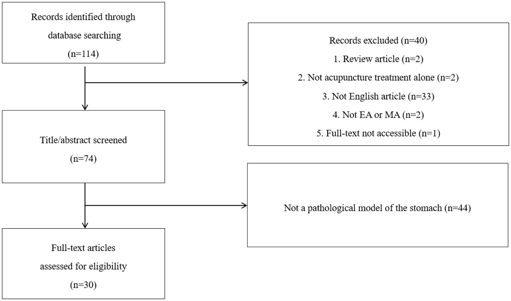
Figure 1. Flowchart of the study.
Quality assessment was performed by two independent researchers using a total of 10 questions, as previously described (9). For the sequence generation domain, the description of whether studies generate allocation sequences in sufficient detail was assessed. The item of baseline characteristics was whether prognostic factors or animal characteristics were described to determine the similarity between the intervention and control groups. For allocation concealment, whether it describes the method used to conceal the allocation sequence in detail was evaluated. Next, the item on random housing describes all measures of housing animals randomly in animal rooms. The fifth item is whether it describes all measures used to blind trial caregivers and researchers from knowing which intervention each animal has received. The item on random outcome assessment asks whether animals were randomly selected for outcome assessment and which methods were used to select the animal. The seventh item is whether it describes all measures used to blind outcome assessors from knowing which intervention each animal has received. Additionally, the item for incomplete outcome data is whether the analysis describes the completeness of the outcome data for each main outcome, including the number or reason for attrition and exclusion. For the selective outcome reporting domain, we assessed whether the examination method of selective result reporting and the identified contents were explained. Finally, for the other sources of the bias domain, we evaluated whether the acupuncture treatment method was specifically explained. Among the 10 questions, the papers scoring 2, 1, and 0 points for each question were considered as having low, unclear, and high risks of bias, respectively.
The disease models, animals, acupuncture methods, and acupuncture points used in each study are listed in Table 1. The 30 papers had the following disease models: five gastroparesis models caused by diabetes mellitus, four functional dyspepsia (FD) models, two gastric ulcer models, one peptic ulcer disease (PUD) model, one GI dysmotility model caused by chronic complicated stress, one GI dysmotility model caused by postoperative ileus, two chronic atrophic gastritis (CAG) models, five gastric distention (GD) models, two gastric mucosal injury models, three gastric mucosal lesion (GML) models, two gastric dysrhythmia models, one reduced gastric electrical activity model caused by acute myocardial ischemia, and one acute inflammatory stomach ache model. In total, 26 papers used EA, and 4 papers used MA. The acupuncture points ST2, ST6, ST21, ST36, ST37, CV12, GB14, GB24, GB34, PC5, PC6, SP4, LI10, LI11, SI18, BL21, RN12, and auricular acupunture (ear point ‘stomach’) were used.
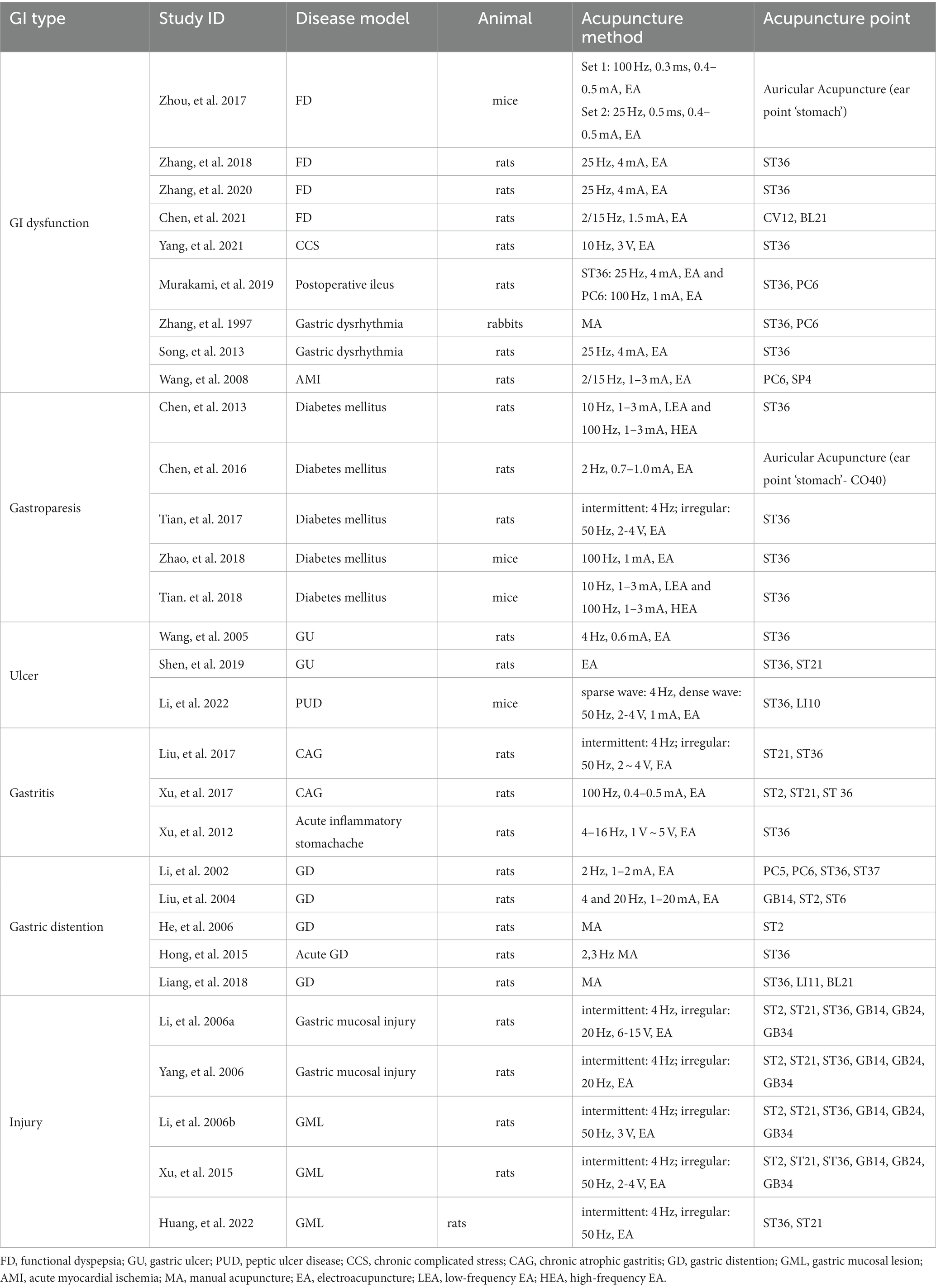
Table 1. Study characteristics.
Figure 2 presents the results of the quality assessment of the 30 research papers. Two papers received 12 points, four papers received 13 points, 16 papers received 14 points, six papers received 15 points, one paper received 16 points, and one paper received 17 points.
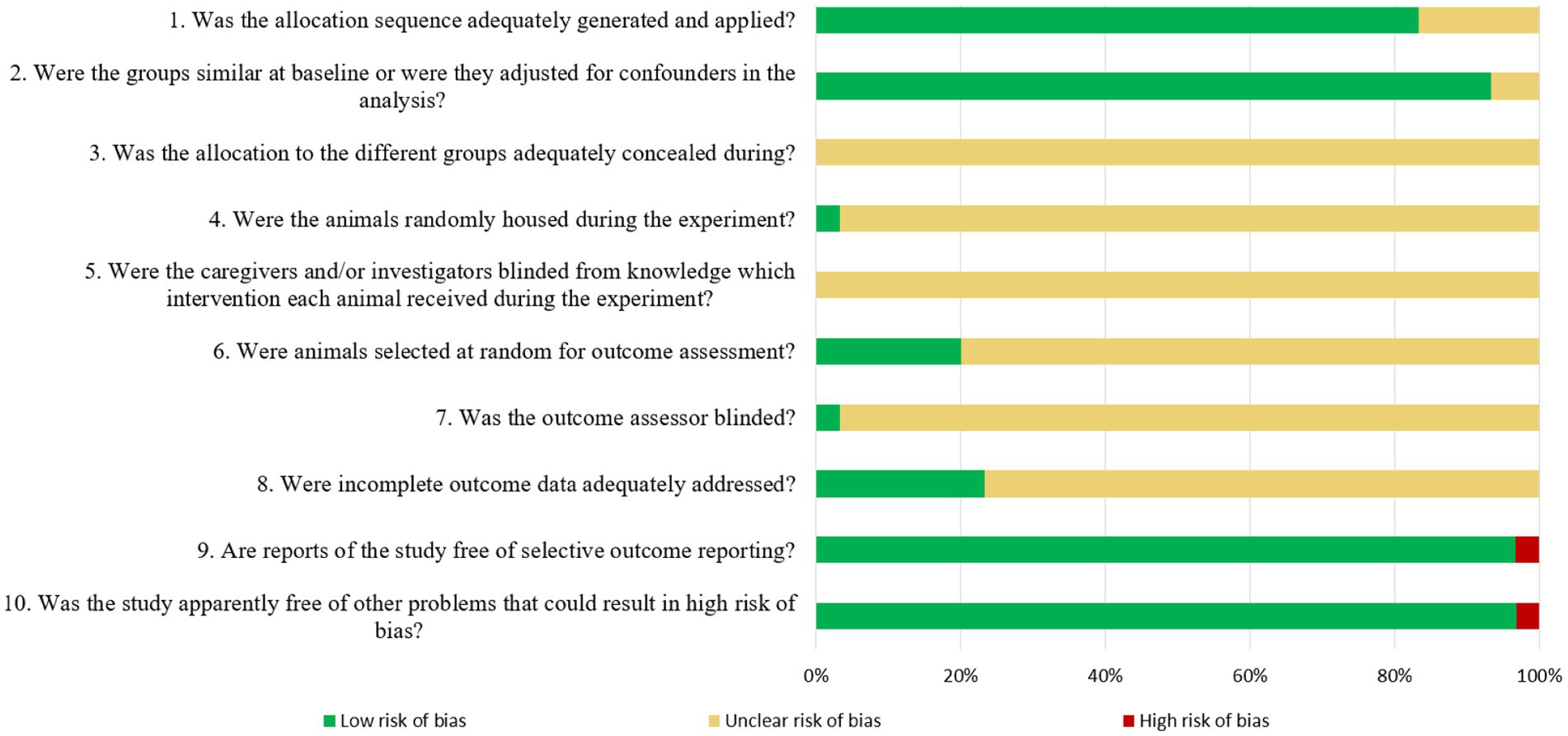
Figure 2. Quality assessment of the literatures.
Table 2 summarizes the efficacy and mechanism of acupuncture treatment for GI diseases.
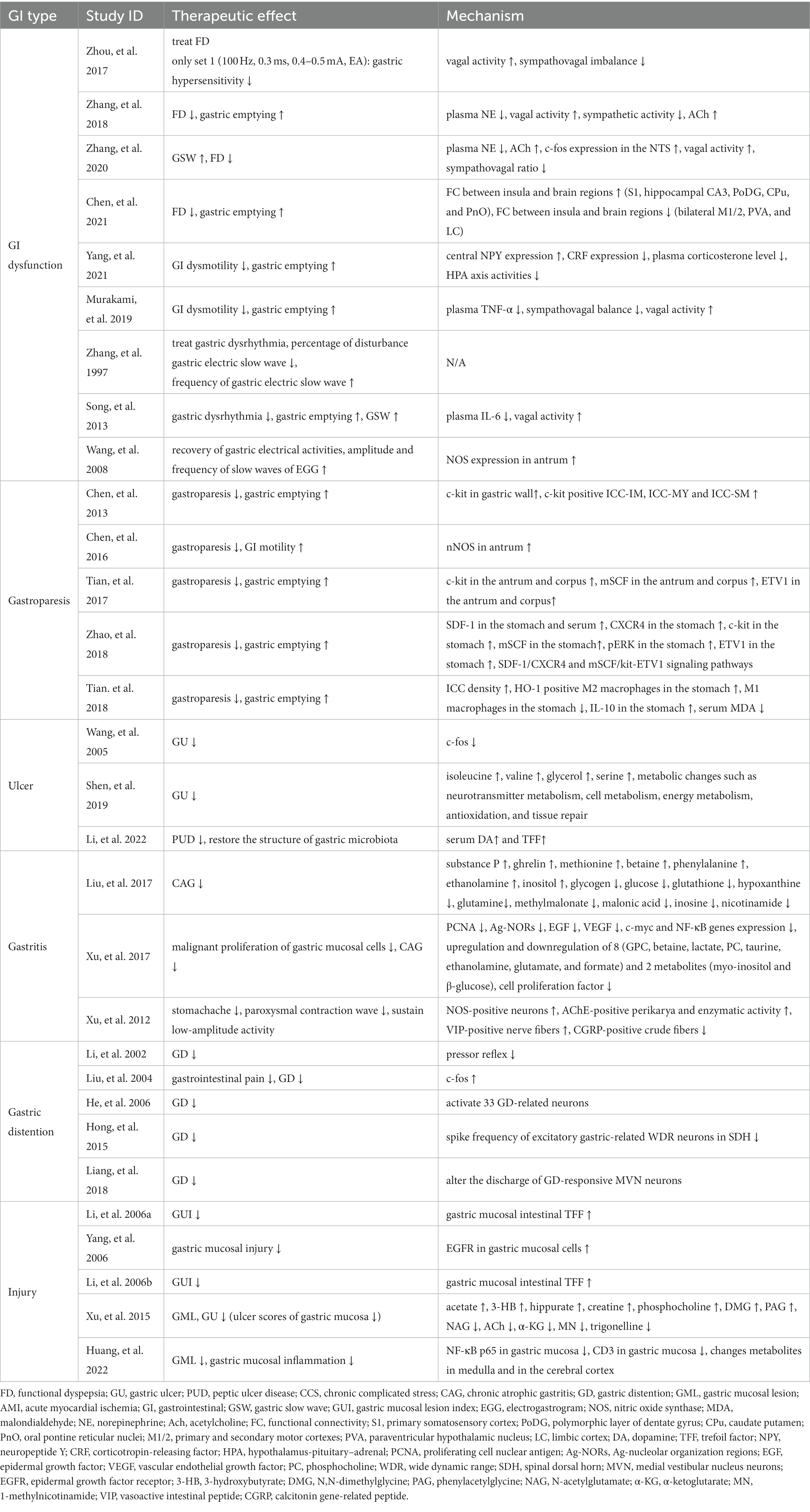
Table 2. Therapeutic effects and underlying mechanisms of the studies.
FD is a chronic dysfunction of the GI tract that is difficult to treat and affects social function and quality of life (17). Its main symptoms include epigastric pain, epigastric burns, postprandial fullness, and initial satiety without any specific cause (12). EA upregulates the vagal nerve and downregulates sympathetic activity, thereby reducing gastric hypersensitivity (13). Moreover, EA lowers plasma norepinephrine (NE) and acetylcholine (ACh), resulting in the activation of vagal activity and lower sympathetic activity (15, 16). EA also increases c-Fos expression in the nuclei of the solitary tract (NTS) and increases the gastric slow wave (GSW) (16). In another study, EA changed the functional connectivity (FC) between the insula and brain regions. The regions with increased FC are the primary somatosensory cortex, hippocampal CA3, polymorphic layer of the dentate gyrus, caudate putamen, and oral pontine reticular nuclei, and the regions with decreased FC are the bilateral primary and secondary motor cortices, paraventricular hypothalamic nucleus, and limbic cortex (14).
GI dysmotility is characterized by delayed gastric emptying, which frequently occurs in critically ill patients (18). Patients with GI dysmotility exhibit various non-discriminatory symptoms including vomiting, abdominal pain, abdominal distention, and constipation (21). In chronic complicated stress models, EA increased the expression of central neuropeptide Y (NPY) and gamma-aminobutyric acid (GABA) A receptor and decreased central corticotropin-releasing factor (CRF), plasma corticosterone levels, and hypothalamus–pituitary–adrenal (HPA) axis activity. NPY inhibits CRF, which inhibits gastric motility through GABA receptors, reduces HPA axis activity, and further reduces plasma corticosterone levels to restore gastric motility (22).
Postoperative ileus is a widely known complication characterized by transient impairment of GI function after abdominal surgery (19). In a postoperative ileus model, EA increased gastric emptying and treated GI dysmotility by decreasing the sympathetic balance and increasing vagus nerve activity. EA also increases the release of ACh, which inhibits the release of TNF-α, an inflammatory factor, through the α7 nicotinic acetylcholine receptor (20).
Gastric dysrhythmia represents the underlying cause of nausea; thus, it is an objective biomarker of nausea (26). In a gastric dysrhythmia model, MA decreased the percentage of disturbance of gastric electric slow waves, increased the frequency of gastric electric slow waves, and treated gastric dysrhythmia (23). Additionally, EA lowered plasma IL-6 levels, activated vagal activity, and increased gastric emptying and GSW in a gastric dysrhythmia model (25).
The electrical activity of the stomach serves as the basis for GI motility, which functions in digestion, absorption, excretion, and protection (27). In an acute myocardial ischemia model, EA restored gastric electrical activity, increased nitric oxide synthase (NOS) expression in the stomach, and increased the amplitude and frequency of slow waves in the electrogastrogram. The generated NO activates guanylate cyclase, and the resulting cyclic guanosine monophosphate can exert biological effects such as increasing gastric electrical activity by regulating myocardial blood pumping (24).
Gastroparesis is characterized by delayed gastric emptying and upper GI symptoms (28). Its symptoms include nausea, vomiting, early satiety, postprandial fullness, bloating, and upper abdominal pain (29). All studies on gastroparesis used a diabetes mellitus-induced gastroparesis model (31–33). Interestingly, the c-Kit signaling pathway is a common therapeutic mechanism in acupuncture treatment for gastroparesis. EA also expressed the tyrosine kinase receptor c-Kit in the gastric wall and restored the ICC network, leading to decreased inflammation (31). Moreover, EA increases the expression of nNOS in the antrum, thus increasing GI motility (30). In another diabetes mellitus model, EA increased the c-Kit, mSCF, and ETV1 expressions in the antrum and corpus, thereby accelerating gastric emptying (34). Moreover, EA increased the SDF-1, CXCR4, and pERK expressions in stomach tissues (32). EA increased the density of ICC and the expression of HO-1 positive M2 macrophages and decreased that of M1 macrophages. Additionally, EA increases the IL-10 expression and decreases serum malondialdehyde levels (33).
Gastic ulcer (GU) is defined as any damage to the mucosa of the stomach lining, which extends >5 mm in diameter and pierces through the muscular mucosa (39). Studies on GU have demonstrated that acupuncture induces metabolic changes and increases vagal activity. EA decreased the expression of c-Fos, which serves as a marker for activity in the central nervous system and can be induced by GU. The vagus nerve mediates c-Fos generation in the NTS, parasympathetic afferents promote harmful visceral stimulation, and EA prevents the formation and increase of Fos-Li neuronal products (38). In the other GU model, isoleucine, valine, glycerol, and serine levels increased regardless of the EA treatment time. EA has therapeutic effects on GU by causing several metabolic changes such as neurotransmitter metabolism, cell metabolism, energy metabolism, antioxidation, and tissue repair (37).
PUD is an interruption of the lining of the GI tract lining due to abnormal secretion of gastric acid or pepsin (36). EA restores the structure of the gastric mucosa and muscle layer by increasing the levels of dopamine and trefoil factor (TFF). EA also improves the diversity of gastric microbiota, such as Firmicutes and Bacteroidetes, through which EA exerts therapeutic effects on PUD (35).
CAG is the result of an inflammatory process that ultimately leads to the loss of appropriate mucosal glands (41). In the CAG model, EA repairs the nervous system of the stomach and brain, which may modulate energy metabolism, membrane metabolism, and various neurotransmitters in the nervous system (40). EA also blocked the malignant proliferation of gastric mucosal cells by reducing the expression of epidermal growth factor, vascular endothelial growth factor, proliferating cell nuclear antigen, Ag-nucleolar organization regions, c-Myc, and NF-κB in gastric mucosal cells. Additionally, eight metabolites (glycerophosphocholine, lactate, phosphocholine, myo-inositol, ethanolamine, glutamate, formate, and β-glucose) were increased, and two metabolites (betaine and taurine) were decreased in gastric tissue by EA (42).
Corrosive substances cause severe GI inflammation and acute stomach pain (43). In a model of acute gastritis caused by corrosive substances, EA controlled GI motility by increasing the number of NOS-positive neurons in the GI wall and restoring ACh activity. EA also increased the number of vascular active intestinal peptide-positive nerve fibers and decreased the number of calcitonin gene-related peptide-positive crude fibers, which affect the smooth muscle relaxation function of the GI (44).
Normally, the gastric mechanical expansion signal occurs after meals and induces GD (45). However, in pathological situations, abnormal GD can be caused by air entering the stomach during strong or fast rescue breathing (49). EA reduces the pressor reflex and has a therapeutic effect on GD. This study suggests that EA inhibits GD-induced pressor reflex, which includes interactions between afferent inputs in various regions of the brain (50). In another GD model, EA reduced GI pain by increasing the c-Fos expression in the NTS and demonstrated therapeutic effects in GD (57). Interestingly, acupuncture also regulates the number of neurons associated with GD (47–55). MA exhibited a therapeutic effect on GD by activating 33 of 47 GD-relative neurons (47) and by lowering the spike frequency of excitatory gastric-related wide dynamic range neurons in the spinal dorsal horn, which is increased by acute GD (53). Moreover, MA alters the discharge of GD-responsive medial vestibular nucleus neurons (55).
Gastric mucosal injury or GML is caused by an imbalance in a series of defense mechanisms that protect the mucous membrane from external aggressive factors (51). EA restores gastric mucosal injury by increasing the expression of epidermal growth factor receptors in gastric mucosal cells (48). In another gastric mucosal injury model, EA decreased the GML index and increased intestinal TFF expression in the gastric mucosa, which restored the wound to the GI mucosa (46, 52). In the GML model, the ulcer scores of the gastric mucosa were decreased by EA. EA increases acetate, 3-hydroxybutyrate, hippurate, creatine, phosphocholine, N,N-dimethylglycine, phenylacetylglycine, N-acetylglutamate, and Ach levels. Additionally, EA lowers α-ketoglutarate, 1-methylnicotinamide, and trigonelline levels (56). In another GML model, EA inhibited gastric mucosal inflammation by lowering the NF-κB p65 and CD3 expressions in the gastric mucosa. Furthermore, 13 metabolite concentrations in the medulla and 9 metabolite concentrations in the cerebral cortex changed after EA (54).
Acupuncture, a complementary and alternative therapy (58), has been used to treat various diseases (59). Its use for GI conditions has been demonstrated in previous studies (60). Clinical trials on acupuncture management for GI diseases have been established (61). However, previous studies are insufficient to clarify the acute therapeutic mechanisms of acupuncture in GI diseases. Therefore, in this study, we aimed to determine the underlying mechanism of acupuncture treatment for GI diseases in animal models.
In our report, 30 studies were selected, and we divided the various types of GI diseases of the included studies into six categories of disease groups (GI dysfunction, gastroparesis, ulcers, gastritis, GD, and injury). Moreover, the therapeutic mechanisms of acupuncture that were obtained from our investigation were changes in gene expression, regulation of anti-inflammatory substances, metabolic changes, increase in vagal activity, change in FC between the insula and brain regions, and control of neuron numbers. Interestingly, studies included in the gastroparesis group identified a common mechanism of the c-Kit signaling pathway. Meanwhile, some mechanisms, such as downregulation of plasma NE and upregulation of ETV1 expression, were duplicated in different studies. Although the present review has a deficiency in representing the entire therapeutic mechanism of acupuncture treatment for GI diseases, this study is meaningful because it provides an overview of the underlying mechanism of acupuncture therapy on stomach-related problems and gastric disease for the first time.
Among the 30 studies, 26 used EA and 4 used MA. The methods of acupuncture treatment differs between MA and EA. EA methods include frequency, waveform, time, and current intensity, whereas MA methods include twisting, twirling, thrusting, lifting, or a combination of these motions (61). Therefore, the efficacy of acupuncture also changes depending on the method used (62). In our study, one study has reported the opposite outcome of the mechanism of different acupuncture methods depending on the frequency of acupuncture stimulation. The spike frequency was further excited by 0.5 Hz and 1 Hz MA stimulation. Meanwhile, the activity significantly decreased for 2 Hz and 3 Hz MA stimulation (53). By contrast, another study has revealed an equivalent mechanism, although the acupoints and acupuncture stimulation were different (20). According to our study results, biological mechanisms exist or change under different conditions of disease and acupuncture methods. Further studies are needed to clarify the therapeutic mechanism of acupuncture in GI diseases and to reveal the difference in the mechanism depending on the acupuncture method.
This study aimed to investigate the effect of acupuncture treatment on stomach-related problems and to clarify the therapeutic mechanism. Although our study is limited by the study size and diverse types of GI diseases, our findings are significant because various mechanisms of the effects of acupuncture on GI diseases have been demonstrated. Further research on the role of acupuncture in GI diseases with a larger sample size is necessary. Therefore, our study results will help reveal the potential mechanisms and therapeutic effects of acupuncture for GI diseases in the future. We expect that our report will contribute to further experimental and clinical trials of acupuncture treatment for stomach-related diseases and digestive system disorders.
M-JK and SL wrote the paper. M-JK investigated the study. S-NK designed and supervised the study. SL and S-NK analyzed the data and revised the paper. All authors contributed to the article and approved the submitted version.
This work was supported by the National Research Foundation of Korea funded by the Korean government (MSIT; NRF-2020R1C1C1004107) and from the Ministry of Health and Welfare through the Korea Health Industry Development Institute (KHIDI; grant no. HF21C0018).
The authors declare that the research was conducted in the absence of any commercial or financial relationships that could be construed as a potential conflict of interest.
All claims expressed in this article are solely those of the authors and do not necessarily represent those of their affiliated organizations, or those of the publisher, the editors and the reviewers. Any product that may be evaluated in this article, or claim that may be made by its manufacturer, is not guaranteed or endorsed by the publisher.
1. Zhang, M, and Yang, XJ. Effects of a high fat diet on intestinal microbiota and gastrointestinal diseases. World J Gastroenterol. (2016) 22:8905–9. doi: 10.3748/wjg.v22.i40.8905
2. Russo, M, Miraglia, C, Nouvenne, A, Leandro, G, Meschi, T, De’Angelis, GL, et al. Approach to gastroenterological diseases in primary care. Acta Biomed. (2018) 89:5–11. doi: 10.23750/abm.v89i8-S.7973
3. Jones, RH. Clinical economics review: gastrointestinal disease in primary care. Aliment Pharmacol Ther. (1996) 10:233–9. doi: 10.1111/j.0953-0673.1996.00233.x
4. Jones, R. Primary care research and clinical practice: gastroenterology. Postgrad Med J. (2008) 84:454–8. doi: 10.1136/pgmj.2008.068361
5. Ifrim Chen, F, Antochi, AD, and Barbilian, AG. Acupuncture and the retrospect of its modern research. Romanian J Morphol Embryol. (2019) 60:411–8.
6. Zhu, J, Li, J, Yang, L, and Liu, S. Acupuncture, from the ancient to the current. Anat Rec (Hoboken). (2021) 304:2365–71. doi: 10.1002/ar.24625
7. Takahashi, T. Acupuncture for functional gastrointestinal disorders. J Gastroenterol. (2006) 41:408–17. doi: 10.1007/s00535-006-1773-6
8. Xuefen, W, Ping, L, Li, L, Xiaoli, C, and Yue, Z. A clinical randomized controlled trial of acupuncture treatment of gastroparesis using different acupoints. Pain Res Manag. (2020) 2020:1–14. doi: 10.1155/2020/8751958
9. Ouyang, H, and Chen, JD. Review article: therapeutic roles of acupuncture in functional gastrointestinal disorders. Aliment Pharmacol Ther. (2004) 20:831–41. doi: 10.1111/j.1365-2036.2004.02196.x
10. Li, H, He, T, Xu, Q, Li, Z, Liu, Y, Li, F, et al. Acupuncture and regulation of gastrointestinal function. World J Gastroenterol. (2015) 21:8304–13. doi: 10.3748/wjg.v21.i27.8304
11. Hooijmans, CR, Rovers, MM, de Vries, RB, Leenaars, M, Ritskes-Hoitinga, M, and Langendam, MW. SYRCLE's risk of bias tool for animal studies. BMC Med Res Methodol. (2014) 14:43. doi: 10.1186/1471-2288-14-43
12. Ford, AC, Mahadeva, S, Carbone, MF, Lacy, BE, and Talley, NJ. Functional dyspepsia. Lancet. (2020) 396:1689–702. doi: 10.1016/S0140-6736(20)30469-4
13. Talley, NJ. Functional dyspepsia: advances in diagnosis and therapy. Gut Liver. (2017) 11:349–57. doi: 10.5009/gnl16055
14. Zhou, J, Li, S, Wang, Y, Lei, Y, Foreman, RD, Yin, J, et al. Effects and mechanisms of auricular electroacupuncture on gastric hypersensitivity in a rodent model of functional dyspepsia. PLoS One. (2017) 12:e0174568. doi: 10.1371/journal.pone.0174568
15. Zhang, S, Li, S, Liu, Y, Ye, F, Yin, J, Foreman, RD, et al. Electroacupuncture via chronically implanted electrodes improves gastric dysmotility mediated by autonomic-cholinergic mechanisms in a rodent model of functional dyspepsia. Neurogastroenterol Motil. (2018) 30:e13381. doi: 10.1111/nmo.13381
16. Zhang, S, Liu, Y, Li, S, Ye, F, Foreman, RD, and Chen, JDZ. Effects of electroacupuncture on stress-induced gastric dysrhythmia and mechanisms involving autonomic and central nervous systems in functional dyspepsia. Am J Physiol Regul Integr Comp Physiol. (2020) 319:R106–13. doi: 10.1152/ajpregu.00256.2019
17. Chen, Y, Zhao, Y, Tan, RY, Zhang, PY, Long, T, Shi, Y, et al. The influence of stomach back-shu and front-mu points on insular functional connectivity in functional dyspepsia rat models. Evid Based Complement Alternat Med. (2021) 2021:1–14. doi: 10.1155/2021/2771094
18. Ladopoulos, T, Giannaki, M, Alexopoulou, C, Proklou, A, Pediaditis, E, and Kondili, E. Gastrointestinal dysmotility in critically ill patients. Ann Gastroenterol. (2018) 31:273–81. doi: 10.20524/aog.2018.0250
19. Vasant, DH, and Lal, S. Recent advances in the management of severe gastrointestinal dysmotility. Clin Exp Gastroenterol. (2021) 14:163–72. doi: 10.2147/CEG.S249877
20. Yang, Y, Yu, H, Babygirija, R, Shi, B, Sun, W, Zheng, X, et al. Electro-acupuncture attenuates chronic stress responses via up-regulated central NPY and GABA (a) receptors in rats. Front Neurosci. (2020) 14:629003. doi: 10.3389/fnins.2020.629003
21. Mazzotta, E, Villalobos-Hernandez, EC, Fiorda-Diaz, J, Harzman, A, and Christofi, FL. Postoperative ileus and postoperative gastrointestinal tract dysfunction: pathogenic mechanisms and novel treatment strategies beyond colorectal enhanced recovery after surgery protocols. Front Pharmacol. (2020) 11:583422. doi: 10.3389/fphar.2020.583422
22. Murakami, H, Li, S, Foreman, R, Yin, J, Hirai, T, and Chen, JDZ. Ameliorating effects of electroacupuncture on dysmotility, inflammation, and pain mediated via the autonomic mechanism in a rat model of postoperative ileus. J Neurogastroenterol Motil. (2019) 25:286–99. doi: 10.5056/jnm18094
23. Koch, KL. Gastric dysrhythmias: a potential objective measure of nausea. Exp Brain Res. (2014) 232:2553–61. doi: 10.1007/s00221-014-4007-9
24. Zhang, AL, Chen, RX, Kang, MF, Fan, HL, and Wang, WL. Study of the regulatory effect of acupuncture on rotation-induced gastric dysrhythmia in rabbits. World J Gastroenterol. (1997) 3:54–5. doi: 10.3748/wjg.v3.i1.54
25. Song, J, Yin, J, Sallam, HS, Bai, T, Chen, Y, and Chen, JD. Electroacupuncture improves burn-induced impairment in gastric motility mediated via the vagal mechanism in rats. Neurogastroenterol Motil. (2013) 25:807–e635. doi: 10.1111/nmo.12183
26. Cheng, LK, O’Grady, G, Du, P, Egbuji, JU, Windsor, JA, and Pullan, AJ. Gastrointestinal system. Wiley Interdiscip Rev Syst Biol Med. (2010) 2:65–79. doi: 10.1002/wsbm.19
27. Wang, SB, Chen, SP, Gao, YH, Luo, MF, and Liu, JL. Effects of electroacupuncture on cardiac and gastric activities in acute myocardial ischemia rats. World J Gastroenterol. (2008) 14:6496–502. doi: 10.3748/wjg.14.6496
28. Bharucha, AE, Kudva, YC, and Prichard, DO. Diabetic gastroparesis. Endocr Rev. (2019) 40:1318–52. doi: 10.1210/er.2018-00161
29. Camilleri, M, Parkman, HP, Shafi, MA, Abell, TL, and Gerson, L. Clinical guideline: management of gastroparesis. Am J Gastroenterol. (2013) 108:18–37. doi: 10.1038/ajg.2012.373
30. Chen, Y, Xu, JJ, Liu, S, and Hou, XH. Electroacupuncture at ST36 ameliorates gastric emptying and rescues networks of interstitial cells of Cajal in the stomach of diabetic rats. PLoS One. (2013) 8:e83904. doi: 10.1371/journal.pone.0083904
31. Chen, H, Zhu, W, Lu, J, Fan, J, Sun, L, Feng, X, et al. The effects of auricular electro-acupuncture on ameliorating the dysfunction of interstitial cells of Cajal networks and nNOSmRNA expression in antrum of STZ-induced diabetic rats. PLoS One. (2016) 11:e0166638. doi: 10.1371/journal.pone.0166638
32. Tian, L, Zhu, B, and Liu, S. Electroacupuncture at ST36 protects ICC networks via mSCF/kit-ETV1 signaling in the stomach of diabetic mice. Evid Based Complement Alternat Med. (2017) 2017:1–13. doi: 10.1155/2017/3980870
33. Zhao, J, An, J, and Liu, S. Electroacupuncture at ST36 increases bone marrow-derived interstitial cells of Cajal via the SDF-1/CXCR4 and mSCF/kit-ETV1 pathways in the stomach of diabetic mice. Evid Based Complement Alternat Med. (2018) 2018:1–14. doi: 10.1155/2018/7878053
34. Tian, L, Song, S, Zhu, B, and Liu, S. Electroacupuncture at ST-36 protects interstitial cells of Cajal via sustaining heme oxygenase-1 positive M2 macrophages in the stomach of diabetic mice. Oxidative Med Cell Longev. (2018) 2018:1–9. doi: 10.1155/2018/3987134
35. Woolf, A, Rehman, RB, and Rose, R. Gastric ulcer In:. StatPearls. Treasure Island (FL): StatPearls Publishing LLC (2022)
36. Wang, H, Wang, CY, Zhang, JS, Sun, L, Sun, JP, Tian, QH, et al. Acupuncture therapy for experimental stomach ulcer and c-Fos expression in rats. World J Gastroenterol. (2005) 11:5517–20. doi: 10.3748/wjg.v11.i35.5517
37. Shen, JC, Lian, LY, Zhang, Y, He, QD, Chen, JL, Zhang, LB, et al. Dynamic analysis of metabolic response in gastric ulcer (GU) rats with electroacupuncture treatment using (1)H NMR-based metabolomics. Evid Based Complement Alternat Med. (2019) 2019:1–12. doi: 10.1155/2019/1291427
38. Malik, TF, Gnanapandithan, K, and Singh, K. Peptic ulcer disease In:. StatPearls. Treasure Island (FL): StatPearls Publishing LLC (2022)
39. Li, X, He, F, Tuo, X, Qiu, Y, Guo, J, Wu, Y, et al. Electroacupuncture ameliorates peptic ulcer disease in association with gastroduodenal microbiota modulation in mice. Front Cell Infect Microbiol. (2022) 12:935681. doi: 10.3389/fcimb.2022.935681
40. Rodriguez-Castro, KI, Franceschi, M, Noto, A, Miraglia, C, Nouvenne, A, Leandro, G, et al. Clinical manifestations of chronic atrophic gastritis. Acta Biomed. (2018) 89:88–92. doi: 10.23750/abm.v89i8-S.7921
41. Liu, CC, Chen, JL, Chang, XR, He, QD, Shen, JC, Lian, LY, et al. Comparative metabolomics study on therapeutic mechanism of electro-acupuncture and moxibustion on rats with chronic atrophic gastritis (CAG). Sci Rep. (2017) 7:14362. doi: 10.1038/s41598-017-13195-5
42. Xu, J, Zheng, X, Cheng, KK, Chang, X, Shen, G, Liu, M, et al. NMR-based metabolomics reveals alterations of electro-acupuncture stimulations on chronic atrophic gastritis rats. Sci Rep. (2017) 7:45580. doi: 10.1038/srep45580
43. Mueller, PD, and Benowitz, NL. Toxicologic causes of acute abdominal disorders. Emerg Med Clin North Am. (1989) 7:667–82. doi: 10.1016/S0733-8627(20)30760-4
44. Xu, X, Li, Q, Zhou, L, and Ru, L. Neurochemical mechanism of the gastrointestinal interdigestive migrating motor complex in rats with acute inflammatory stomach ache. Neural Regen Res. (2012) 7:2136–43. doi: 10.3969/j.issn.1673-5374.2012.27.008
45. Hajishafiee, M, Bitarafan, V, and Feinle-Bisset, C. Gastrointestinal sensing of meal-related signals in humans, and dysregulations in eating-related disorders. Nutrients. (2019) 11:1298. doi: 10.3390/nu11061298
46. Spoormans, I, Van Hoorenbeeck, K, Balliu, L, and Jorens, PG. Gastric perforation after cardiopulmonary resuscitation: review of the literature. Resuscitation. (2010) 81:272–80. doi: 10.1016/j.resuscitation.2009.11.023
47. Li, P, Rowshan, K, Crisostomo, M, Tjen, ALSC, and Longhurst, JC. Effect of electroacupuncture on pressor reflex during gastric distension. Am J Physiol Regul Integr Comp Physiol. (2002) 283:R1335–45. doi: 10.1152/ajpregu.00192.2002
48. Liu, JH, Li, J, Yan, J, Chang, XR, Cui, RF, He, JF, et al. Expression of c-fos in the nucleus of the solitary tract following electroacupuncture at facial acupoints and gastric distension in rats. Neurosci Lett. (2004) 366:215–9. doi: 10.1016/j.neulet.2004.05.068
49. He, J, Yan, J, Chang, X, Liu, J, Li, J, Yi, S, et al. Neurons in the NTS of rat response to gastric distention stimulation and acupuncture at body surface points. Am J Chin Med. (2006) 34:427–33. doi: 10.1142/S0192415X06003965
50. Hong, S, Ding, S, Wu, F, Xi, Q, Li, Q, Liu, Y, et al. Strong manual acupuncture manipulation could better inhibit spike frequency of the dorsal horn neurons in rats with acute visceral nociception. Evid Based Complement Alternat Med. (2015) 2015:675437:1–9. doi: 10.1155/2015/675437
51. Liang, C, Wang, Y, Xu, B, and Yu, Z. Effect of acupuncture at three different acupoints on electrical activity of gastric distention-affected neurons in rat medial vestibular nucleus. J Tradit Chin Med. (2018) 38:125–31. doi: 10.1016/j.jtcm.2018.02.002
52. Suzuki, H, Miura, S, Suzuki, M, Terada, S, Nakamura, M, and Tsuchiya, M. Gastric mucosal injury: microcirculation and Helicobacter pylori. Keio J Med. (1994) 43:1–8. doi: 10.2302/kjm.43.1
53. Li, XP, Yan, J, Yi, SX, Chang, XR, Lin, YP, Huang, A, et al. The effect of EA on the gastric mucosal histology and ITF mRNA expression in stress-induced rats. Am J Chin Med. (2006) 34:1005–14. doi: 10.1142/S0192415X06004478
54. Yang, ZB, Yan, J, Zou, XP, Yi, SX, Chang, XR, Lin, YP, et al. Enhanced expression of epidermal growth factor receptor gene in gastric mucosal cells by the serum derived from rats treated with electroacupuncture at stomach meridian acupoints. World J Gastroenterol. (2006) 12:5557–61. doi: 10.3748/wjg.v12.i34.5557
55. Li, XP, Yan, J, Yi, SX, Chang, XR, Lin, YP, Yang, ZB, et al. Effect of electroacupunture on gastric mucosal intestinal trefoil factor gene expression of stress-induced gastric mucosal injury in rats. World J Gastroenterol. (2006) 12:1962–5. doi: 10.3748/wjg.v12.i12.1962
56. Xu, J, Cheng, KK, Yang, Z, Wang, C, Shen, G, Wang, Y, et al. (1) H NMR metabolic profiling of biofluids from rats with gastric mucosal lesion and electroacupuncture treatment. Evid Based Complement Alternat Med. (2015) 2015:801691:1–11. doi: 10.1155/2015/801691
57. Huang, M, Peng, Y, He, Q, Lian, L, Wang, Y, Zhang, L, et al. NMR-based metabonomics reveals the dynamic effect of electro-acupuncture on central nervous system in gastric mucosal lesions (GML) rats. Chin Med. (2022) 17:37. doi: 10.1186/s13020-022-00593-9
58. Dossett, ML, Davis, RB, Lembo, AJ, and Yeh, GY. Complementary and alternative medicine use by US adults with gastrointestinal conditions: results from the 2012 National Health Interview Survey. Am J Gastroenterol. (2014) 109:1705–11. doi: 10.1038/ajg.2014.108
59. Huang, CY, Lai, WY, Sun, MF, Lin, CC, Chen, BC, Lin, HJ, et al. Prescription patterns of traditional Chinese medicine for peptic ulcer disease in Taiwan: a nationwide population-based study. J Ethnopharmacol. (2015) 176:311–20. doi: 10.1016/j.jep.2015.11.002
60. Li, X, Liu, S, Liu, H, and Zhu, JJ. Acupuncture for gastrointestinal diseases. Anat Rec (Hoboken). (2022) 1–9. doi: 10.1002/ar.24871
61. Wang, H, Jiang, H, Zhao, J, Liu, X, Li, T, Chai, J, et al. Acupuncture therapy for gastric ulcer: a protocol for systematic review and meta-analysis. Medicine (Baltimore). (2021) 100:e27656. doi: 10.1097/MD.0000000000027656
Keywords: gastrointestinal diseases, stomach, acupuncture, animal studies, literature review
Citation: Kim M-J, Lee S and Kim S-N (2023) Effects of acupuncture on gastrointestinal diseases and its underlying mechanism: a literature review of animal studies. Front. Med. 10:1167356. doi: 10.3389/fmed.2023.1167356
Edited by:
Angel Lanas, University of Zaragoza, SpainReviewed by:
Jacqueline Nelisis Zanoni, State University of Maringá, BrazilCopyright © 2023 Kim, Lee and Kim. This is an open-access article distributed under the terms of the Creative Commons Attribution License (CC BY). The use, distribution or reproduction in other forums is permitted, provided the original author(s) and the copyright owner(s) are credited and that the original publication in this journal is cited, in accordance with accepted academic practice. No use, distribution or reproduction is permitted which does not comply with these terms.
*Correspondence: Seung-Nam Kim, c25raW1AZG9uZ2d1ay5lZHU=
†These authors have contributed equally to this work
Disclaimer: All claims expressed in this article are solely those of the authors and do not necessarily represent those of their affiliated organizations, or those of the publisher, the editors and the reviewers. Any product that may be evaluated in this article or claim that may be made by its manufacturer is not guaranteed or endorsed by the publisher.
Research integrity at Frontiers
Learn more about the work of our research integrity team to safeguard the quality of each article we publish.