 Ying Liang†
 Yi-Cong Pan†
 Hui-Ye Shu†Xue-Mei ChouQian-Min GeLi-Juan ZhangQiu-Yu LiRong-Bing LiangHan-Lin Li*
 Yi Shao*
- Department of Ophthalmology, The First Affiliated Hospital of Nanchang University, Jiangxi Province Ophthalmology Institute and Ocular Disease Clinical Research Centre, Nanchang, China
A Corrigendum on
Characteristics of the Fractional Amplitude of Low-Frequency Fluctuation in Ocular Hypertension Patients: A Resting-State fMRI Study
by Liang, Y., Pan, Y.-C., Shu, H.-Y., Chou, X.-M., Ge, Q.-M., Zhang, L.-J., Li, Q.-Y., Liang, R.-B., Li, H.-L., and Shao, Y. (2022). Front. Med. 8:687420. doi: 10.3389/fmed.2021.687420
In the original article, there was a mistake in Table 2. Demographics and behavioral results of OH and HCs groups as published. The age of HC was incorrectly listed as 50.36 ± 54.22. The correct age of HC is 50.36 ± 5.42. The corrected Table 2 appears below.
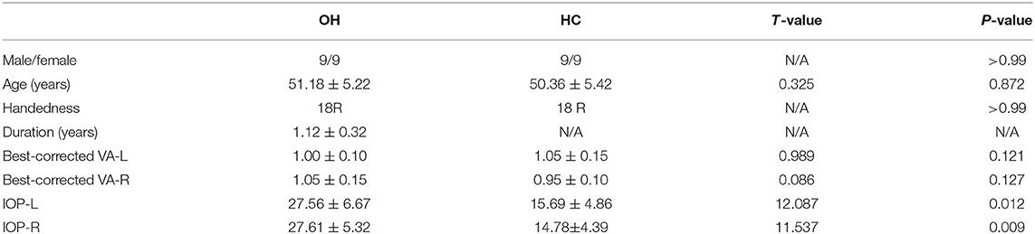
Table 2. Demographics and behavioral results of OH and HCs groups.
In the original article, there was an error in the section Materials and Methods, sub-section Subjects. The subject characteristics: “18 HC patients (8 males and 8 females; mean age 50.36 ± 54.22 years; age range 20–60 years)” is incorrect. The correct sentence is: “18 HCs (9 males and 9 females; mean age 50.36 ± 5.42 years; age range 20–60 years)”.
In the original article, there was an error in the section Results, sub-section The Use of ROC. The sentence: “To test whether the ROC curve has potential as a diagnostic marker for OH, ROC curves were constructed using mean fALFF values of the two brain regions in the OH and HC groups. An area under the curve of 0.5–0.7 is considered to indicate low accuracy, 0.7–0.9 higher accuracy, and an area higher than 0.9 represents good accuracy. Accuracy was low in the left anterior cingulate gyrus (LAC) (0.9892; P < 0.0001; Figure 4A) and higher in the left anterior cuneiform (LP) > HC (0.8834; P < 0.0001; Figure 4B).” is incorrect. The sentence should be: “To test whether the fALFF value has potential as a diagnostic marker for OH, ROC curves were constructed using mean fALFF values of the two brain regions in the OH and HC groups. An area under the curve of 0.5–0.7 is considered to indicate low accuracy, 0.7–0.9 higher accuracy, and an area higher than 0.9 represents good accuracy. Accuracy was high in the left anterior cingulate gyrus (LAC) (0.9892; P < 0.0001; Figure 4A) and lower in the left precuneus (LP) (0.8834; P < 0.0001; Figure 4B).”
In the original article, there was an error in section Results, sub-section The fALFF Value. The values in the sentence: “The fALFF value of LP in the OH group was higher than in the HC group (t = 5.5535), while the fALFF value of LAC in the OH group was decreased (t = −3.7520; Figure 5)” are incorrect. They should be: “The fALFF value of LP in the OH group was higher than in the HC group (t = −3.7520); while the fALFF value of LAC in the OH group was decreased (t = 5.5535) (Figure 5).”
The authors apologize for these errors and state that they do not change the scientific conclusions of the article in any way. The original article has been updated.
Publisher's Note
All claims expressed in this article are solely those of the authors and do not necessarily represent those of their affiliated organizations, or those of the publisher, the editors and the reviewers. Any product that may be evaluated in this article, or claim that may be made by its manufacturer, is not guaranteed or endorsed by the publisher.
Keywords: ocular hypertension, resting state functional magnetic resonance imaging, fractional amplitude of low-frequency fluctuation, left anterior cingulate cortex, left precuneus
Citation: Liang Y, Pan Y-C, Shu H-Y, Chou X-M, Ge Q-M, Zhang L-J, Li Q-Y, Liang R-B, Li H-L and Shao Y (2022) Corrigendum: Characteristics of the Fractional Amplitude of Low-Frequency Fluctuation in Ocular Hypertension Patients: A Resting-State fMRI Study. Front. Med. 9:958937. doi: 10.3389/fmed.2022.958937
Received: 01 June 2022; Accepted: 13 June 2022;
Published: 29 June 2022.
Edited and reviewed by: Jodhbir Mehta, Singapore National Eye Center, Singapore
Copyright © 2022 Liang, Pan, Shu, Chou, Ge, Zhang, Li, Liang, Li and Shao. This is an open-access article distributed under the terms of the Creative Commons Attribution License (CC BY). The use, distribution or reproduction in other forums is permitted, provided the original author(s) and the copyright owner(s) are credited and that the original publication in this journal is cited, in accordance with accepted academic practice. No use, distribution or reproduction is permitted which does not comply with these terms.
*Correspondence: Han-Lin Li, MTMxNzc4MjAxMzNAMTYzLmNvbQ==; Yi Shao, ZnJlZWJlZTk5QDE2My5jb20=
†These authors share first authorship