- 1Department of Neurology, Renmin Hospital of Wuhan University, Wuhan, China
- 2School of Nursing, Tongji Medical College, Huazhong University of Science and Technology, Wuhan, China
Objective: To describe the clinical manifestations and outcomes of COVID-19, and explore the risk factors of deterioration and death of the disease.
Methods: In this retrospective study, we collected data from 121 COVID-19 cases confirmed by RT-PCR and next-generation sequencing in Renmin Hospital of Wuhan University from January 30, 2019, to March 23, 2020, and conducted statistical analysis.
Results: A total of 121 patients were included in our study, the median age was 65 years (IQR, 55.0–71.5 years), and 54.5% cases were men. Among those cases, 52 (43.0%) cases progressed to severe, and 14 (11.6%) died. Overall, the most common manifestations were fever (78.5%) and respiratory symptoms (77.7%), while neurological symptoms were found in only 9.9% of the patients. 70.2% of all the cases had comorbidities, including hypertension (40.5%) and diabetes (20.7%). On admission, cases usually show elevated levels of neutrophils (27.3%), D-dimer (72.6%), Interleukin-6 (35.2%), Interleukin-10 (64.4%), high-sensitivity C-reactive protein (82.6%), and lactate dehydrogenase (62.0%), and decreased levels of lymphocytes (66.9%), CD3 cells (67.2%), and CD4 cells (63.0%). The proportional hazard Cox models showed that the risk factors for severity progression and death included comorbidities (HR: 4.53, 95% CI: 1.78–11.55 and HR: 7.81, 95% CI: 1.02–59.86), leukocytosis (HR: 1.13; 95% CI: 1.05–1.22 and HR: 1.25, 95% CI: 1.10–1.42), neutrophilia (HR: 1.15, 95% CI: 1.07–1.13 and HR: 1.28, 95% CI: 1.13–1.46, and elevated LDH (HR: 1.14, 95% CI: 1.12–1.15 and HR: 1.11, 95% CI: 1.10–1.12). Elevated D-dimer (HR: 1.02, 95% CI: 1.01–1.03), IL-6 (HR: 1.01, 95% CI: 1.00–1.02) and IL-10 levels (HR: 1.04, 95% CI: 1.01–1.07) were also risk factors for the progression of disease severity. Meanwhile, lymphopenia and wake immune responses [e.g., lower CD3, CD4, or CD19 counts (all HR < 1)] were associated with disease deterioration and death.
Conclusions: Severe cases and death of COVID-19 are associated with older age, comorbidities, organ dysfunction, lymphopenia, high cytokines, and weak immune responses.
Introduction
Coronavirus disease 2019 (COVID-19) was first reported in Wuhan, China, and is now spreading in more than 37 countries, including the United States, Japan, South Korea, Australia, and France (1). The pathogen of COVID-19 was identified to be severe acute respiratory syndrome coronavirus 2 (SARS-CoV-2), which is closely related to SARS-like coronavirus (2). In the phylogenetic tree, SARS-CoV-2 was identified as an β-coronavirus 2b lineage. By examining the full-length genome of SARS-CoV-2, it was found that this novel virus shares 87.99% homologous sequences with SARS-like coronavirus (3). Early research has shown that SARS-CoV-2, like SARS-CoV, can infect humans cells expressing angiotensin-converting enzyme 2 (ACE2) (4). Based on the information up to date, SARS-CoV-2 is believed to be the third zoonotic human coronavirus in this century (4). Early reports from Wuhan found that the COVID-19 was SARS-like atypical pneumonia, and can be transmitted from person to person, mainly through respiratory droplets, but also through contact with eye mucosa. Although the virus has been detected in feces, the transmission through the digestive tract remains to be further confirmed. The incubation period is generally 2–14 days. Mild cases are characterized by fever, muscle pain, fatigue, and dry cough. A few patients had neurological and digestive symptoms. In severe cases, dyspnea can occur after a week. Ultimately, the severe patients develop acute respiratory distress syndrome, septic shock, and metabolic acidosis that are difficult to manage (5–7). According to the early epidemiological studies, the basic reproduction number (R0) of SARS-CoV-2 was estimated to be about 2.2 and even more (ranging from 1.4 to 6.5). The low fatality and high morbidity of COVID-19 make it difficult to prevent the spread of the virus (8–10).
Description of clinical features, laboratory indicators, and radiological characteristics of the disease is crucial for early diagnosis of the disease, quarantine of patients, and identification of close contacts. Our goal is to describe the epidemiological, clinical, and laboratory characteristics of patients diagnosed with COVID-19, to compare the clinical characteristics of severe and non-severe cases, and to describe the potential risk factors for disease deterioration and death.
Methods
Patients Enrollment and Data Collection
Our study included 121 inpatients diagnosed as COVID-19 who was hospitalized in Renmin Hospital of Wuhan University in China. The diagnosis was based on clinical symptoms, computed tomography (CT), real-time RT-PCR and next-generation sequencing. The admission date was from January 16 to March 3, 2020. All the patients involved in this study lived in Wuhan during the COVID-19 outbreak. We collected data including demographic information (age, gender, and address of usual residence), clinical characteristics (including medical history, comorbidities, symptoms, and signs), initial laboratory findings (hematologic, blood biochemicals, coagulation function, infection-related, and immune-related indices), and clinical outcomes (survival and death). This study was approved by the Hospital Ethics Committee of the Renmin Hospital of Wuhan University [WDRY2020-K136].
According to the diagnostic and treatment guideline for SARS-CoV-2 issued by the Chinese National Health Committee (Version 3-7), the severity of COVID-19 patients was defined. Severe cases were designated when the patients had one of the following criteria: (1) respiratory distress with RR ≥30/min; (2) oxygen saturation ≤ 93% at rest; (3) arterial partial oxygen pressure (PaO2)/oxygen absorption concentration (FiO2) ≤ 300 mmHg; (4) respiratory failure that needs mechanical ventilation; (5) shock or with organ failure requiring ICU care.
Procedures
Epidemiological, clinical and outcome data from electronic medical records were reviewed and collected by professionals. Throat swabs samples were collected from all patients and texted by real-time RT-PCR and next-generation sequencing. Laboratory tests on admission included routine blood test [including leucocytes, lymphocytes, eosinophils, hs-CRP, procalcitonin (PCT), serum amyloid A (SAA), and others], coagulation function, serum biochemical tests (including renal and liver function, creatine kinase and LDH), and immunological indicators (including lymphocyte differential counts, inflammatory factors, and SARS-CoV-2-specific antibodies IgM and IgG). These findings are the initial laboratory results of the patient during hospitalization. Thus, these results have not been affected by the treatment.
Statistical Analysis
Continuous variables were described as medians and interquartile range (IQR), and categorical variables were described as frequency rates and percentages. We used the chi-square (χ2) test or the Fisher's exact test to compare categorical data. Mann-Whitney-Wilcoxon test was applied to compare non-normally continuous variables. The proportional hazard Cox models were used to determine HR and 95% CIs between individual factors on progression to severe or death. The sample size varied due to missing data, and missing data were not imputed. The analyses regarding different factors were based on non-missing data. All statistical analyses were performed using SPSS software (V.23.0). Two-tailed P values were considered statistically significant at <0.05.
Results
Demographics and Clinical Characteristics
The demographic and clinical characteristics are shown in Table 1. A total of 121 patients were included in this study. The median age was 65 years (IQR, 55.0–71.5 years) (Figure 1A), of which 54.5% were male. On admission, 69 and 52 patients met the diagnostic criteria for non-severe and severe disease, respectively. The age distribution of these two groups was different (Figure 1C). Of the 121 patients, most (107, 88.4%) survived to discharge, and 14 patients (11.6%) dead (Figure 1B). The most common self-reported symptoms at the time of onset were fever (78.5%) and respiratory symptoms (77.7%), whereas chest distress, fatigue, gastrointestinal symptoms, and neurological symptoms were found in 28.1, 36.4, 23.1, and 9.9% of the patients, respectively. Among the 121 patients, 70.2% had at least one comorbidity, including hypertension, diabetes, cardiovascular and cerebrovascular diseases (excluding hypertension), cancers, and chronic pulmonary diseases. The median age of the severe group was higher than that of the non-severe group (69.5 vs. 60.0, P < 0.001). Additionally, the proportion of subjects with at least one comorbidity is more common in the severe group as compared with the non-severe group (88.5 vs. 56.5%, P < 0.001). Especially, the incidence of hypertension, cardiovascular and cerebrovascular diseases, and chronic pulmonary diseases in severe cases were significantly higher than that in non-severe cases (all P < 0.05). There was no significant difference in demographics and initial clinical characteristics between the survivors and non-survivors.
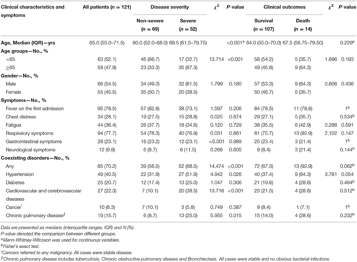
Table 1. Demographic characteristics and initial clinical manifestations of patients with COVID-19 in Wuhan.
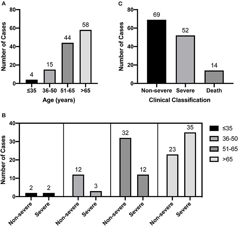
Figure 1. Patient age distribution (A), clinical classification (B), and age distribution of severe and non-serious patients (C).
Laboratory Findings
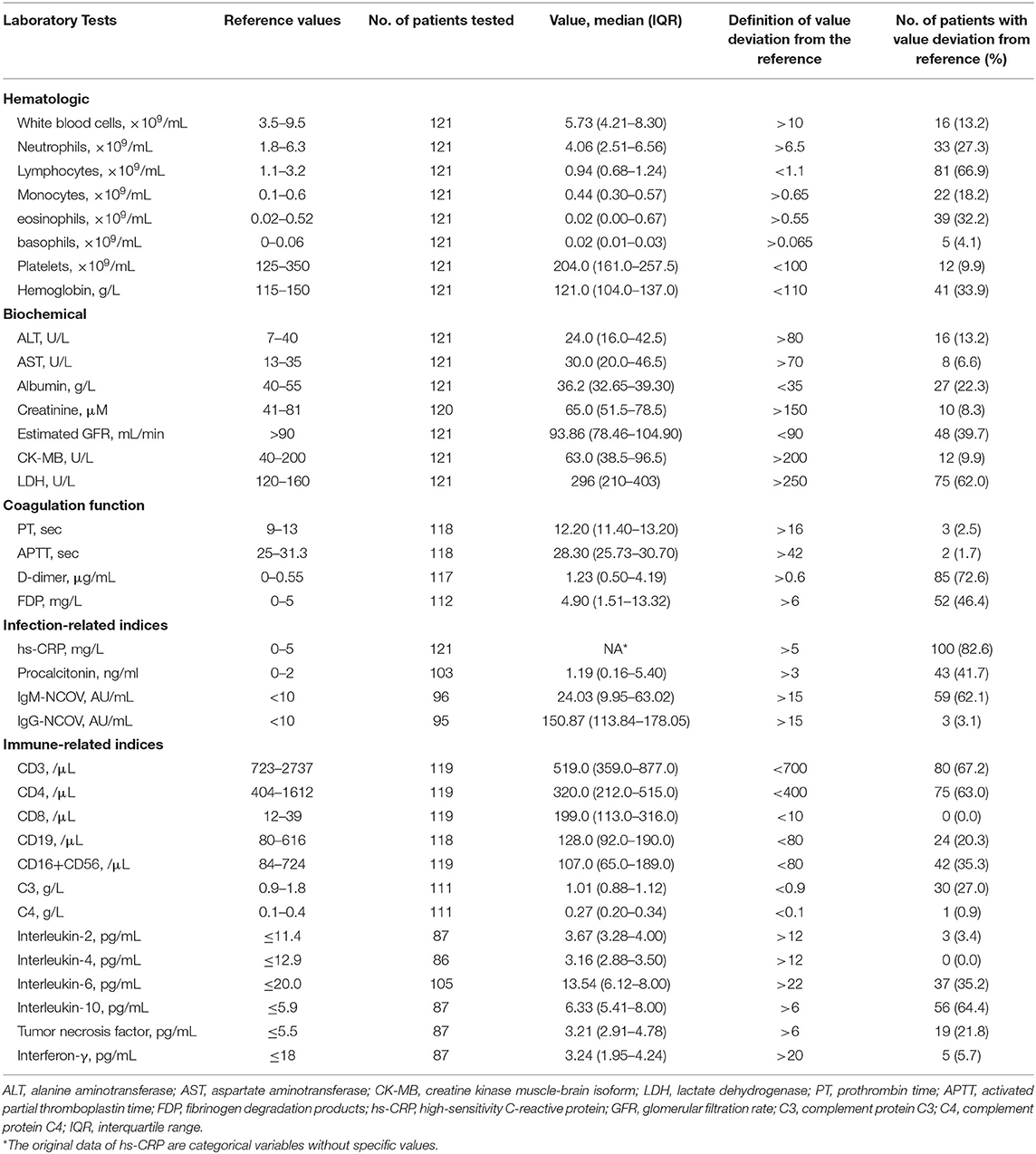
Table 2 shows the initial laboratory findings of 121 patients on admission. On admission, 64.0, 27.3, and 18.2% of all patients had lymphopenia, neutrophilia and eosinophilia, respectively. Monocytosis and basophilia were found in 18.2 and 4.1% of all 121 patients, respectively. Some patients showed impaired liver and renal function, as indicated by increased serum aspartate aminotransferase (AST, 6.6%), alanine aminotransferase (ALT, 13.2%), and serum creatinine (sCr, 8.3%), and decreased estimated glomerular filtration rate (eGFR, 39.7%). Most patients showed increased myocardial injury markers. Among 121 cases, 62.0% had elevated serum lactate dehydrogenase (LDH), while only 4.5% had increased creatine kinase muscle-brain isoform (CK-MB). Most patients showed abnormal inflammatory markers. Of 121 patients, 100 had elevated hs-CRP. Meanwhile, 43 of 103 had elevated PCT, and 59 of 96 patients were positive for IgM antibody against SARS-CoV2. Most patients imply a decline of immune function. One hundred and nineteen patients had decreased CD3 (67.2%) and CD4 (67.2%) cell counts. The cell counts of CD3 and CD4 were closely related to the patients' clinical outcomes. In addition, some patients showed abnormal cytokines: 37 of 105 patients (35.2%) showed increased interleukin-6 (IL-6), 56 of 87 patients (64.4%) showed increased interleukin-10 (IL-10), and 19 of 87 patients (21.8%) showed increased tumor necrosis factor (TNF).
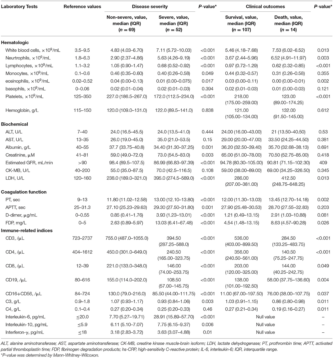
The differences in laboratory findings between non-severe cases and severe cases, as well as survivors and non-survivors are shown in Table 3. Compared with the non-severe group, the severe group showed higher white blood cell (median, 4.83 vs. 7.11, P < 0.001) and neutrophil counts (2.90 vs. 5.63, P < 0.001) (Figure 2A), as well as higher sCr (59.0 vs. 73.0, P < 0.001) and LDH (395.0 vs. 238.0, P < 0.001). As for coagulation function, the severe group showed longer PT and APTT, and higher levels of D-dimer and FDP, as well as lower platelet count (all P < 0.01) as compared with non-severe group. When compared to the non-severe cases, the severe cases showed abnormal immune function, including fewer CD3, CD4, CD8, CD19, CD16+CD56 T-cells (Figure 2B), and complement C3 levels, as well as higher levels of IL-6 and IL-10 (all P < 0.01). Moreover, lower median values of lymphocyte (0.68 vs. 1.05, P < 0.001), albumin (34.40 vs. 37.70, P < 0.001), and eGFR (86.99 vs. 98.4, P < 0.001) were found in severe cases compared to non-severe cases.
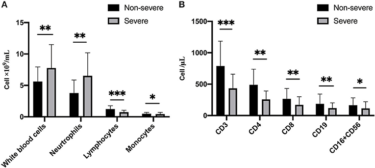
Figure 2. Comparison of immune cells between non-severe and severe patients. (A) The total number and types of white blood cells are different between non-severe and severe patients. (B) The counts of leukocyte classified by differentiation antigen are different between non-severe and severe patients.
Clinical Outcomes
The differences of laboratory indicators between the survivors and non-survivors are shown in Table 3. The non-survivors showed higher leucocyte and neutrophil counts, higher levels of LDH and FDP, longer PT, and fewer counts of lymphocytes, platelets, CD3, CD4, CD8, CD19 T-cells, and lower levels of complement C3 and C4 as compared to the survivors (all P < 0.05).
Table 4 shows the bivariate cox regression of factors associated with disease progression from onset to severe disease or to death. Patients over 65 were at higher risk of developing severe disease than those under 65 (HR, 2.26; 95% CI, 1.23–4.05; P < 0.01). Cases with comorbidities were 4.53 times more likely to progress to severe than cases without comorbidities (HR, 4.53; 95% CI, 1.78–11.55, P < 0.01). In addition, Cox regression models illustrated that comorbidities (HR, 7.81; 95% CI, 1.02–59.86; P < 0.05), leukocytosis (HR, 1.25; 95% CI, 1.10–1.42; P < 0.01), and neutrophilia (HR, 1.28; 95% CI 1.13–1.46; P < 0.001) were risks factors for mortality. Elevated LDH was also a risk factor for the severity deterioration and the development of death (HR, 1.14; 95% CI 1.12–1.15; P < 0.001 and HR, 1.11; 95% CI 1.10–1.12; P < 0.001). Fewer platelets, CD3 or CD4 counts, lower complement C3 level, lymphopenia, and elevated APTT, IL-6 or IL-10 also were related to the progression from oneset to death (all P < 0.05).
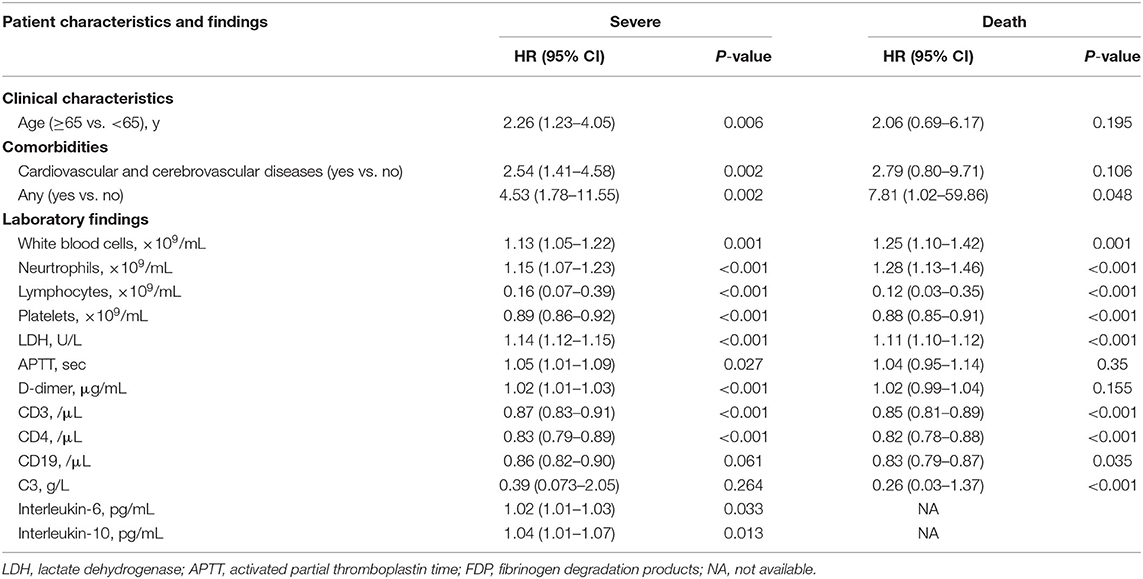
Table 4. Bivariate Cox regression of factors associated with disease progression from onset to severe disease or to death.
Discussion
This retrospective descriptive study included 121 confirmed cases of COVID-19 with a median age of 65. The median age of the severe group was 9.5 years older than that of the non-severe group. The proportion of males in the severe group was as high as 61.5%, compared with 49.3% in the non-severe group. Moreover, according to the bivariate Cox regression analysis, the risk of adverse outcomes in patients with age over 65 years was 2.28 times that of patients under 65 years of age. Fever and respiratory symptoms were the dominant initial symptoms while neurological symptoms and gastrointestinal symptoms are not as common as respiratory symptoms, indicating the difference in viral tropism as compared with influenza. This result is consistent with other reports (11, 12).
In addition, there is no obvious difference in the initial clinical manifestations between non-severe and severe cases, or survivors and non-survivors, suggesting that the progression of the disease cannot be predicted only based on the symptoms on admission. This conclusion was also confirmed by the proportional hazard Cox models (data not shown). In our study, 70.2% of all cases had at least one comorbidity, including hypertension, diabetes, cardiovascular and cerebrovascular diseases, cancers, or chronic pulmonary diseases. Likewise, the adopted Cox models showed that cardiovascular and cerebrovascular diseases were the vital risk factors to predict the severity of the disease.
Analysis of the laboratory indicators shows that the increase of leukocytes, neutrophils, D-dimer, LDH, IL-6, IL-10, TNF, and IFN-γ, and the decrease of lymphocytes, CD3, CD4, and platelets counts were related to disease progression and adverse clinical outcomes. Interestingly, we found eosinophilia in 18.2% of the patients, which seems to be indicator of better outcomes. This observation is different from an early study (13). The eosinophil counts in non-severe cases are significantly higher than that in severe cases. When compare the survivors and non-survivors, the former also show higher eosinophil counts. All cases did not have history of allergic diseases such as asthma. It is conceivable that inflammatory factors released by the eosinophils can trigger cough, sputum discharge, and sneezing, which are beneficial for the patients to expel the virus and fight against viral toxicity. The exact role of eosinophils in COVID-19 remains to be further illustrated.
In our study, leukocytosis was often accompanied by neutropenia and lymphopenia, and this phenomenon was more common in severe cases. Consistently, the median value of neutrophils was higher in the severe group than in the non-severe group, which could be a sign of excessive inflammatory response. According to the bivariate Cox regression analysis, neutropenia was a risk factor for severe cases and adverse clinical outcomes. Lymphocytosis has been reported by many clinical investigators and is also common in severe cases. We also found that CD3, CD4, CD19, and CD16 + CD56 T-cell counts decreased to varying degrees and were more pronounced in severe cases and deaths. Significantly reduced lymphocyte counts in the severe cases indicate impaired immune function in these patients.
The pathological and anatomical examination of patients with COVID-19 also showed that the number of CD4 and CD8 T-cells was greatly reduced (14). Therefore, we speculate that impaired immune function is one of the risk factors for elderly patients with poor clinical outcomes. Although recent studies have emphasized that SARS-CoV-2 can infect immune cells through CD147, thereby affecting the immune function, the cause of immune function impairment in patients with COVID-19 needs to be further studied (15).
Another notable finding was that IL-6, IL-10, TNF, and IFN-γ were generally elevated in severe cases. Studies showed the that inflammatory factor storm was one of the crucial causes of disease progression and adverse clinical outcomes (16–18). Therefore, the prevention of inflammatory factor storm should be considered for patients with severe COVID-19 (19, 20).
Many subjects show increased serum CK-MB and LDH, indicating the SARS-CoV-2 can cause myocardial damage. This coincides with the latest COVID-19 research findings of the Department of Cardiology and Cardiac Ultrasound (21, 22). Furthermore, the bivariate Cox regression analysis emphasized higher LDH as a risk factor for the development of severity disease and the progression of death. It is worth noting that myocardial damage should be treated properly to reduce mortality.
Coagulation dysfunction was another risk factor for death. There are also reports of vascular embolism in patients with COVID-19 (23, 24). In this study, 72.6% and 46.4% of the cases showed high D-dimer and FDP, respectively. Compared with non-severe cases (median, 3.93), the D-dimer of severe cases (median, 0.85) is significantly increased, suggesting that the increase of D-dimer may be one of the risk factors for the deterioration of the disease, and attention should be paid to monitor coagulation function. Longer PT and APTT, with higher D-dimer and FDP in the initial laboratory findings of dead cases may suggest the possibility of disseminated intravascular coagulation (DIC).
The strengths of the present study as follows: The inpatients of this research came from Wuhan, where is the hit-hardest region of China in this epidemic. Understanding the clinical manifestations and prognosis of cases, and analyzing the risk factors of disease progression and death can provide a theoretical basis for clinical diagnosis and intervention. Clinicians, especially critical care physicians, have a deep understanding of the condition of COVID patients upon admission, which is more helpful in controlling the progression of the disease and improving the prognosis. Limitations of this study were that this study conducted in a single hospital with limited sample size. Information and selection bias may influence the result. A larger-scale multicenter study of COVID-19 patients will help to further determine the clinical features and risk factors of this disease.
Conclusion
This study included 121 confirmed COVID-19 cases with a median age of 65. Fever and respiratory symptoms were the most common manifestations. Age and comorbidity were the risk factors for death and disease progression. Myocardial injury markers and renal function also suggest the possibility of deterioration. Abnormal coagulation function suggests the risk of DIC in severe cases. The immune-related laboratory results indicate the presence of inflammatory factor storm and impaired immune function, which are closely related to the adverse clinical outcomes. Thus, these indicators deserved special attention. Early intervention should be considered to treat impaired immune functions to reduce mortality. Most factors that are related to the development of disease deterioration were associated with death, indicating the same pathophysiological mechanisms mediate the progression from hospital admission to severity worsening and from onset to death.
Data Availability Statement
All datasets presented in this study are included in the article/supplementary material.
Ethics Statement
This study was approved by the Hospital Ethics Committee of the Renmin Hospital of Wuhan University [WDRY2020-K136]. Written informed consent for participation was not required for this study in accordance with the national legislation and the institutional requirements.
Author Contributions
LZ had full access to all of the data in the study and take responsibility for the integrity of the data and the accuracy of the data analysis. WF were helped statistical analysis and gave technical support. LD and LZ draft the manuscript. LZ and YZ designed the study. YZ and YG helped in the acquisition of data. ZheZ and ZhaZ were responsible for data interpretation and the critical revision of the manuscript. All authors contributed to the article and approved the submitted version.
Funding
This work was supported by grants from the National Natural Science Foundation of China (Nos. 81822016 and 81771382) to ZheZ.
Conflict of Interest
The authors declare that the research was conducted in the absence of any commercial or financial relationships that could be construed as a potential conflict of interest.
The reviewer XW declared a shared affiliation with one of the reviewers WF to the handling editor at time of review.
Acknowledgments
We thank members of the Medical Team from Qinghai Province and Shanghai City for their efforts in recruiting patients.
References
1. World Health Organization. WHO Coronavirus Disease 2019(COVID-19): Situation Report−39. World Health Organization (2020). Available online at: https://www.who.int/docs/default-source/coronaviruse/situation-reports/20200422-sitrep-93-covid-19.pdf?sfvrsn=35cf80d7_4) (accessed April 22, 2020).
2. Lu R, Zhao X, Li J, Niu P, Yang B, Wu H, et al. Genomic characterisation and epidemiology of 2019 novel coronavirus: implications for virus origins and receptor binding. Lancet. (2020) 395:565–74. doi: 10.1016/S0140-6736(20)30251-8
3. Zhou P, Yang X-L, Wang X-G, Hu B, Zhang L, Zhang W, et al. Discovery of a novel coronavirus associated with the recent pneumonia outbreak in humans and its potential bat origin. Nature. (2020) 579:270–3. doi: 10.1038/s41586-020-2012-7
4. Guo Y-R, Cao Q-D, Hong Z-S, Tan Y-Y, Chen S-D, Jin H-J, et al. The origin, transmission and clinical therapies on coronavirus disease 2019 (COVID-19) outbreak–an update on the status. Military Med Res. (2020) 7:1–10. doi: 10.1186/s40779-020-00240-0
5. Chen N, Zhou M, Dong X, Qu J, Gong F, Han Y, et al. Epidemiological and clinical characteristics of 99 cases of 2019 novel coronavirus pneumonia in Wuhan, China: a descriptive study. Lancet. (2020) 395:507–13. doi: 10.1016/S0140-6736(20)30211-7
6. Huang C, Wang Y, Li X, Ren L, Zhao J, Hu Y, et al. Clinical features of patients infected with 2019 novel coronavirus in Wuhan, China. Lancet. (2020) 395:497–506. doi: 10.1016/S0140-6736(20)30183-5
7. Wang D, Hu B, Hu C, Zhu F, Liu X, Zhang J, et al. Clinical characteristics of 138 hospitalized patients with 2019 novel coronavirus-infected pneumonia in Wuhan, China. JAMA. (2020) 323:1061–9. doi: 10.1001/jama.2020.1585
8. Adhikari SP, Meng S, Wu YJ, Mao YP, Ye RX, Wang QZ, et al. Epidemiology, causes, clinical manifestation and diagnosis, prevention and control of coronavirus disease (COVID-19) during the early outbreak period: a scoping review. Infect Dis Poverty. (2020) 9:29. doi: 10.1186/s40249-020-00646-x
9. Imai N, Cori A, Dorigatti I, Baguelin M, Donnelly CA, Riley S, et al. Report 3: Transmissibility of 2019-nCov. London, UK: Imperial College London (2020).
10. Shen M, Peng Z, Xiao Y, Zhang L. Modelling the epidemic trend of the 2019 novel coronavirus outbreak in China. bioRxiv [Preprint]. (2020). doi: 10.1101/2020.01.23.916726
11. Chang HJ, Burke AE, Glass RM. JAMA patient page. Influenza. JAMA. (2009) 302:1926. doi: 10.1001/jama.302.17.1926
12. Li H, Cao B. Pandemic and avian influenza A viruses in humans: epidemiology, virology, clinical characteristics, and treatment strategy. Clin Chest Med. (2017) 38:59–70. doi: 10.1016/j.ccm.2016.11.005
13. Zhang JJ, Dong X, Cao YY, Yuan YD, Yang YB, Yan YQ, et al. Clinical characteristics of 140 patients infected with SARS-CoV-2 in Wuhan, China. Allergy. (2020) 75:1730–41. doi: 10.1111/all.14238
14. Xu Z, Shi L, Wang Y, Zhang J, Huang L, Zhang C, et al. Pathological findings of COVID-19 associated with acute respiratory distress syndrome. Lancet Respir Med. (2020) 8:420–2. doi: 10.1016/S2213-2600(20)30076-X
15. Bian H, Zheng Z-H, Wei D, Zhang Z, Kang W-Z, Hao C-Q, et al. Meplazumab treats COVID-19 pneumonia: an open-labelled, concurrent controlled add-on clinical trial. medRxiv [Preprint]. (2020). doi: 10.1101/2020.03.21.20040691
16. Hunter CA, Jones SA. IL-6 as a keystone cytokine in health and disease. Nat Immunol. (2015) 16:448–57. doi: 10.1038/ni.3153
17. Liu Q, Zhou YH, Yang ZQ. The cytokine storm of severe influenza and development of immunomodulatory therapy. Cell Mol Immunol. (2016) 13:3–10. doi: 10.1038/cmi.2015.74
18. Channappanavar R Perlman S. Pathogenic human coronavirus infections: causes and consequences of cytokine storm and immunopathology. Semin Immunopathol. (2017) 39:529–39. doi: 10.1007/s00281-017-0629-x
19. Wu C, Chen X, Cai Y, Xia J, Zhou X, Xu S, et al. Risk factors associated with acute respiratory distress syndrome and death in patients with coronavirus disease 2019 pneumonia in Wuhan, China. JAMA Intern Med. (2020) 180:1–11. doi: 10.1001/jamainternmed.2020.0994
20. Ye Q, Wang B, Mao J. The pathogenesis and treatment of the ‘Cytokine Storm’ in COVID-19. J Infect. (2020) 80:607–13. doi: 10.1016/j.jinf.2020.03.037
21. Deng Q, Hu B, Zhang Y, Wang H, Zhou X, Hu W, et al. Suspected myocardial injury in patients with COVID-19: evidence from front-line clinical observation in Wuhan, China. Int J Cardiol. (2020) 311:116–21. doi: 10.1016/j.ijcard.2020.03.087
22. Shi S, Qin M, Shen B, Cai Y, Liu T, Yang F, et al. Association of cardiac injury with mortality in hospitalized patients with COVID-19 in Wuhan, China. JAMA Cardiol. (2020) 5:802–10. doi: 10.1001/jamacardio.2020.0950
23. Leonard-Lorant I, Delabranche X, Severac F, Helms J, Pauzet C, Collange O, et al. Acute pulmonary embolism in COVID-19 patients on CT angiography and relationship to D-dimer levels. Radiology. (2020). doi: 10.1148/radiol.2020201561. [Epub ahead of print].
Keywords: COVID-19, pneumonia, inflammation, lymphopenia, risk factors
Citation: Zou L, Dai L, Zhang Y, Fu W, Gao Y, Zhang Z and Zhang Z (2020) Clinical Characteristics and Risk Factors for Disease Severity and Death in Patients With Coronavirus Disease 2019 in Wuhan, China. Front. Med. 7:532. doi: 10.3389/fmed.2020.00532
Received: 16 June 2020; Accepted: 28 July 2020;
Published: 13 August 2020.
Edited by:
Junxiong Pang, National University of Singapore, SingaporeReviewed by:
Zhihao Wang, Wuhan University, ChinaXiaochuan Wang, Huazhong University of Science and Technology, China
Ji-chun Zhang, Jinan University, China
Copyright © 2020 Zou, Dai, Zhang, Fu, Gao, Zhang and Zhang. This is an open-access article distributed under the terms of the Creative Commons Attribution License (CC BY). The use, distribution or reproduction in other forums is permitted, provided the original author(s) and the copyright owner(s) are credited and that the original publication in this journal is cited, in accordance with accepted academic practice. No use, distribution or reproduction is permitted which does not comply with these terms.
*Correspondence: Zhaohui Zhang, emh6aHFpbmcxOTkwQDE2My5jb20=; Zhentao Zhang, emhlbnRhb3poYW5nQHdodS5lZHUuY24=
†These authors have contributed equally to this work
 Li Zou
 Lijun Dai
 Yangyang Zhang
 Wenning Fu
 Yan Gao
 Zhaohui Zhang
 Zhentao Zhang