95% of researchers rate our articles as excellent or good
Learn more about the work of our research integrity team to safeguard the quality of each article we publish.
Find out more
ORIGINAL RESEARCH article
Front. Mater. , 31 October 2023
Sec. Biomaterials and Bio-Inspired Materials
Volume 10 - 2023 | https://doi.org/10.3389/fmats.2023.1288717
Objective: A visual analysis of the literature in the field of dental adhesives is conducted in order to explore the current state of research, cutting-edge areas of interest, and future development trends in this domain.
Methods: English literature related to dental adhesives published between 2000 and 2023 was searched in the Web of Science Core Collection database. The retrieved results were then imported into VOSviewer and CiteSpace software in plain text format. Various data, such as journal names, authors, institutions, countries, and keywords, were extracted for further bibliometric analysis.
Results: A total of 19,403 publications were retrieved, featuring 42,365 authors, 7,359 institutions, 121 countries, and 1,523 journals. The annual publication and cumulative publication rates in this field are both on the rise. Among them, DENTAL MATERIALS is the journal with the highest publication rate, cumulative publication rate, and number of citations. Ozcan M is the author with the most publications and within the limitations of this study, is considered an influential author in the field (with the highest intermediary centrality score) and Meerbeek B is the author with the highest number of citations. UNIV SAO PAULO is the institution with the highest publication rate. The United States is the country with the highest publication rate and has the most collaborative partnerships with other countries. Collaboration between different authors, institutions, and countries in this field is indeed close, which has greatly contributed to the rapid development of dental adhesives. Current research focuses on various aspects such as the types of dental adhesives, adhesive strength, dental diseases, and clinical trials. Future research directions mainly concentrate on aspects such as nanoparticles, 3D printing, universal adhesives, antibacterial properties of adhesives, and adhesive strength.
Conclusion: Within the defined scope of this study, we have conducted a quantitative and objective analysis of the current research status and emerging trends in dental adhesives. This analysis establishes a knowledge foundation and introduces novel perspectives for future in-depth investigations in this field.
Dental adhesives are an integral component of modern restorative dentistry, primarily filling micro-gaps between the restorative material and tooth structure to bond various restorative materials to the tooth structure through mechanical interlocking, chemical adhesion, or a combination of both (Hill and Lott, 2011). Currently available adhesives have been developed for bonding dental enamel, dentin, silver-mercury amalgam, metals, ceramics, and more. The development of various dental adhesives has greatly transformed the field of conservative dentistry, offering possibilities for preserving tooth structure and creating aesthetically pleasing and durable restorations (Hill, 2007).
Since the introduction of acid etching technology by Buonocore in 1955, there have been significant breakthroughs in the field of dental adhesives. This groundbreaking technique allows for the retention of composite resins on the enamel surface through micro-mechanical means (Buonocore, 1955). From then onwards, various adhesive systems have been developed, ranging from etch-and-rinse adhesives to self-etching adhesive systems, which have been popular for their simplified application techniques and potential for reduced post-operative sensitivity (Swift, 1998; Tay and Pashley, 2002; Sofan et al., 2017). However, the wide application of etch-and-rinse adhesives is limited by concerns over decreased bond strength and long-term durability due to factors such as marginal breakdown and microleakage, polymerization stress, and degradation of resin components (Shirai et al., 2005; Van Meerbeek et al., 2010; Carvalho et al., 2012). The drawbacks of self-etch adhesive systems have also become increasingly apparent, including sensitivity to contamination, limitations in indirect restorations, poor bond with enamel, and lack of control over etching depth (Velo et al., 2002; Townsend and Dunn, 2004). In recent years, universal adhesives have emerged as a novel choice that can be used flexibly in both etch-and-rinse and self-etch modes. universal adhesives simplify the bonding procedure while maintaining acceptable bond strength and reducing the chances of post-operative sensitivity (Muñoz et al., 2013; Rosa et al., 2015; Cuevas-Suárez et al., 2019; Hardan et al., 2021).
Despite making significant progress, dental adhesives still face challenges that affect their long-term clinical performance. Over time, issues such as degradation of the adhesive, postoperative sensitivity, marginal discoloration, and limited bonding durability in complex clinical situations continue to be areas of concern (Cho et al., 2022). Despite the use of dental adhesives and novel composite restorative materials, the lifespan of dental restorations remains unsatisfactory. Recent research has shown a failure rate of dental restorations reaching 15%–20% after 12 years, with the most common causes of failure being secondary caries, marginal fractures and defects, wear, and postoperative sensitivity (Duke, 1993; Opdam et al., 2010; Opdam et al., 2014). To address these challenges, researchers have explored various innovative strategies. Novel adhesive formulations, such as those based on nanotechnology and adhesive monomers, have shown promising results in enhancing bonding strength and longevity (Bin-Jardan et al., 2023). Bio-inspired adhesives draw inspiration from the structure of natural teeth to replicate the adhesive properties of the enamel interface (Yan et al., 2023). Bioactive materials with the ability to release active factors and promote remineralization offer additional benefits for adhesive dentistry (Li et al., 2018). In conclusion, the development and routine use of dental adhesives have brought about revolutionary changes in the restoration and prevention of various aspects of dental care.
Dental adhesives are an important component of modern restorative dentistry and have been developed for bonding enamel, dentin, amalgam, metals, and ceramics, among others. In recent years, the field of dental adhesives has experienced significant growth and development, with a notable increase in the number of published articles. However, it is unclear which countries, institutions, and authors have made the greatest number of peer-reviewed published contributions, what are the current research hotspots and future directions in dental adhesives. To date, a large number of literature reviews and expert opinions have summarized research and frontiers in dental adhesives, but these studies are relatively fragmented. Bibliometrics is an emerging discipline that summarizes the characteristics of literature through qualitative and quantitative methods (Shang et al., 2023). In the current investigation, we used bibliometric methods to analyze the dental adhesive literature from 2000 to 2023. The bibliometric methods allowed us to explore research hotspots and trends while also, providing references for future in-depth research in the field.
The Web of Science Core Collection (WoSCC) database was searched using a computer. The search was conducted from 1 January 2000, to 18 August 2023. The reason for including only literature published after the year 2000 is primarily due to the limited research content, reporting standards, and availability of literature before 2000. By only including literature after 2000, it helps standardize the analysis between studies and ensures consistency in data selection, analysis, and interpretation. The following search strategy was employed (((TS=(dental adhesives OR dental adhesive materials OR dental cements OR dental bonding agent OR dental bonding OR ((adhesive OR binder) AND (dentistry OR teeth OR dental OR tooth))))). Only articles or reviews published in English language were selected. The literature that met the research criteria was exported in Refworks format and saved as “download.txt”. Two researchers independently validated the data entry and analysis.
The software CiteSpace 6.2. R4 was used to conduct co-occurrence, clustering, and burst detection analyses on the authors, institutions, countries, and keywords included in the literature. The parameter settings were as follows: the time period was set from 2000 to 2023, with a time slice of 1. In the Node Types column, the node type “keyword” was selected, with a top N value of 50. The pruning method options “Pathfinder” and “Pruning sliced networks” were selected in the Pruning column. A trend chart for the annual publication volume was created using Excel 2021 software. The Bibliometrix package in the R language was used to perform data statistics and analysis on the publication volume of countries, institutions, and journals. In the graph, the size of nodes and their names represents their frequency of occurrence, the thickness of the connecting lines between nodes represents their degree of association, and the thickness of the node’s annual ring is proportional to the frequency of the literature’s occurrence. The purple outer rings of the nodes represent intermediary centrality, indicating the number of times a node serves as a bridge in the shortest path between two other nodes. The thickness of the purple ring border indicates the strength of the intermediary centrality, with a thicker border indicating a stronger intermediary centrality (Chen, 2006).
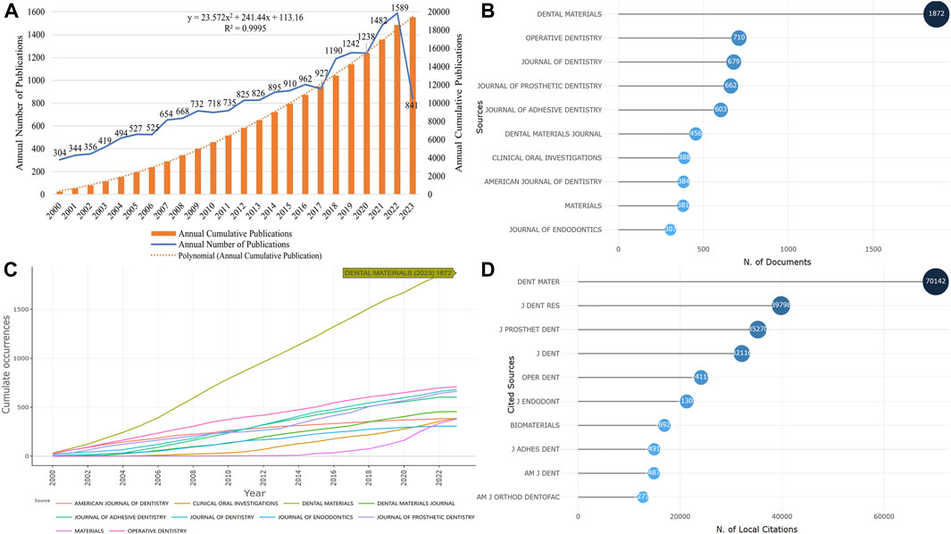
The number of publications reflects the development of the dental adhesive field in the past 20 years. Since 2000, a total of 19,403 articles related to dental adhesives have been published. From Figure 1A, it can be observed that the overall publication volume in this field has been on the rise, with a peak of 1,589 articles in 2022. The continuous increase in publication volume indicates that research on dental adhesives is receiving increasing attention.

FIGURE 1. Publication time and core journals. (A) The change of annual and cumulative number of publications. (B) The top 10 journals in volume of publication. (C) The top 10 journals with the fastest growth in cumulative publication. (D) The top 10 journals cited.).
Research related to dental adhesives has been published in 1,523 journals. The top 10 journals in terms of publication volume are shown in Figure 1B, accounting for approximately 33.20% of the total publication volume. DENTAL MATERIALS, OPERATIVE DENTISTRY, JOURNAL OF DENTISTRY, JOURNAL OF PROSTHETIC DENTISTRY, and JOURNAL OF ADHESIVE DENTISTRY, these five journals have all published over 500 articles, and they are all specialized journals in the field of dentistry. The top 10 journals with the fastest cumulative publication growth are shown in Figure 1C, with DENTAL MATERIALS leading the way in terms of publication growth rate. The top 10 journals with the highest number of citations are shown in Figure 1D, with DENTAL MATERIALS having a remarkable citation count of 70,142 articles. This indicates that the journal is experiencing rapid development and has a significant impact in the current field.
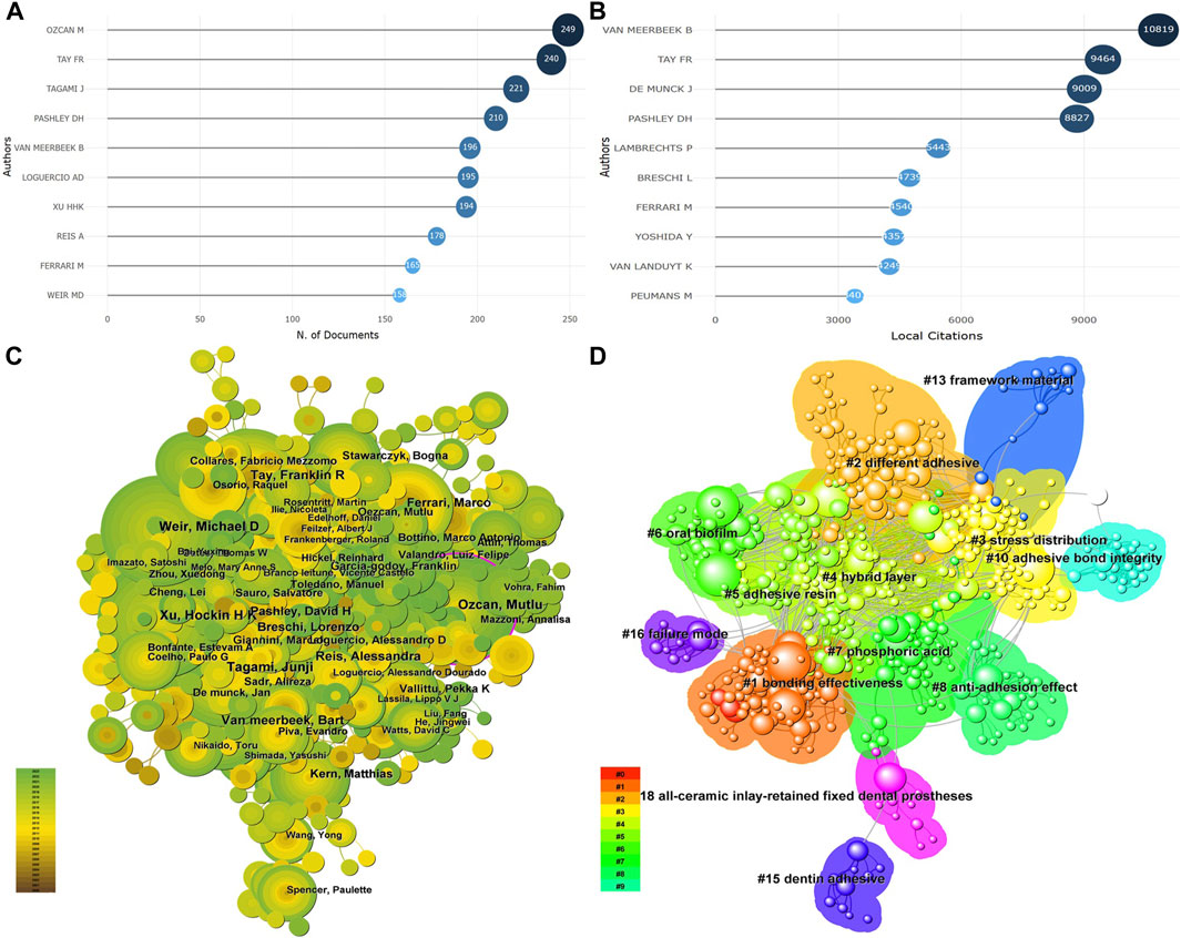
A total of 42,365 authors participated in the publication of the literature, with the author’s publication frequency ranging from 1 to 249 articles. Among them, authors who only participated in the publication of one article accounted for 68% (28,810/42,365). The top 10 authors in terms of publication volume are shown in Figure 2A, with OZCAN M (249 articles), TAY FR (240 articles), and TAGAMI J (221 articles) ranking the highest. The top 10 authors in terms of citation count are shown in Figure 2B, with VAN MEERBEEK B (10,819 citations), TAY FR (9,464 citations), and DE MUNCK J (9,009 citations) ranking the highest. Through co-occurrence analysis of the authors, it was found that there are many influential authors in the current research field, such as OZCAN M, TAY FR, TAGAMI J, VAN MEERBEEK B, and DE MUNCK J, and the collaboration between different authors is very close. This is consistent with the results of publication volume and citation count, indicating that these authors have made significant contributions to the development of dental adhesives, as shown in Figure 2C. In addition, Figure 2C also reveals that only OZCAN M has a larger intermediary centrality (purple outer ring), indicating that OZCAN M occupies the most important position in the current field. Further cluster analysis revealed differences in research areas among different authors. For example, authors led by OZCAN M mainly focus on adhesive bond integrity and stress distribution of dental adhesives, authors led by TAGAMI J emphasize the bonding effectiveness of dental adhesives, and authors led by TAY FR primarily focus on different adhesives, as detailed in Figure 2D.

FIGURE 2. The core author (A) The author of the top 10 publications. (B) The author of the top 10 cited amount. (C) The co-occurrence analysis of the author. (D) Cluster analysis of the author).
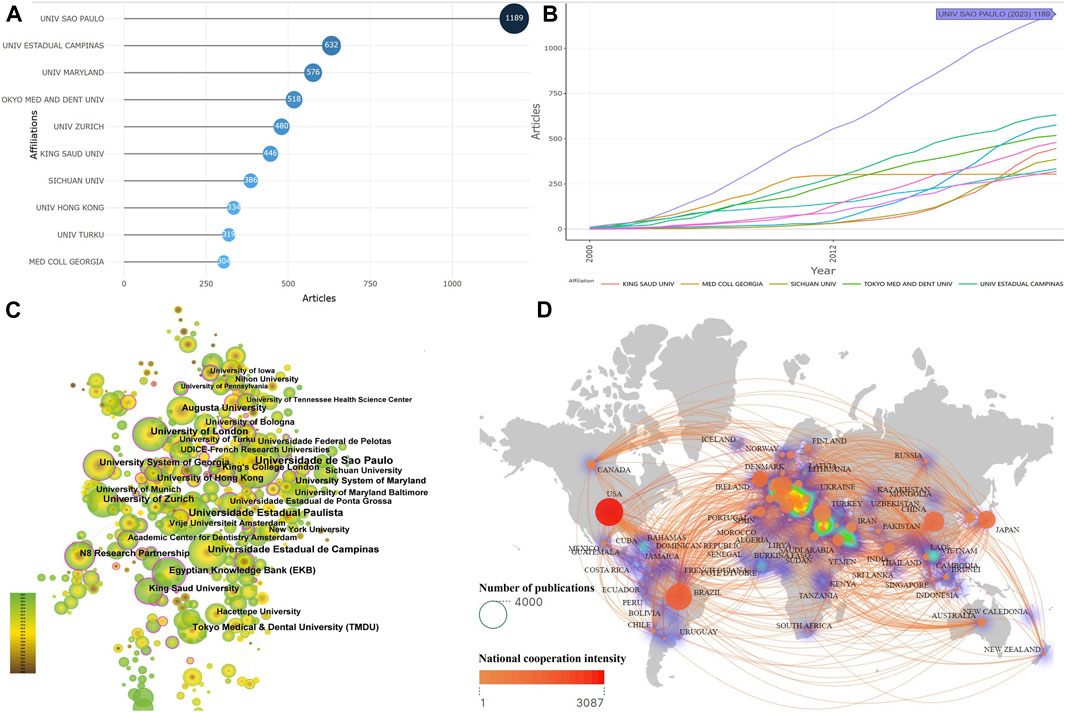
A total of 7,359 institutions participated in the publication of literature. The top 10 institutions in terms of publication volume are shown in Figure 3A. Among them, UNIV SAO PAULO had the highest number of publications, reaching 1,189 articles, followed by UNIV ESTADUAL CAMPINAS (632 articles) and UNIV MARYLAND (576 articles). The institutions with the fastest growth in publication volume are demonstrated in Figure 3B, with UNIV SAO PAULO exhibiting a significantly faster growth rate compared to other institutions. Co-occurrence analysis of these institutions revealed several research-active institutions, indicated by the presence of a purple outer ring, such as UNIV SAO PAULO, Egyptian Knowledge Bank, University of London, and University System of Georgia, detailed in Figure 3C. Furthermore, a total of 121 countries published literature, with the United States , Brazil, and China occupying the top three spots in terms of publication volume. The collaboration between different countries appears to be close, with the US having the tightest collaborations with other nations. Additionally, Europe emerged as the current research hub, as depicted in Figure 3D.

FIGURE 3. The core institutions and countries (A) Institutions with the top 10 publications. (B) The top 10 institutions with the fastest growth in the number of publications. (C) The co-occurrence analysis of the organization. (D) Network map of national cooperation).
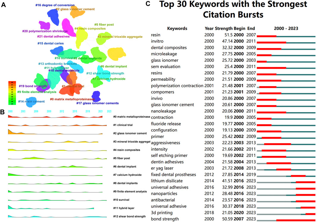
Keywords provide a concise overview of the literature content and visualization of keywords using CiteSpace software can present research hotspots in the related field. There are a total of 22,347 keywords, and the top five keywords with the highest frequency are bond strength, dentin, adhesion, shear bond strength, and zirconia. After clustering the keywords, a total of 22 clusters were formed, mainly focusing on dental adhesive types (clusters 2, 3, 4, 5, 7, 11, 14, 17, 18, 20, 21), bond strength (clusters 0, 9, 12, 16, 19), dental diseases (clusters 6, 8, 10, 13, 15), and clinical trials (cluster 1), as shown in Figure 4A. The landscape diagram also revealed that dental adhesive types, bond strength, and clinical trials have consistently been the research focus in this field and have recently gained wider attention, as depicted in Figure 4B. Keyword emergence analysis clearly reflects the most focused topics and future research trends. Analysis of keyword emergence reveals that topics such as lithium disilicate, nanoparticles, universal adhesives, antibacterial, 3D printing, and bond strength have consistently emerged in recent years, as shown in Figure 4C.

FIGURE 4. Keyword analysis (A) Key words cluster analysis. (B) Keyword growth and decline, the larger the shadow area, the more important the keyword in a given year. (C) The keyword emerges, red stands for emergence, dark green represents the year in which the keyword appears, and light green represents the keyword does not appear.).
The field of dental adhesives has experienced significant growth and development, as evidenced by the increasing number of publications over time. The present study, based on the WoSCC database, retrieved a total of 19,403 articles involving 42,365 authors, 7,359 institutions, 121 countries, and 1,523 journals. Overall, the literature in the field of dental adhesives has shown a continuous upward trend, with the number of published articles gradually increasing from 304 in 2000 to 1,589 in 2022. This indicates a growing interest among researchers in dental adhesives. As research on adhesive dentistry becomes more in-depth and new materials and techniques emerge, researchers strive to disseminate their findings and share knowledge, leading to a constant growth in the number of publications. Researchers from diverse backgrounds have contributed to the expanding literature on dental adhesives. Furthermore, the interdisciplinary nature of dental adhesive research has contributed to this upward trend. Researchers from fields such as chemical engineering, materials engineering, biomaterial sciences, mechanical engineering, computer science, computational mechanics, restorative dentistry, chemistry, dental materials, and polymer chemistry are actively involved in the study and advancement of dental adhesives. This interdisciplinary collaboration provides a rich and diverse research landscape, fostering innovation and driving continuous progress in the field. Among the 1,523 journals surveyed, the top 10 journals accounted for 33.20% of the total publications, indicating that the literature in this field is mainly concentrated in a few specialized journals. Among them, DENTAL MATERIALS had the highest cumulative number of publications, growth rate in publications, and citation count, indicating its significant standing in the field of dental adhesives and deserving special attention. In conclusion, with the continuous development of this field and the emergence of new challenges and opportunities, it is expected that the number of publications related to dental adhesives will continue to increase.
The collaboration between different authors, institutions, and countries is crucial to advancing the field of dental adhesives. In the research field of dental adhesives, collaboration facilitates knowledge exchange, obtaining different perspectives, and pooling resources and expertise. Visual analysis of authors and their institutions can provide potential collaborators and team information for scholars with research interests in the future. From the visualization graph of authors, it can be observed that the current research field is dominated by a research team consisting of core members such as OZCAN M, TAY FR, TAGAMI J, VAN MEERBEEK B, and DE MUNCK J, with close collaboration among different authors. Although there are a large number of authors in this field, 68% of them have only published one article, indicating that most authors are new to the field and have not conducted in-depth research. This also proves the tremendous potential of the field, and with the accumulation of time, the field of dental adhesives will experience new breakthroughs. Among them, OZCAN M, as the author with the highest number of publications, also has the largest intermediary centrality, indicating that OZCAN M occupies the most important position in the current field and to some extent leads the development direction of dental adhesives. Currently, many research institutions in the field are very active, and cooperation between different institutions is also close. UNIV SAO PAULO, as the institution with the fastest growing publication volume and significant intermediation centrality, may become an important research force in the field of dental adhesives in the future. In addition, international collaboration in dental adhesive research is becoming increasingly important as it allows for a global perspective and the sharing of research findings and best practices among different countries. Through international collaboration, it is possible to explore different patient populations, cultural differences, and unique clinical scenarios, thus gaining a broader understanding of the performance and clinical outcomes of dental adhesives. Currently, cooperation between different countries is also very frequent, and although the United States has the closest collaboration with other countries, Europe is the current research center and may dominate the development direction of the field. Scholars from top institutions can often access more resources and funding, making them perfect candidates for collaboration. Among the top ten productive institutions, two each are from the United States, China, and Brazil. The remaining four institutions come from Japan, Switzerland, Saudi Arabia, and Finland, and there is no doubt that these countries’ institutions have the world’s top dental research resources. In conclusion, collaboration between different authors, institutions, and countries in the field is very close, and this is one of the reasons for the significant progress in the field in recent years. In the future, more communication and collaboration focusing on key issues of dental adhesives are needed to better promote the generation of major academic achievements, translate research findings into clinical practice, and ultimately benefit patients around the world.
The keywords in literature serve as important labels through which authors express the research topics, methodologies, and core viewpoints, and they encapsulate and summarize the research content to a great extent. By analyzing the frequency of keyword occurrence and examining keyword co-occurrence, one can gain insight into the scope and hot topics within a specific discipline and further explore the knowledge structure and evolving trends of research topics. In the current field, there are a total of 22,347 keywords, with the highest frequency occurring in bond strength, dentin, adhesion, shear bond strength, and zirconia, respectively. This indicates that the current field primarily focuses on the bonding strength of dental adhesives and related bonding materials. Further clustering analysis of these 22,347 keywords reveals that the types of dental adhesives, bonding strength, dental diseases, and clinical trials are the key areas of research attention at present.
The current state of different types of dental adhesives reflects some important advancements achieved in the field. Various bonding systems have been developed to address the challenges associated with durable and reliable bonding to tooth structure. From traditional acid-etching-rinse (total-etch) adhesive systems to self-etch adhesives, and further evolving to universal adhesives, clinicians are now able to apply adhesives in any bonding strategy based on specific clinical scenarios. Although adhesives allow for more conservative restorative approaches, achieving durable bond strength remains a concern, primarily due to degradation at the resin-dentin interface in challenging oral environments. To address these issues, various improvements have been proposed to develop adhesive systems with special functionalities, such as incorporating matrix metalloproteinase inhibitors, antimicrobial agents, and remineralizing agents into the adhesive system, as well as enhancing its mechanical and chemical properties (Zhou et al., 2019). With advancements in adhesive technology, novel dental adhesives have also emerged. Nanoparticles have been incorporated into adhesive systems based on nanotechnology to enhance mechanical properties and antimicrobial activity (Jandt and Watts, 2020). Biomimetic adhesives that release bioactive factors or promote remineralization contribute to long-term success in restorative procedures (Bhadila et al., 2020). However, degradation over time due to factors such as hydrolysis, enzymatic degradation, and polymer aging remains a significant challenge. Nano-leakage caused by nanoscale gaps and water infiltration can potentially affect bond stability and result in bond failure. Insufficient adhesive strength to certain substrates, such as dentin and non-precious alloys, poses challenges for achieving long-lasting and reliable adhesion. Moreover, chemical, and mechanical challenges associated with adhesion in humid and contaminated environments warrant further research. In conclusion, the development of dental adhesives has significantly improved adhesive performance and enabled durable and aesthetic restorations. However, limitations surrounding bond stability, nanoleakage, and adhesion to challenging substrates present challenges that need to be addressed. Future research should focus on developing strategies to overcome these limitations, including the use of novel materials, improvements in bonding durability testing methods, and enhancement of bonding techniques. Additionally, researchers from diverse fields should prioritize providing safe, biocompatible, and durable dentin adhesives that can restore damaged tissues while also possessing antibacterial activity. Novel research areas include combining experimental and modeling approaches to facilitate the translation of new dentin adhesive formulations from the laboratory to clinical practice. Thus far, verification models require extensive laboratory testing and corresponding clinical investigations. The clinical effectiveness, lifespan, and biocompatibility of dental adhesives also warrant further evaluation. Currently, there are a variety of adhesive choices, each with its unique advantages and potential limitations. Continued research and assessment of different types of adhesives are crucial for optimizing clinical outcomes and providing evidence-based choices for dentists to achieve successful and long-lasting restorations.
An ideal dental adhesive should be biocompatible, possess antimicrobial activity, provide marginal seal, form a minimum film thickness, easy to apply, resistant to dissolution, exhibit semi-transparency and radiopacity, and have optimal working and curing times (Kamposiora et al., 1994; Wingo, 2018; Leung et al., 2022). Various efforts are being made to overcome the issue of bonding strength with dental adhesives. On one hand, researchers are exploring the influence of adhesive components on bonding strength, including the types and concentrations of monomers, fillers, and additives (Cramer et al., 2011). On the other hand, optimizing surface treatment processes to enhance bonding strength and further exploring innovative methods such as laser irradiation, air abrasion, and plasma treatment to improve the tooth surface and enhance bonding efficiency are current research focuses (Ye et al., 2022; Liu et al., 2023; Peng et al., 2023). These techniques aim to create a micromechanical interlocking or alter the chemical properties of the tooth structure to enhance bonding strength. In addition, factors related to adhesive aging, storage conditions, thermal cycling, and oral environment also play a significant role in bonding strength. Currently, the development of novel adhesive systems has facilitated effective bonding to tooth structure with improved mechanical performance, garnering significant attention. However, despite a large amount of in vitro research, only a few clinical trials have evaluated the bonding performance of adhesives, and the available evidence thus far has been collected during short-term follow-up periods (Perdigão et al., 2014; Ruschel et al., 2018; Liu et al., 2023). These published data from clinical trials suggest that the clinical performance of these adhesives is not dependent on bonding strategies over a follow-up period of up to 36 months. However, over time, the reported performance remains less satisfactory compared to marginal discoloration, indicating a need for longer follow-up periods for further clinical research.
Research on the shelf life of dental adhesives is a topic worthy of attention, but there is limited related research. The shelf life of dental adhesives refers to the length of time the adhesive system maintains ideal bond strength, typically 2 years from the production date (Ferracane, 2017; Mazzitelli et al., 2020). In clinical practice, only a small amount of dental adhesive is usually needed for restorative procedures, and a significant quantity of materials may expire before their intended use (Garcia Lda et al., 2010). Even just 3 months past the expiration date, the clinical effectiveness of the adhesive is reduced (Mazzitelli et al., 2020). However, some dental practitioners continue to use these expired materials, which is ethically unacceptable and unsafe (Talreja et al., 2017). In fact, expired materials can still be used for diagnostic purposes, stabilizing matrix bands, temporary crowns, and so on (Bagis et al., 2009; Talreja et al., 2017). Currently, the storage and shelf life of adhesives are considered key factors in their degradation outside the oral cavity (Hardan et al., 2021). Issues related to adhesive storage, such as difficulties in monomer polymerization, component degradation and evaporation, repeated opening of adhesive bottles, and prolonged storage time, can potentially weaken the effectiveness and stability of the adhesive (Van Landuyt et al., 2007; Van Landuyt et al., 2009; Hardan et al., 2021; Kharouf et al., 2021). Additionally, the transportation and storage conditions of adhesives before clinical application are not always optimal (Cardoso et al., 2014). Despite the strong potential impact of these clinical realities on the final quality of adhesives, relevant research remains limited. Future research should focus on improving the lifespan and stability of dental adhesives to ensure their reliable performance during the shelf life and clinical use.
Currently, dental adhesives are widely applied in various dental diseases, primarily in the field of restorative dentistry. They can be extensively used for bonding dental crowns, inlays, onlays, veneers, composite fixed restorations, pulp canal posts, and orthodontic appliances (Edelhoff et al., 2019). Among these, dental caries is one of the common dental diseases where adhesives are widely used. Adhesives play a vital role in bonding restorative materials with the affected tooth structure, allowing conservative removal of decayed tooth tissues and restoring form and function. Additionally, dental adhesives are also utilized in the treatment of tooth erosion, which is a loss of tooth structure caused by chemical dissolution, often associated with acidic foods and beverages or gastric reflux (Mazzoleni et al., 2023). Adhesives provide structural support and aesthetic improvement to dental materials by bonding, aiding in the restoration of eroded tooth surfaces. Another extensively used dental adhesive for dental conditions is tooth wear. Tooth wear can be caused by various factors such as attrition and erosion (Lussi et al., 2019). Adhesives are used to bond restorative materials like composite resins or ceramics to the worn tooth surfaces, thereby restoring both aesthetics and functionality. Besides restoration purposes, dental adhesives are also used in orthodontic treatment. Adhesives are used to bond orthodontic brackets to the tooth surfaces, facilitating the application of orthodontic forces and tooth movement. Additionally, dental adhesives demonstrate potential in controlling dental hypersensitivity. They are used to seal or block dentinal tubules, which are responsible for transmitting sensations, thereby reducing or alleviating tooth sensitivity (Huang et al., 2022). In conclusion, dental adhesives have a wide range of applications in restorative dentistry, including the management of dental caries, erosion, tooth wear, orthodontic treatment, and dental hypersensitivity. Advancing adhesive technologies have brought improvements in the treatment and management of these dental diseases, providing better clinical outcomes and patient satisfaction.
The term “emerging keywords” refers to key terms that exhibit a significant increase in frequency of use during a certain period and possess a strong emergence intensity. These keywords can describe the forefront issues in a research field. A keyword emergence analysis, based on keyword co-occurrence, can visually represent the focal points, hot topics, and trends of advancement in this field. Our analysis results reveal that the emergence of keywords, such as lithium disilicate, nanoparticles, universal adhesives, antibacterial, 3D printing, and bonding strength, has persisted until 2023. Among these keywords, lithium disilicate is associated with new materials in dental restorations, whereas the others mainly revolve around novel universal adhesives, bonding strength in dental adhesives, antibacterial properties, nanoparticles, and 3D printing.
The presence of residual bacteria during the tooth restoration process increases the risk of recurrent dental caries, which remains a challenge in the field of dentistry (Feuerstein et al., 2007; Walter et al., 2007). In recent years, there has been an increasing focus on the development of dental adhesives with antibacterial properties to address the issues of recurrent dental caries and secondary infections. Extensive research has investigated the addition of antimicrobial agents to dental adhesives to inhibit microbial growth and prevent biofilm formation. The antimicrobial properties of formulations such as quaternary ammonium salts, chlorhexidine, and streptomycin have been widely studied (Dionysopoulos et al., 2022). Another method for producing antibacterial dental composites is by incorporating antibacterial particles into the composite material. These particles include polymer nanocomposites, bioactive glass, and metal/metal oxide particles. Polymer antibacterial particles have several advantages over leachable antimicrobial agents, including non-volatility, chemical stability, long-term activity, and non-permeability to the skin (Beyth et al., 2010; Shvero et al., 2015). After these agents have been added to dental adhesives, they have been shown to exhibit varying degrees of antibacterial efficacy against common oral pathogens. Multiple in vitro studies have demonstrated that antibacterial dental adhesives can significantly reduce bacterial adhesion, biofilm formation, and bacterial vitality (Li et al., 2009; Melo et al., 2013; Winkler et al., 2014). Additionally, these adhesives can prevent the development of secondary caries around the margins of adhesive restorations. However, the long-term antibacterial effects and potential development of bacterial resistance are still areas of ongoing research. Indeed, the mechanisms of action of antibacterial dental adhesives are also highly scrutinized. These mechanisms include direct disruption of bacterial membranes, inhibition of bacterial enzymes, and interference with bacterial metabolism (Wang et al., 2017). Despite the promising achievements, challenges and limitations still exist. The extent of antibacterial activity depends on the type and concentration of antimicrobial agents, the interaction with other components in adhesive formulations, and the specific bacterial strain being tested. Furthermore, further research is needed to investigate the compatibility with other dental materials (such as composite materials and cements), as well as the influence of long-term aging and oral environmental conditions on the antibacterial performance of these adhesives.
Currently, the addition of particles to dental adhesive systems to enhance antimicrobial activity is a new direction (Cheng et al., 2013; Zhang et al., 2013). The latest advancements in nanotechnology allow for better control of particle characteristics, resulting in more customized and effective nanoparticle-based dental adhesives (Balhaddad et al., 2019; Jandt and Watts, 2020). Researchers have explored the incorporation of nanoparticles such as silver, zinc oxide, and titanium dioxide into dental adhesives to improve their mechanical, antimicrobial, and remineralization properties (Moradpoor et al., 2021; Nizami et al., 2021). Nanoparticles can enhance bond strength, reduce microleakage, and provide antibacterial effects against oral pathogens. The optimal concentration, size, and distribution of nanoparticles in adhesive formulations need to be closely examined to achieve the desired performance. Attention should also be given to surface modifications of nanoparticles, such as coating with silanes or functional groups, to enhance their compatibility with adhesive matrices and improve their dispersibility. In addition, further investigation is needed to understand the interactions between nanoparticles and other adhesive components, such as monomers and fillers, and their impact on polymerization kinetics, bond formation, and long-term stability. Recent studies have shown that combining nanosilver with antimicrobial monomers can enhance antimicrobial activity without compromising mechanical properties (Cheng et al., 2013; Zhang et al., 2013). Silver is well-known for its low toxicity and good biocompatibility with human cells. It also exhibits antimicrobial, antifungal, and antiviral activities (Monteiro et al., 2009; Allaker, 2010). Therefore, nanosilver particles have been incorporated into dental materials to enhance antimicrobial activity or amplify its effects (Imazato et al., 2003a; Cheng et al., 2013; Li et al., 2014).
As previously mentioned, universal adhesives have revolutionized dental bonding by providing simplified bonding solutions and expanded clinical applications. These adhesives offer a one-stop solution for bonding various dental substrates, including enamel, dentin, and even indirect restorative materials. They are favored for their flexibility in choosing bonding strategies and the number of steps involved (Van Meerbeek et al., 2011; Hanabusa et al., 2012; de Goes et al., 2014; Muñoz et al., 2014; Wagner et al., 2014). Ongoing research aims to further improve the characteristics and clinical performance of universal adhesives in dentistry. This includes exploring new monomers, nanoparticles, additives, and functional agents to enhance bonding efficacy, antimicrobial properties, and bioactivity. Most tested monomers do not affect material performance (Imazato et al., 2003b; Xiao et al., 2009), although some studies have observed a decrease in mechanical properties due to the addition of a large amount of monomers (He et al., 2013). While they are capable of inactivating oral bacteria in vitro (Esteves et al., 2010; da Silva et al., 2010; Türkün et al., 2005), no longitudinal clinical studies have confirmed the role of this antimicrobial effect in controlling secondary caries. Furthermore, advances in adhesive techniques, such as the application of selective etching and optimization of adhesive layer thickness, are being studied to achieve more predictable and reliable bonding outcomes. However, as previously mentioned, the durability, stability, and performance of universal adhesives in clinical trials remain unclear and require careful use.
It is worth noting that restorative dentistry is currently undergoing a revolution brought about by digital technology, such as intraoral scanning (Jurado et al., 2019), computer-aided design/computer-aided manufacturing (CAD-CAM) (Tsujimoto et al., 2017) and 3D printing technology (Jurado et al., 2020). The challenge in this field lies in how to seamlessly integrate these technologies. In addition to existing materials like CAD/CAM resins (Jurado et al., 2020), ceramics (Yoshida et al., 2015) and zirconia (Fischer et al., 2018), there is ongoing development of restorative materials for 3D printing (Kessler et al., 2020). Researchers have explored various 3D printing techniques, such as stereolithography, digital light processing, and selective laser sintering, to create adhesive components with complex designs and internal features to enhance bonding performance (Ngo et al., 2018). The ability to precisely control the shape, size, and surface morphology of the bonding interface optimizes mechanical stability and adhesive strength. The versatility of 3D printing allows for the manufacturing of multi-material structures in dental adhesives. This enables the incorporation of functional additives, such as antimicrobial agents, bioactive compounds, or reinforcing particles, directly into the adhesive structure (Ngo et al., 2018; Ahmed et al., 2021). By embedding these additives, 3D printed dental adhesives can enhance their bonding performance while providing additional functionalities, such as antimicrobial effects or remineralization capabilities. Research has demonstrated the feasibility and potential clinical applications of 3D printed dental adhesives. Customized adhesive structures can be designed and printed for various restorative procedures. 3D printing has improved the efficiency, accuracy, and aesthetics of dental treatment, ultimately leading to better patient outcomes. Concurrently, researchers are actively exploring the development of printable adhesive materials compatible with 3D printing technology. Novel resin formulations, including resin composites and resin binders, are being developed to optimize their printability, mechanical properties, biocompatibility, and long-term stability. Adapting material characteristics to meet the specific requirements of dental adhesives is currently a focal point of research. Despite significant progress, challenges still exist in optimizing material performance, ensuring biocompatibility, and evaluating long-term clinical performance. Research in the field of 3D printed dental adhesives continues to rapidly evolve, with emphasis on refining materials, printability, and expanding the scope of clinical applications.
In conclusion, as the first bibliometric study in the field of dental adhesives, we have objectively and quantitatively analyzed the current state and Frontier hotspots in this field, identifying key research areas, influential authors, collaborative networks, and emerging trends, which provide a knowledge base for future in-depth research in this field. This not only assists researchers in gaining a deeper understanding of the current knowledge landscape but also guides future research directions by identifying emerging areas of interest, such as novel universal adhesives, bond strength and antibacterial properties of dental adhesives, nanoparticles, and 3D printing. Moreover, the insights gained from this study contribute to resource allocation, funding decisions, and policy-making related to dental adhesive research, providing information for researchers, clinicians, and manufacturers regarding the development, optimization, and application of dental adhesive materials and techniques. Ultimately, this study plays a crucial role in advancing in-depth research on dental adhesives, promoting evidence-based practices, and ultimately improving outcomes in dental care for patients.
It is important to acknowledge that this study is not without limitations. Firstly, the literature included only English publications in the WoSCC database, excluding other types of research such as commentaries, letters, conference abstracts, and grey literature, apart from monographs and reviews. This limitation may result in incomplete statements and potential biases in the analysis. Additionally, due to the inability of VOSviewer and Citespace to analyze the full text of publications, certain information may be overlooked, and some recently published articles may be ignored due to their low citation frequency. Furthermore, the results and corresponding discussions of this study are limited to a certain range, which is dependent on the keywords and search strategy formulated at the beginning of the research. For example, based on the search terms we devised, although Ozcan M has the highest number of publications, his work primarily focuses on bond strength testing of adhesives rather than any aspect of dental adhesives specifically. Despite our efforts in formulating a relatively comprehensive search strategy, the omission of important literature is still inevitable, which may affect the analysis results of this study. It is important to note that the scope of this study remains limited. We do not consider published research focused on the development of new monomers, polymerization systems, and emerging areas. The published research in these areas is paramount to the development of the next-generation of dental adhesives. Furthermore, the limitations of bibliometrics as a discipline should not be disregarded. Bibliometric studies primarily focus on published literature, which may lead to underrepresentation of negative or null results, as well as non-English publications, thereby impacting the interpretation of research trends and impact. Bibliometrics mainly captures quantitative information, such as citation counts and publication trends, while neglecting qualitative information, such as the actual content and impact of publications. This limitation can hinder a comprehensive understanding of research domains.
Dental adhesives have received significant attention in the research field since the 21st century. The collaboration between different authors, institutions, and countries within this field has been closely knit, facilitating the rapid development of dental adhesives. While the current research focuses on the types of dental adhesives, bonding strength, applicable dental diseases, and clinical trials, it is anticipated that in the future there will be heightened attention towards nanoparticles, 3D printing, universal adhesives, antibacterial properties of adhesives, and bonding strength.
The original contributions presented in the study are included in the article/supplementary material, further inquiries can be directed to the corresponding author.
XH: Conceptualization, Data curation, Formal Analysis, Investigation, Writing–original draft. TS: Investigation, Methodology, Project administration, Writing–review and editing. BK: Investigation, Methodology, Project administration, Writing–review and editing. YS: Project administration, Software, Supervision, Validation, Writing–review and editing. LC: Investigation, Project administration, Supervision, Writing–review and editing. HD: Conceptualization, Funding acquisition, Investigation, Project administration, Visualization, Writing–review and editing.
The author(s) declare financial support was received for the research, authorship, and/or publication of this article. We acknowledge the financial support from National Traditional Chinese Medicine Advantage Specialty Construction Project ((2022) No.48) and Lanzhou Science and Technology Plan Project (2023-2-71).
The authors declare that the research was conducted in the absence of any commercial or financial relationships that could be construed as a potential conflict of interest.
All claims expressed in this article are solely those of the authors and do not necessarily represent those of their affiliated organizations, or those of the publisher, the editors and the reviewers. Any product that may be evaluated in this article, or claim that may be made by its manufacturer, is not guaranteed or endorsed by the publisher.
Ahmed, W., Siraj, S., and Al-Marzouqi, A. H. (2021). Embracing additive manufacturing technology through fused filament fabrication for antimicrobial with enhanced formulated materials. Polymers 13 (9), 1523. doi:10.3390/polym13091523
Allaker, R. P. (2010). The use of nanoparticles to control oral biofilm formation. J. Dent. Res. 89 (11), 1175–1186. doi:10.1177/0022034510377794
Bagis, Y. H., Baltacioglu, I. H., and Kahyaogullari, S. (2009). Comparing microleakage and the layering methods of silorane-based resin composite in wide Class II MOD cavities. Oper. Dent. 34 (5), 578–585. doi:10.2341/08-073-lr
Balhaddad, A. A., Kansara, A. A., Hidan, D., Weir, M. D., Xu, H. H., and Melo, M. A. S. (2019). Toward dental caries: exploring nanoparticle-based platforms and calcium phosphate compounds for dental restorative materials. Bioact. Mater. 4 (1), 43–55. doi:10.1016/j.bioactmat.2018.12.002
Beyth, N., Yudovin-Farber, I., Perez-Davidi, M., Domb, A. J., and Weiss, E. I. (2010). Polyethyleneimine nanoparticles incorporated into resin composite cause cell death and trigger biofilm stress in vivo. Proc. Natl. Acad. Sci. U. S. A. 107 (51), 22038–22043. doi:10.1073/pnas.1010341107
Bhadila, G., Filemban, H., Wang, X., Melo, M. A. S., Arola, D. D., Tay, F. R., et al. (2020). Bioactive low-shrinkage-stress nanocomposite suppresses S. mutans biofilm and preserves tooth dentin hardness. Acta biomater. 114, 146–157. doi:10.1016/j.actbio.2020.07.057
Bin-Jardan, L. I., Almadani, D. I., Almutairi, L. S., Almoabid, H. A., Alessa, M. A., Almulhim, K. S., et al. (2023). Inorganic compounds as remineralizing fillers in dental restorative materials: narrative review. Int. J. Mol. Sci. 24 (9), 8295. doi:10.3390/ijms24098295
Buonocore, M. G. (1955). A simple method of increasing the adhesion of acrylic filling materials to enamel surfaces. J. Dent. Res. 34 (6), 849–853. doi:10.1177/00220345550340060801
Cardoso, S. A., Oliveira, H. L., Münchow, E. A., Carreño, N. L., Gonini Junior, A., and Piva, E. (2014). Effect of shelf-life simulation on the bond strength of self-etch adhesive systems to dentin. Appl. Adhesion Sci. 2 (1), 26–10. doi:10.1186/s40563-014-0026-9
Carvalho, R. M., Manso, A. P., Geraldeli, S., Tay, F. R., and Pashley, D. H. (2012). Durability of bonds and clinical success of adhesive restorations. Dent. Mater. 28 (1), 72–86. doi:10.1016/j.dental.2011.09.011
Chen, C. (2006). CiteSpace II: detecting and visualizing emerging trends and transient patterns in scientific literature. J. Am. Soc. Inf. Sci. Technol. 57 (3), 359–377. doi:10.1002/asi.20317
Cheng, L., Weir, M. D., Zhang, K., Arola, D. D., Zhou, X., and Xu, H. H. (2013). Dental primer and adhesive containing a new antibacterial quaternary ammonium monomer dimethylaminododecyl methacrylate. J. Dent. 41 (4), 345–355. doi:10.1016/j.jdent.2013.01.004
Cho, K., Rajan, G., Farrar, P., Prentice, L., and Prusty, B. G. (2022). Dental resin composites: a review on materials to product realizations. Compos. Part B Eng. 230, 109495. doi:10.1016/j.compositesb.2021.109495
Cramer, N. B., Stansbury, J., and Bowman, C. (2011). Recent advances and developments in composite dental restorative materials. J. Dent. Res. 90 (4), 402–416. doi:10.1177/0022034510381263
Cuevas-Suárez, C. E., da Rosa, W. L. D. O., Lund, R. G., da Silva, A. F., and Piva, E. (2019). Bonding performance of universal adhesives: an updated systematic review and meta-analysis. J. adhesive Dent. 21 (1), 7–26. doi:10.3290/j.jad.a41975
da Silva, B. M., França, F. M. G., Flório, F. M., and Basting, R. T. (2010). In situ anticariogenic effect of adhesive systems containing fluoride and MDPB. Am. J. Dent. 23 (2), 75–80.
de Goes, M. F., Shinohara, M. S., and Freitas, M. S. (2014). Performance of a new one-step multi-mode adhesive on etched vs non-etched enamel on bond strength and interfacial morphology. J. adhesive Dent. 16 (3), 243–250. doi:10.3290/j.jad.a32033
Dionysopoulos, D., Gerasimidou, O., and Papadopoulos, C. (2022). Current modifications of dental adhesive systems for composite resin restorations: a review in literature. J. Adhesion Sci. Technol. 36 (5), 453–468. doi:10.1080/01694243.2021.1924499
Duke, E. S. (1993). Adhesion and its application with restorative materials. Dent. Clin. N. Am. 37 (3), 329–340. doi:10.1016/s0011-8532(22)00256-7
Edelhoff, D., Stimmelmayr, M., Schweiger, J., Ahlers, M. O., and Güth, J. F. (2019). Advances in materials and concepts in fixed prosthodontics: a selection of possible treatment modalities. Br. Dent. J. 226 (10), 739–748. doi:10.1038/s41415-019-0265-z
Esteves, C. M., Ota-Tsuzuki, C., Reis, A. F., and Rodrigues, J. A. (2010). Antibacterial activity of various self-etching adhesive systems against oral streptococci. Oper. Dent. 35 (4), 448–453. doi:10.2341/09-297-l
Ferracane, J. L. (2017). Models of caries formation around dental composite restorations. J. Dent. Res. 96 (4), 364–371. doi:10.1177/0022034516683395
Feuerstein, O., Matalon, S., Slutzky, H., and Weiss, E. I. (2007). Antibacterial properties of self-etching dental adhesive systems. J. Am. Dent. Assoc. 138 (3), 349–354. doi:10.14219/jada.archive.2007.0167
Fischer, N. G., Tsujimoto, A., and Baruth, A. (2018). Effects of polishing bur application force and reuse on sintered zirconia surface topography. Oper. Dent. 43 (4), 437–446. doi:10.2341/17-105-lr
Garcia Lda, F., Roselino, L. D. M. R., Pires-de-Souza, F. D. C. P., and Consani, S. (2010). Evaluation of the conversion degree, microhardness, and surface roughness of composite resins used after their expiration date. General Dent. 58 (6), e262–e267.
Hanabusa, M., Mine, A., Kuboki, T., Momoi, Y., Van Ende, A., Van Meerbeek, B., et al. (2012). Bonding effectiveness of a new 'multi-mode' adhesive to enamel and dentine. J. Dent. 40 (6), 475–484. doi:10.1016/j.jdent.2012.02.012
Hardan, L., Bourgi, R., Cuevas-Suárez, C. E., Zarow, M., Kharouf, N., Mancino, D., et al. (2021). The bond strength and antibacterial activity of the universal dentin bonding system: a systematic review and meta-analysis. Microorganisms 9 (6), 1230. doi:10.3390/microorganisms9061230
He, J., Söderling, E., Vallittu, P., and Lassila, L. (2013). Preparation and evaluation of dental resin with antibacterial and radio-opaque functions. Int. J. Mol. Sci. 14 (3), 5445–5460. doi:10.3390/ijms14035445
Hill, E. E. (2007). Dental cements for definitive luting: a review and practical clinical considerations. Dent. Clin. N. Am. 51 (3), 643–658. doi:10.1016/j.cden.2007.04.002
Hill, E. E., and Lott, J. (2011). A clinically focused discussion of luting materials. Aust. Dent. J. 56 (1), 67–76. doi:10.1111/j.1834-7819.2010.01297.x
Huang, M., Huang, Y., Liu, H., Tang, Z., Chen, Y., Huang, Z., et al. (2022). Hydrogels for the treatment of oral and maxillofacial diseases: current research, challenges, and future directions. Biomaterials Sci. 10 (22), 6413–6446. doi:10.1039/d2bm01036d
Imazato, S., Ebi, N., and Takahashi, Y. (2003a). Antibacterial activity of bactericide-immobilized filler for resin-based restoratives. Biomaterials 24 (20), 3605–3609. doi:10.1016/s0142-9612(03)00217-5
Imazato, S., Kinomoto, Y., Tarumi, H., Ebisu, S., and R. Tay, F. (2003b). Antibacterial activity and bonding characteristics of an adhesive resin containing antibacterial monomer MDPB. Dent. Mater. 19 (4), 313–319. doi:10.1016/s0109-5641(02)00060-x
Jandt, K. D., and Watts, D. C. (2020). Nanotechnology in dentistry: present and future perspectives on dental nanomaterials. Dent. Mater. 36 (11), 1365–1378. doi:10.1016/j.dental.2020.08.006
Jurado, C. A., Tsujimoto, A., Alhotan, A., Villalobos-Tinoco, J., and AlShabib, A. (2020). Digitally fabricated immediate complete dentures: case reports of milled and printed dentures. Int. J. Prosthodont. 33 (2), 232–241. doi:10.11607/ijp.6305
Jurado, C. A., Tsujimoto, A., Tanaka, K., Watanabe, H., Fischer, N. G., Barkmeier, W. W., et al. (2019). 3D printed coping for intraoral evaluation: a clinical report. Quintessence Int. Berl. Ger. 1985) 50 (7), 534–538. doi:10.3290/j.qi.a42655
Kamposiora, P., Papavasilious, G., Bayne, S. C., and Felton, D. A. (1994). Finite element analysis estimates of cement microfracture under complete veneer crowns. J. Prosthet. Dent. 71 (5), 435–441. doi:10.1016/0022-3913(94)90179-1
Kessler, A., Hickel, R., and Reymus, M. (2020). 3D printing in dentistry-state of the art. Oper. Dent. 45 (1), 30–40. doi:10.2341/18-229-l
Kharouf, N., Eid, A., Hardan, L., Bourgi, R., Arntz, Y., Jmal, H., et al. (2021). Antibacterial and bonding properties of universal adhesive dental polymers doped with pyrogallol. Polymers 13 (10), 1538. doi:10.3390/polym13101538
Leung, G. K., Wong, A. W. Y., Chu, C. H., and Yu, O. Y. (2022). Update on dental luting materials. Dent. J. 10 (11), 208. doi:10.3390/dj10110208
Li, F., Chen, J., Chai, Z., Zhang, L., Xiao, Y., Fang, M., et al. (2009). Effects of a dental adhesive incorporating antibacterial monomer on the growth, adherence and membrane integrity of Streptococcus mutans. J. Dent. 37 (4), 289–296. doi:10.1016/j.jdent.2008.12.004
Li, F., Weir, M. D., Fouad, A. F., and Xu, H. H. (2014). Effect of salivary pellicle on antibacterial activity of novel antibacterial dental adhesives using a dental plaque microcosm biofilm model. Dent. Mater. 30 (2), 182–191. doi:10.1016/j.dental.2013.11.004
Li, Y., Hu, X., Xia, Y., Ji, Y., Ruan, J., Weir, M. D., et al. (2018). Novel magnetic nanoparticle-containing adhesive with greater dentin bond strength and antibacterial and remineralizing capabilities. Dent. Mater. 34 (9), 1310–1322. doi:10.1016/j.dental.2018.06.001
Liu, J., Xue, Y., Dong, X., Fan, Y., Hao, H., and Wang, X. (2023). Review of the surface treatment process for the adhesive matrix of composite materials. Int. J. Adhesion Adhesives 126, 103446. doi:10.1016/j.ijadhadh.2023.103446
Lussi, A., Buzalaf, M. A. R., Duangthip, D., Anttonen, V., Ganss, C., João-Souza, S. H., et al. (2019). The use of fluoride for the prevention of dental erosion and erosive tooth wear in children and adolescents. Eur. archives Paediatr. Dent. 20 (6), 517–527. doi:10.1007/s40368-019-00420-0
Mazzitelli, C., Maravic, T., Sebold, M., Checchi, V., Josic, U., Breschi, L., et al. (2020). Effect of shelf-life of a universal adhesive to dentin. Int. J. Adhesion Adhesives 102, 102673. doi:10.1016/j.ijadhadh.2020.102673
Mazzoleni, S., Gargani, A., Parcianello, R. G., Pezzato, L., Bertolini, R., Zuccon, A., et al. (2023). Protection against dental erosion and the remineralization capacity of non-fluoride toothpaste, fluoride toothpaste and fluoride varnish. Appl. Sci. 13 (3), 1849. doi:10.3390/app13031849
Melo, M. A., Cheng, L., Weir, M. D., Hsia, R., Rodrigues, L. K. A., and Xu, H. H. (2013). Novel dental adhesive containing antibacterial agents and calcium phosphate nanoparticles. J. Biomed. Mater. Res. Part B, Appl. biomaterials 101 (4), 620–629. doi:10.1002/jbm.b.32864
Monteiro, D. R., Gorup, L. F., Takamiya, A. S., Ruvollo-Filho, A. C., Camargo, E. R. D., and Barbosa, D. B. (2009). The growing importance of materials that prevent microbial adhesion: antimicrobial effect of medical devices containing silver. Int. J. Antimicrob. agents 34 (2), 103–110. doi:10.1016/j.ijantimicag.2009.01.017
Moradpoor, H., Safaei, M., Mozaffari, H. R., Sharifi, R., Imani, M. M., Golshah, A., et al. (2021). An overview of recent progress in dental applications of zinc oxide nanoparticles. RSC Adv. 11 (34), 21189–21206. doi:10.1039/d0ra10789a
Muñoz, M. A., Luque, I., Hass, V., Reis, A., Loguercio, A. D., and Bombarda, N. H. C. (2013). Immediate bonding properties of universal adhesives to dentine. J. Dent. 41 (5), 404–411. doi:10.1016/j.jdent.2013.03.001
Muñoz, M. A., Sezinando, A., Luque-Martinez, I., Szesz, A. L., Reis, A., Loguercio, A. D., et al. (2014). Influence of a hydrophobic resin coating on the bonding efficacy of three universal adhesives. J. Dent. 42 (5), 595–602. doi:10.1016/j.jdent.2014.01.013
Ngo, T. D., Kashani, A., Imbalzano, G., Nguyen, K. T., and Hui, D. (2018). Additive manufacturing (3D printing): a review of materials, methods, applications and challenges. Compos. Part B Eng. 143, 172–196. doi:10.1016/j.compositesb.2018.02.012
Nizami, M. Z. I., Xu, V. W., Yin, I. X., Yu, O. Y., and Chu, C. H. (2021). Metal and metal oxide nanoparticles in caries prevention: a review. Nanomater. (Basel). 11 (12), 3446. doi:10.3390/nano11123446
Opdam, N. J., Bronkhorst, E., Loomans, B., and Huysmans, M. C. (2010). 12-year survival of composite vs. amalgam restorations. J. Dent. Res. 89 (10), 1063–1067. doi:10.1177/0022034510376071
Opdam, N. J., van de Sande, F., Bronkhorst, E., Cenci, M., Bottenberg, P., Pallesen, U., et al. (2014). Longevity of posterior composite restorations: a systematic review and meta-analysis. J. Dent. Res. 93 (10), 943–949. doi:10.1177/0022034514544217
Peng, T. Y., Shimoe, S., Higo, M., Kato, M., Hirata, I., Iwaguro, S., et al. (2023). Effect of laser engraving on shear bond strength of polyetheretherketone to indirect composite and denture-base resins. J. Dent. Sci. doi:10.1016/j.jds.2023.06.005
Perdigão, J., Kose, C., Mena-Serrano, A., De Paula, E., Tay, L., Reis, A., et al. (2014). A new universal simplified adhesive: 18-month clinical evaluation. Oper. Dent. 39 (2), 113–127. doi:10.2341/13-045-c
Rosa, W. L., Piva, E., and Silva, A. F. D. (2015). Bond strength of universal adhesives: a systematic review and meta-analysis. J. Dent. 43 (7), 765–776. doi:10.1016/j.jdent.2015.04.003
Ruschel, V. C., Shibata, S., Stolf, S., Chung, Y., Baratieri, L., Heymann, H., et al. (2018). Eighteen-month clinical study of universal adhesives in noncarious cervical lesions. Oper. Dent. 43 (3), 241–249. doi:10.2341/16-320-c
Shang, Z., Wanyan, P., Wang, M., Zhang, B., Cui, X., and Wang, X. (2023). Bibliometric analysis of stem cells for spinal cord injury: current status and emerging frontiers. Front. Pharmacol. 14, 1235324. doi:10.3389/fphar.2023.1235324
Shirai, K., De Munck, J., Yoshida, Y., Inoue, S., Lambrechts, P., Suzuki, K., et al. (2005). Effect of cavity configuration and aging on the bonding effectiveness of six adhesives to dentin. Dent. Mater. 21 (2), 110–124. doi:10.1016/j.dental.2004.01.003
Shvero, D. K., Zatlsman, N., Hazan, R., Weiss, E. I., and Beyth, N. (2015). Characterisation of the antibacterial effect of polyethyleneimine nanoparticles in relation to particle distribution in resin composite. J. Dent. 43 (2), 287–294. doi:10.1016/j.jdent.2014.05.003
Sofan, E., Sofan, A., Palaia, G., Tenore, G., Romeo, U., and Migliau, G. (2017). Classification review of dental adhesive systems: from the IV generation to the universal type. Ann. Stomatol. 8 (1), 1–17. doi:10.11138/ads/2017.8.1.001
Swift, E. J. (1998). Bonding systems for restorative materials--a comprehensive review. Pediatr. Dent. 20 (2), 80–84.
Talreja, N., Singla, S., and Shashikiran, N. D. (2017). Comparative evaluation of bond strength and microleakage of standard and expired composite at resin-dentin interface: an in vitro study. Int. J. Clin. Pediatr. Dent. 10 (1), 1–4. doi:10.5005/jp-journals-10005-1396
Tay, F. R., and Pashley, D. H. (2002). Dental adhesives of the future. J. adhesive Dent. 4 (2), 91–103.
Townsend, R. D., and Dunn, W. J. (2004). The effect of saliva contamination on enamel and dentin using a self-etching adhesive. J. Am. Dent. Assoc. 135 (7), 895–901. doi:10.14219/jada.archive.2004.0335
Tsujimoto, A., Barkmeier, W., Takamizawa, T., Latta, M., and Miyazaki, M. (2017). Influence of thermal cycling on flexural properties and simulated wear of computer-aided design/computer-aided manufacturing resin composites. Oper. Dent. 42 (1), 101–110. doi:10.2341/16-046-l
Türkün, L. S., Ateş, M., Türkün, M., and Uzer, E. (2005). Antibacterial activity of two adhesive systems using various microbiological methods. J. adhesive Dent. 7 (4), 315–320.
Van Landuyt, K. L., Mine, A., De Munck, J., Jaecques, S., Peumans, M., Lambrechts, P., et al. (2009). Are one-step adhesives easier to use and better performing? Multifactorial assessment of contemporary one-step self-etching adhesives. J. adhesive Dent. 11 (3), 175–190.
Van Landuyt, K. L., Snauwaert, J., De Munck, J., Peumans, M., Yoshida, Y., Poitevin, A., et al. (2007). Systematic review of the chemical composition of contemporary dental adhesives. Biomaterials 28 (26), 3757–3785. doi:10.1016/j.biomaterials.2007.04.044
Van Meerbeek, B., Peumans, M., Poitevin, A., Mine, A., Van Ende, A., Neves, A., et al. (2010). Relationship between bond-strength tests and clinical outcomes. Dent. Mater. 26 (2), e100–e121. doi:10.1016/j.dental.2009.11.148
Van Meerbeek, B., Yoshihara, K., Yoshida, Y., Mine, A., and K.l., V. L. (2011). State of the art of self-etch adhesives. Dent. Mater. 27 (1), 17–28. doi:10.1016/j.dental.2010.10.023
Velo, S., Carano, A., and Carano, A. (2002). Self-etching vs. traditional bonding systems in orthodontics: an in vitro study. Orthod. craniofacial Res. 5 (3), 166–169. doi:10.1034/j.1600-0544.2002.02193.x
Wagner, A., Wendler, M., Petschelt, A., Belli, R., and Lohbauer, U. (2014). Bonding performance of universal adhesives in different etching modes. J. Dent. 42 (7), 800–807. doi:10.1016/j.jdent.2014.04.012
Walter, R., Duarte, W. R., Pereira, P. N. R., Heymann, H. O., Swift, E. J., and Arnold, R. R. (2007). In vitro inhibition of bacterial growth using different dental adhesive systems. Oper. Dent. 32 (4), 388–393. doi:10.2341/06-115
Wang, L., Hu, C., and Shao, L. (2017). The antimicrobial activity of nanoparticles: present situation and prospects for the future. Int. J. nanomedicine 12, 1227–1249. doi:10.2147/ijn.S121956
Wingo, K. (2018). A review of dental cements. J. veterinary Dent. 35 (1), 18–27. doi:10.1177/0898756418755339
Winkler, C., Schäfer, L., Felthaus, O., Allerdings, J., Hahnel, S., Behr, M., et al. (2014). The bacterial adhesion on and the cytotoxicity of various dental cements used for implant-supported fixed restorations. Acta odontol. Scand. 72 (4), 241–250. doi:10.3109/00016357.2013.828320
Xiao, Y. H., Ma, S., Chen, J., Chai, Z., Li, F., and Wang, Y. (2009). Antibacterial activity and bonding ability of an adhesive incorporating an antibacterial monomer DMAE-CB. J. Biomed. Mater. Res. Part B, Appl. biomaterials 90 (2), 813–817. doi:10.1002/jbm.b.31350
Yan, Y., Niu, K., Zhang, W., Sun, W., Jiang, Y., Jiang, Z., et al. (2023). A biomimetic tooth replicate that is hard, damage-tolerant, and self-healable. CCS Chem. 5 (1), 95–105. doi:10.31635/ccschem.022.202101648
Ye, X. Y., Liu, M. Y., Li, J., Liu, X. Q., Liao, Y., Zhan, L. L., et al. (2022). Effects of cold atmospheric plasma treatment on resin bonding to high-translucency zirconia ceramics. Dent. Mater. J. 41 (6), 896–904. doi:10.4012/dmj.2022-068
Yoshida, F., Tsujimoto, A., Ishii, R., Nojiri, K., Takamizawa, T., Miyazaki, M., et al. (2015). Influence of surface treatment of contaminated lithium disilicate and leucite glass ceramics on surface free energy and bond strength of universal adhesives. Dent. Mater. J. 34 (6), 855–862. doi:10.4012/dmj.2015-123
Zhang, K., Li, F., Imazato, S., Cheng, L., Liu, H., Arola, D. D., et al. (2013). Dual antibacterial agents of nano-silver and 12-methacryloyloxydodecylpyridinium bromide in dental adhesive to inhibit caries. J. Biomed. Mater. Res. Part B, Appl. biomaterials 101 (6), 929–938. doi:10.1002/jbm.b.32898
Keywords: dental adhesives, bonding strength, antibacterial, bibliometric analysis, research status, hotspots
Citation: Hu X, Sun T, Kuang B, Sun Y, Chen L and Ding H (2023) Bibliometric analysis of dental adhesives: research status and frontier development. Front. Mater. 10:1288717. doi: 10.3389/fmats.2023.1288717
Received: 04 September 2023; Accepted: 19 October 2023;
Published: 31 October 2023.
Edited by:
Marcin Wozniak, Silesian University of Technology, PolandReviewed by:
Carlos Enrique Cuevas-Suárez, Autonomous University of the State of Hidalgo, MexicoCopyright © 2023 Hu, Sun, Kuang, Sun, Chen and Ding. This is an open-access article distributed under the terms of the Creative Commons Attribution License (CC BY). The use, distribution or reproduction in other forums is permitted, provided the original author(s) and the copyright owner(s) are credited and that the original publication in this journal is cited, in accordance with accepted academic practice. No use, distribution or reproduction is permitted which does not comply with these terms.
*Correspondence: Huanyang Ding, MTczOTMxMjI2OTVAMTYzLmNvbQ==
Disclaimer: All claims expressed in this article are solely those of the authors and do not necessarily represent those of their affiliated organizations, or those of the publisher, the editors and the reviewers. Any product that may be evaluated in this article or claim that may be made by its manufacturer is not guaranteed or endorsed by the publisher.
Research integrity at Frontiers

Learn more about the work of our research integrity team to safeguard the quality of each article we publish.