 Charles A. Hirst
 Cody A. Dennett
95% of researchers rate our articles as excellent or good
Learn more about the work of our research integrity team to safeguard the quality of each article we publish.
Find out more
MINI REVIEW article
Front. Mater. , 13 May 2022
Sec. Environmental Degradation of Materials
Volume 9 - 2022 | https://doi.org/10.3389/fmats.2022.888356
This article is part of the Research Topic Environmental Degradation in Nuclear Materials View all 4 articles
Quantifying the population of nanoscale defects that are formed in metals and alloys exposed to extreme radiation environments remains a pressing challenge in materials science. These defects both fundamentally alter material properties and seed long-timescale performance degradation, which often limits the lifespan of engineering systems. Unlike ceramic and semiconducting materials, these defects in metals and alloys are not spectroscopically active, forcing characterization to rely on indirect measurements from which the distribution of nanoscale defects may be inferred. In this mini-review, different experimental methodologies which have been employed for defect inference are highlighted to capture the current state of the art. Future directions in this area are proposed, which, by combining data streams from multiple and complementary characterization methods in concert with multi-scale modeling and simulation, will enable the ultimate goal of quantifying the full spectrum of defects in irradiated metals and alloys.
Materials in extreme radiation environments—from nuclear energy systems, to particle accelerators, to satellites—experience some of the most demanding sets of conditions for components in-service (Allen et al., 2010; Gilbert et al., 2021). In addition to elevated temperatures, stresses, and corrosive species, materials must withstand fluxes of high energy particles, often over long operational lifetimes. These particles collide with atoms, creating cascades of displacements on very short timescales (Zinkle and Singh, 1993). The formation and evolution of these primary, nanoscale defects eventually leads to microstructural changes across all length scales and results in the degradation of material properties.
Techniques used to investigate structural defects in irradiated materials include, but are not limited to, electron microscopy (Jenkins and Kirk, 2001), optical spectroscopies (Rickert et al., 2022), X-ray and neutron scattering (Albertini and Coppola, 1992; Ehrhart, 1994), ion-beam analysis (Swanson, 1982), field ion microscopy (Seidman, 1978), and positron annihilation spectroscopy (Selim, 2021). While transmission electron microscopy (TEM) has been extensively used to characterize radiation-affected microstructures (Jenkins, 1994), it is fundamentally unable to detect the full spectrum of defects in a material (Jenkins and Kirk, 2001) due to a practical resolution limit of ∼1 nm (Zhou et al., 2007). Simulations show that displacement cascades create a power law-scaled distribution of defect clusters (Yi et al., 2015), implying that the smallest defects are the most prevalent in irradiated metals. A void with adiameter of 1 nm corresponds to a cluster of ∼350 vacancies (Caturla et al., 2000). With TEM unable to reliably resolve clusters of this size and below, electron microscopy drastically underestimates the total defect density in irradiated materials (Meslin et al., 2010; Reza et al., 2020; Ungár et al., 2021a).
In order to understand the mechanisms which govern irradiation-induced changes in properties, it is crucial to characterize the formation and evolution of defects on these smallest scales. In addition to seeding larger scale defect formation, nanoscale defects can have a significant effect on the properties of irradiated materials. Reza et al. show that defects below the resolution limit of TEM play a dominant role in the decrease of thermal diffusivity for self-ion irradiated W (Reza et al., 2020). Li et al. report that the ultrahigh hardening of He-irradiated Nb results from the presence of vacancies (V) and He-V complexes (Li et al., 2022). Thus, for accurate prediction of irradiated materials’ properties at the macroscale, it is critical to characterize defects at the nanoscale.
In non-metals, these defects can often be characterized through optical spectroscopic techniques such as Raman spectroscopy (Shelyug et al., 2018), optical ellipsometry (Khanolkar et al., 2022), photoluminescence (Khanolkar et al., 2022), and others. In Raman-active materials, the vibrational modes of different bonds give unique signatures that can be probed optically and provide insight into defects as small as isolated vacancies and interstitials. Similarly, these point defects in many ceramics are charged, leaving them optically accessible through absorption, excitation, or emission. Due to charge screening from free electrons in metals and alloys, these spectroscopic methods may not be readily applied to the study of defects. Thus, nanoscale defects must be detected through alternative means. Their presence, type, and density can be determined indirectly through their effect on certain material properties and structural features. The purpose of this mini-review is to highlight developments in these inference-based techniques and report progress towards the quantitative characterization of nanoscale defects in irradiated metals and alloys.
Some of the oldest methods deployed to infer populations of nanoscale defects in metals are those relying on changes in electrical resistivity. Perturbations of the crystalline lattice increase resistivity through enhanced scattering of electrons (Broom, 1954). Net changes in resistivity can be expressed as the sum of contributions from all types of defects under the principle developed by (Matthiessen and Vogt, 1864), which considers contributions from each defect type through their concentration and specific resistivity.
Many studies have used resistivity measurements to infer the Frenkel pair concentration after cryogenic irradiation. These include experiments seeking to determine threshold displacement energies (Lucasson and Walker, 1962), defect production rates, spontaneous relaxation volumes, and the saturation concentration of defects (Nakagawa et al., 1977; Nakagawa et al., 1979; Nakagawa, 1982). With defect migration deactivated at low temperature, this method has enabled the evaluation of primary damage formation from different irradiation particles in FCC metals (Iwase et al., 1992) and Fe (Chimi et al., 2000), and the validation of displacement cross sections in Cu (Iwamoto et al., 2015).
Often these studies also use isochronal annealing to evaluate the recovery of specific defect populations with increasing temperature (Lucasson and Walker, 1962; Nakagawa et al., 1977; Nakagawa, 1982; Iwase et al., 1992; Chimi et al., 2000; Iwamoto et al., 2015; Horak and Blewitt, 1975). These experiments can be coupled to kinetic Monte Carlo (kMC) simulations (Fu et al., 2004; Fluss et al., 2004) of the defect evolution to gain insight into the precise recovery mechanism, shown in Figure 2A. Additionally, several studies have correlated resistivity to stored energy measurements in order to validate the change in defect concentration (Kinchin and Thompson, 1958; Isebeck et al., 1966; Delaplace et al., 1968; Nicoud et al., 1968; Losehand et al., 1969). Resistivity measurements have also been used to evaluate the effect of solutes on the recovery of radiation damage, including studies on C-doped Fe (Takaki et al., 1983) and Fe-Cr (Gómez-Ferrer, 2016).
Alternative measurement schemes have also been developed. For example, Briggmann et al. use the 1/f nature of noise from a resistivity measurement to characterize defects through their migration rather than through their annihilation (Briggmann et al., 1994). This method reveals the presence of recovery stages, shown in Figure 1A, that do not appear in conventional resistivity measurements, which are attributed to de-trapping of crowdions. Nikolaev has also championed the use of differential resistivity recovery measurements to decouple the effects of changing defect concentration and specific resistivity (Nikolaev, 2007). This has been used to determine the effect of short range order in Fe-based alloys (Nikolaev, 2009), and to deconvolve resistivity contributions from vacancies and interstitials (Nikolaev, 2018).
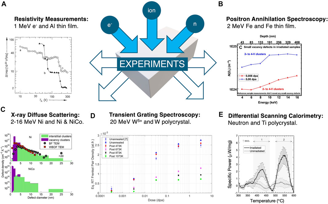
FIGURE 1. Many experimental techniques can be used to infer nanoscale defects in irradiated metals and alloys. (A) Novel resistivity measurements reveal recovery stages unseen in conventional experiments. From (Briggmann et al., 1994), Copyright 2006 John Wiley and Sons. (B) Variable energy PAS can resolve vacancy-type defects with depth. From (Agarwal et al., 2020), CC BY-NC 4.0. (C) XDS can determine the size distribution of dislocation loops down to <1 nm. From (Olsen et al., 2016), Copyright 2016 Elsevier. (D) TGS coupled to kinetic theory allows estimation of the Frenkel pair defect density as a function of dose. From (Reza et al., 2022), CC BY 4.0. (E) DSC can characterize previously unreported defect annealing stages. From (Hirst et al., 2021), CC BY 4.0.
One major limitation of this family of techniques includes the inability to directly simulate the resistivity of large defects, as density-functional theory (DFT) is computationally-limited to small supercell volumes. Additionally, many instances of deviation from Matthiessen’s rule have been reported (Bass, 1972; Fluss et al., 2004). The effect of defect clustering on the specific resistivity has been studied by Zinkle et al. (Zinkle, 1988) who report that the Frenkel pair resistivity for defects in small dislocation loops is similar to that of isolated Frenkel pairs, although this may not hold for larger loop sizes. These factors hinder the accurate determination of defect densities.
Positrons annihilation spectroscopy (PAS) is able to detect open-volume in a crystalline lattice by virtue of locally reduced electron density, allowing incident positrons to probe vacancy-type defects (Selim, 2021). Similar to resistivity measurements, positron annihilation lifetime spectroscopy (PALS) has been used for many years to investigate the formation and evolution of primary radiation damage in metals after irradiation at cryogenic temperatures and subsequent isochronal annealing (Mantl and Triftshäuser, 1978; Eldrup and Singh, 1997; Eldrup and Singh, 2003). Decomposition of the positron lifetime spectrum into multiple components can be used to characterize the size distribution of vacancy clusters (Eldrup and Singh, 2003; Hu et al., 2016), although there is a limit to the number of components that can be identified.
Additionally, the local chemical environment around vacancy-type defects can be explored through analysis of the momentum of the annihilating electron in Coincidence Doppler Broadening (CDB) experiments (Selim, 2021). This method has enabled the detection of solute-vacancy complexes in electron-irradiated reactor pressure vessel (RPV) model alloys. (Nagai et al., 2003) used CDB experiments to estimate the local concentration of solute around vacancies and attribute the irradiation-induced hardening of RPV steels to the formation of these features.
By varying the energy of the incident positrons, the depth-dependence of defect populations can be resolved in materials with heterogeneous microstructures (Lynn et al., 1986; Siemek et al., 2021). Recently, Agarwal et al. (Agarwal et al., 2020) used this method to probe the depth dependence of vacancy clusters created following 2 MeV self-ion irradiation of Fe thin-films, shown in Figure 1B. Their work reports an increase in the density of small vacancy clusters with depth and an associated decrease in large vacancy clusters. Most strikingly, they report a decrease in the diameter of cavities with increasing dose, which is attributed to a new mechanism of interstitial-induced shrinkage of voids and resultant intra-cascade nucleation of small vacancy clusters.
The ability of PALS to detect individual vacancies allows direct comparisons to be made to primary radiation damage simulations Tuomisto et al. (Tuomisto et al., 2020) studied Ni-ion irradiation damage in NiCoFeCr and its derivative alloys through PALS, molecular dynamics (MD), and density functional theory (DFT) simulations, shown in Figure 2B. They compare the modeled fraction of vacancies as a function of dose to their experimental data to rule out potential mechanisms and in doing so reveal the segregation of Ni and Co to vacancies. Soneda et al. (Soneda et al., 2003) use kMC simulations to model defect accumulation in Fe and demonstrate strong agreement between their predicted number densities of vacancy clusters and earlier PAS work. Further information on the use of PAS for irradiation materials can be found in a comprehensive review by Selim (Selim, 2021).
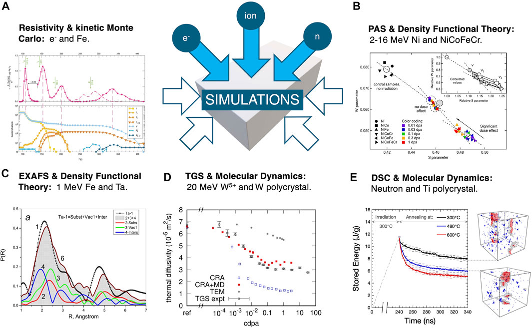
FIGURE 2. Simulation techniques can be used to aid the inference of nanoscale defects in irradiation experiments. (A) kMC can predict the isochronal evolution of defects and thus simulate the resistivity recovery spectrum. From (Fu et al., 2004), Copyright 2004 Springer Nature. (B) DFT (inset) can determine the effect of defects on positron lifetime characteristics. From (Tuomisto et al., 2020), CC BY-NC-ND 4.0. (C) DFT can be used to predict the radial probability distribution and compared to extended X-ray absorption fine structure experiments. From (Andrianov et al., 2021), Copyright 2021 Elsevier. (D) MD schemes can simulate the degradation in thermal diffusivity with dose and can be compared to TGS experiments. From (Mason et al., 2021), CC BY 4.0. (E) MD can also determine the system stored energy following PKA cascades and subsequent isothermal annealing. From (Hirst et al., 2021), CC BY 4.0.
X-ray scattering has been extensively used to probe radiation damage in metals due to its sensitivity to defects below the resolution limit of TEM and the ability to directly compare measurements to scattering theory. Many studies have used X-ray diffuse scattering (XDS) to characterize the size distribution of dislocation loops in irradiated Cu (Larson, 1975; Larson and Young, 1987; Ehrhart and Averback, 1989), Ni (Narayan and Larson, 1977; Larson and Young, 1987; Ehrhart and Averback, 1989; Yuya et al., 1999; Olsen et al., 2016), and W (Sun et al., 2018). Most of these studies compare their XDS results to the size distribution of loops determined from TEM measurements. These comparisons explicitly demonstrate a) the presence of a significant fraction of loops with radii below 1 nm, and b) the inability of TEM to characterize this population of defects. An example from (Olsen et al., 2016) is shown in Figure 1C.
More recently, the convolutional multiple whole profile (CMWP) X-ray diffraction Line Profile Analysis (XLPA) method (Ribárik, 2008; Ribárik et al., 2020) has been developed to characterize dislocation loops in irradiated materials (Seymour et al., 2017; Ungár et al., 2021b). Through calculation of the broadening of X-ray diffraction (XRD) peaks, the type, density, and size distribution of dislocation loops can be determined. Ungár et al. (Ungár et al., 2021a) demonstrated this in proton-irradiated Zircaloy-2 where they combined TEM and synchrotron XRD to calculate the power law size density function of
In addition to X-ray scattering, the local environment around atoms can be investigated using extended X-ray absorption fine structure (EXAFS) spectroscopy. EXAFS allows calculation of the radial distribution function which reveals the presence of defects through changes in the local coordination. This enables experiments to be directly compared to simulations, as demonstrated by Andrianov et al. (Andrianov et al., 2021) in Figure 2C. EXAFS has been used to study the impact of self-irradiation in Pu and its alloys as a function of dose (Booth et al., 2007; Booth et al., 2013; Olive et al., 2016; Booth and Olive, 2017). Booth et al. use EXAFS to determine the fraction of a sample that is damaged and compare these measurements to estimates based on point defect production and annealing (Booth et al., 2013). From this comparison, they report that classical defect production models (NRT-dpa and Kinchin-Pease) are insufficient to capture the effect of different bonding on defect evolution in intermetallics. Further validation of this finding with MD simulations --- as demonstrated by Okamoto et al. (Okamoto et al., 1991) for N-ion irradiated amorphous Pd80Si20, and Booth and Olive (Booth and Olive, 2017) for pure Pu --- would be insightful, but subject to the availability of accurate interatomic potentials.
The ability of EXAFS to probe the local structure also extends to chemical environments. Kuri et al. investigated neutron-irradiated, annealed and re-irradiated, RPV steels to analyze the coordination of solutes Cu, Ni, and Mn (Kuri et al., 2009). They report similar atomic environments for each of the solute atoms, with attenuation in the radial distribution function peaks attributed to local disorder. The nearest neighbor coordination of Fe atoms does not decrease, which suggests the presence of vacancy-solute complexes, as has been demonstrated in PAS (Nagai et al., 2003).
Rutherford backscattering in the channeling condition (RBS/C) is part of a family of ion beam analysis techniques that have been used to characterize disorder in irradiated materials (Chu et al., 1973; Swanson, 1982; Matzke, 1985). Placing a reference material in a channeling condition under light ion bombardment dramatically reduces the backscattered ion yield. However, by introducing defects or disorder into the lattice via irradiation, that backscattered yield increases. By varying the energy of the incident ions, the depth dependence of radiation damage can be probed. Comparing these data to the predicted damage profile allows for interpretation of the mobility of irradiation-induced defects, as shown in (Lu et al., 2016).
The radiation resistance of Ni-based complex solid solution alloys (CSSAs) has been extensively explored using RBS/C (Zhang et al., 2015; Jin et al., 2016; Lu et al., 2016; Velişa et al., 2017; Fan et al., 2019). Through comparison of the backscattering yield of different alloys irradiated to the same dose, the effect of chemical complexity on recombination can be deduced. The morphology of larger defect clusters is found to evolve as a function of dose, with dislocation loops forming at higher dose levels, which reduce the irradiation-induced strain in the material (Jin et al., 2016; Velişa et al., 2017).
Most studies use measures of the relative disorder between pristine and as-irradiated specimens to investigate the accumulated defects. However, further insight into nanoscale defect populations can be gained from RBS/C spectra simulated from atomistic configurations (Zhang et al., 2016; Zhang et al., 2017; Levo et al., 2021). Zhang et al.’s simulations of ion-irradiated Ni indicate that the contribution to the RBS/C signal from extended defects is stronger than the contribution from Frenkel pairs. While this appears to conflict with previous studies which report a decrease in irradiation-induced strain and thus decrease in backscattering signal with defect agglomeration, this work demonstrates the insight that can be gained from a combination of RBS/C experiments and simulations.
Recently, local thermophysical properties as measured using transient grating spectroscopy (TGS) have been applied to the measurement of radiation effects on metals and alloys (Short et al., 2015; Dennett et al., 2016; Hofmann et al., 2019). In these experiments, a micron-scale 1D periodic laser excitation causes local heating and thermoelastic generation of surface-confined acoustic waves (Käding et al., 1995; Johnson et al., 2012; Hofmann et al., 2019). By optically measuring the dynamics of these excitations, elastic and thermal properties can be extracted simultaneously from a single non-destructive measurement (Dennett and Short, 2018). The measurement sampling depth is on the order of single microns, a length scale particularly relevant for the study of metals with defects induced through ion beam irradiation (Hofmann et al., 2015a; Hofmann et al., 2015b; Dennett et al., 2018; AlMousa et al., 2021). Capabilities for in situ TGS measurements during high temperature ion beam exposure have also been developed (Dennett et al., 2019).
TGS has been used for nanoscale defect inference in several studies. Ferry et al. used changes of thermal diffusivity in Si-ion irradiated Nb to infer the agglomeration of point defects into clusters, reducing total thermal carrier scattering (Ferry et al., 2019). The most direct demonstration of nanoscale defect inference through TGS has come from Reza and coworkers (Reza et al., 2020; Reza et al., 2022). Initially, measurements of thermal diffusivity in self-ion irradiated W specimens were used in concert with a point defect-electron scattering model to infer the population of Frenkel defects retained after room temperature irradiation to dose levels spanning five orders of magnitude (Reza et al., 2020). Comparing with TEM-measured defect populations, a total defect density approximately an order of magnitude higher across all conditions was inferred. By using MD defect generation simulations to predict the nanoscale defect populations missed by TEM, the TGS-inferred and TEM/MD combination defect densities become comparable. In follow-on work, Reza et al. use in situ TGS during thermal annealing of previously-irradiated samples to infer point defect densities and explore their recombination kinetics (Reza et al., 2022), shown in Figure 1D. They then compared their work to MD estimations of the thermal diffusivity degradation with increasing dose, shown in Figure 2D.
Using local elastic properties measured with in situ TGS during high temperature ion irradiation, Dennett et al. studied the accumulation of point defects, vacancy clusters, and eventually void swelling in a series of Ni-based CSSAs up to and including 5-component, single phase Cantor alloy (Dennett et al., 2021). Alloy chemistries known to retain a higher density of nanoscale vacancy clusters, such as ternary NiCoCr, showed a much stronger elastic signature of vacancy-type defect density. Although TGS returns both elastic and thermal properties simultaneously, no work has yet demonstrated an inference method based on the combination of these properties together.
The characteristic energies of formation and migration can be used to infer the density of irradiation-induced defects through calorimetric experiments. Annealing irradiated materials leads to multiple stages of recovery (Schilling, 1978) where the recovery onset temperature can be used to determine the migration energy, therefore defect type, and the enthalpy change divided by the formation energy, therefore defect density. Due to their high stored energy density (∼ 103 J/g) (Snead et al., 2019) most prior literature focuses on the recovery of irradiated ceramics, including studies on graphite (Iwata, 1985), UO2 (Staicu et al., 2010), SiC (Snead et al., 2019), and NaCl (Vainshtein and Hartog, 2000). However, this strategy is also applicable to metals.
After Eugene Wigner first postulated that energy could be stored in metals as irradiation-induced defects (Wigner, 1942), many works investigated defect recovery at cryogenic temperatures. These include studies on Cu (Losehand et al., 1969; Blewitt et al., 1959; Richard et al., 1990), Al (Isebeck et al., 1966), Mg (Delaplace et al., 1968), and Be (Nicoud et al., 1968). Often the focus of these works was the fundamental science behind primary radiation damage production, rather than investigating defect evolution at engineering-relevant temperatures. Research has been conducted following ambient temperature irradiation, including studies on Mo (Kinchin and Thompson, 1958; Pedchenko and Karasev, 1971; Lambri et al., 2009), Cu (Pedchenko and Karasev, 1971; Blewitt et al., 1961), Ni and Fe (Toktogulova et al., 2010), and Pu (Ennaceur and Migliori, 2018). But little work has been conducted following irradiation at elevated temperatures, with Lee et al.’s analysis of Zr-U (Lee et al., 2007) and Hirst and colleagues’ recent study of Ti (Hirst et al., 2021), shown in Figure 1E, the only research to date. In addition, very few prior studies have leveraged the information contained within the fine structure of annealing spectra, with the exception of (Richard et al., 1990). The authors of this study derived a kinetic model which was fit to the experimental recovery of neutron-irradiated Cu. This model yielded information on the activation energies of multiple substages and was used to (in)validate the proposed recovery mechanism for reactor-irradiated metals.
Calorimetric measurements have the key advantage of being directly comparable to simulations of radiation damage. However, until recently, this comparison had only been conducted for ceramics (Béland et al., 2013). Hirst et al. used MD simulations to investigate the evolution of defects below the resolution limit of TEM in neutron-irradiated Ti (Hirst et al., 2021). Their isothermal annealing simulations, shown in Figure 2E, reveal a new mechanism for radiation damage recovery, with dislocation loops sweeping up point defects as they glide. This process was correlated to differential scanning calorimetry (DSC) measurements of the stored energy release between 300 and 480°C, where TEM micrographs show little change in the microstructure. This work reports that TEM is unable to account for 80% of the stored energy release and demonstrates the use of DSC as a tool to characterize defects that may be hidden to microscopy techniques.
Table 1 compares some characteristic features of each of the techniques highlighted above. While great insight into the populations of nanoscale defects can be gained from these methods, their accuracy depends on the quality of data interpretation. Without the ability to directly image defects, inference-based techniques rely on theoretical models to deduce the type, size distribution, and number density of defects in materials.
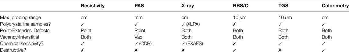
TABLE 1. Comparison of some salient features of the reviewed defect inference techniques. All methods listed can be applied to both pure metals as well as alloys and for irradiation with both neutrons and ions. Note: classifications should be understood as generalizations and abilities thus far demonstrated, not as absolute limitations.
One strategy that can be used to strengthen the inference model is to combine data streams from multiple methodologies. Disparate techniques often have unique sensitivities to different defect features, offering some degree of measurement orthogonality. Correlative experiments thus allow for a more comprehensive characterization of defect populations. This can be done ex situ, as in Meslin et al.’s study of neutron-irradiated ferritic alloys (Meslin et al., 2010), or in situ, demonstrated by Jackson’s simultaneous measurement of resistivity recovery and stored energy release in deuteron-irradiated metals (Jackson, 1980).
Many inference-based techniques are non-destructive, which allows for both concurrent multi-modal characterization and also the ability to observe nanoscale defect generation and evolution in real-time. Combining inference-based techniques and ion-accelerators allows for significant insight, but requires detailed consideration of the experimental geometries, as highlighted by Dennett et al. (Dennett et al., 2019). The ability to investigate the dynamics of defect accumulation and recovery in situ can uncover fundamental mechanisms and also accelerate nuclear materials development. It is under this promise that recent efforts have been directed in this area (Selim, 2021) and continued expansion of such capabilities is a necessity.
Combining experimental methods with simulated data, as shown in Figure 2, can also greatly benefit the interpretation of inference-based methods. This can be done directly, as in the comparison of simulated and measured atomic radial distribution functions (Andrianov et al., 2021), or indirectly, through the measurement and estimation of thermal diffusivity degradation using TGS and MD (Reza et al., 2020; Mason et al., 2021). The limitations of this strategy remain the considerable range of time and length scales that radiation damage encompasses. Atomistic simulations are typically restricted to timescales below microsecond and coarser methods rely on accurate parameterization. Recent advances in kMC methods (Béland et al., 2015) have demonstrated the ability to couple displacement cascades to experimental timescales and warrant further study.
In summary, the development of inference-based techniques has significantly advanced the ability to characterize the entire spectrum of defects formed in metals and alloys under irradiation. However, truly quantitative measurement of defects at the smallest scales remains a grand challenge. By designing future experimentation and simulation to function in concert, rapid progress towards this goal may be achieved.
CAD conceived the manuscript scope. CAH wrote the first draft of the manuscript. CAH and CAD jointly revised and approved the manuscript for publication.
CAH acknowledges support from the National Science Foundation (NSF) Faculty Early Career Development Program (CAREER) Grant DMR-1654548. CAH and CAD acknowledge support from the INL Laboratory Directed Research & Development Program under U.S. Department of Energy Idaho Operations Office Contract DE-AC07-05ID14517.
The authors declare that the research was conducted in the absence of any commercial or financial relationships that could be construed as a potential conflict of interest.
All claims expressed in this article are solely those of the authors and do not necessarily represent those of their affiliated organizations, or those of the publisher, the editors and the reviewers. Any product that may be evaluated in this article, or claim that may be made by its manufacturer, is not guaranteed or endorsed by the publisher.
Agarwal, S., Liedke, M. O., Jones, A. C. L., Reed, E., Kohnert, A. A., Uberuaga, B. P., et al. (2020). A New Mechanism for Void-Cascade Interaction from Nondestructive Depth-Resolved Atomic-Scale Measurements of Ion Irradiation-Induced Defects in Fe. Sci. Adv. 6, eaba8437. doi:10.1126/sciadv.aba8437
Albertini, G., and Coppola, R. (1992). Small-Angle Neutron Scattering Studies of Irradiated Metallic Materials. Nucl. Instr. Methods Phys. Res. Section A: Acc. Spectrometers, Detectors Associated Equipment 314, 352–365. doi:10.1016/0168-9002(92)90979-e
Allen, T., Busby, J., Meyer, M., and Petti, D. (2010). Materials Challenges for Nuclear Systems. Mater. Today 13, 14–23. doi:10.1016/s1369-7021(10)70220-0
AlMousa, N., Dacus, B., Woller, K. B., Shin, J. H., Jang, C., Shao, L., et al. (2021). On the Use of Non-Destructive, Gigahertz Ultrasonics to Rapidly Screen Irradiated Steels for Swelling Resistance. Mater. Characterization 174, 111017. doi:10.1016/j.matchar.2021.111017
Andrianov, V. A., Bedelbekova, K. A., and Trigub, A. L. (2021). Study of Radiation Defects in Metal Mo and Ta by Mössbauer Effect and EXAFS. Vacuum 193, 110521. doi:10.1016/j.vacuum.2021.110521
Bass, J. (1972). Deviations from Matthiessen's Rule. Adv. Phys. 21, 431–604. doi:10.1080/00018737200101308
Béland, L. K., Anahory, Y., Smeets, D., Guihard, M., Brommer, P., Joly, J.-F., et al. (2013). Replenish and Relax: Explaining Logarithmic Annealing in Ion-Implanted C-Si. Phys. Rev. Lett. 111, 105502. doi:10.1103/PhysRevLett.111.105502
Béland, L. K., Osetsky, Y. N., Stoller, R. E., and Xu, H. (2015). Slow Relaxation of Cascade-Induced Defects in Fe. Phys. Rev. B 91, 054108. doi:10.1103/PhysRevB.91.054108
Blewitt, T. H., Coltman, R. R., and Klabunde, C. E. (1959). Stored Energy of Irradiated Copper. Phys. Rev. Lett. 3, 132–134. doi:10.1103/physrevlett.3.132
Blewitt, T. H., Sekula, S. T., and Diehl, J. (1961). Energy Release in Reactor-Irradiated Copper. II. 600° to 700°K Release. Phys. Rev. 122, 53–57. doi:10.1103/physrev.122.53
Booth, C. H., Bauer, E. D., Daniel, M., Wilson, R. E., Mitchell, J. N., Morales, L. A., et al. (2007). Quantifying Structural Damage from Self-Irradiation in a Plutonium Superconductor. Phys. Rev. B 76, 064530. doi:10.1103/physrevb.76.064530
Booth, C. H., Jiang, Y., Medling, S. A., Wang, D. L., Costello, A. L., Schwartz, D. S., et al. (2013). Self-Irradiation Damage to the Local Structure of Plutonium and Plutonium Intermetallics. J. Appl. Phys. 113, 093502. doi:10.1063/1.4794016
Booth, C. H., and Olive, D. T. (2017). Effect of Temperature and Radiation Damage on the Local Atomic Structure of Metallic Plutonium and Related Compounds. Adv. Phys. X 2, 1–21. doi:10.1080/23746149.2016.1243994
Briggmann, J., Dagge, K., Frank, W., Seeger, A., Stoll, H., and Verbruggen, A. H. (1994). Irradiation-Induced Defects in Thin Aluminium Films Studied by 1/f Noise. Phys. Stat. Sol. (A) 146, 325–335. doi:10.1002/pssa.2211460128
Broom, T. (1954). Lattice Defects and the Electrical Resistivity of Metals. Adv. Phys. 3, 26–83. doi:10.1080/00018735400101163
Caturla, M. J., Soneda, N., Alonso, E., Wirth, B. D., Dı́az de la Rubia, T., and Perlado, J. M. (2000). Comparative Study of Radiation Damage Accumulation in Cu and Fe. J. Nucl. Mater. 276, 13–21. doi:10.1016/s0022-3115(99)00220-2
Chimi, Y., Iwase, A., Ishikawa, N., Kuroda, N., and Kambara, T. (2000). Radiation Annealing Induced by Electronic Excitation in Iron. Nucl. Instr. Methods Phys. Res. Section B: Beam Interactions Mater. Atoms 164-165, 408–414. doi:10.1016/s0168-583x(99)01139-8
Chu, W. K., Mayer, J. W., Nicolet, M.-A., Buck, T. M., Amsel, G., and Eisen, F. (1973). Principles and Applications of Ion Beam Techniques for the Analysis of Solids and Thin Films. Thin Solid Films 17, 1–41. doi:10.1016/0040-6090(73)90002-3
Delaplace, J., Hillairet, J., Nicoud, J. C., Schumacher, D., and Vogl, G. (1968). Low Temperature Neutron Radiation Damage and Recovery in Magnesium. Phys. Stat. Sol. (B) 30, 119–126. doi:10.1002/pssb.19680300115
Dennett, C. A., Buller, D. L., Hattar, K., and Short, M. P. (2019). Real-Time Thermomechanical Property Monitoring during Ion Beam Irradiation Using In Situ Transient Grating Spectroscopy. Nucl. Instr. Methods Phys. Res. Section B: Beam Interactions Mater. Atoms 440, 126–138. doi:10.1016/j.nimb.2018.10.025
Dennett, C. A., Cao, P., Ferry, S. E., Vega-Flick, A., Maznev, A. A., Nelson, K. A., et al. (2016). Bridging the Gap to Mesoscale Radiation Materials Science with Transient Grating Spectroscopy. Phys. Rev. B 94, 214106. doi:10.1103/physrevb.94.214106
Dennett, C. A., Dacus, B. R., Barr, C. M., Clark, T., Bei, H., Zhang, Y., et al. (2021). The Dynamic Evolution of Swelling in Nickel Concentrated Solid Solution Alloys through In Situ Property Monitoring. Appl. Mater. Today 25, 101187. doi:10.1016/j.apmt.2021.101187
Dennett, C. A., and Short, M. P. (2018). Thermal Diffusivity Determination Using Heterodyne Phase Insensitive Transient Grating Spectroscopy. J. Appl. Phys. 123, 215109. doi:10.1063/1.5026429
Dennett, C. A., So, K. P., Kushima, A., Buller, D. L., Hattar, K., and Short, M. P. (2018). Detecting Self-Ion Irradiation-Induced Void Swelling in Pure Copper Using Transient Grating Spectroscopy. Acta Materialia 145, 496–503. doi:10.1016/j.actamat.2017.12.007
Ehrhart, P., and Averback, R. S. (1989). Diffuse X-ray Scattering Studies of Neutron- and Electron-Irradiated Ni, Cu and Dilute Alloys. Philosophical Mag. A 60, 283–306. doi:10.1080/01418618908213863
Ehrhart, P. (1994). Investigation of Radiation Damage by X-Ray Diffraction. J. Nucl. Mater. 216, 170–198. doi:10.1016/0022-3115(94)90012-4
Eldrup, M., and Singh, B. N. (2003). Accumulation of Point Defects and Their Complexes in Irradiated Metals as Studied by the Use of Positron Annihilation Spectroscopy - a Brief Review. J. Nucl. Mater. 323, 346–353. doi:10.1016/j.jnucmat.2003.08.011
Eldrup, M., and Singh, B. N. (1997). Studies of Defects and Defect Agglomerates by Positron Annihilation Spectroscopy. J. Nucl. Mater. 251, 132–138. doi:10.1016/s0022-3115(97)00221-3
Ennaceur, S. M., and Migliori, A. (2018). Toward an Understanding of Aging in Plutonium from Direct Measurements of Stored Energy. Philos. Mag. Lett. 98, 502–510. doi:10.1080/09500839.2019.1583392
Fan, Z., Velisa, G., Jin, K., Crespillo, M. L., Bei, H., and Weber, W. J. (2019). Temperature-Dependent Defect Accumulation and Evolution in Ni-Irradiated NiFe Concentrated Solid-Solution Alloy. J. Nucl. Mater. 519, 1–9. doi:10.1016/j.jnucmat.2019.03.031
Ferry, S. E., Dennett, C. A., Woller, K. B., and Short, M. P. (2019). Inferring Radiation-Induced Microstructural Evolution in Single-Crystal Niobium Through Changes in Thermal Transport. J. Nucl. Mater. 523, 378–382. doi:10.1016/j.jnucmat.2019.06.015
Fluss, M. J., Wirth, B. D., Wall, M., Felter, T. E., Caturla, M. J., Kubota, A., et al. (2004). Temperature-dependent Defect Properties from Ion-Irradiation in Pu(Ga). J. Alloys Compd. 368, 62–74. doi:10.1016/j.jallcom.2003.08.080
Fu, C.-C., Torre, J. D., Willaime, F., Bocquet, J.-L., and Barbu, A. (2004). Multiscale Modelling of Defect Kinetics in Irradiated Iron. Nat. Mater. 4, 68–74. doi:10.1038/nmat1286
Gilbert, M. R., Arakawa, K., Bergstrom, Z., Caturla, M. J., Dudarev, S. L., Gao, F., et al. (2021). Perspectives on Multiscale Modelling and Experiments to Accelerate Materials Development for Fusion. J. Nucl. Mater. 554, 153113. doi:10.1016/j.jnucmat.2021.153113
Gómez-Ferrer, B. (2016). Resistivity Recovery in Fe and FeCr Alloys. Berlin, Germany: Springer Briefs in Applied Sciences and Technology.
Hirst, C. A., Granberg, F., Kombaiah, B., Cao, P., Middlemas, S., Kemp, R. S., et al. (2021). Revealing Hidden Defects through Stored Energy Measurements of Radiation Damage. Available at: https://arxiv.org/abs/2109.15138.
Hofmann, F., Mason, D. R., Eliason, J. K., Maznev, A. A., Nelson, K. A., and Dudarev, S. L. (2015). Non-contact Measurement of thermal Diffusivity in Ion-Implanted Nuclear Materials. Sci. Rep. 5, 16042. doi:10.1038/srep16042
Hofmann, F., Nguyen-Manh, D., Gilbert, M. R., Beck, C. E., Eliason, J. K., Maznev, A. A., et al. (2015). Lattice Swelling and Modulus Change in a Helium-Implanted Tungsten alloy: X-ray Micro-diffraction, Surface Acoustic Wave Measurements, and Multiscale Modelling. Acta Materialia 89, 352–363. doi:10.1016/j.actamat.2015.01.055
Hofmann, F., Short, M. P., and Dennett, C. A. (2019). Transient Grating Spectroscopy: An Ultrarapid, Nondestructive Materials Evaluation Technique. MRS Bull. 44, 392–402. doi:10.1557/mrs.2019.104
Horak, J. A., and Blewitt, T. H. (1975). Fast- and Thermal-Neutron Irradiation and Annealing of Cu, Ni, Fe, Ti, Pd. Nucl. Technol. 27, 416.
Hu, X., Koyanagi, T., Fukuda, M., Katoh, Y., Snead, L. L., and Wirth, B. D. (2016). Defect Evolution in Single Crystalline Tungsten Following Low Temperature and Low Dose Neutron Irradiation. J. Nucl. Mater. 470, 278–289. doi:10.1016/j.jnucmat.2015.12.040
Isebeck, K., Rau, F., Schilling, W., Sonnenberg, K., Tischer, P., and Wenzl, H. (1966). Stored Energy, Volume, and Resistivity Change in Neutron Irradiated Aluminium. Phys. Status Solidi B 17, 259–268. doi:10.1515/9783112498088-031
Iwamoto, Y., Yoshiie, T., Yoshida, M., Nakamoto, T., Sakamoto, M., Kuriyama, Y., et al. (2015). Measurement of the Displacement Cross-Section of Copper Irradiated with 125 MeV Protons at 12 K. J. Nucl. Mater. 458, 369–375. doi:10.1016/j.jnucmat.2014.12.125
Iwase, A., Iwata, T., Nihira, T., and Sasaki, S. (1992). Defect Recovery and Radiation Annealing in Fcc Metals Irradiated with High Energy Ions. Radiat. Effects Defects Sol. 124, 117–126. doi:10.1080/10420159208219833
Iwata, T. (1985). Fine Structure of Wigner Energy Release Spectrum in Neutron Irradiated Graphite. J. Nucl. Mater. 133-134, 361–364. doi:10.1016/0022-3115(85)90168-0
Jackson, J. J. (1980). Calorimeter for Simultaneous, Low‐Temperature Measurements of Energy Release and of Resistivity Recovery in Irradiated Metals. Rev. Scientific Instr. 51, 35–41. doi:10.1063/1.1136052
Jenkins, M. L. (1994). Characterisation of Radiation-Damage Microstructures by TEM. J. Nucl. Mater. 216, 124–156. doi:10.1016/0022-3115(94)90010-8
Jenkins, M. L., and Kirk, M. A. (2001). Characterisation of Radiation Damage by Transmission Electron Microscopy. Bristol, UK: IoP Publishing.
Jin, K., Bei, H., and Zhang, Y. (2016). Ion Irradiation Induced Defect Evolution in Ni and Ni-Based Fcc Equiatomic Binary Alloys. J. Nucl. Mater. 471, 193–199. doi:10.1016/j.jnucmat.2015.09.009
Johnson, J. A., Maznev, A. A., Bulsara, M. T., Fitzgerald, E. A., Harman, T. C., Calawa, S., et al. (2012). Phase-Controlled, Heterodyne Laser-Induced Transient Grating Measurements of thermal Transport Properties in Opaque Material. J. Appl. Phys. 111, 023503. doi:10.1063/1.3675467
Käding, O. W., Skurk, H., Maznev, A. A., and Matthias, E. (1995). Transient Thermal Gratings at Surfaces for Thermal Characterization of Bulk Materials and Thin Films. Appl. Phys. A. 61, 253–261.
Khanolkar, A., Dennett, C. A., Hua, Z., Mann, J. M., Hurley, D. H., and Khafizov, M. (2022). Inferring Relative Dose-Dependent Color Center Populations in Proton Irradiated Thoria Single Crystals Using Optical Spectroscopy. Phys. Chem. Chem. Phys. 24, 6133–6145. doi:10.1039/d1cp05191a
Kinchin, G. H., and Thompson, M. W. (1958). Irradiation Damage and Recovery in Molybdenum and Tungsten. J. Nucl. Energ. (1954) 6, 275–284. doi:10.1016/0891-3919(58)90054-8
Kuri, G., Cammelli, S., Degueldre, C., Bertsch, J., and Gavillet, D. (2009). Neutron Induced Damage in Reactor Pressure Vessel Steel: An X-ray Absorption fine Structure Study. J. Nucl. Mater. 385, 312–318. doi:10.1016/j.jnucmat.2008.12.037
Lambri, O. A., Zelada-Lambri, G. I., Cuello, G. J., Bozzano, P. B., and García, J. A. (2009). Study of the Temperature Evolution of Defect Agglomerates in Neutron Irradiated Molybdenum Single Crystals. J. Nucl. Mater. 385, 552–558. doi:10.1016/j.jnucmat.2008.12.312
Larson, B. C. (1975). X-ray Studies of Defect Clusters in Copper. J. Appl. Cryst. 8, 150–160. doi:10.1107/s0021889875009946
Larson, B. C., and Young, F. W. (1987). X-ray Diffuse Scattering Study of Irradiation Induced Dislocation Loops in Copper. Phys. Stat. Sol. (A) 104, 273–286. doi:10.1002/pssa.2211040120
Lee, B., Cheon, J.-S., Koo, Y.-H., Oh, J.-Y., Yim, J.-S., Sohn, D.-S., et al. (2007). Measurement of the Specific Heat of Zr–40 Wt%U Metallic Fuel. J. Nucl. Mater. 360, 315–320. doi:10.1016/j.jnucmat.2006.10.023
Levo, E., Granberg, F., Nordlund, K., and Djurabekova, F. (2021). Temperature Effect on Irradiation Damage in Equiatomic Multi-Component Alloys. Comput. Mater. Sci. 197, 110571. doi:10.1016/j.commatsci.2021.110571
Li, J.-T., Beyerlein, I. J., and Han, W.-Z. (2022). Helium Irradiation-Induced Ultrahigh Hardening in Niobium. Acta Materialia 226, 117656. doi:10.1016/j.actamat.2022.117656
Losehand, R., Rau, F., and Wenzl, H. (1969). Stored Energy and Electrical Resistivity of Frenkel Defects in Copper. Radiat. Effects 2, 69–74. doi:10.1080/00337576908235586
Lu, C., Jin, K., Béland, L. K., Zhang, F., Yang, T., Qiao, L., et al. (2016). Direct Observation of Defect Range and Evolution in Ion-Irradiated Single Crystalline Ni and Ni Binary Alloys. Sci. Rep. 6, 19994. doi:10.1038/srep19994
Lucasson, P. G., and Walker, R. M. (1962). Production and Recovery of Electron-Induced Radiation Damage in a Number of Metals. Phys. Rev. 127, 485–500. doi:10.1103/physrev.127.485
Lynn, K. G., Chen, D. M., Nielsen, B., Pareja, R., and Myers, S. (1986). Variable-Energy Positron-Beam Studies of Ni Implanted with He. Phys. Rev. B 34, 1449–1458. doi:10.1103/physrevb.34.1449
Mantl, S., and Triftshäuser, W. (1978). Defect Annealing Studies on Metals by Positron Annihilation and Electrical Resitivity Measurements. Phys. Rev. B 17, 1645–1652. doi:10.1103/physrevb.17.1645
Mason, D. R., Reza, A., Granberg, F., and Hofmann, F. (2021). Estimate for thermal Diffusivity in Highly Irradiated Tungsten Using Molecular Dynamics Simulation. Phys. Rev. Mater. 5, 125407. doi:10.1103/physrevmaterials.5.125407
Matthiessen, A., and Vogt, C. (1864). Ueber den Einfluss der Temperatur auf die elektrische Leitungsfähigkeit der Legirungen. Ann. Phys. Chem. 198, 19–78. doi:10.1002/andp.18641980504
Matzke, H. (1985). Application of Ion Beam Techniques to Solid State Physics and Technology of Nuclear Materials. J. Nucl. Mater. 136, 143–153. doi:10.1016/0022-3115(85)90001-7
Meslin, E., Lambrecht, M., Hernández-Mayoral, M., Bergner, F., Malerba, L., Pareige, P., et al. (2010). Characterization of Neutron-Irradiated Ferritic Model Alloys and a RPV Steel from Combined APT, SANS, TEM and PAS Analyses. J. Nucl. Mater. 406, 73–83. doi:10.1016/j.jnucmat.2009.12.021
Nagai, Y., Takadate, K., Tang, Z., Ohkubo, H., Sunaga, H., Takizawa, H., et al. (2003). Positron Annihilation Study of Vacancy-Solute Complex Evolution in Fe-Based Alloys. Phys. Rev. B 67, 224202. doi:10.1103/physrevb.67.224202
Nakagawa, M., Böning, K., Rosner, P., and Vogl, G. (1977). High-Dose Neutron-Irradiation Effects in Fcc Metals at 4.6 K. Phys. Rev. B 16, 5285–5302. doi:10.1103/physrevb.16.5285
Nakagawa, M., Mansel, W., Böning, K., Rosner, P., and Vogl, G. (1979). Spontaneous Recombination Volumes of Frenkel Defects in Neutron-Irradiated Non-fcc Metals. Phys. Rev. B 19, 742–748. doi:10.1103/physrevb.19.742
Nakagawa, M. (1982). Saturation Phenomena in Irradiated Metals at Low Temperature. J. Nucl. Mater. 108-109, 194–200. doi:10.1016/0022-3115(82)90487-1
Narayan, J., and Larson, B. C. (1977). Defect Clusters and Annealing in Self‐Ion‐Irradiated Nickel. J. Appl. Phys. 48, 4536–4539. doi:10.1063/1.323468
Nicoud, J. C., Bonjour, E., Schumacher, D., and Delaplace, J. (1968). Restauration dans le stade i du beryllium irradie par des neutrons et par des electrons. Phys. Lett. A 26, 228–229. doi:10.1016/0375-9601(68)90616-6
Nikolaev, A. L. (2009). Difference Approach to the Analysis of Resistivity Recovery Data for Irradiated Short-Range Ordered Alloys. Philos. Mag. 89, 1017–1033. doi:10.1080/14786430902835651
Nikolaev, A. L. (2007). Specificity of Stage III in Electron-Irradiated Fe-Cr Alloys. Philos. Mag. 87, 4847–4874. doi:10.1080/14786430701468977
Nikolaev, A. L. (2018). The Unambiguous Identification of Vacancy Migration Stage in Resistivity Recovery of Irradiated Metals Based on Trapping of Vacancies at Probe Impurity Atoms. Philos. Mag. 98, 2481–2494. doi:10.1080/14786435.2018.1492158
Okamoto, Y., Takagi, R., and Kawamura, K. (1991). Molecular Dynamics of Radiation Damage in Amorphous Pd80Si20Alloy Using N+Ions. Mol. Simulation 6, 353–362. doi:10.1080/08927029108022443
Olive, D. T., Wang, D. L., Booth, C. H., Bauer, E. D., Pugmire, A. L., Freibert, F. J., et al. (2016). Isochronal Annealing Effects on Local Structure, Crystalline Fraction, and Undamaged Region Size of Radiation Damage in Ga-stabilized δ-Pu. J. Appl. Phys. 120, 035103. doi:10.1063/1.4958856
Olsen, R. J., Jin, K., Lu, C., Beland, L. K., Wang, L., Bei, H., et al. (2016). Investigation of Defect Clusters in Ion-Irradiated Ni and NiCo Using Diffuse X-ray Scattering and Electron Microscopy. J. Nucl. Mater. 469, 153–161. doi:10.1016/j.jnucmat.2015.11.030
Pedchenko, K. S., and Karasev, V. S. (1971). Stored Energy in Neutron-Bombarded Metals. Mater. Sci. 4, 332–336. doi:10.1007/bf00722627
Reza, A., He, G., Dennett, C. A., Yu, H., Mizohata, K., and Hofmann, F. (2022). Thermal Diffusivity Recovery and Defect Annealing Kinetics of Self-Ion Implanted Tungsten Probed by In Situ Transient Grating Spectroscopy. Acta Materialia 232, 117926. doi:10.1016/j.actamat.2022.117926
Reza, A., Yu, H., Mizohata, K., and Hofmann, F. (2020). Thermal Diffusivity Degradation and Point Defect Density in Self-Ion Implanted Tungsten. Acta Materialia 193, 270–279. doi:10.1016/j.actamat.2020.03.034
Ribárik, G., Jóni, B., and Ungár, T. (2020). The Convolutional Multiple Whole Profile (CMWP) Fitting Method, a Global Optimization Procedure for Microstructure Determination. Crystals 10, 623. doi:10.3390/cryst10070623
Ribárik, G. (2008). Modeling of Diffraction Patterns Based on Microstructural Properties. Ph.D. thesis (Budapest, Hungary: Eötvös Loránd University).
Richard, R. T., Chaplin, R. L., Coltman, R. R., Kerchner, H. R., and Klabunde, C. E. (1990). Stored Energy Recovery of Irradiated Copper. Radiat. Effects Defects Sol. 112, 161–179. doi:10.1080/10420159008213042
Rickert, K., Prusnick, T. A., Hunt, E., French, A., Turner, D. B., Dennett, C. A., et al. (2022). Raman and Photoluminescence Evaluation of Ion-Induced Damage Uniformity in ThO2. Nucl. Instr. Methods Phys. Res. Section B: Beam Interactions Mater. Atoms 515, 69–79. doi:10.1016/j.nimb.2022.01.011
Schilling, W. (1978). Radiation Induced Damage in Metals. J. Nucl. Mater. 72, 1–4. doi:10.1016/0022-3115(78)90384-7
Seidman, D. N. (1978). The Study of Radiation Damage in Metals with the Field-Ion and Atom-Probe Microscopes. Surf. Sci. 70, 532–565. doi:10.1016/0039-6028(78)90430-2
Selim, F. A. (2021). Positron Annihilation Spectroscopy of Defects in Nuclear and Irradiated Materials- a Review. Mater. Characterization 174, 110952. doi:10.1016/j.matchar.2021.110952
Seymour, T., Frankel, P., Balogh, L., Ungár, T., Thompson, S. P., Jädernäs, D., et al. (2017). Evolution of Dislocation Structure in Neutron Irradiated Zircaloy-2 Studied by Synchrotron X-ray Diffraction Peak Profile Analysis. Acta Materialia 126, 102–113. doi:10.1016/j.actamat.2016.12.031
Shelyug, A., Palomares, R. I., Lang, M., and Navrotsky, A. (2018). Energetics of Defect Production in Fluorite-Structured CeO2 Induced by Highly Ionizing Radiation. Phys. Rev. Mat. 2, 093607. doi:10.1103/physrevmaterials.2.093607
Short, M. P., Dennett, C. A., Ferry, S. E., Yang, Y., Mishra, V. K., Eliason, J. K., et al. (2015). Applications of Transient Grating Spectroscopy to Radiation Materials Science. JOM 67, 1840–1848. doi:10.1007/s11837-015-1496-3
Siemek, K., Horodek, P., Skuratov, V. A., Waliszewski, J., and Sohatsky, A. (2021). Positron Annihilation Studies of Irradiation Induced Defects in Nanostructured Titanium. Vacuum 190, 110282. doi:10.1016/j.vacuum.2021.110282
Snead, L. L., Katoh, Y., Koyanagi, T., and Terrani, K. (2019). Stored Energy Release in Neutron Irradiated Silicon Carbide. J. Nucl. Mater. 514, 181–188. doi:10.1016/j.jnucmat.2018.12.005
Soneda, N., Ishino, S., Takahashi, A., and Dohi, K. (2003). Modeling the Microstructural Evolution in Bcc-Fe during Irradiation Using Kinetic Monte Carlo Computer Simulation. J. Nucl. Mater. 323, 169–180. doi:10.1016/j.jnucmat.2003.08.021
Staicu, D., Wiss, T., Rondinella, V. V., Hiernaut, J. P., Konings, R. J. M., and Ronchi, C. (2010). Impact of Auto-Irradiation on the Thermophysical Properties of Oxide Nuclear Reactor Fuels. J. Nucl. Mater. 397, 8–18. doi:10.1016/j.jnucmat.2009.11.024
Sun, P., Wang, Y., Frost, M., Schönwälder, C., Levitan, A. L., Mo, M., et al. (2018). Characterization of Defect Clusters in Ion-Irradiated Tungsten by X-ray Diffuse Scattering. J. Nucl. Mater. 510, 322–330. doi:10.1016/j.jnucmat.2018.07.062
Swanson, M. L. (1982). The Study of Lattice Defects by Channelling. Rep. Prog. Phys. 45, 47–93. doi:10.1088/0034-4885/45/1/002
Takaki, S., Fuss, J., Kuglers, H., Dedek, U., and Schultz, H. (1983). The Resistivity Recovery of High Purity and Carbon Doped Iron Following Low Temperature Electron Irradiation. Radiat. Effects 79, 87–122. doi:10.1080/00337578308207398
Toktogulova, D. A., Gusev, M. N., Maksimkin, O. P., and Garner, F. A. (2010). Influence of Neutron Irradiation on Energy Accumulation and Dissipation during Plastic Flow and Hardening of Metallic Polycrystals. J. ASTM Int. 7, 238. doi:10.1520/jai102175
Tuomisto, F., Makkonen, I., Heikinheimo, J., Granberg, F., Djurabekova, F., Nordlund, K., et al. (2020). Segregation of Ni at Early Stages of Radiation Damage in NiCoFeCr Solid Solution Alloys. Acta Materialia 196, 44–51. doi:10.1016/j.actamat.2020.06.024
Ungár, T., Frankel, P., Ribárik, G., Race, C. P., and Preuss, M. (2021). Size-distribution of Irradiation-Induced Dislocation-Loops in Materials Used in the Nuclear Industry. J. Nucl. Mater. 550, 152945. doi:10.1016/j.jnucmat.2021.152945
Ungár, T., Ribárik, G., Topping, M., Jones, R. M. A., Xu, X. D., Hulse, R., et al. (2021). Characterizing Dislocation Loops in Irradiated Polycrystalline Zr Alloys by X-ray Line Profile Analysis of Powder Diffraction Patterns with Satellites. J. Appl. Cryst. 54, 803–821. doi:10.1107/S1600576721002673
Vainshtein, D. I., and Hartog, H. W. D. (2000). Explosive Decomposition of Heavily Irradiated NaCl. Radiat. Effects Defects Sol. 152, 23–37. doi:10.1080/10420150008211811
Velişa, G., Ullah, M. W., Xue, H., Jin, K., Crespillo, M. L., Bei, H., et al. (2017). Irradiation-Induced Damage Evolution in Concentrated Ni-Based Alloys. Acta Mater. 135, 54–60. doi:10.1016/j.actamat.2017.06.002
Wigner, E. (1942). Report for Month Ending December 15. Chicago: Tech. Rep. (US Atomic Energy Commission Report CP-387), Univ. of Chicago.
Yi, X., Sand, A. E., Mason, D. R., Kirk, M. A., Roberts, S. G., Nordlund, K., et al. (2015). Direct Observation of Size Scaling and Elastic Interaction Between Nano-Scale Defects in Collision Cascades. Euro. Phys. Lett. 110, 36001. doi:10.1209/0295-5075/110/36001
Yuya, H., Maeta, H., Ohtsuka, H., Matsumoto, N., Sugai, H., Iwase, A., et al. (1999). Diffuse X-ray Scattering Studies of Radiation Defects in Ni and Dilute Ni Alloys. J. Nucl. Mater. 271-272, 7–10. doi:10.1016/s0022-3115(98)00642-4
Zhang, S., Nordlund, K., Djurabekova, F., Zhang, Y., Velisa, G., and Wang, T. S. (2016). Simulation of Rutherford Backscattering Spectrometry from Arbitrary Atom Structures. Phys. Rev. E 94, 043319. doi:10.1103/PhysRevE.94.043319
Zhang, S., Nordlund, K., Djurabekova, F., Granberg, F., Zhang, Y., and Wang, T. S. (2017). Radiation Damage Buildup by Athermal Defect Reactions in Nickel and Concentrated Nickel Alloys. Mater. Res. Lett. 5, 433–439. doi:10.1080/21663831.2017.1311284
Zhang, Y., Stocks, G. M., Jin, K., Lu, C., Bei, H., Sales, B. C., et al. (2015). Influence of Chemical Disorder on Energy Dissipation and Defect Evolution in Concentrated Solid Solution Alloys. Nat. Commun. 6, 8736. doi:10.1038/ncomms9736
Zhou, Z., Dudarev, S. L., Jenkins, M. L., Sutton, A. P., and Kirk, M. A. (2007). Diffraction Imaging and Diffuse Scattering by Small Dislocation Loops. J. Nucl. Mater. 367-370, 305–310. doi:10.1016/j.jnucmat.2007.03.135
Zinkle, S. J. (1988). Electrical Resistivity of Small Dislocation Loops in Irradiated Copper. J. Phys. F: Met. Phys. 18, 377–391. doi:10.1088/0305-4608/18/3/009
Keywords: Defects, irradiated, metals, resistivity, positron annihilation spectroscopy, X-ray scattering, Rutherford backscattering, transient grating spectroscopy
Citation: Hirst CA and Dennett CA (2022) Towards Quantitative Inference of Nanoscale Defects in Irradiated Metals and Alloys. Front. Mater. 9:888356. doi: 10.3389/fmats.2022.888356
Received: 02 March 2022; Accepted: 11 April 2022;
Published: 13 May 2022.
Edited by:
Long Xin, University of Science and Technology Beijing, ChinaReviewed by:
Ke Jin, Beijing Institute of Technology, ChinaCopyright © 2022 Hirst and Dennett. This is an open-access article distributed under the terms of the Creative Commons Attribution License (CC BY). The use, distribution or reproduction in other forums is permitted, provided the original author(s) and the copyright owner(s) are credited and that the original publication in this journal is cited, in accordance with accepted academic practice. No use, distribution or reproduction is permitted which does not comply with these terms.
*Correspondence: Charles A. Hirst, Y2FoaXJzdEBtaXQuZWR1 Cody A. Dennett, Y2Rlbm5ldHRAbWl0LmVkdQ==
Disclaimer: All claims expressed in this article are solely those of the authors and do not necessarily represent those of their affiliated organizations, or those of the publisher, the editors and the reviewers. Any product that may be evaluated in this article or claim that may be made by its manufacturer is not guaranteed or endorsed by the publisher.
Research integrity at Frontiers
Learn more about the work of our research integrity team to safeguard the quality of each article we publish.