
95% of researchers rate our articles as excellent or good
Learn more about the work of our research integrity team to safeguard the quality of each article we publish.
Find out more
REVIEW article
Front. Mater. , 28 July 2021
Sec. Biomaterials
Volume 8 - 2021 | https://doi.org/10.3389/fmats.2021.708945
 Alfredo Ayala-Ham1,2Jorge López-Gutierrez2
 Mercedes Bermúdez3
 Maribel Aguilar-Medina3Juan Ignacio Sarmiento-Sánchez4
 César López-Camarillo5
 Guzman Sanchez-Schmitz6
 Rosalio Ramos-Payan3*
Regenerative therapy in dentistry has gained interest given the complexity to restore dental and periodontal tissues with inert materials. The best approach for regeneration requires three elements for restoring functions of affected or diseased organ tissues: cells, bioactive molecules, and scaffolds. This triad is capable of modulating the processes to replace lost or damaged tissues and restore function, as it has an impact on diverse cellular processes, influencing cell behavior positively to induce the complete restoration of function and morphology of such complex tissues. Hydrogels (HG) have shown advantages as scaffolds as they are soft and elastic three-dimensional (3D) networks formed from hydrophilic homopolymers, copolymers, or macromers. Besides simple or hybrid, HG show chemical, mechanical and biological activities such as the incorporation of cells in their structures, the retention of high-water content which enhances the transportation of cell nutrients and waste, and elastic and flexible characteristics that emulate the native extracellular matrix (ECM). HG can induce changes in cellular processes such as chemotaxis, proliferation, angiogenesis, biomineralization, and expression of specific tissue biomarkers, enhancing the regeneration process. Besides some of them have anti-inflammatory and anti-bacterial effects. This review aims to show an extensive overview of the most used hydrogels in tissue engineering, emphasizing those that are studied for the regeneration of oral tissues, their biological effects, and their clinical implications. Even though most of the HG are still under investigation, some of them have been studied in vitro and in vivo with outstanding results that may lead to preclinical studies. Besides there are HG that have shown their efficacy in patients such as hyaluronan HG that enhances the healing of gingival tissue.
In recent years, advances have been made in oral tissue engineering and regenerative dentistry thanks to the growing amount of research in fields such as, stem cell biology, genetic, molecular engineering, and pathologies that affect the dental organ and its supporting tissues (Miran et al., 2016). Nevertheless, regeneration is still ruled by the need for three elements that have the goal of restoring functions of affected or diseased organ tissues (Cavalcanti et al., 2013; Lee et al., 2014; Fukushima et al., 2019): ECM, which serves as a scaffold; active biomolecules, that regulate cell growth and differentiation; and mesenchymal cells, needed for the new tissue formation (Sakata et al., 2015). Thus, in tissue engineering, regeneration will be achieved if a structural scaffold is provided, conferring mechanical properties (Goker et al., 2019), but also, inducing migration of cell populations. Besides, biomaterials used in tissue engineering must be degraded in a time range comparable to the growth of the new tissue (Colombo et al., 2014).
Consequently, dental biomaterials have passed from passive bioinert structures to bioactive materials that have the final goal to return form and function to oral tissues (Schmalz and Smith, 2014). Despite progress, oral tissue regeneration is challenging as the dental organ is made up of different tissues as well as the periodontium. In this regard, hydrogels (HG), have been gaining interest since they possess three-dimensional (3D) polymeric networks with characteristics similar to tissues (Athirasala et al., 2018).
According to Slaughter et al., HG are defined as 3D insoluble polymer matrices created from crosslinked hydrophilic homopolymers, copolymers, or macromers (Slaughter et al., 2009). Due to their thermodynamic compatibility, these polymers are soft and elastic with water and have been used in many biomedical applications (Peppas et al., 2000; Slaughter et al., 2009). In the case of biological HG, they can be formed mainly from agarose, alginate, chitosan, hyaluronan, fibrin, and collagen (Lee and Mooney, 2001; Malafaya et al., 2007).
Such HG, either simple or hybrid, have shown excellent chemical, mechanical and biological activities (Moussa and Aparicio, 2019). They work incorporating cells in their structures, and at the same time, degrading themselves to let place to new healthy tissue. HG, been porous structures, can retain high-water content enhancing the transportation of cell nutrients and waste. Besides, HG are elastic and flexible emulating the native ECM (Mantha et al., 2019).
HG are considered the biomaterial of choice for tissue engineering in dentistry and biomedicine. According to Eelkema et al., at present, HG are used in personal care products, biomaterials, coatings, and plant fertilizers. They also are considered for future applications such as sensing, drug delivery, soft robotics, and biohybrid or biointerfacing materials (Chai, 2009). When HG are compared with other types of biomaterials, they are superior showing proper mechanical strength, porous structure, enhanced biocompatibility, and adjustable biodegradability (Chai et al., 2017). HG have emerged as a promising biomaterial for therapeutic delivery of cells and bioactive molecules for tissue regeneration in dentistry, showing the capacity to conform to the three-dimensional defect and the adaptability for minimally invasive surgical procedures. Given that HG have been considered the biomaterial of choice for tissue engineering applications (Fan and Wang, 2017), review aims to show an extensive overview of the most used hydrogels in tissue engineering, emphasizing those that are studied for the regeneration of oral tissues , their biological effects, and their clinical implications. This is the first review that compiles the use of hydrogels in regenerative dentistry, taking into consideration all dental and periodontal tissues.
The success of tissue regeneration relies on the design of biofunctional scaffolds. Lately, HG are considered the best candidates given their characteristics that emulate the natural ECM, enabling proliferation, vascularization, and survival of cells. Here we summarize the most common HG used in tissue engineering, their characteristics and its current uses.
Collagen-based HG mimic interactions between cells and ECM in vivo and can be biofunctionalized. Collagen is the most studied fibrous protein, it confers unique properties to the ECM allowing cell adhesion, migration, and proliferation (Lodish et al., 2000). One of the most common methods to obtain it for therapeutic purposes is by decellularization of organs or tissues (Magno et al., 2020). Currently, collagen-based HG can be found commercially in different concentrations, alone or mixed with various molecules, such as cinnamaldehyde (Kwon et al., 2017), riboflavin (that confers resistance, compression, shortens the setting time, and also confers photocurable properties), and chondroitin sulfate-polyethylene glycol (CS PEG) adhesive (Chae et al., 2014). Collagen can be biofunctionalized using growth factors (Momose et al., 2016a, 2016b), bone morphogenetic proteins (BMPs) (Yamamoto et al., 2003), cytokines, or exomes potentiating their use (Barthes et al., 2021).
Collagen-based HG can mimic cell-cell and cell-matrix interactions in vivo and regulate more orderly cell growth, for this reason, it have been applied to the engineering of cardiac tissues (Kaiser et al., 2018), corneal and corneoscleral regions (Chae et al., 2014), alveolar bone, and periodontal tissues (Kato et al., 2015). Also in the aesthetic area, cosmetic formulations are widely used due to their moisturizing, regenerating, and film-forming properties, for filling wrinkles and facial expression lines (Li et al., 2005).
Hyaluronic acid or hyaluronan (HyA) HG can be combined to improve their characteristics and have multiple clinical uses in human. HyA is a biopolymer that can be modified and processed to form HG for biomedical applications (Burdick and Prestwich, 2011). It has great biological importance since it has a high affinity to CD44 and is degradable through oxidative species and various enzymes such as hyaluronidase, beta-D glucuronidase and beta D-N-acetyl-hexosaminidase (Stern, 2004). HyA HG can combine with DNA/polyethyleneimine molecules and MMP-sensitive peptides to modulate the delivery of particles to cells (Gojgini et al., 2011). HyA can be directly combined with 2-Aminoethyl methacrylate (AEMA) (Niloy et al., 2020a), platelet lysate (HAPL) (Almeida et al., 2018a), polyethylene glycol diacrylate (PEGDA) + Gelatin (HySistem-C) (Jones et al., 2016a, 2016b), and catechol (enhancing adhesion in vivo) (Shin et al., 2015). HyA HG systems have been successful for clinical use in human patients in areas such as ophthalmology (Chang et al., 2021), orthopedics (Pitarresi et al., 2013), oncology (Suo et al., 2019), gynecology (Chen et al., 2017), dentistry (Ni et al., 2019), and plastic surgery (Borzacchiello et al., 2015) for the repair of skin (as dermal fillers) and neural tissues, (Yeom et al., 2010), in gingival tissue (Jentsch et al., 2003), corneal tissue (Williams et al., 2017), for inter-articular viscosupplementation (Narkbunnam et al., 2012) and prevention of postoperative adhesions (Li et al., 2014a).
Gelatin based HG offer a variety of benefits for tissue engineering that enable their use as scaffold and drug delivery system. Gelatin is an insipid protein derived from collagen used in food and pharmaceutical industries (Mariod et al., 2011). It is obtained from the skin, scales, bones, ligaments, and tendons of bovine and porcine livestock (Okonkwo et al., 2007). Its abundance, low price, biodegradability, and non-toxicity (Ulfa et al., 2015) make it ideal for therapeutical use. Gelatin-based HG retain their structure as basic polymer chains, which can be combined, allowing biocompatibility, low immunogenicity, rapid biodegradation, allowing their use in biomedical applications such as in the manufacture of contact lenses, matrices for tissue engineering, and systems drug administration (Hoffman, 2012).
Nowadays, methacrylate gelatin HG (GelMA), is also used and polymerizes under ultraviolet light, in the presence of a photoinitiator, in a thermosetting cross-linked HG (Yue et al., 2015). It is biocompatible and preserves the RGD binding motifs to cells and the MMP binding domain, allowing its biodegradation (Sun et al., 2018). Besides, can be used as an injectable 3D bioprinted scaffold through electrospun fibrous membrane by light-induced crosslinking, extrusion, and microfluidics (Radhakrishnan et al., 2017). GelMA has been used also for the construction of cell-loaded microspheres for bone regeneration in vivo (Zhao et al., 2016), as multicomposite with polyethylene glycol (PEG), gelatin, and heparin (PGH) scaffold to induce chondrogenesis (Chen et al., 2020), in combination with sodium alginate, and poly (ethylene glycol)-tethacrylate (PEGTA) as a bio-link to print highly organized and perfusible 3D biologically relevant vessels with significant potential in tissue construct engineering vascularized (Jia et al., 2016).
Another HG used is the microbial transglutaminase (mTG) enzymatically crosslinked gelatin. mTG catalyzes the formation of covalent N e-(g-glutamyl) lysineamide bonds between individual gelatin strands to form a permanent network of polypeptides, thus, creating physiologically biocompatible scaffolds adequate for live surgery procedures, as well as a slow-release HG to deliver antibiotics and prevent bacterial colonization on the surface of implants for orthopedic surgery, as is feasible for intraoperative manipulation and can resist the pressure force in the insertion of intramedullary implants (Yung et al., 2007).
Chitosan (CS)-based scaffolds provide mucoadhesive characteristics through interactions between opposite charges and can be combined with other synthetic polymers such as methyl acroloyl glycin (CS-MAG), acquiring photosensitive properties (Qi et al., 2013), with PEG (Li et al., 2009), with fibrinogen and different types of proteins such as BMPs or amelogenin, among others, demonstrating a tissue binding capacity, able to control the release of many drugs or organo-specific signaling molecules. These chitosan-based HG have been gaining ground in biomedicine, cosmetics, immunotherapy, cell therapy, and tissue engineering. At present, it is used in the repair of arteriovenous (Kim et al., 2020), bone (Liu et al., 2017), skin (Huang et al., 2019), dental (Park et al., 2013), and even ocular tissues (Ozcelik et al., 2013).
These type of HG lack of biological activity but can be biofunctionalized for medical applications. The brown algae produces a series of anionic biopolymers known as alginates (unbranched polysaccharides of mannuronic β-d acid (M) and α-L guluronic acid (G) covalently linked in different sequences and blocks along the polymer chain) that retain water, which offers advantages in terms of temperature and pH, enabling encapsulation and cell recruitment (Augst et al., 2006).
The crosslinked alginate HG have low mechanical rigidity and are progressively absorbed in vivo due to the release of divalent cations that crosslink the HG in the surrounding environment in exchange with monovalent cations (Lee and Mooney, 2012). Although alginate HG lack biological activity, they can suffer chemical and biochemical modifications (Augst et al., 2006; Bidarra et al., 2014) that enable biomedicals applications such as drug delivery (Loebsack et al., 2001) (Supramaniam et al., 2018), vehicle for Schwan cell transplantation (Mosahebi et al., 2001), and bulking agent (Loebsack et al., 2001).
Agarose is a linear polysaccharide found in marine algae, composed of -β- d galactopyran, -3,6-anhydrous β-l-galactopyranose units with thermo-reversible capacity when solubilized in water that can be combined with chitosan, gelatin, fibrin, ECM, minerals, and synthetic particles for biomedical applications (López-Marcial et al., 2018). Agarose HG are thermosensitive to a temperature between 32 and 40°C, which enable their use as bioprinting materials in vitro and in vivo for skin, peripheral nerves, and skeletal tissue (Gopinathan and Noh, 2018).
ECM scaffolds are widely used in tissue engineering given their well-preserved native characteristics. Decellularized ECM-based scaffolds are generally of porcine or bovine origin and that preserve the biochemical structure, nanostructure, and bioinductive properties of the native matrix (Badylak et al., 2009), structural and functional proteins, such as glycosaminoglycans, proteoglycans, and growth factors (GF) (Voytik-Harbin et al., 1998). ECM-derived materials are FDA approved and have been used in millions of patients (Zantop et al., 2006) since they can be solubilized in injectable HG by enzymatic digestion mainly by pepsin-mediated solubilization. Then the matrix obtained is transported to physiological pH and salt conditions to adapt to in vivo conditions, as well as to inactivate pepsin. The digested ECM is cross-linked on a nano-fibrous hydrogel after incubation at 37°C (Saldin et al., 2017; Spang and Christman, 2018).
The biggest disadvantage of the ECM HG are the manufacturing time and the addition of aggressive proteins for digestion. Nevertheless, they are widely used for various tissue engineering applications (Saldin et al., 2017) such as photothrombotic cortical and other soft tissue ischemic lesion in rats (Kočí et al., 2017) and percutaneous transendocardial injections for cardiac repair (Traverse et al., 2019).
Keratin is a structural fibrous protein associated with epithelial cells, is also found in hair, wool, claws, and nails (Coulombe et al., 2000). It is considered one of the most abundant natural polymers (Donato and Mija, 2020) and its products are keratein and keratose (obtained when extracted at low pH in the presence of reducing agents) (Hill et al., 2010; Rouse and Van Dyke, 2010). The extracted keratin proteins can self-assemble preserving the RGD Leu Asp-Val (LDV) and Leu-Asp-Ser (LDS) motifs, which favor cell union and growth (Verma et al., 2008; Ajay Sharma et al., 2017a). Keratin HG influence cell behavior, allowing innate response modulation, as well as epithelial cell polarization (Salas et al., 2016). Furthermore, these HG promotes adhesion, proliferation, and differentiation of ad-MSCs in adipocytes, osteoblasts, vascular endothelial cells, and myocytes in vitro and improves skin wound healing in vivo (Lin et al., 2019).
Peptide-based HG are biomedical materials that have great stability due to their self-assembly capacity, their high-water content allows an application based on infiltrates. These can be obtained by various methods, such as enzyme controlled hydrogelation, Spontaneous self-assembly, hydrogelation, Chemical, and physical crosslinking enhanced hydrogelation, presenting microporous structure, mechanical stability, biocompatibility, injectability, and tissue elasticity (Li et al., 2019). Furthermore, they can be combined with other types of molecules that make them suitable for biomedical applications such as drug delivery (Paladini et al., 2013), antitumor therapy (Altunbas et al., 2011), 3D bioprinting, 3D culture neural tissue, tissue engineering, and wound healing (Altunbas et al., 2011).
Synthetic hydrogels are a group of materials with diverse biomedical applications that possess thermostability and durability in comparison with natural hydrogels (Varaprasad et al., 2017). Furthermore, they are reasonably inexpensive (Munim and Raza, 2019). They can be divided into degradable and non-degradable HG.
Polylactic acid (PLA) is a synthetic, hydrophobic, bio-based polymer obtained from the bacterial fermentation of renewable carbohydrates of vegetable origin, it is biocompatible and biodegradable (Jain et al., 2016). It has adequate mechanical properties, is safe and non-toxic (Reichert et al., 2020). PLA can be conjugated to PEG and polysaccharides to form HG(92).
Polyvinyl alcohol (vinyl alcohol) (PVA) is a polymer consisting of repeating units of vinyl alcohol, with a constant relative weight allowing a high polydispersity index (Goodship and Jacobs, 2009). Biomaterials based on PVA have biocompatibility and are sensitive to pH changes, allowing commercial availability and facilitating the synthesis of HG (Peixoto et al., 2006). These PVA HG have particular physical properties that preserve microstructure, being ideal for tissue substitution and other biomedical applications (Jiang et al., 2011) such as administration through nanoparticles that encapsulate the drug that respond to changes in temperature, changes in pH, and the presence of an oscillating magnetic field (Koetting et al., 2015).
PEG is widely used to produce constructs for biological applications due to hydrophilicity, non-toxicity, low protein adhesion, and non-immunogenicity (Yang et al., 2005; Buxton et al., 2007). The PEG HG must be cross-linked to achieve a high-water content construction. Furthermore, the terminal hydroxyl groups of PEG molecules can be easily functionalized by thiol, carboxyl and acrylate groups. It also can bind to other molecules or bioactive agents (Zhu, 2010). PEG-based HG can be synthesized by radiation crosslinking and covalent crosslinking allowing reactive chain ends (Zhu and Marchant, 2011).
HG have emerged as an alternative for the regeneration of dental organ tissues (Figure 1) and their characteristics make them a promising alternative for clinical application (Table 1). Tooth loss is a global health problem that affects socially and economically (Righolt et al., 2018). It causes affections in vital functions such as biting, chewing, smiling, speaking, and also have psychosocial implications. Dental caries and periodontal disease are major causes of tooth loss (Angelova Volponi et al., 2018). Caries is the most common disease reported worldwide (Petersen, 2003; Kassebaum et al., 2015) and is given by an imbalance in the demineralization-remineralization equilibrium caused by the presence of dental biofilm leading to a clear loss of tooth minerals, progressing to a cavity through the dissolution of the subsurface mineral tooth structure, followed by progression to irreversible cavitation of the enamel and/or dentine layers (Han et al., 2017; Jablonski-Momeni et al., 2019; Pandya and Diekwisch, 2019).
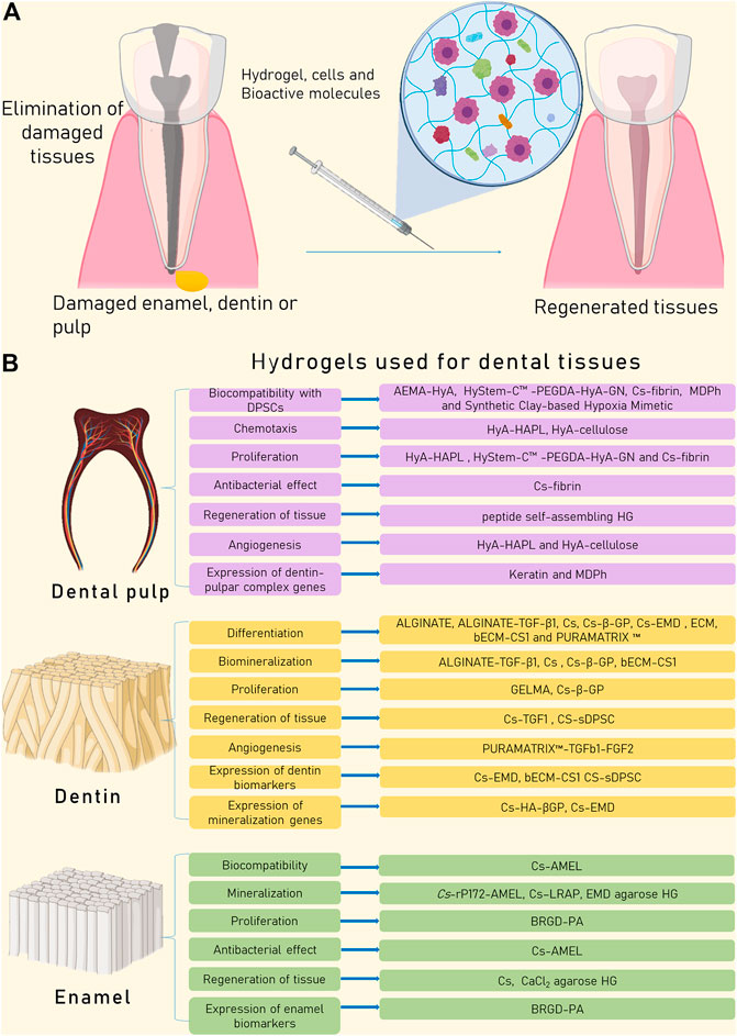
FIGURE 1. The combined use of cells, bioactive molecules, and scaffolds such as HG is considered the best approach to achieve tissue regeneration. (A) Dental organs can be damaged principally by caries and trauma, injuring enamel, dentin, or pulp. Advances in the field of HG could be beneficial to find the ideal scaffold to regenerate every lost tissue. (B) HG can induce changes in cellular processes such as chemotaxis, proliferation, angiogenesis, biomineralization, and expression of specific tissue biomarkers, enhancing the regeneration process.
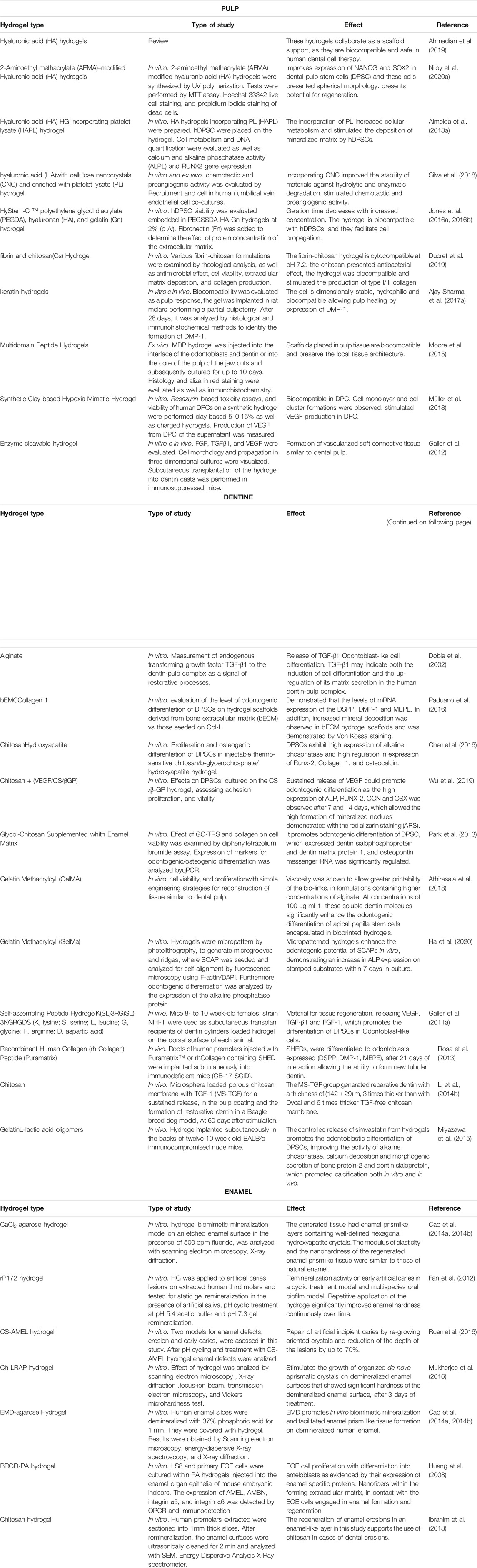
TABLE 1. Hydrogel-based scaffolds for dental tissue regeneration.
When a tooth is cavitated in the dentin limit with the pulp chamber, the odontoblast layer secretes bioactive molecules to induce dentinogenesis. Nevertheless, the processes of reparative dentine formation, as well as enamelogenesis, are still unclear, and their regeneration is a clinical challenge (Smith et al., 1995; Bleicher, 2014; Wang et al., 2014). Additionally, in the case of pulp involvement, one of the main intentions of clinical treatment is the preservation of pulp vitality by using direct capping with biocompatible materials to form reparative dentin (Huang, 2011; Hilton et al., 2013). Although this treatment is in some cases effective, so far there is not a successful treatment to induce the full regeneration of the enamel, dentin, and pulp.
The dental pulp is one of the most difficult tissue to regenerate, given its complex nature. The dental pulp is a dynamic connective tissue that has an innate capacity to respond to infections or injuries (Morotomi et al., 2019). This property has provided the basis for the design of regenerative treatments. Research on pulp regeneration increases steadily in the literature, applying biological principles to protect the pulp (Nakashima et al., 2017). According to Ahmadian et al., successful pulp therapy depends on the form and site of injury, the age of the tooth, and the therapeutic approach and capping material (Ahmadian et al., 2019). The investigations have focused on the use of HG, in studies with pulp tissue cells, in vitro and in vivo. This knowledge is applied in clinical treatments, which include; indirect or direct pulp capping, pulpotomies, and pulp revascularization, with emphasis on dentin formation, sensory nerves, and blood vessel development (Moussa and Aparicio, 2019). Here we present an overview of all HG studied for dental pulp regeneration.
HyA favors stem cell therapy in dentin/pulp lesions, although in vivo studies showed a high degradation rate and poor mechanical resistance (Ahmadian et al., 2019). HyA can be modified with 2-Aminoethyl methacrylate (AEMA) to form a non-cytotoxic HG for dental pulp stem cells (DPSCs). This HG mimics the in vivo 3D environment to cell growth and maintains their native morphology and stemness, increasing the expression of stemness factors such as NANOG and SOX2, in comparison with cells grown on conventional culture plates (Niloy et al., 2020b). On the other hand, the HyA HG biofunctionalized withHAPL enhances the recruitment of DPSCs isolated from permanent teeth and stimulates the mineral matrix deposition, evidencing its potential for repairing damaged pulp/dentin tissue (Almeida et al., 2018b). Also, when HyA is combined with cellulose nanocrystals improves the stability of the material against hydrolytic and enzymatic degradation, producing an HG that can be injected in situ. Besides, the addition of HAPL to this HG results in the release of the chemotactic and pro-angiogenic growth factors such as platelet-derived growth factor (PDGF) and vascular endothelial growth factor (VEGF) which function as chemotactic and pro-angiogenic agents promoting DPSCs recruitment and proliferation, in co-cultures of DPSCs and human umbilical vein endothelial cells. Besides, in a chorioallantoic membrane assay (CAM) the arrangement of DPSCs nearby the new vessels promoted by the CAM infiltration into the HG was seen (Silva et al., 2018).
In the case of the commercially available HyStem-C™ injectable HG PEGDA, hyaluronan, and gelatin (Gn)], the modification of PEGDA by adding a disulfide bond results in a biocompatible HG that can hold DPSCs, meanwhile, increased ratios of HyA: Gn improves cell viability for 14 days. Besides, when fibronectin is added, cell proliferation increases over time (only when concentrations of 1.0 and 10.0 μg/ml in PEGDA-HyA-Gn HG are used) (Jones et al., 2016a, 2016b).
Chitosan (Cs) is a biodegradable natural biopolymer of amino polysaccharides used in pharmaceuticals, biomedicine, tissue repair, and tissue engineering, given its biocompatibility (Li et al., 2009). Its residual products are not toxic nor immunogenic (Tamura et al., 2011). Cs-based HG (CsHG) can be easily modified or coupled with various molecules and proteins that potentiate the repair effect or act synergistically (Giri et al., 2012). In oral research, CsHG has been used both in vitro and in vivo, adding hydroxyapatite (HA), β-glycerophosphate (β-GP), VEGF, or extracellular enamel matrix.
CsHG combined with fibrin shows an excellent capacity to maintain DPSCs viability, fibroblast-like morphology, proliferation rate, and type I/III collagen production. Besides, it has an antibacterial effect against Enterococcus faecalis (Ducret et al., 2019); meanwhile, Keratin HG has shown biocompatibility with the dental pulp of upper molars in partial pulpotomy treatments, and after 28 days the expression of dentin matrix protein 1 (DMP-1) was detected, suggesting that keratin HG may be a source of biomaterial for biological treatment of pulp tissue (Ajay Sharma et al., 2017b).
The multidomain peptide HG (MDPh), in ex vivo organ culture, shows compatibility with the dental pulp and stimulates odontoblasts to be synthetically active. Also, MDPh can stimulate the expression of dentin-matrix proteins as dentin sialophosphoprotein (DSPP). An important finding is that when MDPh is used in dental pulp did not induce mineralization, but preserved pulp tissue architecture (Moore et al., 2015). On the other hand, the synthetic clay-based HG can maintain the viability of dental pulp cells (DPCs), without increasing VEGF levels in the supernatant when DPCs are cultured on the clay-HG loaded with hypoxia mimetics agents such as dimethyloxalylglycine (DMOG), desferrioxamine, L-mimosine, and CoCl2. In contrast, the supernatant of DMOG-loaded HG alone stimulated VEGF production (Müller et al., 2018). In the case of the enzyme-cleavable HG (made from self-assembling peptide nanofibers), it can be combined with basic fibroblast growth factor (FGF2), transforming growth factor (TGF-β1), and VEGF, maintaining DPSCs morphology and enhancing their spreading in 3D cultures. Besides, the subcutaneous transplantation of this HG within dentin cylinders into immunocompromised mice enhances the formation of a vascularized soft connective tissue similar to the dental pulp (Galler et al., 2012).
In the regenerative dentistry area, it has been implemented the use of HG for dentin regeneration, testing different biomaterials such as alginate alone or in combination with GelMa, Cs, glycol-chitin, ECM, self-assembled peptides, and collagen. Dentin is considered the second hardest mineralized tissue in the body after enamel and corresponds to the largest part of the tooth crown. During odontogenesis, cells from the neural crest migrate into the mesenchyme and reach the branchial arch where they contribute to the formation of maxillary dental germs. The odontoblast precursors migrate from the front-nasal bud where interactions between the epithelial cells of the dental lamina and mesenchymal cells contribute to the formation of pulp. Initially, these cells do not appear to be functional, but later, they can constitute a deposit for the renewal of apoptotic odontoblasts, becoming prepolarized osteoblasts (Mao and Prockop, 2012). Dentin is formed by two simultaneous processes, the formation of a collagen matrix, which is associated with soluble and insoluble signaling molecules that stimulate odontogenesis (Chun et al., 2011), and the formation of mineral crystals in this matrix. These processes are orchestrated mainly by Ca2+ where they interact with ATPase, Na+, calcium channels, and intercellular binding proteins. This specialized tissue has specific functions such as root formation and the formation of new dentin throughout life, this is a physiological process known as secondary dentin formation; meanwhile, the repair induced by environmental stimulus generating an interface between pulp and damaged dentin (Couve et al., 2014) or restoration (Sloan and Waddington, 2009) is known as tertiary dentin. The HG used for dentin are summarized below.
Alginate HG have been applied for wound healing, as they can structurally emulate ECM and can be combined with various drugs or molecules for sustained release (Lee and Mooney, 2012). Currently, printable alginate HG combined with dentin matrix have been designed, showing that this biomaterial increases cell viability, due to their ability to retain soluble dentin molecules within itself, improving their regeneration capacity (Athirasala et al., 2018). Besides, when is combined with GelMa (known as micropatterned HG) induces a synergistic effect, increasing the odontogenic potential of stem cells from apical papilla (SCAPs) in vitro, increasing alkaline phosphatase (ALP) expression in stamped substrates within 7 days of culture (Ha et al., 2020). Alternatively, the effect of the alginate HG loaded with TGF-β1 on root discs shows a sustained release of this growth factor, increases dentin matrix deposition, and induces odontoblast cell differentiation with subsequent secretion of tubular dentin matrix on the cut surface (Dobie et al., 2002).
CsHG coupled with HA and β-GP induces a high ALP expression and a positive regulation in the expression of Runx-2, type I collagen (COL1), and osteocalcin (OCN) in DPSCs (Chen et al., 2016). Also, the effects of the CsHG/β-GP as a sustained release system for VEGF in DPSCs has been evaluated, demonstrating that this biomaterial enables adhesion, cell proliferation, and odontogenic differentiation with high expression of ALP, RUNX-2, OCN, and osterix (OSX) after 7 and 14 days, allowing the formation of mineralized nodules in vitro (Wu et al., 2019).
In the case of ChHG supplemented with enamel matrix derivatives (EMD), it promotes the odontogenic differentiation of DPSCs, showing a porous and well-interconnected structure, allowing proliferation, odontogenic differentiation of DPSCs and the expression of DSPP, DMP-1, and osteopontin (OPN) (Park et al., 2013). In vivo studies using a porous material loaded with microspheres of ChHG with TGF-β1, a sustained release over 7 days of more than 40% was shown. After days of stimulation, reparative dentin was formed, resulting in 3 times thicker dentin than the Dycal group and 6 times thicker than ChHG without TGF-β1(137).
The ECM provides physical scaffolding to cells and structural support to tissues, offering specific signals that are needed for regeneration and homeostasis, through its proteins, proteoglycans, and anchor sites (Hussey et al., 2018). ECM allows the release of cytokines and signaling molecules that induce cell activation and differentiation (Johnson et al., 2019). EMC-based HG are 3D networks capable of absorbing large amounts of water or physiological fluids from the implantation site (Rosales and Anseth, 2016). In vitro studies have shown odontogenic differentiation of DPSCs in HG scaffolds derived from decellularized bone ECM (bECM) or COL1, demonstrating that the levels of mRNA expression of DSPP, DMP-1, and matrix extracellular phosphoglycoprotein (MEPE) increases significantly in cells grown in bEMC vs those grown in COL1 scaffolds. In addition, increased mineral deposition has been observed in bECM HG scaffolds (Paduano et al., 2016).
Peptides are short amino acid chains that can be used in biomaterials without the need to replicate the entire natural sequence. In this regard, the supramolecular self-assembled and patented HG, Puramatrix™, has been used for in vivo research, injected into the human root canals in the back of immunodeficient mice (CB-17 SCID), containing human SHED (stem human exfoliated deciduous teeth), resulting in differentiation to odontoblasts that express DSPP, DMP-1, and MEPE, after 21 days of interaction, allowing the formation of new tubular dentin (Rosa et al., 2013). Additionally, conjugates with TGF-β1, FGF2, VEGF in DPSCs have also been used, showing not only dentin reparation after 6 weeks, but also vascularized and soft connective tissue like dental pulp within the dentin cylinder. Dental Sialoprotein (DSP) was used to detect odontoblastic-like cells at the dentin interface, confirming cell differentiation (Galler et al., 2012).
Collagen is the most abundant protein within the ECM (Ricard-Blum, 2011). Collagen-based HG have been used as immunomodulators, drug carriers, healing agents, chelators, and in tissue regeneration (Wei et al., 2019). This type of HG can promote adhesion, proliferation, and differentiation in cardiac, corneal epithelial, endothelial, liver, bone, and mesenchymal stem cells (MSCs) (Agmon and Christman, 2016) and have been studied in a Yucatecan mini-porcine model, where the porcine DPSCs were transferred through the collagen HG directly to the root canal, previously prepared through pulpectomy, then, after 4 months, the analysis showed that vascularized soft tissue was recovered, creating a structure similar to a dentin bridge covering it. In addition, immunohistochemical analysis detected the expression of nestin, DSPP, DMP-1 and bone sialoprotein in cells similar to odontoblasts (Zhu et al., 2018).
HG-based scaffolds, provide the essential support needed during tissue regeneration (Abbass et al., 2020) and can be used for repairing enamel. This tissue forms the outermost covering of teeth, being a nanostructured mineralized tissue. The ameloblasts, epithelial cells derived from the enamel organ of the developing tooth, are the cells involved in the production of enamel (Chatzistavrou et al., 2012) in a highly regulated process known as amelogenesis, in which proteins are secreted into the extracellular space to then initiate biomineralization (Roveri and Iafisco, 2010). Amelogenin (AMEL) is the most abundant protein, acting as master control of the orientation and elongation growth of enamel rods during the mineralization process. Then, is ameloblastin (AMBN) as the second most abundant non-amelogenin enamel-specific glycoprotein, acting as a cell adhesion molecule for ameloblasts (Moradian-Oldak, 2009). Lastly, enamelin and tuftelin proteins (found in lower proportions) that control apatite nucleation and growth in conjunction with amelogenin. AMEL and other enamel proteins are eventually degraded by proteinases such as the matrix metalloproteinase 20 (MMP-20) and kallikrein 4 (KLK4) at different stages of amelogenesis (Moradian-Oldak, 2009; Moradian-Oldak, 2012). At the end of the process, the enamel is 96% crystalline calcium phosphate in form of HA and 4% organic components (products of degradation of enamel proteins) and water (Baldassarri et al., 2008).
Since mature enamel does not contain cells, it cannot be remodeled by itself. Therefore, biomaterials mimicking enamel are now under revision to regenerate decayed or traumatized dental organs (Mann, 1997). Nevertheless, HA crystals isolated from mature enamel are longer and different in shape from those synthetic. In this regard, the synthesis of enamel-like hydroxyapatite nanorods has been only possible by applying extreme laboratory conditions such as high temperature, high pressure, and extremely acidic pH (Chen et al., 2005; Yamagishi et al., 2005). Moreover, replacing enamel is difficult since none of the materials can mimic all its physical, mechanical, and aesthetic properties (Moradian-Oldak, 2012). Thus, the development of improved biomaterials that could be used in physiological conditions is needed. Here we present the emerging biomaterials in HG presentation that are used for enamel regeneration.
In an exploratory study, a biocompatible recombinant amelogenin (rP172) HG was prepared to release phosphate, Ca2+, and F−. This HG applied to artificial enamel caries lesions, displayed remineralization activity on early artificial caries in a cyclic treatment model and multispecies oral biofilm model. Repetitive application of the HG significantly improved enamel hardness continuously over time (Fan et al., 2012).
Ibrahim, et al., have used CsHG in enamel slices etched with 37% phosphoric acid. After application, morphological changes on the enamel surface were observed by scanning electron microscopy showing the regeneration of an enamel-like layer in the treated samples with a difference between the Ca/P ratios. They showed that Cs provided a substrate to immobilize the nano-building units acting as a template for the structured assembly of the regenerated enamel layer (Ibrahim et al., 2018).
The amelogenin-CsHG (CS-AMEL) is biocompatible, biodegradable, and has antimicrobial and adhesion properties (Ruan et al., 2013; Ruan and Moradian-Oldak, 2014). It has been used for biomimetic repair of human enamel with erosive or carious lesions in two pH-cycling systems. CS-AMEL HG showed effectiveness at pH 4.6 (similar to the pH of the mouth after food consumption) and pH 6.5 (average pH during the nighttime). This HG forms a new organized layer of enamel-like crystals on the surface of erosive lesions and could repair artificial incipient caries by regrowing oriented crystals and reducing the depth of the lesions by up to 50–70% under pH-cycling conditions (Ruan et al., 2016).
Cs with leucine-rich amelogenin peptide (Ch-LRAP) HG system acts as an organic template on acid-treated enamel surfaces and allows faster mineral induction and results in organized crystal growth of hydroxyapatite. The demineralized tooth slices treated with a single application of Ch-LRAP HG for 3 days showed a dense mineralized layer comprising very well-organized enamel-like apatite crystals with a continuous growth at the interface between the repaired layer and natural enamel. There was an improvement in the surface hardness after treatment of the demineralized sample with almost 87% recovery of the hardness value (Mukherjee et al., 2016).
An HG biomimetic mineralization model consisting of an enamel slice, CaCl2 agarose HG, ion-free agarose HG, and a phosphate solution was designed to regenerate enamel prism-like crystals. The CaCl2 agarose HG regulated the pattern, size, and mineral phase of the growing crystals through cooperative interactions with calcium, phosphate, and fluoride ions. The regenerated apatite crystals were found to be highly oriented along the c-axis with good crystallinity (Cao et al., 2014a, 2014b). Agarose HG has been also combined with the enamel matrix derivative (EMD) and applied to demineralized enamel, showing enamel prism-like crystals with hexagonal structures denser, thicker, and more orderly packed than the crystals formed without EMD (being calcium, phosphorus, and fluorine the main elements found in their composition), confirming that they were fluorinated hydroxyapatite. Moreover, the degree of crystallinity of the hexagonal crystals formed is improved in the presence of EMD. Nevertheless, the degree of structural perfection in the regenerated enamel prism-like tissue without EMD was higher than that with EMD (Cao et al., 2014a, 2014b).
The self-assembled peptide amphiphile (PA), containing the peptide motif Arg-Gly-Asp, or “RGD” (BRGD-PA), were used in ameloblast-like cells (line LS8) and primary enamel organ epithelial (EOE). Cells were cultured within PA HG, and the PA was injected into the enamel organ epithelia of mouse embryonic incisors. In vitro, LS8 cells and primary EOE cells responded to the BRGD-PA nanostructures with enhanced proliferation and higher amelogenin, ameloblastin, and integrin expression levels. In the organ culture model, the site of BRGD-PA injection showed EOE cell proliferation with differentiation into ameloblasts, expressing enamel-specific proteins such as AMEL and new discovered proteins involved such as thrombospondin 2 (TSP2). Ultrastructural analysis showed the nanofibers within the forming ECM, in contact with the EOE cells engaged in enamel formation and regeneration (Galler et al., 2011b).
HG can be easily administered into irregular shapes of tissue defects as those caused by periodontal disease (Li and Guan, 2011; Shi et al., 2017; Xu et al., 2019). Periodontal disease is a prevalent infectious disease worldwide, that causes the damage of periodontal support tissues, which can eventually lead to tooth loss. Periodontal treatments aim to first control the infections and then reconstruct the architecture and function of periodontal tissues including cementum, periodontal ligament (PDL), and alveolar bone (Liu et al., 2019). Nevertheless, clinical periodontal therapy fails in regenerating the lost periodontal tissues, thus alternative treatments are needed (Sowmya et al., 2017a).
The engineering of periodontal tissues is a challenging approach given the complex hierarchical architecture comprising of a soft tissue interspersed between two distinct hard-tissue structures (Carranza et al., 2006). The attempts for simultaneous regeneration of the lost tooth-supporting structures are ultimately dependent on the interplay among scaffold, cells, and bioactive signals (Sowmya et al., 2017b) (Figure 2). Another limiting factor is the stiffness of scaffolds since they may lead to poor adaptation of the construct on the root surface (Ivanovski et al., 2014). Hence, HG can be used as an alternative, as they are a widely employed carrier in drug-controlled release, and possesses distinctive qualities, such as easy preparation, low cost, low toxicity and eases of use (Ahadian et al., 2015; Merino et al., 2015) (Table 2).
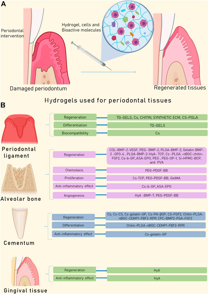
FIGURE 2. The complete regeneration of the periodontium is not possible yet given the complex characteristics of the tissues. (A) HG are a promising alternative to restore the structure and function of the periodontal complex. (B) HG in periodontal cells can induce differentiation, proliferation, chemotaxis, and angiogenesis, enabling the formation of new tissue. Besides some HG in periodontium show anti-inflammatory effect.
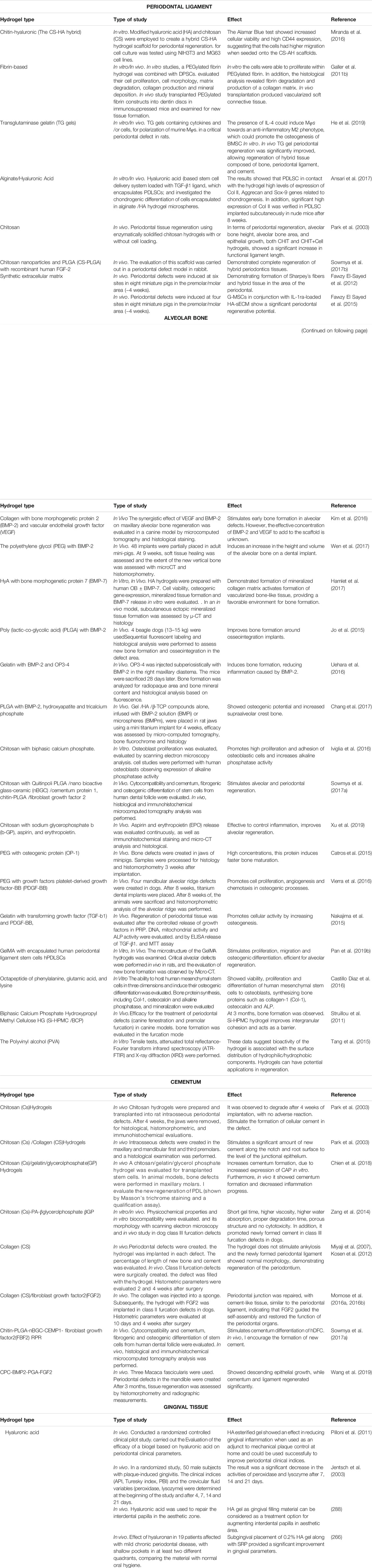
TABLE 2. Hydrogel-based scaffolds for periodontal tissue regeneration.
Currently, various materials have been used to regenerate the periodontal ligament (Tomokiyo et al., 2019), among them, are fibrin, transglutaminase gelatin, alginate, Cs, chitin-HyA, chitin-PGLA, peptides, and synthetic ECM. The PDL is conformed of fibers arranged in different spatial directions. PDL allows anchoring, provides firmness and cushioning capacity, and is located in the space between the dental organ and alveolar bone (Mehrotra and Singh, 2019). It is a specialized connective tissue, mainly composed of extrinsic collagen fibers known as Sharpey´s fibers, mesenchymal cells, undifferentiated fibroblasts, as well as by the vasculature of the dentinal arteries, which allows rapid healing and remodeling (Yamamoto et al., 2016). Chronic periodontitis and aggressive periodontitis cause loss of function of PDL and later loss of support of the dental organ, ending in their premature loss. The HG used for periodontal ligament regeneration are presented below.
High rigidity matrix-based materials such as TG-gels can allow osteogenic differentiation in vitro. New findings demonstrate macrophage modulation and stem cell recruitment in highly rigid HG for regeneration of the periodontal complex, where macrophages tend to undergo M1 polarization. Furthermore, when they were prepared in 3D and combined with IL-4 or stromal cell-derived factor 1 (SDF1), they repaired periodontal defects 4 weeks after surgery, allowing regeneration of hybrid tissue composed of bone, periodontal ligament, and cementum (He et al., 2019).
CsHG have been studied for regeneration of the periodontal complex. In vivo, CsHG with PDL cells transplanted in rat intraosseous periodontal defects is highly biocompatible and biodegradable. Histological analysis showed that the CsHG is degraded after 4 weeks of implantation and instead a cell complex, cement, PDL, and new bone formation were formed at the margin of the defect without any adverse reaction in the surrounding tissue (Park et al., 2003).
Chitin-derived biomaterials can mimic the structure of the ECM, providing excellent scaffolding for cells, and creating a favorable immune environment for accurate periodontal healing and regeneration (Xie et al., 2020). In this regard, the chitin-HyA hybrid HG generates a perfect scaffold material that can be used to support osteoblasts, gingival fibroblasts, cementoblasts, and periodontal fibroblast cells (Miranda et al., 2016; Orellana et al., 2018). Also, HG composed of chitin nanoparticles and PLGA can benefit tissue regeneration when is functionalized with recombinant human FGF-2, since it can achieve total closure of the periodontal defect in a rabbit model, where histologically periodontal ligament-like characteristics can be seen (Sowmya et al., 2017b).
These new generation HG have been designed from synthetic HyA scaffolds (Espandar et al., 2012) to improve the results of periodontal treatment. These HG can be modified by adding IL1 receptor antagonist (IL-1ra) that binds with high affinity to cell surface IL-1 receptors (Gorth et al., 2012). As it is known that in periodontitis there is an increase in IL-1 (Suwatanapongched et al., 2000), a favorable effect in periodontal wound healing is seen when the HG containing IL-1ra is administered (Zhang et al., 2004). In vivo studies confirm the periodontal regenerative potential of this HG with gingival margin stem cells (GMSCs) demonstrating the formation of Sharpey´s fibers and hybrid tissue in the area of the periodontal defect (Fawzy El Sayed et al., 2015).
Tissue engineering in alveolar bone regeneration has developed new alternatives for the treatment of bone defects. The alveolar bone is essential to guarantee the success of dental organ preservation, and also if the use of dental implants is required (Warnke et al., 2004). The close relationship between alveolar bone, periodontal ligament, and cement, hinders successful regeneration. To solve this problem, It is necessary to develop HG in combination with biomolecules (Lee and Shin, 2007). Alveolar scaffolds must be osteoinductive or osteoconductive, have mechanical resistance, adequate degradation, avoid the collapse of the area to be regenerated, good flexibility, adapt to different morphologies, and be easily managed. However, until now, biomaterials with all of these characteristics are not available (Kaigler et al., 2011). Here we summarize the HG used for alveolar bone regeneration.
HG with different BMPs have been used, due to their commercial availability and their easy incorporation into different HG. For instance, the collagen HG with BMP-2 and VEGF, in vivo has shown a favorable early bone formation in dogs with alveolar defects. However, the effective concentration of BMP-2 and VEGF to add to the scaffold is unknown, and long-term studies are required. (Kim et al., 2016). The polyethylene glycol (PEG) HG with BMP-2 in vivo induces an increase of the height and volume of the alveolar and cortical bone over an implant placed in the jaw of pigs, indicating that it could enhance the retention of the implants when the alveolar crest is small. (Wen et al., 2017). In the case of HyA HG with bone BMP-7, it has the capability of forming a mineralized collagen matrix in vitro. Interestingly, in vivo, this HG can form vascularized bone-like tissue after 4 weeks in a rat model, since it provides a favorable environment for bone formation. (Hamlet et al., 2017). The PLGA HG with BMP-2 has been used in beagle dogs creating irregular bone defects around implants resulting in the improvement of bone formation and osseointegration when compared with control without BMP-2 (Jo et al., 2015). The Gelatin HG with BMP-2 and the osteoprotegerin mimic peptide OP3-4 (YCEIEFCYLIR) was created to avoid the side effects of BMPs, such as inflammation and carcinogenesis. This combination in a murine model applied by subperiosteal injection shows the induction of bone formation, reducing inflammation caused by BMP-2 (Uehara et al., 2016). The PLGA HG with BMP-2, hydroxyapatite and tricalcium phosphate has been used in rats for supra-alveolar crest augmentation treatment, showing osteogenic potential and increasing supra-alveolar crest bone (Pilloni et al., 2011).
CsHG can be combined with biphasic calcium phosphate. The incorporation of ceramic particles in this material makes it stable at different pH and increases the elastic modulus and maximum tensile strength. Besides, it promotes high proliferation, osteoblastic cell adhesion and increases ALP activity (Iviglia et al., 2016). Other additions that have been made to the CsHG such as Quitinpoli PLGA/nano bioactive glass-ceramic (nBGC)/CEMP-1, chitin-PLGA/FGF2 as PDL layer, and platelet-rich plasma-derived growth factors and chitin-PLGA/nBGC. This three-layer nanocomposite HG is cytocompatible and favors osteogenic differentiation of stem cells from the human dental follicle, and its content of growth factors helps the formation of bone trabeculae, stimulating alveolar and periodontal regeneration (Sowmya et al., 2017a). CsHG can be used also with β-GP, aspirin, and erythropoietin. In vivo experiments revealed that this HG is effective in controlling inflammation, enhances alveolar regeneration, and could be used in the future for clinical application (Xu et al., 2019).
The PEG HG has been used in defects in the mandibular alveolar ridge of dogs showing unpredictable bone formation and, in most cases, a negative impact on alveolar regeneration (Vierra et al., 2016). The PEG with osteogenic protein (OP-1) in a mini-pig alveolar defects model shows that in high concentrations, this protein induces faster bone maturation (Catros et al., 2015). In the case of HG with growth factors such as platelet-derived growth factor-BB (PDGF-BB), it promotes cell proliferation, angiogenesis, and chemoattraction in osteogenic processes (Pan et al., 2019a). Meanwhile, gelatin HG with TGF-β1 and PDGF-BB promotes cellular activity by increasing osteogenesis (Nakajima et al., 2015)
The GelMA HG with encapsulated human periodontal ligament stem cells (PDLSCs) presents a highly porous and interconnected network providing an optimal niche for proliferation, migration, and osteogenic differentiation. In vivo, performing alveolar bone defects in rats, the PDLSCs encapsulated in GelMA HG show to be more efficient for alveolar regeneration and it may represent a new strategy for therapy of alveolar bone defects (Pan et al., 2019b).
The use of biodegradable HG with octapeptide of phenylalanine, glutamic acid, and lysine has shown the formation of an ideal niche, apt for cell physiological functions. In vitro tests showed viability, proliferation, and differentiation of human mesenchymal stem cells to osteoblasts, synthesizing bone proteins such COL1, OCN, and ALP. Furthermore, mineralization within the HG was evident, showing potential to regenerate hard tissues such as alveolar bone (Castillo Diaz et al., 2016).
Regarding Biphasic Calcium Phosphate Hydroxypropyl Methyl Cellulose (Si-HPMC/BCP) HG, due to its BCP particle components helps to osteoconduction process, while Si-HPMC improves intergranular cohesion. This biomaterial is easy to use and simplifies the filling process for bone lesions. However, more research is needed to be used on alveolar defects. (Struillou et al., 2011).
The PVA HG has chemical stability, biocompatibility and is porous facilitating cell adhesion. In vitro tests showed excellent biocompatibility, and in X-ray Diffraction tests, it was observed that it has affinity to form HA. This HG may have potential applications in alveolar bone regeneration, as it was also associated with the surface distribution of hydrophilic/hydrophobic components (Tang et al., 2015).
Cementum neoformation is esencial for periodontal regeneration. Precementum formation starts following the fragmentation of Hertwig’s root sheath, the matrix of proteins formed subsequently provides an adequate attachment substrate to support differentiation of cementoblasts from ectomesenchymal progenitors of the dental follicle (Hammarström et al., 1996). Cementum deposition continues with the apical development of the root allowing the integration of Sharpey´s fibers collagen bundles, into the newly generated cementum (Menicanin et al., 2015). The formed tissue is a hard and avascular connective tissue located on the dental root that emerges from the alveolar bone to the dental organ. It has been described two types of cementum depending on the content of inner cells: cellular and acellular (Veis, 2002). Dental cementum participates in key processes such as maintaining stability and chewing charge distribution, thus, when cementum is infected or suffers trauma, treatments require to provide a disinfected root surface to promote the reinsertion of collagen fibers and avoid the formation of pathological spaces that would promote reinfection. Consequently, the success of periodontal regenerative therapy relies on the health of the dental cementum, its health, and its capacity to regenerate (Grzesik and Narayanan, 2002).
Dental cementum is composed of 50% of organic matrix, of which, 90% is COL1. Another cement-related collagen protein is type III collagen, involved in the development, repair, and regeneration of periodontal tissues (Alvarez-Pérez et al., 2006). Besides, within its organic composition, there are two specific proteins of this tissue 3-Hydroxyacyl coenzyme A dehydratase 1 (HACD1)/Cementum Attachment Protein (CAP) and CEMP-1. HACD/CAP has been implicated in the nucleation, regulation, and direction of hydroxyapatite crystal growth and regeneration of critical bone defects in rat calvaria, meanwhile, CEMP-1 regulates the activity of cementoblasts by inducing differentiation and mediates in the mineralization process, migration, and proliferation of gingival fibroblasts (Arzate et al., 1992; Bermúdez et al., 2015; Villegas-Mercado et al., 2018); Furthermore, CEMP1 also promotes the nucleation of octacalcium phosphate crystals, which is a mineral phase and a precursor of HA crystals (Alvarez-Pérez et al., 2006). The other 50% are minerals, mostly HA [Ca10 (PO4)6 (OH)2] (Nanci and Bosshardt, 2000). Here we show the HG able to sustain at the same time biomineralization for cementum regeneration and proliferation of cells for periodontal ligament formation.
CsHG with or without fluorescently labeled PDL cells were prepared and transplanted into rat intrabony periodontal defects; untreated defects were used as empty controls. After 4 weeks the results showed that PDL cells remained viable upon encapsulation within CsHG before transplantation but histological analysis demonstrated that the CsHG were degraded after 4 weeks without affection of the adjacent tissue. Interestingly, CsHG without cell loading showed a newly formed cellular cementum limited to the apical part of the defect (Yan et al., 2015). In another study, one‐wall intrabony defects of beagle dogs were surgically created in the bilateral maxillary first and third, and the mandibular second and fourth premolars and treated with a Cs/collagen sponge, at 8 weeks after the operation, a significant amount of new cementum formed along the notch and root surface to the level of the junctional epithelium was found (Park et al., 2003).
In 2018 Chien et al., applied an injectable and thermosensitive Cs/gelatin/glycerol phosphate HG to enhance stem cell delivery and engraftment. Synergistic effects of iPSCs and BMP-6 increased cementum formation which was confirmed by CAP expression since it was significantly up-regulated in vitro. Besides, iPSCs-BMP-6-HG in vivo showed the formation of cementum and minimized the progress of inflammation in rats with maxillary-molar defects. These findings suggest that this combination can promote stem cell-derived graft engraftment with an anti-inflammatory effect, which resulted in highly possible periodontal regeneration (Chien et al., 2018). Interestingly, a thermosensitive CsHG using autoclaved Cs powder (121°C, 10 min) and β-GP (Cs-PA/GP) were compared to the physicochemical properties and biocompatibility in vitro. According to their results, the Cs-PA/GP HG had a shortened gelation time, higher viscosity, increased water absorption, appropriate degradation time, porous structure, and no obvious cytotoxicity on human periodontal ligament cells. Besides, the Cs-PA/GP HG promoted cell-like newly formed cementum in dog class III furcation defects (Zang et al., 2014).
In 2017, Miyaji et al., studied collagen HG on periodontal wound healing in beagles. Sixty-four periodontal dehiscence type defects were created on the buccal roots of four beagles for collagen HG implantation. After 8 weeks new cementum was found in a higher proportion when compared with the control group indicating that periodontal regeneration was stimulated by collagen HG implantation (Miyaji et al., 2007). Another study in surgical-created defects was developed by Kosen et al., where collagen HG/sponge scaffold was prepared by injecting collagen HG, cross-linked to the ascorbate-copper ion system, and placed in class II furcation defects in beagle dogs. At 2 weeks, the collagen HG/sponge scaffold displayed high biocompatibility and biodegradability with numerous infiltrating cells. In the experimental group, reconstruction of alveolar bone and cementum was frequently observed 4 weeks after surgery. The study showed that cell-rich connective tissue had reformed along the root surfaces after the application of HG. Also, the 4-week specimens in the HG group showed no ankylosis, and the newly formed periodontal ligament showed functional morphology, demonstrated by the connection of newly formed cementum to the alveolar bone with Sharpey´s fibers (Kosen et al., 2012)
Momose et al., evaluated the application of collagen HG scaffold in combination with FGF2 in furcation defects in beagle dogs. Collagen HG was associated with FGF2 and injected into sponge-form collagen and then implanted into class II furcation defects in dogs. This combination improved cell and tissue growth where cells and blood vessel-like structures were found on day 10. At 4 weeks, periodontal attachment, consisting of cementum-like tissue, periodontal ligament-like tissue, and Sharpey’s fibers, was repaired, indicating that FGF2-loaded scaffold led to self-assembly and then re-established the function of periodontal organs (Momose et al., 2016a, 2016b).
Sowmya et al., in 2017 used the strategy of a tri-layered nanocomposite HGl scaffold that was developed by assembling chitin PLGA/nano bioactive glass-ceramic (nBGC)/CEMP-1 as the cementum layer, chitin–PLGA/FGF2 as the PDL layer, and chitin–PLGA/nBGC/platelet-rich plasma-derived growth factors as the alveolar bone layer layers in the order of periodontal tissue location, i.e., cementum, PDL, and alveolar bone. The cementogenic differentiation of human dental follicle stem cells on the tri-layered nanocomposite HG scaffold with growth factors was comparable to the cellular differentiation on the tri-layered nanocomposite HG scaffold cultured in induction medium. In vivo, complete healing with the formation of new cementum was more pronounced in tri-layered nanocomposite HG scaffold with growth factors in comparison to the other groups (Sowmya et al., 2017a).
The calcium phosphate cement (CPC)/propylene glycol alginate (PGA) with BMP-2 and fibroblast growth factor (FGF)-2 gel (CPC/BMP+PGA/FGF) was studied in a periodontal defect model in Macaca fascicularis where three-wall periodontal defects were surgically created in the mandible. Results showed that epithelial downgrowth, meanwhile cementum, and ligament were regenerated significantly when compared to the control group (Wang et al., 2019).
The gingival tissue can be completely regenerated (Freedman, 2012). The gingival tissue is a fibromucous membrane that covers the mandibular and maxillary bone, designed to withstand constant trauma from chewing. Clinically it is divided into free, inserted, and papillary. According to its structure, it is made up of stratified squamous epithelium, rich in keratinocytes. The gingival epithelium is divided into oral epithelium, sulcus epithelium, and junction epithelium (Andrian et al., 2006). HG used for gingival tissue regeneration are presented below.
Various injuries can cause periodontal damage, in the clinical area, several studies have been able to demonstrate the capacity of new materials based on polysaccharides such as HyA since it is known to be a powerful antioxidant, biocompatible, anti-inflammatory, with viscoelastic and bacteriostatic properties. The efficacy of topical application of HyA HG in the treatment of gingivitis has been evaluated and it turned out to be a potentially useful supplement in therapy against this disease (Pistorius et al., 2005). The gel containing 0.2% hyaluronate has a beneficial effect in the treatment of gingivitis, the result was a significant decrease in the activities of peroxidase and lysozyme after 7, 14, and 21 days (Jentsch et al., 2003), complemented with scaling and root planing (SRP) in patients with chronic periodontitis (Gontiya and Galgali, 2012).
In terms of better healing after nonsurgical therapy, supplemental use of 0.8% hyaluronan HG after complete mechanical debridement potentially has significant clinical benefits (Bolt and Bhupinder, 2007). In the area of dental esthetics, it has been used to repair the interdental papilla, where it is infiltrated until extravasation, after 3 months of follow-up, a completely healthy gum was observed and the papilla was filled (Zatta da Silva et al., 2019).
Given that human dental tissues cannot be biologically repaired once formed, strategies such as the use of cells and biomolecules in 3D scaffolds for regeneration are required. Nonetheless, pulp tissue vulnerability needs a different perspective, since it lacks collateral circulation and requires the revascularization and reinnervation of the new tissue (Bermúdez et al., 2021). Therefore, the development of strategies with the potential for clinical implementations is still a far task to achieve.
From a clinical viewpoint, HG combined or alone can enhance remineralization or increase thickness and mechanical properties of the regenerated enamel and dentin. Furthermore, HG present outstanding chemical, mechanical and biological activities (Moussa and Aparicio, 2019) such as the capacity of incorporating cells in their structures and at the same time degrading themselves to let place to new healthy tissue. Besides, they can retain high-water content which enhances the transportation of cell nutrients and waste, and also are elastic and flexible emulating the native ECM (Mantha et al., 2019). Nevertheless, most of them remain under experimental investigation on in vitro cell models and preclinical models and need to validate their dose and efficiency to be used. Nowadays, only ChHG with TGF-β1and Puramatrix™ have shown promising results in dental tissues in vivo. ChHG with TGF-β1showed a sustained release over 7 days of more than 40% of the TGF-β1 in vivo, resulting in reparative dentin formation (Li et al., 2014b). In the case of Puramatrix™, when is injected into the human root canals in the back of immunodeficient mice (CB-17 SCID), containing human SHED, induces the differentiation to odontoblasts that express DSPP, DMP-1, and MEPE, allowing the formation of new tubular dentin and when conjugates with growth factors TGF-β1, FGF2, VEGF in DPSC, vascularized and soft connective tissue similar to a dental pulp was observed within the dentin cylinder after 6 weeks (Almeida et al., 2018a). These results could, in the future, change the clinical approach, avoiding the use of inert materials for treating dental tissues.
Concerning periodontal tissues, during in vitro or ex vivo tests, normal conditions such as resistance of occlusal mechanical forces of PDL, cementum, and alveolar bone cannot be completely achieved. Due to their complex composition, HG should be capable to interact with hard and soft tissues at the same time (Elango et al., 2020). Many options have been studied in vitro with promising results that may lead to preclinical studies (Nagy et al., 2018), but this closed relationship hinders the use of HG in clinical studies. Thus, in vivo models are mandatory for representing the occlusal state to support the functional regeneration design (Bermúdez et al., 2021). Besides, one of the biggest challenges is the reconstitution of the insertion apparatus (Sharpey’s fiber connecting cementum and alveolar bone) since its absence impacts negatively the capacity of the tooth to support the occlusal forces (Liang et al., 2020).
Regarding HG used to promote bone, PDL, and cementum healing and regeneration, most of them have only been evaluated in vitro or in preclinical animal studies. Among the HG reported for alveolar regeneration, the PEG HG with BMP-2 (Wen et al., 2017) and PLGA HG with BMP-2 (Jo et al., 2015) have proved their value to regenerate bone defects around implants in animal models resulting in the improvement of bone formation and osseointegration. Another promising HG is the collagen HG/sponge achieving the formation of bone, cementum, and periodontal ligament with functional morphology (Kosen et al., 2012). Also, its combination with FGF2 results in periodontal attachment, consisting of cementum-like tissue, periodontal ligament-like tissue (Momose et al., 2016a, 2016b).
In the case of gingival tissue wound healing stimulated by the application of HG, it should benefit the clinical outcome of other oral surgical procedures by guaranteeing that the tissue is sealed off from the contaminated oral environment, conducting to a more rapid connective tissue reorganization and causing a faster gain in wound strength, reducing the potential for scarring and scar tissue contraction, thus gingival restoration and stability is crucial to obtain predictive results (Villa et al., 1943). Fortunately, HG have proved their value in regenerating soft tissues. For instance, the 0.2% hyaluronate HG decreases the peroxidase and lysozyme activity (Jentsch et al., 2003), enhancing wound healing after SRP in patients with chronic periodontitis (Gontiya and Galgali, 2012). Besides, 0.8% hyaluronan HG (Bolt and Bhupinder, 2007) has been used to repair the interdental papilla with completely healthy gum and the papilla after 3 months (Zatta da Silva et al., 2019).
Finally, although the results obtained with HG are promising, we must not lose sight of the fact that oral tissues function unified, and during their development, signal pathways can crosstalk and participate in more than one tissue. Thus, the stimuli given by one HG can be useful for another closely related one (Bermúdez et al., 2021). For instance, some promising HG alone or combined might induce biomineralization in enamel, dentin, cementum, and alveolar bone, and others might function for neovascularization and neoformation of the dental pulp and soft periodontal tissue. Despite this, more research is needed to determine which combinations of biofunctionalizing molecules, cells, and HG can improve clinical results.
HG-based scaffolds used for oral tissue regeneration offer several advantages including the capacity of incorporating cells in their structures and at the same time degrading themselves to let place to new healthy tissue. Besides, they can be injectable and retain high water content which enhances the transportation of cell nutrients and waste. They are elastic and flexible emulating the native ECM. Additionally, HG can be combined with biofunctionalizing molecules to induce cellular processes such as chemotaxis, proliferation, differentiation, neovascularization, and biomineralization.
Even though most of the HG are still under investigation, some of them have been studied in vitro and in vivo with promising results that may lead to preclinical studies. Besides, there are HG that have shown their efficacy in healing gingival tissue in patients, such as hyaluronan HG.
Finally, the use of HG could become a gold standard for the treatment of damaged oral tissues by replacing the use of inert materials that are currently highly used in dental practice, if limitations for in vivo studies are exceeded and well-designed clinical studies support their value to be broadly used.
AA, MB, RR, CL, and MA conceived and designed the content of this review, AA, JL, RR and MB wrote the paper, JS and GS contributed to the final version of the manuscript.
The authors declare that the research was conducted in the absence of any commercial or financial relationships that could be construed as a potential conflict of interest.
All claims expressed in this article are solely those of the authors and do not necessarily represent those of their affiliated organizations, or those of the publisher, the editors and the reviewers. Any product that may be evaluated in this article, or claim that may be made by its manufacturer, is not guaranteed or endorsed by the publisher.
Abbass, M. M. S., El-Rashidy, A. A., Sadek, K. M., Moshy, S. E., Radwan, I. A., Rady, D., et al. (2020). Hydrogels and Dentin-Pulp Complex Regeneration: From the Benchtop to Clinical Translation. Polymers (Basel) 12 (12). doi:10.3390/polym12122935
Agmon, G., and Christman, K. L. (2016). Controlling Stem Cell Behavior with Decellularized Extracellular Matrix Scaffolds. Curr. Opin. Solid State. Mater. Sci. 20 (4), 193–201. doi:10.1016/j.cossms.2016.02.001
Ahadian, S., Sadeghian, R. B., Salehi, S., Ostrovidov, S., Bae, H., Ramalingam, M., et al. (2015). Bioconjugated Hydrogels for Tissue Engineering and Regenerative Medicine. Bioconjug. Chem. 26 (10), 1984–2001. doi:10.1021/acs.bioconjchem.5b00360
Ahmadian, E., Eftekhari, A., Dizaj, S. M., Sharifi, S., Mokhtarpour, M., Nasibova, A. N., et al. (2019). The Effect of Hyaluronic Acid Hydrogels on Dental Pulp Stem Cells Behavior. Int. J. Biol. Macromolecules 140, 245–254. doi:10.1016/j.ijbiomac.2019.08.119
Ajay Sharma, L., Love, R. M., Ali, M. A., Sharma, A., Macari, S., Avadhani, A., et al. (2017). Healing Response of Rat Pulp Treated with an Injectable Keratin Hydrogel. J. Appl. Biomater. Funct. Mater. 15 (3), e244–e250. doi:10.5301/jabfm.5000346
Ajay Sharma, L., Love, R. M., Ali, M. A., Sharma, A., Macari, S., Avadhani, A., et al. (2017). Healing Response of Rat Pulp Treated with an Injectable Keratin Hydrogel. J. Appl. Biomater. Funct. Mater. 15 (3), 244–250. doi:10.5301/jabfm.5000346
Almeida, L. D. F., Babo, P. S., Silva, C. R., Rodrigues, M. T., Hebling, J., Reis, R. L., et al. (2018). Hyaluronic Acid Hydrogels Incorporating Platelet Lysate Enhance Human Pulp Cell Proliferation and Differentiation. J. Mater. Sci. Mater. Med. 29 (6), 88–11. doi:10.1007/s10856-018-6088-7
Almeida, L. D. F., Babo, P. S., Silva, C. R., Rodrigues, M. T., Hebling, J., Reis, R. L., et al. (2018). Hyaluronic Acid Hydrogels Incorporating Platelet Lysate Enhance Human Pulp Cell Proliferation and Differentiation. J. Mater. Sci. Mater. Med. 29 (6), 88. doi:10.1007/s10856-018-6088-7
Altunbas, A., Lee, S. J., Rajasekaran, S. A., Schneider, J. P., and Pochan, D. J. (2011). Encapsulation of Curcumin in Self-Assembling Peptide Hydrogels as Injectable Drug Delivery Vehicles. Biomaterials 32 (25), 5906–5914. doi:10.1016/j.biomaterials.2011.04.069
Alvarez-Pérez, M. A., Narayanan, S., Zeichner-David, M., Rodríguez Carmona, B., and Arzate, H. (2006). Molecular Cloning, Expression and Immunolocalization of a Novel Human Cementum-Derived Protein (CP-23). Bone 38, 409–419. doi:10.1016/j.bone.2005.09.009
Andrian, E., Grenier, D., and Rouabhia, M. (2006). Porphyromonas Gingivalis-Epithelial Cell Interactions in Periodontitis. J. Dent Res. 85 (5), 392–403. doi:10.1177/154405910608500502
Angelova Volponi, A., Zaugg, L. K., Neves, V., Liu, Y., and Sharpe, P. T. (2018). Tooth Repair and Regeneration. Curr. Oral Health Rep. 5 (4), 295–303. doi:10.1007/s40496-018-0196-9
Ansari, S., Diniz, I. M., Chen, C., Aghaloo, T., Wu, B. M., Shi, S., et al. (2017). Alginate/hyaluronic Acid Hydrogel Delivery System Characteristics Regulate the Differentiation of Periodontal Ligament Stem Cells toward Chondrogenic Lineage. J. Mater. Sci. Mater. Med. 28 (10), 1–12. doi:10.1007/s10856-017-5974-8
Arzate, H., Olson, S. W., Page, R. C., Gown, A. M., and Narayanan, A. S. (1992). Production of a Monoclonal Antibody to an Attachment Protein Derived from Human Cementum. FASEB j. 6 (11), 2990–2995. doi:10.1096/fasebj.6.11.1644261
Athirasala, A., Tahayeri, A., Thrivikraman, G., França, C. M., Monteiro, N., Tran, V., et al. (2018). A Dentin-Derived Hydrogel Bioink for 3D Bioprinting of Cell Laden Scaffolds for Regenerative Dentistry. Biofabrication 10 (2), 024101. doi:10.1088/1758-5090/aa9b4e
Augst, A. D., Kong, H. J., and Mooney, D. J. (2006). Alginate Hydrogels as Biomaterials. Macromol. Biosci. 6 (8), 623–633. doi:10.1002/mabi.200600069
Badylak, S., Freytes, D., and Gilbert, T. (2009). Extracellular Matrix as a Biological Scaffold Material: Structure and Function. Acta Biomater. 5 (1), 1–13. doi:10.1016/j.actbio.2008.09.013
Baldassarri, M., Margolis, H. C., and Beniash, E. (2008). Compositional Determinants of Mechanical Properties of Enamel. J. Dent Res. 87 (7), 645–649. doi:10.1177/154405910808700711
Barthes, J., Lagarrigue, P., Riabov, V., Lutzweiler, G., Kirsch, J., Muller, C., et al. (2021). Biofunctionalization of 3D-Printed Silicone Implants with Immunomodulatory Hydrogels for Controlling the Innate Immune Response: an In Vivo Model of Tracheal Defect Repair. Biomaterials 268, 120549. doi:10.1016/j.biomaterials.2020.120549
Bermúdez, M., Hoz, L., Montoya, G., Nidome, M., Pérez-Soria, A., Romo, E., et al. (2021). Bioactive Synthetic Peptides for Oral Tissues Regeneration. Front. Mater. 8, 63. doi:10.3389/fmats.2021.655495
Bermúdez, M., Imaz-Rosshandler, I., Rangel-Escareño, C., Zeichner-David, M., Arzate, H., and Mercado-Celis, G. E. (2015). CEMP1 Induces Transformation in Human Gingival Fibroblasts. PLoS One 10 (5), e0127286. doi:10.1371/journal.pone.0127286
Bidarra, S. J., Barrias, C. C., and Granja, P. L. (2014). Injectable Alginate Hydrogels for Cell Delivery in Tissue Engineering. Acta Biomater. 10 (4), 1646–1662. doi:10.1016/j.actbio.2013.12.006
Bleicher, F. (2014). Odontoblast Physiology. Exp. Cel Res. 325 (2), 65–71. doi:10.1016/j.yexcr.2013.12.012
Bolt, R., and Bhupinder, D. B. (2007). A Comparison in Postoperative Healing of Sites Receiving Non-surgical Debridement Augmented with and without a Single Application of Hyaluronan 0.8% Gel. SIGNIFICANCE 1, 1–5030.
Borzacchiello, A., Russo, L., Malle, B. M., Schwach-Abdellaoui, K., and Ambrosio, L. (2015). Hyaluronic Acid Based Hydrogels for Regenerative Medicine Applications. Biomed. Res. Int. 2015, 871218. doi:10.1155/2015/871218
Burdick, J. A., and Prestwich, G. D. (2011). Hyaluronic Acid Hydrogels for Biomedical Applications. Adv. Mater. 23 (12), H41–H56. doi:10.1002/adma.201003963
Buxton, A. N., Zhu, J., Marchant, R., West, J. L., Yoo, J. U., and Johnstone, B. (2007). Design and Characterization of Poly(Ethylene Glycol) Photopolymerizable Semi-interpenetrating Networks for Chondrogenesis of Human Mesenchymal Stem Cells. Tissue Eng. 13 (10), 2549–2560. doi:10.1089/ten.2007.0075
Cao, Y., Mei, M. L., Li, Q.-L., Lo, E. C. M., and Chu, C. H. (2014a). Agarose Hydrogel Biomimetic Mineralization Model for the Regeneration of Enamel Prismlike Tissue. ACS Appl. Mater. Inter. 6 (1), 410–420. doi:10.1021/am4044823
Cao, Y., Mei, M. L., Li, Q.-L., Lo, E. C. M., and Chu, C. H. (2014b). Enamel Prism-like Tissue Regeneration Using Enamel Matrix Derivative. J. Dentistry 42 (12), 1535–1542. doi:10.1016/j.jdent.2014.08.014
Carranza, F. A., Newman, M., Takei, H. H., and Klokkevold, P. R. (2006). Carranza’s Clinical Periodontology. Los Angeles, CA: SAUNDERS ELSEVIER.
Castillo Diaz, L. A., Elsawy, M., Saiani, A., Gough, J. E., and Miller, A. F. (2016). Osteogenic Differentiation of Human Mesenchymal Stem Cells Promotes Mineralization within a Biodegradable Peptide Hydrogel. J. Tissue Eng. 7, 2041731416649789. doi:10.1177/2041731416649789
Catros, S., Molenberg, A., Freilich, M., and Dard, M. (2015). Evaluation of a Polyethylene Glycol-Osteogenic Protein-1 System on Alveolar Bone Regeneration in the Mini-Pig. J. Oral Implantol. 41 (4), e96–e101. doi:10.1563/aaid-joi-d-13-00307
Cavalcanti, B. N., Zeitlin, B. D., and Nör, J. E. (2013). A Hydrogel Scaffold that Maintains Viability and Supports Differentiation of Dental Pulp Stem Cells. Dental Mater. 29 (1), 97–102. doi:10.1016/j.dental.2012.08.002
Chae, J. J., Mulreany, D. G., Guo, Q., Lu, Q., Choi, J. S., Strehin, I., et al. (2014). Application of a Collagen-Based Membrane and Chondroitin Sulfate-Based Hydrogel Adhesive for the Potential Repair of Severe Ocular Surface Injuries. Mil. Med. 179 (6), 686–694. doi:10.7205/milmed-d-13-00360
Chai, Q., Jiao, Y., and Yu, X. (2017). Hydrogels for Biomedical Applications: Their Characteristics and the Mechanisms behind Them. Gels 3 (1), 6. doi:10.3390/gels3010006
Chai, Y. (2009). Characterization of Recombinant Human Cementum Protein 1 (hrCEMP1): Primary Role in Biomineralization. Developmental Biol. 384 (1), 49–54.
Chang, H.-C., Yang, C., Feng, F., Lin, F.-H., Wang, C.-H., and Chang, P.-C. (2017). Bone Morphogenetic Protein-2 Loaded poly(D,L-lactide-co-glycolide) Microspheres Enhance Osteogenic Potential of Gelatin/hydroxyapatite/β-Tricalcium Phosphate Cryogel Composite for Alveolar ridge Augmentation. J. Formos. Med. Assoc. 116 (12), 973–981. doi:10.1016/j.jfma.2017.01.005
Chang, W.-H., Liu, P.-Y., Lin, M.-H., Lu, C.-J., Chou, H.-Y., Nian, C.-Y., et al. (2021). Applications of Hyaluronic Acid in Ophthalmology and Contact Lenses. Molecules 26 (9), 2485. doi:10.3390/molecules26092485
Chatzistavrou, X., Papagerakis, S., Ma, P. X., and Papagerakis, P. (2012). Innovative Approaches to Regenerate Enamel and Dentin. Int. J. Dent 2012, 856470. doi:10.1155/2012/856470
Chen, C.-H., Chen, S.-H., Mao, S.-H., Tsai, M.-J., Chou, P.-Y., Liao, C.-H., et al. (2017). Injectable Thermosensitive Hydrogel Containing Hyaluronic Acid and Chitosan as a Barrier for Prevention of Postoperative Peritoneal Adhesion. Carbohydr. Polym. 173, 721–731. doi:10.1016/j.carbpol.2017.06.019
Chen, H., Clarkson, B. H., Sun, K., and Mansfield, J. F. (2005). Self-assembly of Synthetic Hydroxyapatite Nanorods into an Enamel Prism-like Structure. J. Colloid Interf. Sci. 288 (1), 97–103. doi:10.1016/j.jcis.2005.02.064
Chen, J., Chin, A., Almarza, A. J., and Taboas, J. M. (2020). Hydrogel to Guide Chondrogenesis versus Osteogenesis of Mesenchymal Stem Cells for Fabrication of Cartilaginous Tissues. Biomed. Mater. 15 (4), 045006. doi:10.1088/1748-605x/ab401f
Chen, Y., Zhang, F., Fu, Q., Liu, Y., Wang, Z., and Qi, N. (2016). In Vitro proliferation and Osteogenic Differentiation of Human Dental Pulp Stem Cells in Injectable Thermo-Sensitive Chitosan/β-Glycerophosphate/hydroxyapatite Hydrogel. J. Biomater. Appl. 31 (3), 317–327. doi:10.1177/0885328216661566
Chien, K.-H., Chang, Y.-L., Wang, M.-L., Chuang, J.-H., Yang, Y.-C., Tai, M.-C., et al. (2018). Promoting Induced Pluripotent Stem Cell-Driven Biomineralization and Periodontal Regeneration in Rats with Maxillary-Molar Defects Using Injectable BMP-6 Hydrogel. Sci. Rep. 8 (1), 114. doi:10.1038/s41598-017-18415-6
Chun, S. Y., Lee, H. J., Choi, Y. A., Kim, K. M., Baek, S. H., Park, H. S., et al. (2011). Analysis of the Soluble Human Tooth Proteome and its Ability to Induce Dentin/tooth Regeneration. Tissue Eng. Part. A. 17 (1–2), 181–191. doi:10.1089/ten.TEA.2010.0121
Colombo, J. S., Moore, A. N., Hartgerink, J. D., and D'Souza, R. N. (2014). Scaffolds to Control Inflammation and Facilitate Dental Pulp Regeneration. J. Endod. 40 (4 Suppl. l), S6–S12. doi:10.1016/j.joen.2014.01.019
Coulombe, P. A., Bousquet, O., Ma, L., Yamada, S., and Wirtz, D. (2000). The 'ins' and 'outs' of Intermediate Filament Organization. Trends Cell Biology 10 (10), 420–428. doi:10.1016/s0962-8924(00)01828-6
Couve, E., Osorio, R., and Schmachtenberg, O. (2014). Reactionary Dentinogenesis and Neuroimmune Response in Dental Caries. J. Dent Res. 93 (8), 788–793. doi:10.1177/0022034514539507
Dobie, K., Smith, G., Sloan, A. J., and Smith, A. J. (2002). Effects of Alginate Hydrogels and TGF-Beta 1 on Human Dental Pulp Repair In Vitro. Connect. Tissue Res. 43 (2–3), 387–390. doi:10.1080/03008200290000574
Donato, R. K., and Mija, A. (2020). Keratin Associations with Synthetic, Biosynthetic and Natural Polymers: An Extensive Review. Polymers (Basel) 12 (1), 32. doi:10.3390/polym12010032
Ducret, M., Montembault, A., Josse, J., Pasdeloup, M., Celle, A., Benchrih, R., et al. (2019). Design and Characterization of a Chitosan-Enriched Fibrin Hydrogel for Human Dental Pulp Regeneration. Dental Mater. 35 (4), 523–533. doi:10.1016/j.dental.2019.01.018
Elango, J., Selvaganapathy, P. R., Lazzari, G., Bao, B., and Wenhui, W. (2020). Biomimetic Collagen-Sodium Alginate-Titanium Oxide (TiO2) 3D Matrix Supports Differentiated Periodontal Ligament Fibroblasts Growth for Periodontal Tissue Regeneration. Int. J. Biol. Macromolecules 163, 9–18. doi:10.1016/j.ijbiomac.2020.06.173
Espandar, L., Bunnell, B., Wang, G. Y., Gregory, P., McBride, C., and Moshirfar, M. (2012). Adipose-Derived Stem Cells on Hyaluronic Acid-Derived Scaffold. Arch. Ophthalmol. 130 (2), 202–208. doi:10.1001/archopthalmol.2011.1398
Fan, C., and Wang, D.-A. (2017). Macroporous Hydrogel Scaffolds for Three-Dimensional Cell Culture and Tissue Engineering. Tissue Eng. B: Rev. 23 (5), 451–461. doi:10.1089/ten.teb.2016.0465
Fan, Y., Wen, Z. T., Liao, S., Lallier, T., Hagan, J. L., Twomley, J. T., et al. (2012). Novel Amelogenin-Releasing Hydrogel for Remineralization of Enamel Artificial Caries. J. Bioactive Compatible Polym. 27 (6), 585–603. doi:10.1177/0883911512458050
Fawzy El‐Sayed, K. M., Mekhemar, M. K., Beck‐Broichsitter, B. E., Bähr, T., Hegab, M., Receveur, J., et al. (2015). Periodontal Regeneration Employing Gingival Margin‐derived Stem/progenitor Cells in Conjunction with IL‐1ra‐hydrogel Synthetic Extracellular Matrix. J. Clin. Periodontol. 42 (5), 448–457.
Fawzy El-Sayed, K. M., Paris, S., Becker, S. T., Neuschl, M., De Buhr, W., Sälzer, S., et al. (2012). Periodontal Regeneration Employing Gingival Margin-Derived Stem/progenitor Cells: an Animal Study. J. Clin. Periodontol. 39 (9), 861–870. doi:10.1111/j.1600-051X.2012.01904.x
Fukushima, K. A., Marques, M. M., Tedesco, T. K., Carvalho, G. L., Gonçalves, F., Caballero-Flores, H., et al. (2019). Screening of Hydrogel-Based Scaffolds for Dental Pulp Regeneration-A Systematic Review. Arch. Oral Biol. 98, 182–194. doi:10.1016/j.archoralbio.2018.11.023
Freedman, G. (2012). “Chapter 28 - Perioesthetics,” in Contemporary Esthetic Dentistry (Saint Louis: Mosby), 663–684.
Galler, K. M., Hartgerink, J. D., Cavender, A. C., Schmalz, G., and D'Souza, R. N. (2012). A Customized Self-Assembling Peptide Hydrogel for Dental Pulp Tissue Engineering. Tissue Eng. Part. A. 18 (1–2), 176–184. doi:10.1089/ten.TEA.2011.0222
Galler, K. M., Cavender, A. C., Koeklue, U., Suggs, L. J., Schmalz, G., and D’Souza, R. N. (2011). Bioengineering of Dental Stem Cells in a PEGylated Fibrin Gel. Regenerative Med. 6 (2), 191–200. doi:10.2217/rme.11.3
Galler, K. M., D’Souza, R. N., Federlin, M., Cavender, A. C., Hartgerink, J. D., Hecker, S., et al. (2011). Dentin Conditioning Codetermines Cell Fate in Regenerative Endodontics. J. endodontics 37 (11), 1536–1541. doi:10.1016/j.joen.2011.08.027
Giri, T. K., Thakur, A., Alexander, A., Ajazuddin, H., Badwaik, H., and Tripathi, D. K. (2012). Modified Chitosan Hydrogels as Drug Delivery and Tissue Engineering Systems: Present Status and Applications. Acta Pharmaceutica Sinica B 2 (5), 439–449. doi:10.1016/j.apsb.2012.07.004
Gojgini, S., Tokatlian, T., and Segura, T. (2011). Utilizing Cell-Matrix Interactions to Modulate Gene Transfer to Stem Cells inside Hyaluronic Acid Hydrogels. Mol. Pharmaceutics 8 (5), 1582–1591. doi:10.1021/mp200171d
Goker, F., Larsson, L., Del Fabbro, M., and Asa'ad, F. (2019). Gene Delivery Therapeutics in the Treatment of Periodontitis and Peri-Implantitis: A State of the Art Review. Int. J. Mol. Sci. 20 (14). doi:10.3390/ijms20143551
Gontiya, G., and Galgali, S. (2012). Effect of Hyaluronan on Periodontitis: A Clinical and Histological Study. J. Indian Soc. Periodontol. 16 (2), 184. doi:10.4103/0972-124x.99260
Goodship, V., and Jacobs, D. (2009). Polyvinyl Alcohol: Materials, Processing and Applications. Smithers rapra technology.
Gopinathan, J., and Noh, I. (2018). Recent Trends in Bioinks for 3D Printing. Biomater. Res. 22 (1), 11–15. doi:10.1186/s40824-018-0122-1
Gorth, D. J., Mauck, R. L., Chiaro, J. A., Mohanraj, B., Hebela, N. M., Dodge, G. R., et al. (2012). IL-1ra Delivered from Poly(lactic-Co-Glycolic Acid) Microspheres Attenuates IL-1beta Mediated Degradation of Nucleus Pulposus In Vitro. Arthritis Res. Ther. 14 (4), R179. doi:10.1186/ar3932
Grzesik, W. J., and Narayanan, A. S. (2002). Cementum and Periodontal Wound Healing and Regeneration. Crit. Rev. Oral Biol. Med. 13, 474–484. doi:10.1177/154411130201300605
Ha, M., Athirasala, A., Tahayeri, A., Menezes, P. P., and Bertassoni, L. E. (2020). Micropatterned Hydrogels and Cell Alignment Enhance the Odontogenic Potential of Stem Cells from Apical Papilla Iin-Vvitro. Dental Mater. 36 (1), 88–96. doi:10.1016/j.dental.2019.10.013
Hamlet, S. M., Vaquette, C., Shah, A., Hutmacher, D. W., and Ivanovski, S. (2017). 3-Dimensional Functionalized Polycaprolactone-Hyaluronic Acid Hydrogel Constructs for Bone Tissue Engineering. J. Clin. Periodontol. 44 (4), 428–437. doi:10.1111/jcpe.12686
Hammarström, L., Alatli, I., and Fong, C. D. (1996). Origins of Cementum. Oral Dis. 2 (1), 63–69. doi:10.1111/j.1601-0825.1996.tb00205.x
Han, S., Fan, Y., Zhou, Z., Tu, H., Li, D., Lv, X., et al. (2017). Promotion of Enamel Caries Remineralization by an Amelogenin-Derived Peptide in a Rat Model. Arch. Oral Biol. 73, 66–71. doi:10.1016/j.archoralbio.2016.09.009
He, X.-T., Li, X., Xia, Y., Yin, Y., Wu, R.-X., Sun, H.-H., et al. (2019). Building Capacity for Macrophage Modulation and Stem Cell Recruitment in High-Stiffness Hydrogels for Complex Periodontal Regeneration: Experimental Studies In Vitro and in Rats. Acta Biomater. 88, 162–180. doi:10.1016/j.actbio.2019.02.004
Hill, P., Brantley, H., and Van Dyke, M. (2010). Some Properties of Keratin Biomaterials: Kerateines. Biomaterials 31 (4), 585–593. doi:10.1016/j.biomaterials.2009.09.076
Hilton, T. J., Ferracane, J. L., and Mancl, L. (2013). Comparison of CaOH with MTA for Direct Pulp Capping: a PBRN Randomized Clinical Trial. J. Dent Res. 92 (7 Suppl. l), 16s–22s. doi:10.1177/0022034513484336
Hoffman, A. S. (2012). Hydrogels for Biomedical Applications. Adv. Drug Deliv. Rev. 64, 18–23. doi:10.1016/j.addr.2012.09.010
Huang, G. T.-J. (2011). Dental Pulp and Dentin Tissue Engineering and Regeneration Advancement and challenge. Front. Biosci. E3, 788–800. doi:10.2741/e286
Huang, L., Zhu, Z., Wu, D., Gan, W., Zhu, S., Li, W., et al. (2019). Antibacterial Poly (Ethylene Glycol) Diacrylate/chitosan Hydrogels Enhance Mechanical Adhesiveness and Promote Skin Regeneration. Carbohydr. Polym. 225, 115110. doi:10.1016/j.carbpol.2019.115110
Huang, Z., Sargeant, T. D., Hulvat, J. F., Mata, A., Bringas, P., Koh, C.-Y., et al. (2008). Bioactive Nanofibers Instruct Cells to Proliferate and Differentiate during Enamel Regeneration. J. Bone Mineral Res. 23 (12), 1995–2006. doi:10.1359/jbmr.080705
Hussey, G. S., Dziki, J. L., and Badylak, S. F. (2018). Extracellular Matrix-Based Materials for Regenerative Medicine. Nat. Rev. Mater. 3 (7), 159–173. doi:10.1038/s41578-018-0023-x
Ibrahim, I. E., Karam, S. S., and Aly, H. M. (2018). BIOMIMETIC ENAMEL REMINERALIZATION USING CHITOSAN HYDROGEL (AN In Vitro STUDY). Alexandria Dental J. 43 (3), 116–121. doi:10.21608/adjalexu.2018.58011
Ivanovski, S., Vaquette, C., Gronthos, S., Hutmacher, D. W., and Bartold, P. M. (2014). Multiphasic Scaffolds for Periodontal Tissue Engineering. J. Dent Res. 93 (12), 1212–1221. doi:10.1177/0022034514544301
Iviglia, G., Cassinelli, C., Torre, E., Baino, F., Morra, M., and Vitale-Brovarone, C. (2016). Novel Bioceramic-Reinforced Hydrogel for Alveolar Bone Regeneration. Acta Biomater. 44, 97–109. doi:10.1016/j.actbio.2016.08.012
Jablonski-Momeni, A., Korbmacher-Steiner, H., Heinzel-Gutenbrunner, M., Jablonski, B., Jaquet, W., and Bottenberg, P. (2019). Randomised In Situ Clinical Trial Investigating Self-Assembling Peptide Matrix P11-4 in the Prevention of Artificial Caries Lesions. Sci. Rep. 9 (1), 269. doi:10.1038/s41598-018-36536-4
Jain, A., Kunduru, K. R., Basu, A., Mizrahi, B., Domb, A. J., and Khan, W. (2016). Injectable Formulations of Poly(lactic Acid) and its Copolymers in Clinical Use. Adv. Drug Deliv. Rev. 107, 213–227. doi:10.1016/j.addr.2016.07.002
Jentsch, H., Pomowski, R., Kundt, G., and Göcke, R. (2003). Treatment of Gingivitis with Hyaluronan. J. Clin. Periodontol. 30 (2), 159–164. doi:10.1034/j.1600-051x.2003.300203.x
Jia, W., Gungor-Ozkerim, P. S., Zhang, Y. S., Yue, K., Zhu, K., Liu, W., et al. (2016). Direct 3D Bioprinting of Perfusable Vascular Constructs Using a Blend Bioink. Biomaterials 106, 58–68. doi:10.1016/j.biomaterials.2016.07.038
Jiang, S., Liu, S., and Feng, W. (2011). PVA Hydrogel Properties for Biomedical Application. J. Mech. Behav. Biomed. Mater. 4 (7), 1228–1233. doi:10.1016/j.jmbbm.2011.04.005
Jo, J. H., Choi, S. W., Choi, J. W., Paik, D. H., Kang, S. S., Kim, S. E., et al. (2015). Effects of Different rhBMP-2 Release Profiles in Defect Areas Around Dental Implants on Bone Regeneration. Biomed. Mater. 10 (4), 045007. doi:10.1088/1748-6041/10/4/045007
Johnson, Z. I., Mahoney, C., Heo, J., Frankel, E., Julian, D. R., and Yates, C. C. (2019). The Role of Chemokines in Fibrotic Dermal Remodeling and Wound Healing. Fibrosis in Disease. Springer, 3–24. doi:10.1007/978-3-319-98143-7_1
Jones, T. D., Kefi, A., Sun, S., Cho, M., and Alapati, S. B. (2016a). An Optimized Injectable Hydrogel Scaffold Supports Human Dental Pulp Stem Cell Viability and Spreading. Adv. Med. 2016, 7363579. doi:10.1155/2016/7363579
Jones, T. D., Kefi, A., Sun, S., Cho, M., and Alapati, S. B. (2016b). An Optimized Injectable Hydrogel Scaffold Supports Human Dental Pulp Stem Cell Viability and Spreading. Adv. Med. 2016, 7363579. doi:10.1155/2016/7363579
Kaigler, D., Avila, G., Wisner-Lynch, L., Nevins, M. L., Nevins, M., Rasperini, G., et al. (2011). Platelet-derived Growth Factor Applications in Periodontal and Peri-Implant Bone Regeneration. Expert Opin. Biol. Ther. 11 (3), 375–385. doi:10.1517/14712598.2011.554814
Kaiser, N. J., Kant, R. J., Minor, A. J., and Coulombe, K. L. K. (2018). Optimizing Blended Collagen-Fibrin Hydrogels for Cardiac Tissue Engineering with Human iPSC-Derived Cardiomyocytes. ACS Biomater. Sci. Eng. 5 (2), 887–899. doi:10.1021/acsbiomaterials.8b01112
Kassebaum, N. J., Bernabé, E., Dahiya, M., Bhandari, B., Murray, C. J. L., and Marcenes, W. (2015). Global Burden of Untreated Caries. J. Dent Res. 94 (5), 650–658. doi:10.1177/0022034515573272
Kato, A., Miyaji, H., Ishizuka, R., Tokunaga, K., Inoue, K., Kosen, Y., et al. (2015). Combination of Root Surface Modification with BMP-2 and Collagen Hydrogel Scaffold Implantation for Periodontal Healing in Beagle Dogs. Todentj 9, 52–59. doi:10.2174/1874210601509010052
Kim, M., Ahn, Y., Lee, K., Jung, W., and Cha, C. (2020). In Situ facile-forming Chitosan Hydrogels with Tunable Physicomechanical and Tissue Adhesive Properties by Polymer Graft Architecture. Carbohydr. Polym. 229, 115538. doi:10.1016/j.carbpol.2019.115538
Kim, S. K., Cho, T. H., Han, J. J., Kim, I. S., Park, Y., and Hwang, S. J. (2016). Comparative Study of BMP-2 Alone and Combined with VEGF Carried by Hydrogel for Maxillary Alveolar Bone Regeneration. Tissue Eng. Regen. Med. 13 (2), 171–181. doi:10.1007/s13770-015-0046-y
Kočí, Z., Výborný, K., Dubišová, J., Vacková, I., Jäger, A., Lunov, O., et al. (2017). Extracellular Matrix Hydrogel Derived from Human Umbilical Cord as a Scaffold for Neural Tissue Repair and its Comparison with Extracellular Matrix from Porcine Tissues. Tissue Eng. C: Methods 23 (6), 333–345.
Koetting, M. C., Peters, J. T., Steichen, S. D., and Peppas, N. A. (2015). Stimulus-responsive Hydrogels: Theory, Modern Advances, and Applications. Mater. Sci. Eng. R: Rep. 93, 1–49. doi:10.1016/j.mser.2015.04.001
Kosen, Y., Miyaji, H., Kato, A., Sugaya, T., and Kawanami, M. (2012). Application of Collagen Hydrogel/sponge Scaffold Facilitates Periodontal Wound Healing in Class II Furcation Defects in Beagle Dogs. J. Periodontal Res. 47 (5), 626–634. doi:10.1111/j.1600-0765.2012.01475.x
Kwon, Y. S., Lee, S. H., Hwang, Y. C., Rosa, V., Lee, K. W., and Min, K. S. (2017). Behaviour of Human Dental Pulp Cells Cultured in a Collagen Hydrogel Scaffold Cross-Linked with Cinnamaldehyde. Int. Endod. J. 50 (1), 58–66. doi:10.1111/iej.12592
Lee, E. J., Kasper, F. K., and Mikos, A. G. (2014). Biomaterials for Tissue Engineering. Ann. Biomed. Eng. 42 (2), 323–337. doi:10.1007/s10439-013-0859-6
Lee, K. Y., and Mooney, D. J. (2012). Alginate: Properties and Biomedical Applications. Prog. Polym. Sci. 37 (1), 106–126. doi:10.1016/j.progpolymsci.2011.06.003
Lee, K. Y., and Mooney, D. J. (2001). Hydrogels for Tissue Engineering. Chem. Rev. 101 (7), 1869–1880. doi:10.1021/cr000108x
Lee, S. H., and Shin, H. (2007). Matrices and Scaffolds for Delivery of Bioactive Molecules in Bone and Cartilage Tissue Engineering. Adv. Drug Deliv. Rev. 59 (4-5), 339–359. doi:10.1016/j.addr.2007.03.016
Li, F., Liu, X., Zhao, S., Wu, H., and Xu, H. H. K. (2014). Porous Chitosan Bilayer Membrane Containing TGF-Β1 Loaded Microspheres for Pulp Capping and Reparative Dentin Formation in a Dog Model. Dental Mater. 30 (2), 172–181. doi:10.1016/j.dental.2013.11.005
Li, G. Y., Fukunaga, S., Takenouchi, K., and Nakamura, F. (2005). Comparative Study of the Physiological Properties of Collagen, Gelatin and Collagen Hydrolysate as Cosmetic Materials. Int. J. Cosmet. Sci. 27 (2), 101–106. doi:10.1111/j.1467-2494.2004.00251.x
Li, J., Xing, R., Bai, S., and Yan, X. (2019). Recent Advances of Self-Assembling Peptide-Based Hydrogels for Biomedical Applications. Soft Matter 15 (8), 1704–1715. doi:10.1039/c8sm02573h
Li, L., Wang, N., Jin, X., Deng, R., Nie, S., Sun, L., et al. (2014). Biodegradable and Injectable In Situ Cross-Linking Chitosan-Hyaluronic Acid Based Hydrogels for Postoperative Adhesion Prevention. Biomaterials 35 (12), 3903–3917. doi:10.1016/j.biomaterials.2014.01.050
Li, Q., Yang, D., Ma, G., Xu, Q., Chen, X., Lu, F., et al. (2009). Synthesis and Characterization of Chitosan-Based Hydrogels. Int. J. Biol. macromolecules 44 (2), 121–127. doi:10.1016/j.ijbiomac.2008.11.001
Li, Z., and Guan, J. (2011). Thermosensitive Hydrogels for Drug Delivery. Expert Opin. Drug Deliv. 8 (8), 991–1007. doi:10.1517/17425247.2011.581656
Liang, Y., Luan, X., and Liu, X. (2020). Recent Advances in Periodontal Regeneration: A Biomaterial Perspective. Bioactive Mater. 5 (2), 297–308. doi:10.1016/j.bioactmat.2020.02.012
Lin, C. W., Chen, Y. K., Tang, K. C., Yang, K. C., Cheng, N. C., and Yu, J. (2019). Keratin Scaffolds with Human Adipose Stem Cells: Physical and Biological Effects toward Wound Healing. J. Tissue Eng. Regen. Med. 13 (6), 1044–1058. doi:10.1002/term.2855
Liu, J., Ruan, J., Weir, M. D., Ren, K., Schneider, A., Wang, P., et al. (2019). Periodontal Bone-Ligament-Cementum Regeneration via Scaffolds and Stem Cells. Cells 8 (6), 537. doi:10.3390/cells8060537
Liu, M., Zeng, X., Ma, C., Yi, H., Ali, Z., Mou, X., et al. (2017). Injectable Hydrogels for Cartilage and Bone Tissue Engineering. Bone Res. 5 (1), 17014–17020. doi:10.1038/boneres.2017.14
Lodish, H., Berk, A., Zipursky, S. L., Matsudaira, P., Baltimore, D., and Darnell, J. (2000). Collagen: the Fibrous Proteins of the Matrix. Mol. Cel. Biol. 4
Loebsack, A., Greene, K., Wyatt, S., Culberson, C., Austin, C., Beiler, R., et al. (2001). In Vivo characterization of a Porous Hydrogel Material for Use as a Tissue Bulking Agent. J. Biomed. Mater. Res. 57 (4), 575–581. doi:10.1002/1097-4636(20011215)57:4<575::aid-jbm1204>3.0.co;2-9
López-Marcial, G. R., Zeng, A. Y., Osuna, C., Dennis, J., García, J. M., and O’Connell, G. D. (2018). Agarose-based Hydrogels as Suitable Bioprinting Materials for Tissue Engineering. ACS Biomater. Sci. Eng. 4 (10), 3610–3616. doi:10.1021/acsbiomaterials.8b00903
Peixoto, L. S., Silva, F. M., Niemeyer, M. A., Espinosa, G., Melo, P. A., Nele, M., et al. (2006). Synthesis of Poly (Vinyl Alcohol) And/or Poly (Vinyl Acetate) Particles with Spherical Morphology and Core‐Shell Structure and its Use in Vascular Embolization (Macromolecular SymposiaWiley Online Library).
Magno, V., Meinhardt, A., and Werner, C. (2020). Polymer Hydrogels to Guide Organotypic and Organoid Cultures. Adv. Funct. Mater. 30 (48), 2000097. doi:10.1002/adfm.202000097
Malafaya, P. B., Silva, G. A., and Reis, R. L. (2007). Natural-origin Polymers as Carriers and Scaffolds for Biomolecules and Cell Delivery in Tissue Engineering Applications. Adv. Drug Deliv. Rev. 59 (4-5), 207–233. doi:10.1016/j.addr.2007.03.012
Mantha, S., Pillai, S., Khayambashi, P., Upadhyay, A., Zhang, Y., Tao, O., et al. (2019). Smart Hydrogels in Tissue Engineering and Regenerative Medicine. Materials (Basel) 12 (20). doi:10.3390/ma12203323
Mao, J. J., and Prockop, D. J. (2012). Stem Cells in the Face: Tooth Regeneration and beyond. Cell stem cell 11 (3), 291–301. doi:10.1016/j.stem.2012.08.010
Mariod, A., Abdelwahab, S., Ibrahim, M., Mohan, S., Abd Elgadir, M., and Ain, N. (2011). Preparation and Characterization of Gelatins from Two Sudanese Edible Insects. J. Food Sci. Eng. 1 (1), 45.
Mehrotra, N., and Singh, S. (2019). Periodontitis. StatPearls [Internet]. Treasure Island, FL: StatPearls Publishing.
Menicanin, D., Hynes, K., Han, J., Gronthos, S., and Bartold, P. M. (2015). Cementum and Periodontal Ligament Regeneration. Adv. Exp. Med. Biol. 881, 207–236. doi:10.1007/978-3-319-22345-2_12
Merino, S., Martín, C., Kostarelos, K., Prato, M., and Vázquez, E. (2015). Nanocomposite Hydrogels: 3D Polymer-Nanoparticle Synergies for On-Demand Drug Delivery. ACS Nano 9 (5), 4686–4697. doi:10.1021/acsnano.5b01433
Miran, S., Mitsiadis, T. A., and Pagella, P. (2016). Innovative Dental Stem Cell-Based Research Approaches: The Future of Dentistry. Stem Cell Int 2016, 7231038. doi:10.1155/2016/7231038
Miranda, D. G., Malmonge, S. M., Campos, D. M., Attik, N. G., Grosgogeat, B., and Gritsch, K. (2016). A Chitosan-Hyaluronic Acid Hydrogel Scaffold for Periodontal Tissue Engineering. J. Biomed. Mater. Res. 104 (8), 1691–1702. doi:10.1002/jbm.b.33516
Miyaji, H., Sugaya, T., Kato, K., Kawamura, N., and Kawanami, M. (2007). The Effects of Collagen Hydrogel Implantation in Buccal Dehiscence Defects in Beagles. J. Oral Tissue Eng. 5 (2), 87–95.
Miyazawa, A., Matsuno, T., Asano, K., Tabata, Y., and Satoh, T. (2015). Controlled Release of Simvastatin from Biodegradable Hydrogels Promotes Odontoblastic Differentiation. Dent. Mater. J. 34 (4), 466–474. doi:10.4012/dmj.2014-272
Momose, T., Miyaji, H., Kato, A., Ogawa, K., Yoshida, T., Nishida, E., et al. (2016a). Collagen Hydrogel Scaffold and Fibroblast Growth Factor-2 Accelerate Periodontal Healing of Class II Furcation Defects in Dog. Todentj 10, 347–359. doi:10.2174/1874210601610010347
Momose, T., Miyaji, H., Kato, A., Ogawa, K., Yoshida, T., Nishida, E., et al. (2016b). Collagen Hydrogel Scaffold and Fibroblast Growth Factor-2 Accelerate Periodontal Healing of Class II Furcation Defects in Dog. Todentj 10, 347–359. doi:10.2174/1874210601610010347
Moore, A. N., Perez, S. C., Hartgerink, J. D., D’Souza, R. N., and Colombo, J. S. (2015). Ex Vivo Modeling of Multidomain Peptide Hydrogels with Intact Dental Pulp. J. Dent Res. 94 (12), 1773–1781. doi:10.1177/0022034515600380
Moradian-Oldak, J. (2012). Protein-mediated Enamel Mineralization. Front. Biosci. 17, 1996–2023. doi:10.2741/4034
Morotomi, T., Washio, A., and Kitamura, C. (2019). Current and Future Options for Dental Pulp Therapy. Jpn. Dental Sci. Rev. 55 (1), 5–11. doi:10.1016/j.jdsr.2018.09.001
Mosahebi, A., Simon, M., Wiberg, M., and Terenghi, G. (2001). A Novel Use of Alginate Hydrogel as Schwann Cell Matrix. Tissue Eng. 7 (5), 525–534. doi:10.1089/107632701753213156
Moussa, D. G., and Aparicio, C. (2019). Present and Future of Tissue Engineering Scaffolds for Dentin-Pulp Complex Regeneration. J. Tissue Eng. Regen. Med. 13 (1), 58–75. doi:10.1002/term.2769
Mukherjee, K., Ruan, Q., Liberman, D., White, S. N., and Moradian-Oldak, J. (2016). Repairing Human Tooth Enamel with Leucine-Rich Amelogenin Peptide-Chitosan Hydrogel. J. Mater. Res. 31 (5), 556–563. doi:10.1557/jmr.2016.64
Müller, A. S., Artner, M., Janjić, K., Edelmayer, M., Kurzmann, C., Moritz, A., et al. (2018). Synthetic Clay-based Hypoxia Mimetic Hydrogel for Pulp Regeneration: The Impact on Cell Activity and Release Kinetics Based on Dental Pulp-Derived Cells In Vitro. J. Endodontics 44 (8), 1263–1269. doi:10.1016/j.joen.2018.04.010
Munim, S. A., and Raza, Z. A. (2019). Poly(lactic Acid) Based Hydrogels: Formation, Characteristics and Biomedical Applications. J. Porous Mater. 26 (3), 881–901. doi:10.1007/s10934-018-0687-z
Nagy, K., Láng, O., Láng, J., Perczel-Kovách, K., Gyulai-Gaál, S., Kádár, K., et al. (2018). A Novel Hydrogel Scaffold for Periodontal Ligament Stem Cells. Interv. Med. Appl. Sci. 10 (3), 162–170. doi:10.1556/1646.10.2018.21
Nakajima, D., Tabata, Y., and Sato, S. (2015). Periodontal Tissue Regeneration with PRP Incorporated Gelatin Hydrogel Sponges. Biomed. Mater. 10 (5), 055016. doi:10.1088/1748-6041/10/5/055016
Nakashima, M., Iohara, K., Murakami, M., Nakamura, H., Sato, Y., Ariji, Y., et al. (2017). Pulp Regeneration by Transplantation of Dental Pulp Stem Cells in Pulpitis: a Pilot Clinical Study. Stem Cel Res Ther 8 (1), 61. doi:10.1186/s13287-017-0506-5
Nanci, A., and Bosshardt, D. D. (2000). Structure of Periodontal Tissues in Health and Disease. Periodontol. 2000 40, 11–28. doi:10.1111/j.1600-0757.2005.00141.x
Narkbunnam, R., Chareancholvanich, K., and Pornrattanamaneewong, C. (2012). Efficacy and Safety of Continuous Intra-articular Hyaluronic Acid Injection (Hyalgan ) in Treatment of Knee Osteoarthritis Patients Who Underwent High Tibial Osteotomy, Clinical and MRI Evaluation. Osteoarthritis and Cartilage 20, S292–S293. doi:10.1016/j.joca.2012.02.507
Ni, J., Shu, R., and Li, C. (2019). Efficacy Evaluation of Hyaluronic Acid Gel for the Restoration of Gingival Interdental Papilla Defects. J. Oral Maxillofacial Surg. 77 (12), 2467–2474. doi:10.1016/j.joms.2019.06.190
Niloy, K. K., Gulfam, M., Compton, K. B., Li, D., Huang, G. T-J., and Lowe, T. L. (2020). Methacrylated Hyaluronic Acid–Based Hydrogels Maintain Stemness in Human Dental Pulp Stem Cells. Regenerative Eng. Translational Med. 6, 262–272. doi:10.1007/s40883-019-00115-4
Niloy, K. K., Gulfam, M., Compton, K. B., Li, D., Huang, G. T.-J., and Lowe, T. L. (2020). Methacrylated Hyaluronic Acid-Based Hydrogels Maintain Stemness in Human Dental Pulp Stem Cells. Regen. Eng. Transl. Med. 6 (3), 262–272. doi:10.1007/s40883-019-00115-4
Okonkwo, E., Ezeanyanaso, C., Ofoegbu, O., Ali, E., and Iyamu, N. (2007). Extraction and Characterization of Gelatin from Cow Bones: Using Substitute Demineratizing Agent (H2SO4. Glob. J. Pure Appl. Sci. 13 (2), 189–191. doi:10.4314/gjpas.v13i2.16692
Orellana, S. L., Giacaman, A., Vidal, A., Morales, C., Oyarzun-Ampuero, F., Lisoni, J. G., et al. (2018). Chitosan/chondroitin Sulfate Aerogels with High Polymeric Electroneutralization Degree: Formation and Mechanical Properties. Pure Appl. Chem. 90 (5), 901–911. doi:10.1515/pac-2016-1111
Ozcelik, B., Brown, K. D., Blencowe, A., Daniell, M., Stevens, G. W., and Qiao, G. G. (2013). Ultrathin Chitosan-Poly(ethylene Glycol) Hydrogel Films for Corneal Tissue Engineering. Acta Biomater. 9 (5), 6594–6605. doi:10.1016/j.actbio.2013.01.020
Paduano, F., Marrelli, M., White, L. J., Shakesheff, K. M., and Tatullo, M. (2016). Odontogenic Differentiation of Human Dental Pulp Stem Cells on Hydrogel Scaffolds Derived from Decellularized Bone Extracellular Matrix and Collagen Type I. PloS one 11 (2), e0148225. doi:10.1371/journal.pone.0148225
Paladini, F., Meikle, S. T., Cooper, I. R., Lacey, J., Perugini, V., and Santin, M. (2013). Silver-doped Self-Assembling Di-phenylalanine Hydrogels as Wound Dressing Biomaterials. J. Mater. Sci. Mater. Med. 24 (10), 2461–2472. doi:10.1007/s10856-013-4986-2
Pan, J., Deng, J., Luo, Y., Yu, L., Zhang, W., Han, X., et al. (2019). Thermosensitive Hydrogel Delivery of Human Periodontal Stem Cells Overexpressing Platelet-Derived Growth Factor-BB Enhances Alveolar Bone Defect Repair. Stem Cell Development 28 (24), 1620–1631. doi:10.1089/scd.2019.0184
Pan, J., Deng, J., Yu, L., Wang, Y., Zhang, W., Han, X., et al. (2019). Investigating the Repair of Alveolar Bone Defects by Gelatin Methacrylate Hydrogels-Encapsulated Human Periodontal Ligament Stem Cells. J. Mater. Sci. Mater. Med. 31 (1), 3. doi:10.1007/s10856-019-6333-8
Pandya, M., and Diekwisch, T. G. H. (2019). Enamel Biomimetics-Fiction or Future of Dentistry. Int. J. Oral Sci. 11 (1), 8. doi:10.1038/s41368-018-0038-6
Park, J.-S., Choi, S.-H., Moon, I.-S., Cho, K.-S., Chai, J.-K., and Kim, C.-K. (2003). Eight-week Histological Analysis on the Effect of Chitosan on Surgically Created One-wall Intrabony Defects in Beagle Dogs. J. Clin. Periodontol. 30 (5), 443–453. doi:10.1034/j.1600-051x.2003.10283.x
Park, S.-J., Li, Z., Hwang, I.-N., Huh, K. M., and Min, K.-S. (2013). Glycol Chitin-Based Thermoresponsive Hydrogel Scaffold Supplemented with Enamel Matrix Derivative Promotes Odontogenic Differentiation of Human Dental Pulp Cells. J. endodontics 39 (8), 1001–1007. doi:10.1016/j.joen.2013.04.021
Peppas, N. A., Huang, Y., Torres-Lugo, M., Ward, J. H., and Zhang, J. (2000). Physicochemical Foundations and Structural Design of Hydrogels in Medicine and Biology. Annu. Rev. Biomed. Eng. 2, 9–29. doi:10.1146/annurev.bioeng.2.1.9
Petersen, P. E. (2003). The World Oral Health Report 2003: Continuous Improvement of Oral Health in the 21st century - the Approach of the WHO Global Oral Health Programme. Community Dent Oral Epidemiol. 31 (Suppl. 1), 3–24. doi:10.1046/j.2003.com122.x
Pilloni, A., Annibali, S., Dominici, F., Di Paolo, C., Papa, M., Cassini, M. A., et al. (2011). Evaluation of the Efficacy of an Hyaluronic Acid-Based Biogel on Periodontal Clinical Parameters. A Randomized-Controlled Clinical Pilot Study. Ann. Stomatol (Roma) 2 (3-4), 3–9.
Pistorius, A., Martin, M., Willershausen, B., and Rockmann, P. (2005). The Clinical Application of Hyaluronic Acid in Gingivitis Therapy. Quintessence Int. 36 (7), 531–538.
Pitarresi, G., Palumbo, F. S., Calascibetta, F., Fiorica, C., Di Stefano, M., and Giammona, G. (2013). Medicated Hydrogels of Hyaluronic Acid Derivatives for Use in Orthopedic Field. Int. J. Pharm. 449 (1-2), 84–94. doi:10.1016/j.ijpharm.2013.03.059
Qi, Z., Xu, J., Wang, Z., Nie, J., and Ma, G. (2013). Preparation and Properties of Photo-Crosslinkable Hydrogel Based on Photopolymerizable Chitosan Derivative. Int. J. Biol. macromolecules 53, 144–149. doi:10.1016/j.ijbiomac.2012.10.021
Radhakrishnan, J., Subramanian, A., Krishnan, U. M., and Sethuraman, S. (2017). Injectable and 3D Bioprinted Polysaccharide Hydrogels: from Cartilage to Osteochondral Tissue Engineering. Biomacromolecules 18 (1), 1–26. doi:10.1021/acs.biomac.6b01619
Reichert, C. L., Bugnicourt, E., Coltelli, M.-B., Cinelli, P., Lazzeri, A., Canesi, I., et al. (2020). Bio-based Packaging: Materials, Modifications, Industrial Applications and Sustainability. Polymers 12 (7), 1558. doi:10.3390/polym12071558
Ricard-Blum, S. (2011). The Collagen Family. Cold Spring Harbor Perspect. Biol. 3 (1), a004978. doi:10.1101/cshperspect.a004978
Righolt, A. J., Jevdjevic, M., Marcenes, W., and Listl, S. (2018). Global-, Regional-, and Country-Level Economic Impacts of Dental Diseases in 2015. J. Dent Res. 97 (5), 501–507. doi:10.1177/0022034517750572
Rosa, V., Zhang, Z., Grande, R. H. M., and Nör, J. E. (2013). Dental Pulp Tissue Engineering in Full-Length Human Root Canals. J. Dent Res. 92 (11), 970–975. doi:10.1177/0022034513505772
Rosales, A. M., and Anseth, K. S. (2016). The Design of Reversible Hydrogels to Capture Extracellular Matrix Dynamics. Nat. Rev. Mater. 1 (2), 1–15. doi:10.1038/natrevmats.2015.12
Rouse, J. G., and Van Dyke, M. E. (2010). A Review of Keratin-Based Biomaterials for Biomedical Applications. Materials 3 (2), 999–1014. doi:10.3390/ma3020999
Roveri, N., and Iafisco, M. (2010). Evolving Application of Biomimetic Nanostructured Hydroxyapatite. Nsa 3, 107–125. doi:10.2147/nsa.s9038
Ruan, Q., Liberman, D., Bapat, R., Chandrababu, K. B., Phark, J. H., and Moradian-Oldak, J. (2016). Efficacy of Amelogenin-Chitosan Hydrogel in Biomimetic Repair of Human Enamel in pH-Cycling Systems. J. Biomed. Eng. Inform. 2 (1), 119–128. doi:10.5430/jbei.v2n1p119
Ruan, Q., and Moradian-Oldak, J. (2014). Development of Amelogenin-Chitosan Hydrogel for In Vitro Enamel Regrowth with a Dense Interface. J. Vis. Exp. 89. doi:10.3791/51606
Ruan, Q., Zhang, Y., Yang, X., Nutt, S., and Moradian-Oldak, J. (2013). An Amelogenin-Chitosan Matrix Promotes Assembly of an Enamel-like Layer with a Dense Interface. Acta Biomater. 9 (7), 7289–7297. doi:10.1016/j.actbio.2013.04.004
Mann, S. (1997). The Biomimetics of Enamel: A Paradigm for Organized Biomaterials Synthesis, Ciba Found Symp, 205, 261–269.
Sakata, R., Iwakura, T., and Reddi, A. H. (2015). Regeneration of Articular Cartilage Surface: Morphogens, Cells, and Extracellular Matrix Scaffolds. Tissue Eng. Part B: Rev. 21 (5), 461–473. doi:10.1089/ten.teb.2014.0661
Salas, P. J., Forteza, R., and Mashukova, A. (2016). Multiple Roles for Keratin Intermediate Filaments in the Regulation of Epithelial Barrier Function and Apico-Basal Polarity. Tissue barriers 4 (3), e1178368. doi:10.1080/21688370.2016.1178368
Saldin, L. T., Cramer, M. C., Velankar, S. S., White, L. J., and Badylak, S. F. (2017). Extracellular Matrix Hydrogels from Decellularized Tissues: Structure and Function. Acta Biomater. 49, 1–15. doi:10.1016/j.actbio.2016.11.068
Schmalz, G., and Smith, A. J. (2014). Pulp Development, Repair, and Regeneration: Challenges of the Transition from Traditional Dentistry to Biologically Based Therapies. J. Endod. 40 (4 Suppl. l), S2–S5. doi:10.1016/j.joen.2014.01.018
Shi, L., Wang, F., Zhu, W., Xu, Z., Fuchs, S., Hilborn, J., et al. (2017). Self-Healing Silk Fibroin-Based Hydrogel for Bone Regeneration: Dynamic Metal-Ligand Self-Assembly Approach. Adv. Funct. Mater. 27 (37), 1700591. doi:10.1002/adfm.201700591
Shin, J., Lee, J. S., Lee, C., Park, H.-J., Yang, K., Jin, Y., et al. (2015). Tissue Adhesive Catechol-Modified Hyaluronic Acid Hydrogel for Effective, Minimally Invasive Cell Therapy. Adv. Funct. Mater. 25 (25), 3814–3824. doi:10.1002/adfm.201500006
Silva, C. R., Babo, P. S., Gulino, M., Costa, L., Oliveira, J. M., Silva-Correia, J., et al. (2018). Injectable and Tunable Hyaluronic Acid Hydrogels Releasing Chemotactic and Angiogenic Growth Factors for Endodontic Regeneration. Acta Biomater. 77, 155–171. doi:10.1016/j.actbio.2018.07.035
Slaughter, B. V., Khurshid, S. S., Fisher, O. Z., Khademhosseini, A., and Peppas, N. A. (2009). Hydrogels in Regenerative Medicine. Adv. Mater. 21 (32–33), 3307–3329. doi:10.1002/adma.200802106
Sloan, A. J., and Waddington, R. J. (2009). Dental Pulp Stem Cells: what, where, How? Int. J. Paediatric Dentistry 19 (1), 61–70. doi:10.1111/j.1365-263x.2008.00964.x
Smith, A. J., Cassidy, N., Perry, H., Bègue-Kirn, C., Ruch, J. V., and Lesot, H. (1995). Reactionary Dentinogenesis. Int. J. Dev. Biol. 39 (1), 273–280.
Sowmya, S., Mony, U., Jayachandran, P., Reshma, S., Kumar, R. A., Arzate, H., et al. (2017). Tri-Layered Nanocomposite Hydrogel Scaffold for the Concurrent Regeneration of Cementum, Periodontal Ligament, and Alveolar Bone. Adv. Healthc. Mater. 6 (7). doi:10.1002/adhm.201601251
Sowmya, S., Mony, U., Jayachandran, P., Reshma, S., Kumar, R. A., Arzate, H., et al. (2017). Tri-Layered Nanocomposite Hydrogel Scaffold for the Concurrent Regeneration of Cementum, Periodontal Ligament, and Alveolar Bone. Adv. Healthc. Mater. 6 (7), 1601251. doi:10.1002/adhm.201601251
Spang, M. T., and Christman, K. L. (2018). Extracellular Matrix Hydrogel Therapies: In Vivo Applications and Development. Acta Biomater. 68, 1–14. doi:10.1016/j.actbio.2017.12.019
Stern, R. (2004). Hyaluronan Catabolism: a New Metabolic Pathway. Eur. J. Cel. Biol. 83 (7), 317–325. doi:10.1078/0171-9335-00392
Struillou, X., Boutigny, H., Badran, Z., Fellah, B. H., Gauthier, O., Sourice, S., et al. (2011). Treatment of Periodontal Defects in Dogs Using an Injectable Composite Hydrogel/biphasic Calcium Phosphate. J. Mater. Sci. Mater. Med. 22 (7), 1707–1717. doi:10.1007/s10856-011-4344-1
Sun, M., Sun, X., Wang, Z., Guo, S., Yu, G., and Yang, H. (2018). Synthesis and Properties of Gelatin Methacryloyl (GelMA) Hydrogels and Their Recent Applications in Load-Bearing Tissue. Polymers 10 (11), 1290. doi:10.3390/polym10111290
Suo, A., Xu, W., Wang, Y., Sun, T., Ji, L., and Qian, J. (2019). Dual-degradable and Injectable Hyaluronic Acid Hydrogel Mimicking Extracellular Matrix for 3D Culture of Breast Cancer MCF-7 Cells. Carbohydr. Polym. 211, 336–348. doi:10.1016/j.carbpol.2019.01.115
Supramaniam, J., Adnan, R., Mohd Kaus, N. H., and Bushra, R. (2018). Magnetic Nanocellulose Alginate Hydrogel Beads as Potential Drug Delivery System. Int. J. Biol. macromolecules 118, 640–648. doi:10.1016/j.ijbiomac.2018.06.043
Suwatanapongched, P., Laohapand, P., Surarit, R., Ohmoto, Y., and Ruxrungtham, K. (2000). Interleukin-1beta Level in Gingival Crevicular Fluid of Patients with Active Periodontitis. Asian Pac. J. Allergy Immunol. 18, 201–207.
Tamura, H., Furuike, T., Nair, S. V., and Jayakumar, R. (2011). Biomedical Applications of Chitin Hydrogel Membranes and Scaffolds. Carbohydr. Polym. 84 (2), 820–824. doi:10.1016/j.carbpol.2010.06.001
Tang, C.-M., Tian, Y.-H., and Hsu, S.-H. (2015). Poly(vinyl Alcohol) Nanocomposites Reinforced with Bamboo Charcoal Nanoparticles: Mineralization Behavior and Characterization. Materials 8 (8), 4895–4911. doi:10.3390/ma8084895
Tomokiyo, A., Wada, N., and Maeda, H. (2019). Periodontal Ligament Stem Cells: Regenerative Potency in Periodontium. Stem Cell Dev. 28 (15), 974–985. doi:10.1089/scd.2019.0031
Traverse, J. H., Henry, T. D., Dib, N., Patel, A. N., Pepine, C., Schaer, G. L., et al. (2019). First-in-man Study of a Cardiac Extracellular Matrix Hydrogel in Early and Late Myocardial Infarction Patients. JACC: Basic Translational Sci. 4 (6), 659–669. doi:10.1016/j.jacbts.2019.07.012
Uehara, T., Mise-Omata, S., Matsui, M., Tabata, Y., Murali, R., Miyashin, M., et al. (2016). Delivery of RANKL-Binding Peptide OP3-4 Promotes BMP-2-Induced Maxillary Bone Regeneration. J. Dent Res. 95 (6), 665–672. doi:10.1177/0022034516633170
Ulfa, M., Trisunaryanti, W., Falah, , and Kartini, I. (2015). Characterization of Gelatines Extracted from Cow Bone for Carbon Synthesis. J. Appl. Chem. 8 (8), 57–63.
Varaprasad, K., Raghavendra, G. M., Jayaramudu, T., Yallapu, M. M., and Sadiku, R. (2017). A Mini Review on Hydrogels Classification and Recent Developments in Miscellaneous Applications. Mater. Sci. Eng. C 79, 958–971. doi:10.1016/j.msec.2017.05.096
Veis, A. (2002). Cementum and Periodontal Wound Healing and Regeneration. Connect. Tissue Res. 13 (6), 474–484.
Verma, V., Verma, P., Ray, P., and Ray, A. R. (2008). Preparation of Scaffolds from Human Hair Proteins for Tissue-Engineering Applications. Biomed. Mater. 3 (2), 025007. doi:10.1088/1748-6041/3/2/025007
Vierra, M., Mau, L. P., Huynh-Ba, G., Schoolfield, J., and Cochran, D. L. (2016). A Lateral ridge Augmentation Study to Evaluate a Synthetic Membrane for Guided Bone Regeneration: an experiment in the Canine Mandible. Clin. Oral Impl. Res. 27 (1), 73–82. doi:10.1111/clr.12517
Villa, O., Wohlfahrt, J. C., Mdla, I., Petzold, C., Reseland, J. E., Snead, M. L., et al. (1943). Proline-Rich Peptide Mimics Effects of Enamel Matrix Derivative on Rat Oral Mucosa Incisional Wound Healing, J. Periodontol. 86 (12), 1386–1395. doi:10.1902/jop.2015.150207
Villegas-Mercado, C. E., Agredano-Moreno, L. T., Bermúdez, M., Segura-Valdez, M. L., Arzate, H., Del Toro-Rangel, E. F., et al. (2018). Cementum Protein 1 Transfection Does Not lead to Ultrastructural Changes in Nucleolar Organization of Human Gingival Fibroblasts. J. Periodont Res. 53 (4), 636–642. doi:10.1111/jre.12553
Voytik-Harbin, S. L., Brightman, A. O., Waisner, B. Z., Robinson, J. P., and Lamar, C. H. (1998). Small Intestinal Submucosa: A Tissue-Derived Extracellular Matrix that Promotes Tissue-specific Growth and Differentiation of Cells In Vitro. Tissue Eng. 4 (2), 157–174. doi:10.1089/ten.1998.4.157
Wang, B., Mastrogiacomo, S., Yang, F., Shao, J., Ong, M. M. A., Chanchareonsook, N., et al. (2019). Application of BMP-Bone Cement and FGF-Gel on Periodontal Tissue Regeneration in Nonhuman Primates. Tissue Eng. Part C: Methods 25 (12), 748–756. doi:10.1089/ten.tec.2019.0160
Wang, X., He, H., Wu, X., Hu, J., and Tan, Y. (2014). Promotion of Dentin Regeneration via CCN3 Modulation on Notch and BMP Signaling Pathways. Biomaterials 35 (9), 2720–2729. doi:10.1016/j.biomaterials.2013.12.029
Warnke, P., Springer, I., Wiltfang, J., Acil, Y., Eufinger, H., Wehmöller, M., et al. (2004). Growth and Transplantation of a Custom Vascularised Bone Graft in a Man. The Lancet 364 (9436), 766–770. doi:10.1016/s0140-6736(04)16935-3
Wei, W., Zhang, Q., Zhou, W., Liu, Z., Wang, Y., Alakpa, E. V., et al. (2019). Immunomodulatory Application of Engineered Hydrogels in Regenerative Medicine. Appl. Mater. Today 14, 126–136. doi:10.1016/j.apmt.2018.11.013
Wen, B., Shafer, D., Schleier, P., Pendrys, D., Kuhn, L., and Freilich, M. (2017). Implant-guided Supracrestal Alveolar Bone Growth Using Scaffolds, BMP-2, and Novel Scaffold-Retaining Device. Clin. Oral Impl. Res. 28 (11), 1411–1420. doi:10.1111/clr.13005
Williams, D. L., Wirostko, B. M., Gum, G., and Mann, B. K. (2017). Topical Cross-Linked HA-Based Hydrogel Accelerates Closure of Corneal Epithelial Defects and Repair of Stromal Ulceration in Companion Animals. Invest. Ophthalmol. Vis. Sci. 58 (11), 4616–4622. doi:10.1167/iovs.16-20848
Wu, S., Zhou, Y., Yu, Y., Zhou, X., Du, W., Wan, M., et al. (2019). Evaluation of Chitosan Hydrogel for Sustained Delivery of VEGF for Odontogenic Differentiation of Dental Pulp Stem Cells. Stem Cell Int 2019, 1515040. doi:10.1155/2019/1515040
Xie, C., Huang, W., Sun, W., and Jiang, X. (2020). “Injectable Polymeric Gels Based on Chitosan and Chitin for Biomedical Applications,” in Handbook of Chitin and Chitosan. Editors S. Gopi, S. Thomas, and A. Pius (Elsevier), 281–306. doi:10.1016/b978-0-12-817966-6.00009-1
Xu, X., Gu, Z., Chen, X., Shi, C., Liu, C., Liu, M., et al. (2019). An Injectable and Thermosensitive Hydrogel: Promoting Periodontal Regeneration by Controlled-Release of Aspirin and Erythropoietin. Acta Biomater. 86, 235–246. doi:10.1016/j.actbio.2019.01.001
Yamagishi, K., Onuma, K., Suzuki, T., Okada, F., Tagami, J., Otsuki, M., et al. (2005). A Synthetic Enamel for Rapid Tooth Repair. Nature 433 (7028), 819. doi:10.1038/433819a
Yamamoto, M., Takahashi, Y., and Tabata, Y. (2003). Controlled Release by Biodegradable Hydrogels Enhances the Ectopic Bone Formation of Bone Morphogenetic Protein. Biomaterials 24 (24), 4375–4383. doi:10.1016/s0142-9612(03)00337-5
Yamamoto, T., Hasegawa, T., Yamamoto, T., Hongo, H., and Amizuka, N. (2016). Histology of Human Cementum: Its Structure, Function, and Development. Jpn. dental Sci. Rev. 52 (3), 63–74. doi:10.1016/j.jdsr.2016.04.002
Yan, X. Z., van den Beucken, J. J., Cai, X., Yu, N., Jansen, J. A., and Yang, F. (2015). Periodontal Tissue Regeneration Using Enzymatically Solidified Chitosan Hydrogels with or without Cell Loading. Tissue Eng. Part. A. 21 (5–6), 1066–1076. doi:10.1089/ten.TEA.2014.0319
Yang, F., Williams, C. G., Wang, D.-a., Lee, H., Manson, P. N., and Elisseeff, J. (2005). The Effect of Incorporating RGD Adhesive Peptide in Polyethylene Glycol Diacrylate Hydrogel on Osteogenesis of Bone Marrow Stromal Cells. Biomaterials 26 (30), 5991–5998. doi:10.1016/j.biomaterials.2005.03.018
Yeom, J., Bhang, S. H., Kim, B.-S., Seo, M. S., Hwang, E. J., Cho, I. H., et al. (2010). Effect of Cross-Linking Reagents for Hyaluronic Acid Hydrogel Dermal Fillers on Tissue Augmentation and Regeneration. Bioconjug. Chem. 21 (2), 240–247. doi:10.1021/bc9002647
Yue, K., Trujillo-de Santiago, G., Alvarez, M. M., Tamayol, A., Annabi, N., and Khademhosseini, A. (2015). Synthesis, Properties, and Biomedical Applications of Gelatin Methacryloyl (GelMA) Hydrogels. Biomaterials 73, 254–271. doi:10.1016/j.biomaterials.2015.08.045
Yung, C. W., Wu, L. Q., Tullman, J. A., Payne, G. F., Bentley, W. E., and Barbari, T. A. (2007). Transglutaminase Crosslinked Gelatin as a Tissue Engineering Scaffold. J. Biomed. Mater. Res. 83A (4), 1039–1046. doi:10.1002/jbm.a.31431
Zang, S., Dong, G., Peng, B., Xu, J., Ma, Z., Wang, X., et al. (2014). A Comparison of Physicochemical Properties of Sterilized Chitosan Hydrogel and its Applicability in a Canine Model of Periodontal Regeneration. Carbohydr. Polym. 113, 240–248. doi:10.1016/j.carbpol.2014.07.018
Zantop, T., Gilbert, T. W., Yoder, M. C., and Badylak, S. F. (2006). Extracellular Matrix Scaffolds Are Repopulated by Bone Marrow-Derived Cells in a Mouse Model of Achilles Tendon Reconstruction. J. Orthop. Res. 24 (6), 1299–1309. doi:10.1002/jor.20071
Zatta da Silva, T., Margonar, R., Silveira Faeda, R., de Oliveira, A., Cavalcanti de Souza, I., dos Santos, P. L., et al. (2019). Hyaluronic Acid for Repairing Interdental Papilla in Esthetic Area: Case Report. Rev. Clin. Periodoncia Implantol. Rehabil. Oral 12 (3), 157–158. doi:10.4067/s0719-01072019000300157
Zhang, X., Kohli, M., Zhou, Q., Graves, D. T., and Amar, S. (2004). Short- and Long-Term Effects of IL-1 and TNF Antagonists on Periodontal Wound Healing. J. Immunol. 173 (5), 3514–3523. doi:10.4049/jimmunol.173.5.3514
Zhao, X., Liu, S., Yildirimer, L., Zhao, H., Ding, R., Wang, H., et al. (2016). Injectable Stem Cell-Laden Photocrosslinkable Microspheres Fabricated Using Microfluidics for Rapid Generation of Osteogenic Tissue Constructs. Adv. Funct. Mater. 26 (17), 2809–2819. doi:10.1002/adfm.201504943
Zhu, J. (2010). Bioactive Modification of Poly(ethylene Glycol) Hydrogels for Tissue Engineering. Biomaterials 31 (17), 4639–4656. doi:10.1016/j.biomaterials.2010.02.044
Zhu, J., and Marchant, R. E. (2011). Design Properties of Hydrogel Tissue-Engineering Scaffolds. Expert Rev. Med. devices 8 (5), 607–626. doi:10.1586/erd.11.27
Keywords: hydrogel, oral tissue, dental regeneration, periodontal regeneration, tissue engineering
Citation: Ayala-Ham A, López-Gutierrez J, Bermúdez M, Aguilar-Medina M, Sarmiento-Sánchez JI, López-Camarillo C, Sanchez-Schmitz G and Ramos-Payan R (2021) Hydrogel-Based Scaffolds in Oral Tissue Engineering. Front. Mater. 8:708945. doi: 10.3389/fmats.2021.708945
Received: 12 May 2021; Accepted: 15 July 2021;
Published: 28 July 2021.
Edited by:
Larry D Unsworth, University of Alberta, CanadaReviewed by:
Zhila Izadi, Kermanshah University of Medical Sciences, IranCopyright © 2021 Ayala-Ham, López-Gutierrez, Bermúdez, Aguilar-Medina, Sarmiento-Sánchez, López-Camarillo, Sanchez-Schmitz and Ramos-Payan. This is an open-access article distributed under the terms of the Creative Commons Attribution License (CC BY). The use, distribution or reproduction in other forums is permitted, provided the original author(s) and the copyright owner(s) are credited and that the original publication in this journal is cited, in accordance with accepted academic practice. No use, distribution or reproduction is permitted which does not comply with these terms.
*Correspondence: Rosalio Ramos-Payan, cm9zYWxpb3JwQHVhcy5lZHUubXg=
Disclaimer: All claims expressed in this article are solely those of the authors and do not necessarily represent those of their affiliated organizations, or those of the publisher, the editors and the reviewers. Any product that may be evaluated in this article or claim that may be made by its manufacturer is not guaranteed or endorsed by the publisher.
Research integrity at Frontiers
Learn more about the work of our research integrity team to safeguard the quality of each article we publish.