 Bin Wang
 José A. Paullada-Salmerón
 Alba Vergès-Castillo3,4,5
 José A. Muñoz-Cueto
- 1Key Laboratory of Sustainable Development of Marine Fisheries, Ministry of Agriculture and Rural Affairs, Yellow Sea Fisheries Research Institute, Chinese Academy of Fishery Sciences, Qingdao, China
- 2Laboratory for Marine Fisheries and Food Production Processes, Pilot National Laboratory for Marine Science and Technology, Qingdao, China
- 3Department of Biology, Faculty of Marine and Environmental Sciences, University of Cádiz, Puerto Real, Cádiz, Spain
- 4Marine Research Institute (INMAR), Marine Campus of International Excellence (CEIMAR) and Agrifood Campus of International Excellence (ceiA3), Puerto Real, Cádiz, Spain
- 5The European University of the Seas (SEA-EU), Cádiz, Spain
Neuropeptide FF (NPFF) and gonadotropin-inhibitory hormone (GnIH) are thought to be paralogous, and a recent study has revealed that both NPFF and GnIH peptides can activate the GnIH receptor (GnIHR, also called NPFFR1) in the European sea bass (Dicentrarchus labrax). However, whether GnIH can bind to the NPFF receptor (NPFFR2) is still yet unknown in this species. Accordingly, we further investigated the potential interactions between GnIH and NPFFR2 (two NPFFR2 forms present in sea bass, namely NPFFR2-1 and NPFFR2-2) on the intracellular signaling pathways. Neither GnIH1 nor GnIH2 had any effect on basal CRE-luc activity, while forskolin-stimulated CRE-luc activity was significantly reduced when COS-7 cells expressing sea bass NPFFR2-1 and NPFFR2-2 were challenged with these two GnIH peptides. NPFF and NPAF also inhibited forskolin-induced CRE-luc activity via their cognate receptors. An evident stimulation of SRE-luc activity was observed when COS-7 cells transfected with NPFFR2-1 and NPFFR2-2 were treated with NPFF and NPAF, whereas GnIH peptides had no effect, except a slight but significant increase elicited by 1000 nM of GnIH1 in COS-7 cells expressing NPFFR2-2. Moreover, only GnIH2 exerted an inhibitory action on NFAT-RE-luc activity in COS-7 cells expressing NPFFR2-1. None of GnIH or NPFF peptides altered ERK phosphorylation levels via NPFFR2 receptors. Our results provide new evidence that sea bass GnIH peptides may exert their functions partially via NPFFR2, and PKA, PKC and Ca2+ routes are potential mediators.
1 Introduction
Gonadotropin-inhibitory hormone (GnIH) was the first reported hypothalamic neuropeptide inhibiting reproduction in vertebrates, and neuropeptide FF (NPFF) is considered to be paralogous of GnIH. Both peptides belong to the RFamide peptide family (Osugi et al., 2014b; Tsutsui and Ubuka, 2021). Depending on the species, the GnIH precursor encodes 2 to 4 mature peptides that have a conserved LPXRFa (X=L or Q) motif at their C-termini, while the NPFF precursor encodes 2 mature peptides, namely NPFF and NPAF, which are characterized by a common C-terminal PQRFa motif (Koller et al., 2021; Pinelli et al., 2022). The receptors for GnIH (GnIHR, also known as NPFFR1 or GPR147) and NPFF (NPFFR2, also called GPR74) are also paralogous. They show high sequence similarity that results in receptor cross-reactivity for both GnIH and NPFF peptides (Tsutsui and Ubuka, 2021). In mammals and birds, functional and binding assays indicated that GnIH preferentially activates GPR147, whereas NPFF has a higher binding affinity for GPR74 (Bonini et al., 2000; Liu et al., 2001; Ikemoto and Park, 2005). However, such comparative studies are still very limited in fish.
The physiological role of GnIH in the control of reproduction has been widely investigated in different vertebrates, but the molecular mechanisms of GnIH actions and the interactions on cell signaling induced by other neuroendocrine factors are just emerging (Muñoz-Cueto et al., 2017; Wang et al., 2019a; Teo et al., 2021). Results of previous studies performed in mice (Son et al., 2012), chicken (Shimizu and Bedecarrats, 2010), orange-spotted grouper (Wang et al., 2015) and half-smooth tongue sole (Wang et al., 2018) showed that GnIHR is coupled to Gαi protein. In contrast, GnIHR is coupled to Gαs protein in other fish species such as Nile tilapia (Biran et al., 2014), zebrafish (Spicer et al., 2017) and chub mackerel (Ohga and Matsuyama, 2021). On the other hand, GnIHR is also coupled to Gαq protein in Nile tilapia, half-smooth tongue sole and chub mackerel (Biran et al., 2014; Wang et al., 2018; Ohga and Matsuyama, 2021), but not in chicken, zebrafish and orange-spotted grouper (Shimizu and Bedecarrats, 2010; Wang et al., 2015; Spicer et al., 2017). These data reveal differential involvement of PKA and PKC pathways in GnIH actions among various species, which could account for the functional divergence reported for this neuropeptide. In zebrafish (Spicer et al., 2017) and half-smooth tongue sole (Wang et al., 2017; Wang et al., 2019b), activation of GnIHR by GnIH interfered with kisspeptin signaling. Moreover, GnIH also antagonized GnRH-induced signaling in sheep (Clarke et al., 2008; Sari et al., 2009), mice (Son et al., 2012) and chicken (Shimizu and Bedecarrats, 2010).
Extensive evidence has shown that NPFF is involved in a variety of physiological processes in mammals, including anxiety, feeding, gut motility, cardiovascular regulation, pain and stress response, among others (Lin and Chen, 2019; Nguyen et al., 2020; Koller et al., 2021). Preliminary results also showed that NPFF has been implicated in the regulation of reproduction in fish (Osugi et al., 2011; Shahjahan et al., 2015; Tomihara et al., 2022). However, the signaling of the NPFF/NPFFR2 system is still not well defined (Nguyen et al., 2020). Similar to GnIHR, NPFFR2 is able to couple to Gαi protein (Bonini et al., 2000; Liu et al., 2001) or Gαs protein (Li et al., 2019). Activation of NPFFR2 by its agonist dNPA or NPAF also stimulated ERK phosphorylation levels (Yu et al., 2016; Chen et al., 2020). Nevertheless, information regarding whether and how GnIH exerts its physiological functions via NPFFR2 and vice versa is still very limited in fish, which merits further studies.
We previously identified the European sea bass (Dicentrarchus labrax) gnih gene, whose precursor polypeptide encompassed two RFamide peptides, GnIH1 and GnIH2, that inhibited the reproductive axis of this species (Paullada-Salmeron et al., 2016a; Paullada-Salmeron et al., 2016b; Paullada-Salmeron et al., 2016c). Our recent studies showed that sea bass GnIHR signals are transduced through the PKA and PKC pathways, and GnIH2 antagonized Kiss2/Kiss2R-stimulated PKA signaling via its cognate receptor GnIHR (Wang et al., 2022b). However, neither GnIH1 nor GnIH2 reduced Kiss1/Kiss1R-, Kiss1/Kiss2R-, or Kiss2/Kiss1R-induced PKA signaling (Wang et al., 2022b). It is of note that both NPFF and NPAF could activate sea bass GnIHR coupling to Gαi protein (Wang et al., 2022b), and two NPFFR2 genes (npffr2-1 and npffr2-2) have been predicted by automated computational analysis derived from genomic sequence whole genome shotgun sequencing project (GenBank Accession No. XM_051412533 and XM_051421705). Thus, the current study aimed to further examine the four potential intracellular signaling pathways (PKA, PKC, Ca2+ and ERK) elicited by the two NPFFR2 receptors in response to sea bass GnIH peptides, and to compare the signaling differences between NPFFR2-1 and NPFFR2-2 activation by GnIH and NPFF.
2 Materials and methods
2.1 Peptides and plasmids
All sea bass peptides, GnIH1 (PLHLHANMPMRF-NH2), GnIH2 (SPNSTPNMPQRF-NH2), NPFF (NSVLHQPQRF-NH2) and NPAF (DWEAAPGQIWSMAVPQRF-NH2), were synthesized by ChinaPeptides Co., Ltd. (Shanghai, China) with a C-terminal amidation and purity of 98.09%, 96.18%, 96.18%, and 96.54%, respectively. Each peptide was dissolved in distilled water, and aliquots were stored at -20°C.
Both CRE-luc and SRE-luc plasmids were kindly provided by Dr. Ana Gómez (Institute of Aquaculture of Torre de la Sal, CSIC, Spain). These two plasmids contain the firefly luciferase gene under the control of promoters with cAMP response element or serum response element, respectively (Felip et al., 2015). They are usually used as PKA-specific and PKC-specific reporters, respectively. NFAT-RE-luc and pRL-TK plasmids were purchased from Promega, and they have been validated in a previous study (Wang et al., 2022b). The NFAT-RE-luc plasmid includes an NFAT response element that drives the transcription of the firefly luciferase reporter gene, which allows identification of intracellular Ca2+ mobilization. The pRL-TK plasmid, which constitutively expresses the Renilla reniformis luciferase gene, was used as control of the transfection efficiency. The entire ORFs of sea bass npffr2-1 and npffr2-2 genes were obtained by PCR amplification using Q5® High-Fidelity DNA Polymerase (New England Biolabs, Ipswich, MA, USA) with the specific primers (Table 1). These sequences exhibited high degree of identity with spotted sea bass (Lateolabrax maculatus) npffr2-1 (90.55%) and npffr2-2 (94.46%) sequences, respectively (Li et al., 2019). The plasmids (pUC57-npffr2-1 and pUC57-npffr2-2) containing the full-length ORFs were used as templates. The PCR conditions were as follows: denaturation at 98°C for 30 s, followed by 40 cycles of 98°C for 10 s, 50-68°C for 20 s, and 72°C for 60 s and a final incubation for 10 min at 72°C. The purified PCR products of npffr2-1 and npffr2-2 were subcloned into the HindIII and EcoRI sites of pcDNA3.1/Zeo(+) vector (Invitrogen, Waltham, MA, USA), respectively. Both constructs (pcDNA3.1-NPFFR2-1 and pcDNA3.1-NPFFR2-2) were verified by sequencing.
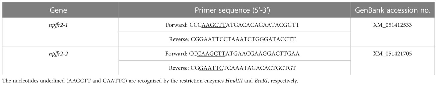
Table 1 Primers for construction of pcDNA3.1-NPFFR2-1 and pcDNA3.1-NPFFR2-2.
2.2 Signal transduction pathways of sea bass NPFFR2 receptors
Transfection experiments were generally performed as described previously (Wang et al., 2022b). In brief, COS-7 cells (ATCC) were seeded in 24-well plates at a density of 1×10⁵ cells/well/mL of DMEM (Gibco, Waltham, MA, USA) supplemented with 10% FBS (Gibco) and 1% penicillin/streptomycin (Gibco) and maintained in an incubator (ThermoFisher Scientific, Waltham, MA, USA) at 37°C with a humidified 5% CO2 atmosphere for 24 h before transfection. Cells were then co-transfected with 200 ng of CRE-luc, SRE-luc, or NFAT-RE-luc, 200 ng of pcDNA3.1-NPFFR2-1 or pcDNA3.1-NPFFR2-2, and 20 ng of pRL-TK using Lipofectamine 3000 (Invitrogen). Following starvation overnight, (1) cells were challenged for 6 h with 10, 100, 1000 nM of GnIH1 and GnIH2, or 1000 nM of NPFF and NPAF, as in our previous study (Wang et al., 2022b); (2) cells were treated for 6 h with 10 μM of forskolin (FSK, Calbiochem, San Diego, CA, USA) alone or co-administration with 1000 nM of GnIH1, GnIH2, NPFF and NPAF. Cells were harvested using 1×Passive Lysis Buffer (Promega, Madison, WI, USA) and luciferase activity in cell extracts was evaluated using Dual-Glo® Luciferase Assay System (Promega) on the LB963 luminometer (Berthold Technologies GmbH & Co.KG, Bad Wildbad, Germany). Each transfection experiment was performed in triplicate and repeated twice.
2.3 Western blot analysis
To examine whether activation of sea bass NPFFR2-1 or NPFFR2-2 by GnIH peptides can increase ERK phosphorylation levels, COS-7 cells were seeded in 24-well plates (2×10⁵ cells/well/mL DMEM) one day before transfection, and then transfected with 200 ng of pcDNA3.1-NPFFR2-1 or pcDNA3.1-NPFFR2-2, starved overnight, and treated with 1 μM of GnIH1, GnIH2, NPFF, and NPAF for 10 min. Cells were lysed and phosphorylated ERK levels were detected by Western blot, as described in details in our recent study (Wang et al., 2022b). In parallel, total ERK levels was also detected as loading controls to normalize the blots. Phosphop44/42 MAPK (Erk1/2) (Thr202/Tyr204) antibody (1:1000, Cell Signaling Technology, Danvers, MA, USA), p44/42 MAPK (Erk1/2) antibody (1:1000, Cell Signaling Technology) and HRP-linked anti-rabbit IgG antibody (1:2000, Cell Signaling Technology) were used for Western blot analysis in this study.
2.4 Statistical analysis
Data were analyzed by one-way ANOVA followed by Duncan’s multiple range test using SPSS17.0 software and presented as the mean ± SEM. Normality and homoscedasticity assumptions were tested prior to the analysis. Differences were considered to be statistically significant when p < 0.05.
3 Results
3.1 CRE-luc activity in COS-7 cells expressing sea bass NPFFR2 receptors
Compared to the control group, no response in CRE-luc activity was observed when COS-7 cells expressing sea bass NPFFR2-1 were challenged with 10, 100, 1000 nM of GnIH1 and GnIH2, respectively (Figure 1A). As a comparative control, neither NPFF nor NPAF (1000 nM) altered CRE-luc activity, either (Figure 1A). Similar results were obtained for sea bass NPFFR2-2 (Figure 1B). However, these four peptides (1000 nM for each) significantly reduced FSK-induced CRE-luc activity in COS-7 cells transfected with NPFFR2-1 (Figure 2A) and NPFFR2-2 (Figure 2B). These data indicate that both NPFFR2-1 and NPFFR2-2 receptors are coupled to Gαi protein and can be activated by GnIH and NPFF peptides.

Figure 1 Effects of GnIH peptides on CRE-luc activity in COS-7 cells expressing sea bass NPFFR2-1 (A) and NPFFR2-2 (B). Cells were challenged with 10, 100, 1000 nM of GnIH peptides for 6 h and then harvested for assays. NPFF and NPAF (1000 nM) acted as a comparative control. Data are presented as the mean ± SEM (n=6). Different letters indicate statistically significant differences between mean values (one-way ANOVA, p < 0.05).
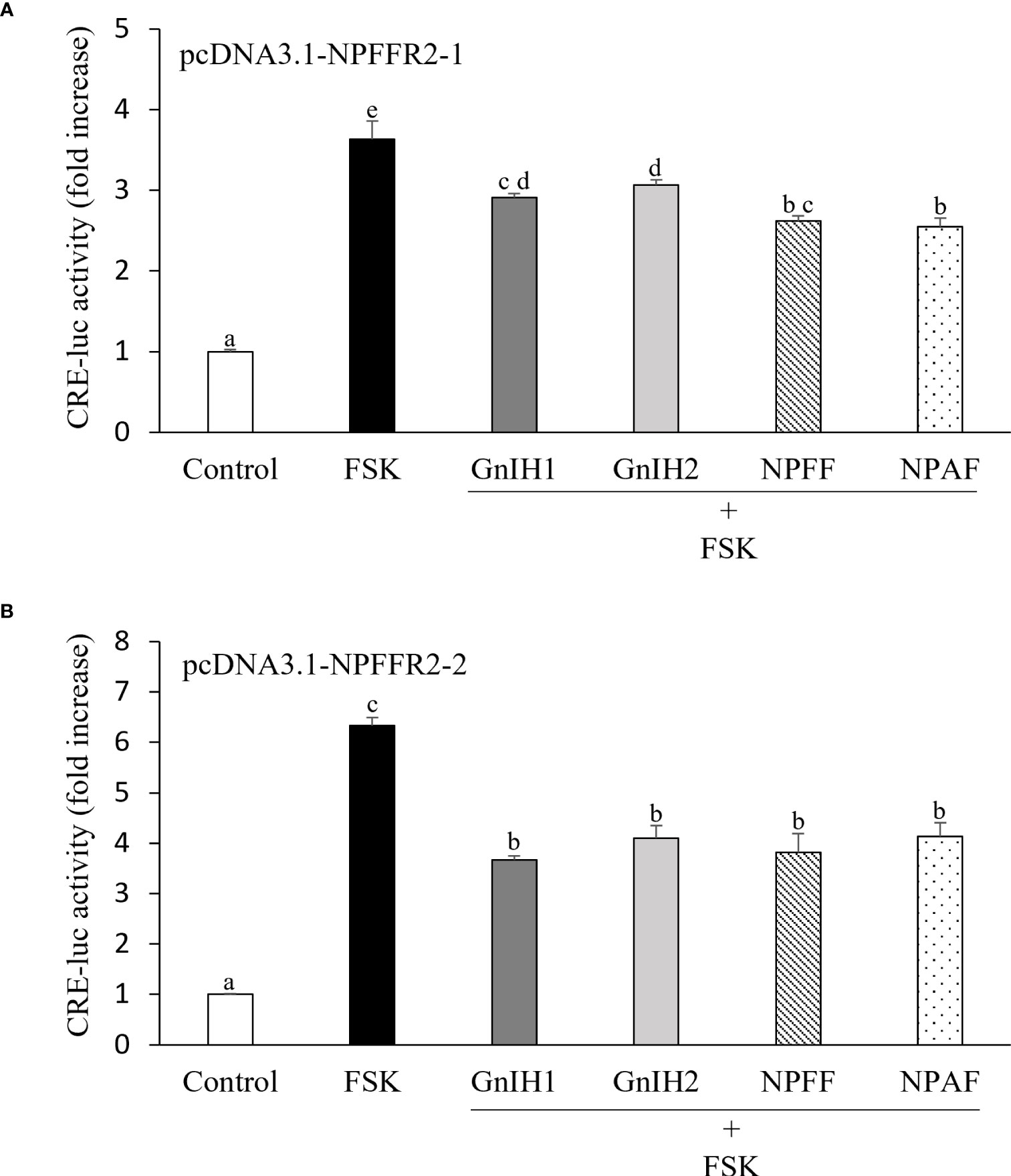
Figure 2 Effects of GnIH and NPFF peptides on FSK-stimulated CRE-luc activity in COS-7 cells expressing sea bass NPFFR2-1 (A) and NPFFR2-2 (B). Cells were challenged with 10 μM of FSK alone or co-treated with 1 μM of GnIH and NPFF peptides for 6 h and then harvested for assays. Data are presented as the mean ± SEM (n=6). Different letters indicate statistically significant differences between mean values (one-way ANOVA, p < 0.05).
3.2 SRE-luc activity in COS-7 cells expressing sea bass NPFFR2 receptors
As shown in Figure 3A, neither GnIH1 nor GnIH2 at any of the three doses modified SRE-luc activity in COS-7 cells transfected with sea bass NPFFR2-1. However, both NPFF and NPAF elicited a slight but significant increase in SRE-luc activity when compared to the control treatment (Figure 3A). On the other hand, an evident increase in SRE-luc activity was noted when COS-7 cells expressing sea bass NPFFR2-2 were treated with 1000 nM of GnIH1, NPFF and NPAF, but NPFF and NPAF showed more potent activity than GnIH1 (Figure 3B). GnIH2 had no effect on SRE-luc activity in COS-7 cells expressing sea bass NPFFR2-2 (Figure 3B). These results showed that both NPFFR2-1 and NPFFR2-2 receptors are coupled to Gαq protein and can be preferentially activated by NPFF peptides.
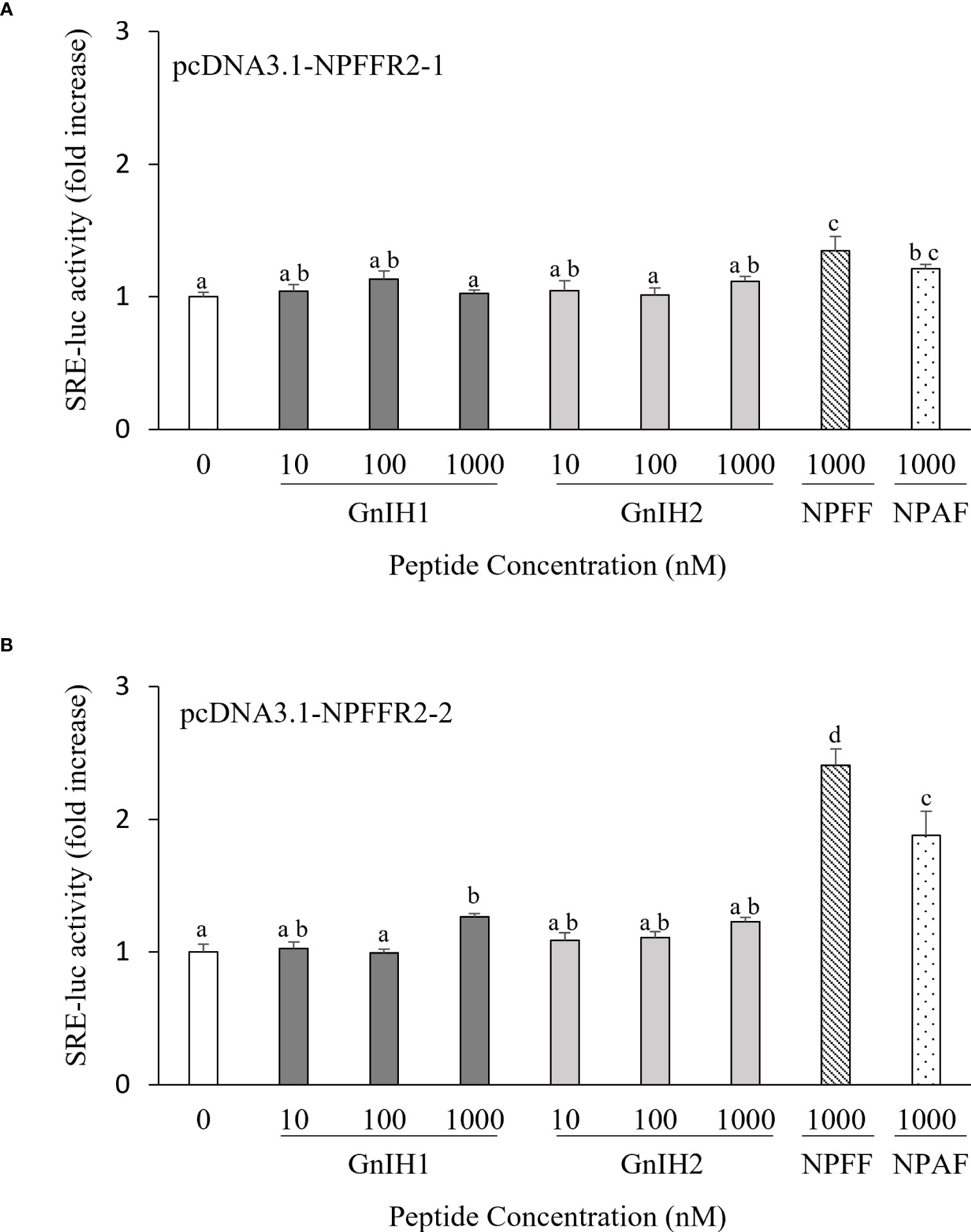
Figure 3 Effects of GnIH peptides on SRE-luc activity in COS-7 cells expressing sea bass NPFFR2-1 (A) and NPFFR2-2 (B). Cells were challenged with 10, 100, 1000 nM of GnIH peptides for 6 h and then harvested for assays. NPFF and NPAF (1000 nM) acted as a comparative control. Data are presented as the mean ± SEM (n=6). Different letters indicate statistically significant differences between mean values (one-way ANOVA, p < 0.05).
3.3 NFAT-RE-luc activity in COS-7 cells expressing sea bass NPFFR2 receptors
The possible involvement of intracellular Ca2+ mobilization in activation of sea bass NPFFR2 receptors was also examined. None of GnIH1, NPFF or NPAF had any effect on NFAT-RE-luc activity in COS-7 cells transfected with sea bass NPFFR2-1, compared to the control group (Figure 4A). A slight but significant reduction of NFAT-RE-luc activity was noticed when cells expressing sea bass NPFFR2-1 were exposed to 10, 100 and 1000 nM of GnIH2 (Figure 4A). None of these four peptides tested changed NFAT-RE-luc activity in COS-7 cells expressing sea bass NPFFR2-2 (Figure 4B).
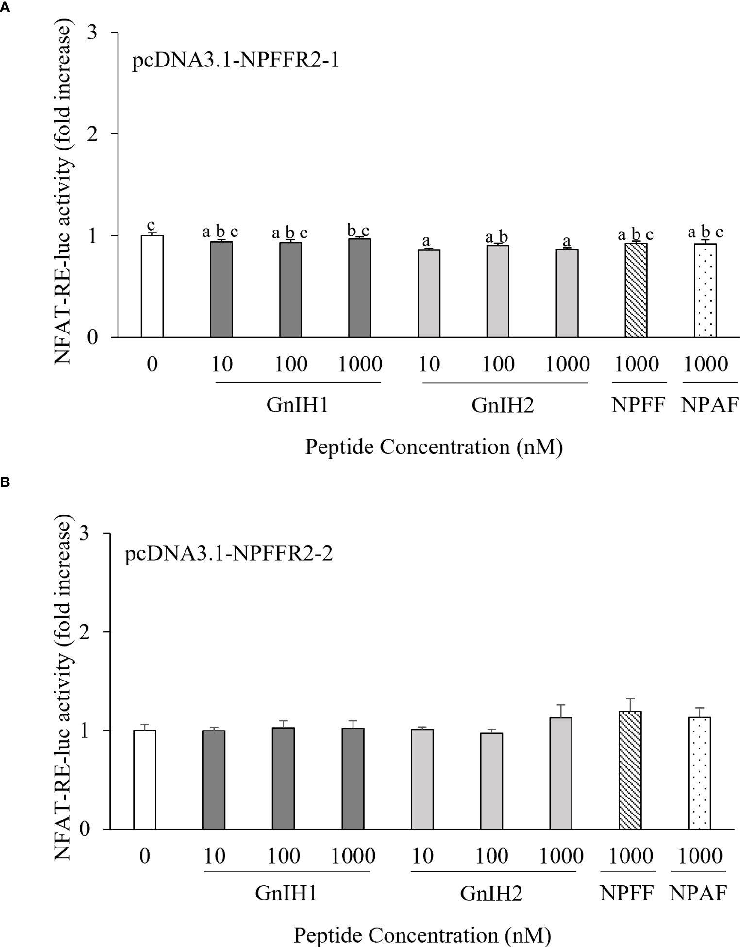
Figure 4 Effects of GnIH peptides on NFAT-RE-luc activity in COS-7 cells expressing sea bass NPFFR2-1 (A) and NPFFR2-2 (B). Cells were challenged with 10, 100, 1000 nM of GnIH peptides for 6 h and then harvested for assays. NPFF and NPAF (1000 nM) acted as a comparative control. Data are presented as the mean ± SEM (n=6). Different letters indicate statistically significant differences between mean values (one-way ANOVA, p < 0.05).
3.4 Effects of GnIH and NPFF on ERK activation
Western blot was employed to verify whether ERK pathway participates in sea bass NPFFR2 activation. There was no significant change in ERK phosphorylation levels when COS-7 cells expressing sea bass NPFFR2-1 (Figure 5A) or NPFFR2-2 (Figure 5B) were stimulated with 1 μM of GnIH1, GnIH2, NPFF and NPAF peptides.
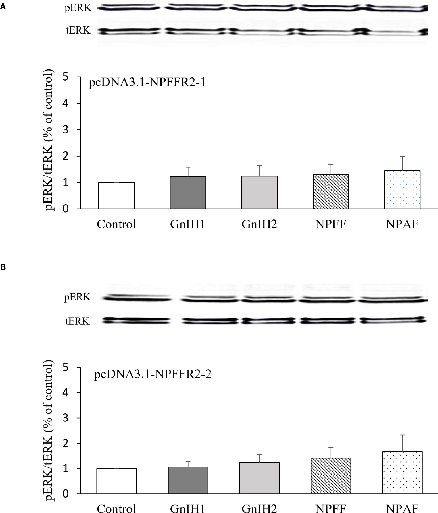
Figure 5 Effects of GnIH and NPFF peptides on ERK phosphorylation levels in COS-7 cells expressing sea bass NPFFR2-1 (A) and NPFFR2-2 (B). Cells were challenged with 1 μM of GnIH and NPFF peptides for 10 min and then harvested for Western blot analysis. Data are presented as the mean ± SEM (n=3).
4 Discussion
It has been evidenced that NPFF and GnIH genes have diverged from a common ancestral gene through whole-genome duplication during vertebrate evolution (Osugi et al., 2014a; Osugi et al., 2014b). Our recent studies showed that GnIH exerted an inhibitory effect on the brain-pituitary-gonadal axis of male sea bass (Paullada-Salmeron et al., 2019), and that GnIHR signals can be transduced via PKA and PKC pathways (Wang et al., 2022b). In addition, both NPFF and NPAF significantly reduced FSK-elicited CRE-luc activity and stimulated SRE-luc activity in COS-7 cells expressing sea bass GnIHR, suggesting that GnIHR is a candidate receptor for these two neuropeptides (Wang et al., 2022b). However, whether GnIH also exerts its actions on the reproductive axis via NPFFR2 and the signaling pathways of NPFFR2 in response to GnIH are still unknown in fish. Accordingly, we further addressed these issues in the current study.
Our results showed that neither GnIH1 nor GnIH2 had any effect on basal CRE-luc activity, but significantly down-regulated FSK-induced CRE-luc activity in COS-7 cells transfected with sea bass NPFFR2-1 and NPFFR2-2, indicating that both NPFFR2 receptors are coupled to Gαi protein, consistently with previous studies in mammals (Bonini et al., 2000; Liu et al., 2001; Mollereau et al., 2005) and chicken (Chen et al., 2020). In contrast, in spotted sea bass, two NPFFR2 receptors are coupled to Gαs protein, because NPFF increased CRE-luc activity in a dose-dependent manner in HEK-293T cells expressing its cognate receptors (Li et al., 2019). The reasons for these apparent discrepancies among the different species are still unclear, perhaps due to variations in conditions of transfection experiments, including cell origins (COS-7 or HEK293-T), plasmid doses, transfection chemicals, time elapsed after challenge, and luciferase assay kits. It is worth mentioning that receptors for sea bass kisspeptin and GnRH have been demonstrated to be coupled to Gαs protein (Servili et al., 2010; Felip et al., 2015). Considering the opposite actions on the release of gonadotropins between GnIH and kisspeptin/GnRH (Espigares et al., 2015; Paullada-Salmeron et al., 2016c; Muñoz-Cueto et al., 2020), it is reasonable to speculate that GnIH could antagonize kisspeptin and GnRH signaling involved in PKA pathway via NPFFR2, as is the case of NPFFR1/GnIHR (Wang et al., 2019a). On the other hand, activation of NPFFR2 by sea bass GnIH may account for the absence of its inhibitory effect on sea bass kisspeptin-stimulated CRE-luc activity in COS-7 cells co-transfected with Kiss1R and GnIHR (Wang et al., 2022b). Further research is being directed in the laboratory to investigate the synergistic effect of GnIHR and NPFFR2 activation by GnIH.
In the current study, both NPFF and NPAF stimulated SRE-luc activity in COS-7 cells expressing sea bass NPFFR2-1 and NPFFR2-2, although NPFFR2-2 showed much higher affinity than NPFFR2-1 by both peptides, indicating that these two receptors couple to the Gαq protein and convey their signaling via the PKC pathway, which is consistent with a recent study in chicken (Chen et al., 2020). Interestingly, a recent study using chimeric G-protein-based TGF-α shedding assays showed that human NPFFR2 receptor exhibited coupling preference for Gi/o when NPFF peptide was used as ligand (Inoue et al., 2019). Neither GnIH1 nor GnIH2 altered SRE-luc activity in NPFFR2-1 transfected COS-7 cells, whereas a slight but significant increase was observed when cells expressing NPFFR2-2 were treated with the highest dose (1000 nM) of GnIH1. In sea bass, GnIHR is also coupled to the Gαq protein (Wang et al., 2022b) and, therefore, GnIHR and NPFFR2-2 may work in concert to convey the actions of GnIH1 via the PKC pathway, which will require further studies in the near future. To the best of our knowledge, there are no comparable studies on the activation of NPFFR2 by GnIH in other teleosts.
The present study also investigated the possible involvement of Ca2+ and ERK pathways in mediating the actions of GnIH through NPFFR2. Only GnIH2 slightly reduced NFAT-RE-luc activity in COS-7 cells expressing NPFFR2-1, but none of the four peptides modified NFAT-RE-luc activity in cells expressing NPFFR2-2. Our recent study also revealed that there was no change in NFAT-RE-luc activity when COS-7 cells transfected with GnIHR were challanged with GnIH1, GnIH2, NPFF and NPAF (Wang et al., 2022b). Similar results were also observed in chicken, where GnIH had no effect on NFAT-RE-luc activity via either GnIHR or NPFFR2, but activation of NPFFR2 by NPAF led to an obvious elevation (Chen et al., 2020). Previous results have provided evidence that Ca2+ signaling is involved in the actions of kisspeptin and GnRH in fish (Chang and Pemberton, 2018; Wang et al., 2022a). Thus, GnIH may antagonize the effects of kisspeptin and GnRH on target cells by lowering Ca2+ levels. In this sense, GnRH-stimulated cytoplasmic Ca2+ mobilization was blocked by GnIH in ovine pituitary gonadotropes (Clarke et al., 2008).
Similar to sea bass GnIHR (Wang et al., 2022b), none of GnIH and NPFF peptides increased ERK phosphorylation levels via NPFFR2 in this study. Likewise, GnIH did not alter basal and kisspeptin-induced ERK phosphorylation levels in GnRH neuronal cell line, GT1-7, which endogenously expresses NPFFR1 and NPFFR2 (Son et al., 2016). GnIH had no effect on ERK phosphorylation in ovine pituitary gonadotropes, but suppressed GnRH-induced phosphorylation of ERK (Sari et al., 2009). On the other hand, ERK phosphorylation levels were enhanced in HEK293 cells expressing chicken NPFFR2 treated with NPAF or in NPFFR1-expressing cells treated with GnIH, respectively (Chen et al., 2020). Moreover, activation of NPFFR2 stimulated neurite outgrowth in Neuro 2A cells through the ERK route (Yu et al., 2016). Thus, whether ERK signaling mediates NPFFR2 actions appears to vary among species.
In summary, we have revealed differential activation of two NPFFR2 receptors by sea bass GnIH peptides, showing that NPFFR2 signals can be transduced through PKA, PKC and Ca2+ pathways. Combined with results from our recent study (Wang et al., 2022b), we propose that sea bass GnIH exerts its actions preferentially via GnIHR, and NPFFR2 may function as an alternative route to mediate GnIH effects, since GnIHR and NPFFR2 share similar signaling pathways (Figure 6). Further studies are urgently needed to evaluate the interactions of GnIH with multiple neuroendocrine factors in cell signaling, via GnIHR, NPFFR2 or both receptors.

Figure 6 Proposed model for GnIH receptor (GnIHR) and NPFF receptor (NPFFR2) signaling and putative interaction with kisspeptin receptor (KissR) signaling in sea bass. Sea bass GnIHR signals can be transduced via both PKA and PKC pathways, and activation of GnIHR can interfere with kisspeptin signaling involving the PKA pathway (Wang et al., 2022b). Sea bass GnIH may exert its actions partially via NPFFR2, and PKA, PKC and Ca2+ routes are potential mediators [this study].
Data availability statement
The original contributions presented in the study are included in the article/supplementary material. Further inquiries can be directed to the corresponding author.
Author contributions
BW and JAM-C designed research. BW, JAP-S, and AV-C performed experiments. BW analyzed data and wrote the paper. JAM-C edited the manuscript and provided funding. All authors contributed to the article and approved the submitted version.
Funding
This study was supported by grants from PAIDI2020 (AQUABASS, Consejería de Economía, Conocimiento, Empresas y Universidad. Junta de Andalucía. Grant no P18-RT-5152), Junta de Andalucía (QUALIFICA, Grant no QUAL21-0019) and Universidad de Cádiz (Plan Propio de Investigación, Grant no PB2023-052) to JAM-C. BW was awarded a scholarship sponsored by the China Scholarship Council (CSC, File no 201903260004).
Acknowledgments
We thank Dr. Ana Gómez (Institute of Aquaculture of Torre de la Sal, CSIC, Spain) for kindly providing us with the CRE-luc and SRE-luc plasmids.
Conflict of interest
The authors declare that the research was conducted in the absence of any commercial or financial relationships that could be construed as a potential conflict of interest.References
Publisher’s note
All claims expressed in this article are solely those of the authors and do not necessarily represent those of their affiliated organizations, or those of the publisher, the editors and the reviewers. Any product that may be evaluated in this article, or claim that may be made by its manufacturer, is not guaranteed or endorsed by the publisher.
References
Biran J., Golan M., Mizrahi N., Ogawa S., Parhar I. S., Levavi-Sivan B. (2014). LPXRFa, the piscine ortholog of GnIH, and LPXRF receptor positively regulate gonadotropin secretion in Tilapia (Oreochromis niloticus). Endocrinology 155, 4391–4401. doi: 10.1210/en.2013-2047
Bonini J. A., Jones K. A., Adham N., Forray C., Artymyshyn R., Durkin M. M., et al. (2000). Identification and characterization of two G protein-coupled receptors for neuropeptide FF. J. Biol. Chem. 275, 39324–39331. doi: 10.1074/jbc.M004385200
Chang J. P., Pemberton J. G. (2018). Comparative aspects of GnRH-Stimulated signal transduction in the vertebrate pituitary - Contributions from teleost model systems. Mol. Cell Endocrinol. 463, 142–167. doi: 10.1016/j.mce.2017.06.002
Chen J., Huang S., Zhang J., Li J., Wang Y. (2020). Characterization of the neuropeptide FF (NPFF) gene in chickens: evidence for a single bioactive NPAF peptide encoded by the NPFF gene in birds. Domest Anim. Endocrinol. 72, 106435. doi: 10.1016/j.domaniend.2020.106435
Clarke I. J., Sari I. P., Qi Y., Smith J. T., Parkington H. C., Ubuka T., et al. (2008). Potent action of RFamide-related peptide-3 on pituitary gonadotropes indicative of a hypophysiotropic role in the negative regulation of gonadotropin secretion. Endocrinology 149, 5811–5821. doi: 10.1210/en.2008-0575
Espigares F., Zanuy S., Gomez A. (2015). Kiss2 as a regulator of Lh and Fsh secretion via paracrine/autocrine signaling in the teleost fish European sea bass (Dicentrarchus labrax). Biol. Reprod. 93, 114. doi: 10.1095/biolreprod.115.131029
Felip A., Espigares F., Zanuy S., Gomez A. (2015). Differential activation of kiss receptors by Kiss1 and Kiss2 peptides in the sea bass. Reproduction 150, 227–243. doi: 10.1530/REP-15-0204
Ikemoto T., Park M. K. (2005). Chicken RFamide-related peptide (GnIH) and two distinct receptor subtypes: identification, molecular characterization, and evolutionary considerations. J. Reprod. Dev. 51, 359–377. doi: 10.1262/jrd.16087
Inoue A., Raimondi F., Kadji F. M. N., Singh G., Kishi T., Uwamizu A., et al. (2019). Illuminating G-protein-coupling selectivity of GPCRs. Cell 177, 1933–1947.e25. doi: 10.1016/j.cell.2019.04.044
Koller J., Herzog H., Zhang L. (2021). The distribution of neuropeptide FF and neuropeptide VF in central and peripheral tissues and their role in energy homeostasis control. Neuropeptides 90, 102198. doi: 10.1016/j.npep.2021.102198
Li Q., Wen H., Li Y., Zhang Z., Zhou Y., Qi X. (2019). Evidence for the direct effect of the NPFF peptide on the expression of feeding-related factors in spotted sea bass (Lateolabrax maculatus). Front. Endocrinol. (Lausanne) 10, 545. doi: 10.3389/fendo.2019.00545
Lin Y. T., Chen J. C. (2019). Neuropeptide FF modulates neuroendocrine and energy homeostasis through hypothalamic signaling. Chin. J. Physiol. 62, 47–52. doi: 10.4103/CJP.CJP_23_19
Liu Q., Guan X. M., Martin W. J., McDonald T. P., Clements M. K., Jiang Q., et al. (2001). Identification and characterization of novel mammalian neuropeptide FF-like peptides that attenuate morphine-induced antinociception. J. Biol. Chem. 276, 36961–36969. doi: 10.1074/jbc.M105308200
Mollereau C., Mazarguil H., Zajac J. M., Roumy M. (2005). Neuropeptide FF (NPFF) analogs functionally antagonize opioid activities in NPFF2 receptor-transfected SH-SY5Y neuroblastoma cells. Mol. Pharmacol. 67, 965–975. doi: 10.1124/mol.104.004614
Muñoz-Cueto J. A., Paullada-Salmeron J. A., Aliaga-Guerrero M., Cowan M. E., Parhar I. S., Ubuka T. (2017). A Journey through the gonadotropin-inhibitory hormone system of fish. Front. Endocrinol. (Lausanne) 8, 285. doi: 10.3389/fendo.2017.00285
Muñoz-Cueto J. A., Zmora N., Paullada-Salmeron J. A., Marvel M., Mañanos E., Zohar Y. (2020). The gonadotropin-releasing hormones: Lessons from fish. Gen. Comp. Endocrinol. 291, 113422. doi: 10.1016/j.ygcen.2020.113422
Nguyen T., Marusich J., Li J. X., Zhang Y. (2020). Neuropeptide FF and its receptors: Therapeutic applications and ligand development. J. Med. Chem. 63, 12387–12402. doi: 10.1021/acs.jmedchem.0c00643
Ohga H., Matsuyama M. (2021). Effects of LPXRFamide peptides on chub mackerel gonadotropin secretion. Biol. Reprod. 105, 1179–1188. doi: 10.1093/biolre/ioab130
Osugi T., Okamura T., Son Y. L., Ohkubo M., Ubuka T., Henmi Y., et al. (2014a). Evolutionary origin of GnIH and NPFF in chordates: insights from novel amphioxus RFamide peptides. PloS One 9, e100962. doi: 10.1371/journal.pone.0100962
Osugi T., Ubuka T., Tsutsui K. (2014b). Review: evolution of GnIH and related peptides structure and function in the chordates. Front. Neurosci. 8, 255. doi: 10.3389/fnins.2014.00255
Osugi T., Uchida K., Nozaki M., Tsutsui K. (2011). Characterization of novel RFamide peptides in the central nervous system of the brown hagfish: isolation, localization, and functional analysis. Endocrinology 152, 4252–4264. doi: 10.1210/en.2011-1375
Paullada-Salmeron J. A., Cowan M., Aliaga-Guerrero M., Gomez A., Zanuy S., Mañanos E., et al. (2016a). LPXRFa peptide system in the European sea bass: A molecular and immunohistochemical approach. J. Comp. Neurol. 524, 176–198. doi: 10.1002/cne.23833
Paullada-Salmeron J. A., Cowan M., Aliaga-Guerrero M., Lopez-Olmeda J. F., Mañanos E. L., Zanuy S., et al. (2016b). Testicular steroidogenesis and locomotor activity are regulated by gonadotropin-inhibitory hormone in male European sea bass. PloS One 11, e0165494. doi: 10.1371/journal.pone.0165494
Paullada-Salmeron J. A., Cowan M., Aliaga-Guerrero M., Morano F., Zanuy S., Muñoz-Cueto J. A. (2016c). Gonadotropin inhibitory hormone down-regulates the brain-pituitary reproductive axis of male European sea bass (Dicentrarchus labrax). Biol. Reprod. 94, 121. doi: 10.1095/biolreprod.116.139022
Paullada-Salmeron J. A., Cowan M. E., Loentgen G. H., Aliaga-Guerrero M., Zanuy S., Mañanos E. L., et al. (2019). The gonadotropin-inhibitory hormone system of fish: The case of sea bass (Dicentrarchus labrax). Gen. Comp. Endocrinol. 279, 184–195. doi: 10.1016/j.ygcen.2019.03.015
Pinelli C., Scandurra A., Tsutsui K., Falvo S., D'Aniello B. (2022). Comparative insights of the neuroanatomical distribution of the gonadotropin-inhibitory hormone (GnIH) in fish and amphibians. Front. Neuroendocrinol. 65, 100991. doi: 10.1016/j.yfrne.2022.100991
Sari I. P., Rao A., Smith J. T., Tilbrook A. J., Clarke I. J. (2009). Effect of RF-amide-related peptide-3 on luteinizing hormone and follicle-stimulating hormone synthesis and secretion in ovine pituitary gonadotropes. Endocrinology 150, 5549–5556. doi: 10.1210/en.2009-0775
Servili A., Lethimonier C., Lareyre J. J., Lopez-Olmeda J. F., Sanchez-Vazquez F. J., Kah O., et al. (2010). The Highly conserved gonadotropin-releasing hormone-2 form acts as a melatonin-releasing factor in the pineal of a teleost fish, the european sea bass Dicentrarchus labrax. Endocrinology 151, 2265–2275. doi: 10.1210/en.2009-1207
Shahjahan M., Doi H., Ando H. (2015). Differential expression patterns of PQRFamide peptide and its two receptor genes in the brain and pituitary of grass puffer during the reproductive cycle. Gen. Comp. Endocrinol. 210, 152–160. doi: 10.1016/j.ygcen.2014.07.005
Shimizu M., Bedecarrats G. Y. (2010). Activation of the chicken gonadotropin-inhibitory hormone receptor reduces gonadotropin releasing hormone receptor signaling. Gen. Comp. Endocrinol. 167, 331–337. doi: 10.1016/j.ygcen.2010.03.029
Son Y. L., Ubuka T., Millar R. P., Kanasaki H., Tsutsui K. (2012). Gonadotropin-inhibitory hormone inhibits GnRH-induced gonadotropin subunit gene transcriptions by inhibiting AC/cAMP/PKA-dependent ERK pathway in LbetaT2 cells. Endocrinology 153, 2332–2343. doi: 10.1210/en.2011-1904
Son Y. L., Ubuka T., Soga T., Yamamoto K., Bentley G. E., Tsutsui K. (2016). Inhibitory action of gonadotropin-inhibitory hormone on the signaling pathways induced by kisspeptin and vasoactive intestinal polypeptide in GnRH neuronal cell line, GT1-7. FASEB J. 30, 2198–2210. doi: 10.1096/fj.201500055
Spicer O. S., Zmora N., Wong T. T., Golan M., Levavi-Sivan B., Gothilf Y., et al. (2017). The gonadotropin-inhibitory hormone (Lpxrfa) system's regulation of reproduction in the brain-pituitary axis of the zebrafish (Danio rerio). Biol. Reprod. 96, 1031–1042. doi: 10.1093/biolre/iox032
Teo C. H., Phon B., Parhar I. (2021). The role of GnIH in biological rhythms and social behaviors. Front. Endocrinol. (Lausanne) 12, 728862. doi: 10.3389/fendo.2021.728862
Tomihara S., Ikegami K., Shimomai R., Umatani C. (2022). Neuropeptide FF indirectly affects testicular morphogenesis and functions in medaka. Proc. Natl. Acad. Sci. USA. 119, e2209353119. doi: 10.1073/pnas.2209353119
Tsutsui K., Ubuka T. (2021). Gonadotropin-inhibitory hormone (GnIH): A new key neurohormone controlling reproductive physiology and behavior. Front. Neuroendocrinol. 61, 100900. doi: 10.1016/j.yfrne.2021.100900
Wang B., Mechaly A. S., Somoza G. M. (2022a). Overview and new insights into the diversity, evolution, role, and regulation of kisspeptins and their receptors in teleost fish. Front. Endocrinol. (Lausanne) 13, 862614. doi: 10.3389/fendo.2022.862614
Wang B., Paullada-Salmeron J. A., Verges-Castillo A., Gomez A., Muñoz-Cueto J. A. (2022b). Signaling pathways activated by sea bass gonadotropin-inhibitory hormone peptides in COS-7 cells transfected with their cognate receptor. Front. Endocrinol. (Lausanne) 13, 982246. doi: 10.3389/fendo.2022.982246
Wang Q., Qi X., Guo Y., Li S., Zhang Y., Liu X., et al. (2015). Molecular identification of GnIH/GnIHR signal and its reproductive function in protogynous hermaphroditic orange-spotted grouper (Epinephelus coioides). Gen. Comp. Endocrinol. 216, 9–23. doi: 10.1016/j.ygcen.2015.04.016
Wang B., Yang G., Liu Q., Qin J., Xu Y., Li W., et al. (2017). Inhibitory action of tongue sole LPXRFa, the piscine ortholog of gonadotropin-inhibitory hormone, on the signaling pathway induced by tongue sole kisspeptin in COS-7 cells transfected with their cognate receptors. Peptides 95, 62–67. doi: 10.1016/j.peptides.2017.07.014
Wang B., Yang G., Liu Q., Qin J., Xu Y., Li W., et al. (2018). Characterization of LPXRFa receptor in the half-smooth tongue sole (Cynoglossus semilaevis): Molecular cloning, expression profiles, and differential activation of signaling pathways by LPXRFa peptides. Comp. Biochem. Physiol. A Mol. Integr. Physiol. 223, 23–32. doi: 10.1016/j.cbpa.2018.05.008
Wang B., Yang G., Xu Y., Li W., Liu X. (2019a). Recent studies of LPXRFa receptor signaling in fish and other vertebrates. Gen. Comp. Endocrinol. 277, 3–8. doi: 10.1016/j.ygcen.2018.11.011
Wang B., Yang G., Xu Y., Zhang Y., Liu X. (2019b). In vitro effects of tongue sole LPXRFa and kisspeptin on relative abundance of pituitary hormone mRNA and inhibitory action of LPXRFa on kisspeptin activation in the PKC pathway. Anim. Reprod. Sci. 203, 1–9. doi: 10.1016/j.anireprosci.2019.01.009
Keywords: GnIH, NPFF, NPAF, NPFF receptor, signaling pathway
Citation: Wang B, Paullada-Salmerón JA, Vergès-Castillo A and Muñoz-Cueto JA (2023) Differential activation of neuropeptide FF receptors by gonadotropin-inhibitory hormone peptides in the European sea bass. Front. Mar. Sci. 10:1199189. doi: 10.3389/fmars.2023.1199189
Received: 03 April 2023; Accepted: 13 July 2023;
Published: 28 July 2023.
Edited by:
Benjamin Costas, University of Porto, PortugalReviewed by:
Ana Rocha, University of Porto, PortugalMatan Golan, Agricultural Research Organization (ARO), Israel
Copyright © 2023 Wang, Paullada-Salmerón, Vergès-Castillo and Muñoz-Cueto. This is an open-access article distributed under the terms of the Creative Commons Attribution License (CC BY). The use, distribution or reproduction in other forums is permitted, provided the original author(s) and the copyright owner(s) are credited and that the original publication in this journal is cited, in accordance with accepted academic practice. No use, distribution or reproduction is permitted which does not comply with these terms.
*Correspondence: José A. Muñoz-Cueto, bXVub3ouY3VldG9AdWNhLmVz