- Department of Biology, University of Dallas, Irving, TX, United States
Ongoing ecological events, such as new and emerging diseases, provide an important platform for education and research. Field courses and undergraduate research projects can be critical to assisting students with learning scientific skills and career discernment as these experiences provide more one-on-one instruction and an immersive learning environment. A novel coral disease called “Stony Coral Tissue Loss Disease” (SCTLD) provided one such opportunity. SCTLD is characterized by rapid progression with entire coral heads dying within 2–3 weeks after initial observation of the onset of symptoms. At a wider geographic scale, the disease has migrated with extreme velocity and has now been documented across the Caribbean from as far North as the Southeast Florida Reef Tract, as far South as St. Lucia, and as far West as Honduras and Belize. Here, I summarize what is currently known about SCTLD and document an educational field course that involved eight undergraduate students with visits to multiple locations along the Florida Keys Reef Tract during the disease progression in March 2019. Students were able to observe sites where SCTLD had been present for over 2 years and sites where the disease was only just emerging for observational comparison. Student educational outcomes from field trips and activities will be discussed. Current research and educational activities can interact to enhance each other, creating a positive feedback loop. Future directions for research, educational opportunities, and their interaction to accelerate understanding of this novel disease are discussed.
Introduction
Utilization of current ecological topics and events are an excellent launching point for undergraduate education and research. Field courses are particularly useful for retention in STEM for underserved populations (Beltran et al., 2020), and provide an immersive experience that can stimulate career discernment and motivate more ecologically-friendly personal decisions (Fleischner et al., 2017). Not all students are able to travel to the field due to cost and time, but could still engage with important current issues through research. Stony Coral Tissue Loss Disease (SCTLD) has recently devastated Caribbean coral reefs after its emergence off the coast of South Florida near Virginia Key in 2014 (Miller et al., 2016; Precht et al., 2016; Walton et al., 2018). This event provided the opportunity to teach students about how scientists investigate and treat emerging diseases, and here I will first provide a summary of what is known about SCTLD, outline a field course developed that utilized this situation for educational purposes, and finally provide some perspectives on potential future directions for integrating research and education particularly with undergraduate students.
SCTLD: What We Know
Stony Coral Tissue Loss Disease is quite virulent, causing whole colony mortality within a few weeks to a few months (Precht et al., 2016; Walton et al., 2018; Rippe et al., 2019; Estrada-Saldívar et al., 2020), can result in high levels of infection (i.e., 30% or higher), rapid mortality (i.e., 1% mortality in June 2018 to 58% mortality in March 2019 at the Fish Market Site, Mexican Caribbean), and expresses high amounts of variability among sites in disease incidence (Alvarez-Filip et al., 2019; Estrada-Saldívar et al., 2020; Meiling et al., 2020). Since its arrival, SCTLD has rapidly spread following the contagion epidemiological model and has been documented in both large- and small-scale biogeographic regions. SCTLD is widespread in the Caribbean and now can be observed as far south as St. Lucia and as far west as Mexico, Belize, and Honduras (Alvarez-Filip et al., 2019; Kramer et al., 2019; Rippe et al., 2019; Estrada-Saldívar et al., 2020; Muller et al., 2020; Sharp et al., 2020). In-shore reefs have lower frequencies of disease and mortality, are more resilient displaying high tolerance to even the most extreme bleaching events, and recover more quickly from stress than off-shore reefs (Kenkel et al., 2013; Manzello et al., 2015; Gintert et al., 2018; Rippe et al., 2019; Sharp et al., 2020). Resilience may play a role in the ability of corals to resist pathogens present as physiologic stress is linked to decreased ability of recovery, immune function, and can alter gene expression (Bruno et al., 2007; Muller et al., 2008; Brandt and McManus, 2009a; Kenkel and Matz, 2016; Haslun et al., 2018; Fuess et al., 2020).
The rapid sweep of SCTLD over large geographic areas has led to the question of how this disease is transmitted, which could be through the water column, vectors (i.e., reef-eating fish), or both. The north-to-south direction of SCTLD spread throughout the Keys is interesting considering the upper Keys generally have a weak northward flow, but in-shore locations experience currents that interact with the curvature orientation of the islands and winds to create a generally southwest alongshore current (Lee and Williams, 1999). This raises the question if SCTLD was primarily transported by in-shore currents with later spread to off-shore reef locations. Although, transmission via vectors (i.e., butterflyfish) should not be ruled out (Noonan and Childress, 2020). A recent laboratory study determined that SCTLD can be transmitted via the water column and vectors (Aeby et al., 2019). Several organisms can feed on both healthy and diseased tissue providing an avenue for transmission (Alvarez-Filip et al., 2019). Given these facts, one hypothesis is that SCTLD may be transmitted differently at large geographic scales, perhaps via water currents, and smaller, local geographic scales via vectors.
A number of environmental factors have been investigated concerning the emergence of the disease, which include increased sea surface temperatures (SSTs), increased turbidity from a concurrent dredging project (Miller et al., 2016; Gintert et al., 2019; Precht, 2021), and eutrophication (Dougan et al., 2020). Eutrophication from nutrient run off in the South Florida region, may have led to a reduction in immune function making corals susceptible to infection (Dougan et al., 2020). Although the exact biological causative agent is still unknown, it seems probable that a bacterial component exists. Examination of the microbiome of diseased corals and the water column have revealed a number of different bacterial taxa (Meyer et al., 2019; Rosales et al., 2020). These data paired with the observation that antibiotic treatments are effective makes a strong case for bacterial involvement (Aeby et al., 2019; Neely et al., 2020), but there are no studies that have determined whether there is a viral component to the disease complex.
Histological analysis has revealed that alterations to the coral host-zooxanthellae associations occur at the cellular level (Landsberg et al., 2020). Physiological disruption between the coral host and their endosymbionts may cause SCTLD, with the bacterial infection being entirely opportunistic (Landsberg et al., 2020). But, this explanation does not explain why so many species of coral would have altered endosymbiont associations nor account for the progression of the disease over larger geographic areas.
Taking all of the factors into account, one plausible hypothesis is that certain environmental factors (i.e., increased SSTs; increased turbidity; nutrient runoff) led to physiological stress, which may have resulted in the disruption of coral host-zooxanthellae associations. This paired with reduced immunity could have resulted in subsequent bacterial (or other pathogen) infection, which then spread along the reef. Due to continued spread throughout winter months and to areas where turbidity is low, it is possible that environmental factors played less of a role in the spread than in the initial emergence of SCTLD. Alteration of physiological equilibrium from environmental perturbation can make animals more susceptible to already present bacteria in hosts or in reef sediments. This hypothesis has already been posited for other coral diseases (Weil, 2004, pg. 36; Muller and van Woesik, 2012) and could be an explanation for the emergence and spread of SCTLD.
Comparison of SCTLD to Other Diseases
Stony coral tissue loss disease is not the first disease to afflict stony corals in the Caribbean. In fact, the Caribbean is home to a disproportionately large number of diseases compared to other reefs around the world (Weil, 2004). Although, SCTLD differs from other coral diseases in several ways. SCTLD clusters following a contagion model (Muller et al., 2020), other Caribbean diseases, such as White Plague, have mixed evidence for clustered distribution (Nugues, 2002; Borger, 2003, 2005; Richardson and Voss, 2005; Voss and Richardson, 2006; Brandt and McManus, 2009b; Muller and van Woesik, 2012). SCTLD is a generalist afflicting well over 20 species of stony coral, whereas other diseases in the Caribbean are more targeted with infection of their hosts ranging from 1 to 11 species (Weil, 2004; pg. 52). At sites where SCTLD occurs the frequency of infection is high (i.e., 57% at Fish Market site in Mexican Caribbean, Estrada-Saldívar et al., 2020) compared to other diseases such as Dark Spot Disease (1.96%, Weil et al., 2002), White Band Disease (up to 12%; Mayor et al., 2006), and Yellow Band (4.8%, Weil and Cróquer, 2009). Although this pattern could be explained by the fact that SCTLD infects significantly more species, even taking all diseases together within sites in the Caribbean still yields lower prevalence than SCTLD (Weil, 2004, pg. 53; Weil and Cróquer, 2009). SCTLD appears to have emerged simultaneously with a period of increased SSTs and although its progression tends to slow during times when temperatures are high, SCTLD persists throughout all seasons (Meiling et al., 2020). Other coral diseases such as White Band (infecting Acropora) and Yellow Band (infecting Orbicella spp.) positively correlated with average STTs (van Woesik and Randall, 2017). One striking similarity is that multiple modes of transmission seem to be plausible: Black Band Disease (Rützler et al., 1983; Aeby and Santavy, 2006) and SCTLD can be transmitted via the water column and vectors (Aeby et al., 2019; Alvarez-Filip et al., 2019; Noonan and Childress, 2020).
Throughout research for this article I could not help but realize how these features are similar to the COVID-19 outbreak we are currently contending with. Its spread is rapid, affecting people all over the world and shows an extraordinarily high level of variability in virulence that has the possibility of expeditiously leading to mortality. The high level of transmissibility of both diseases means that populations, which are geographically distant, are similarly, impacted. I am certain that we will continue to see more novel diseases arise in both wildlife and human populations. Many questions are left unanswered at the moment, which calls for more research.
Student Engagement
Field courses provide students with the opportunity to become immersed in the environment to fully engage in active learning and significantly improve scientific skills such as species identification, experimental design, and oral presentations (Beltran et al., 2020). These courses typically have smaller sizes that encourage more peer discussion, one-on-one attention, and can provide skills and contacts within a field that give students tangible career avenues to pursue (Kim and Sax, 2009; Newman, 2011; Wilson et al., 2012; Ballen et al., 2019). In 2019 I developed a field course called Marine Field Ecology (see Supplementary Figure 1 for course syllabus), which aimed to introduce students to the ecology and evolution of corals throughout the Florida Keys. Students were able to observe first-hand how a selective force, such as disease, can influence the evolution (and/or extinction) of species in the marine environment. During March 2019, eight University of Dallas undergraduates participated, three of which had been in the ocean previously, but only two had visited coral reefs with the third being an avid surfer from California. The students were comprised of seven females and one male, with four coming from marginalized or underrepresented racial backgrounds in STEM.
Course Objectives, Activities and Assessments
Objective #1
Compare reef sites with newly emerging disease to those with more developed disease progress. The goal was for students to gain an understanding of the impact of SCTLD on the ecosystem, learn about the potential for phase shifts in the ecosystem, and observe mitigation strategies both in coral restoration and disease mitigation.
Activity
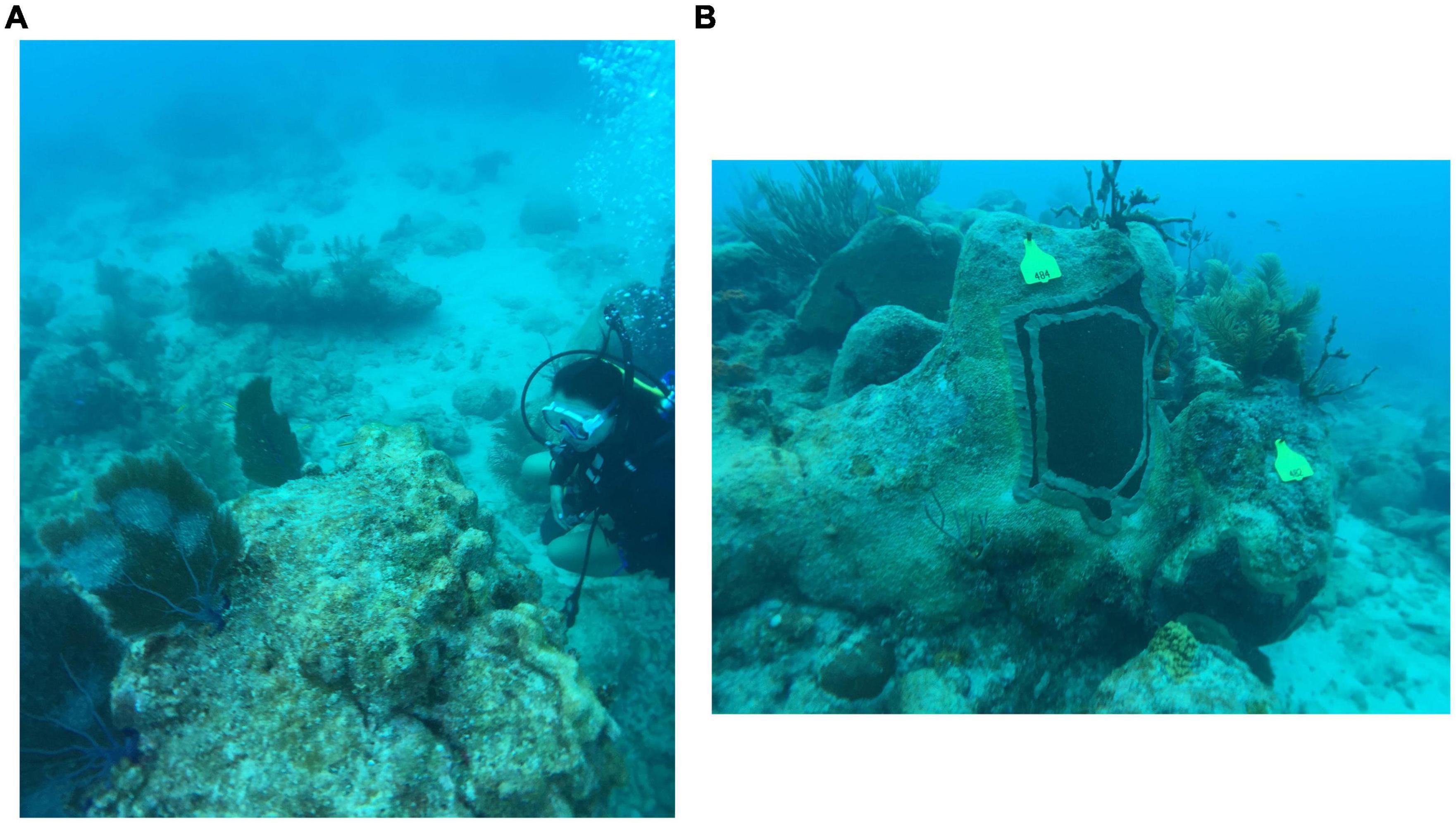
Students visited the upper Keys, staying at MarineLab in Key Largo for 3 days and then traveled to Mote’s Elizabeth Moore International Center for Coral Reef Research and Restoration (Summerland Key). Student observations were conducted via snorkel or SCUBA at Grecian Dry Rocks, the SS Benwood wreck site, and two locations on French Reef in the upper keys and Looe Key in the lower keys. The upper keys were found to have few large stony coral heads alive, and of those present most appeared healthy (likely because SCTLD had already killed susceptible genotypes). Observations on Looe Key included mitigation efforts by Force Blue, a team of military-trained veteran combat divers, who had recently applied antibiotic-laced epoxy to the border between diseased and apparently healthy tissue (Neely et al., 2020) of multiple coral heads (Figure 1).

Figure 1. (A) Student examines the impact of SCTLD on coral head at Looe Key Reef. (B) Example of epoxy treatment administered by Force Blue on Looe Key Reef. Pictures were both taken on March 13, 2019.
Assessment
A final summative presentation using digital media was conducted by students placed in pairs (Supplementary Figure 2). Presentations needed to include their background knowledge and perspectives coming into the course, a discussion of the topics learned throughout the week, and how they integrated their new knowledge into their current framework. A grading rubric was developed to assist with communicating presentation expectations and maintain consistency with grading (Supplementary Figure 3).
Objective #2
Compare biodiversity of fish populations between artificial and natural reef locations. The goal was for students to gain an understanding of how ecologists assess biodiversity in a field setting and how different habitat structures may lead to alterations in species composition and abundance.
Activity
The students also completed a mini-research project that addressed the question of how different habitats can influence fish species diversity. Pictures were taken at three different sites in the upper Keys and fish were classified into major fish groups (i.e., parrotfishes, butterflyfishes, etc.) for comparison of one artificial (Benwood) and two natural reef sites (Grecian Dry Rocks and French Reef). Students then utilized the Shannon-Wiener Diversity Index to quantify differences in group composition among locations.
Assessment
Each student wrote their own laboratory report, fashioned after a scientific article (Supplementary Figure 4). The students found higher biodiversity index scores at both natural sites compared to the artificial reef site (Supplementary Figure 5). Although it is difficult to obtain true fish population size estimates, this study gave students a taste of what it is like to perform such analyses to better understand the health of an ecosystem.
Student Outcomes
The last evening of the trip we sat on Mallory Square in Key West to deconstruct the week and what they had learned in an informal assessment discussion session. The students all agreed that seeing the effects of SCTLD were shocking. They all attested that the experience helped them to gain a better understanding of human impact on the environment, especially the ocean. Students were motivated to make better decisions in their everyday actions that will reduce CO2 emissions and plastic waste. They reported to have learned that although it is unknown what exact role increasing temperatures played in the emergence and spread of SCTLD, it seems likely that increasing physiologic stress, which can lower immune function, is not helpful in disease prevention.
The evidence of their transformation was shown through their presentations, but also in their lab reports. One student remarked in the discussion section of her lab report: “Observing indicators of change such as diversity indexes and levels of species are essential to grasping an idea of just how our human impact looks in the environment.” By showing students restoration efforts and teaching them about the patterns of SCTLD, they gained some solace after viewing the devastation. Another student wrote in her discussion: “One good thing about this phenomenon is that there are two important species of coral that are less affected by this phenomenon elkhorn and staghorn corals. This does not dismiss the fact that there is widespread disease that is killing coral. There are restoration efforts that are being implemented, and research is being done.” This statement shows they learned about the characteristics of the disease (affecting mostly bouldering corals) and that the research and restoration are two good steps forward to assisting with mitigating disease impact.
Discussion
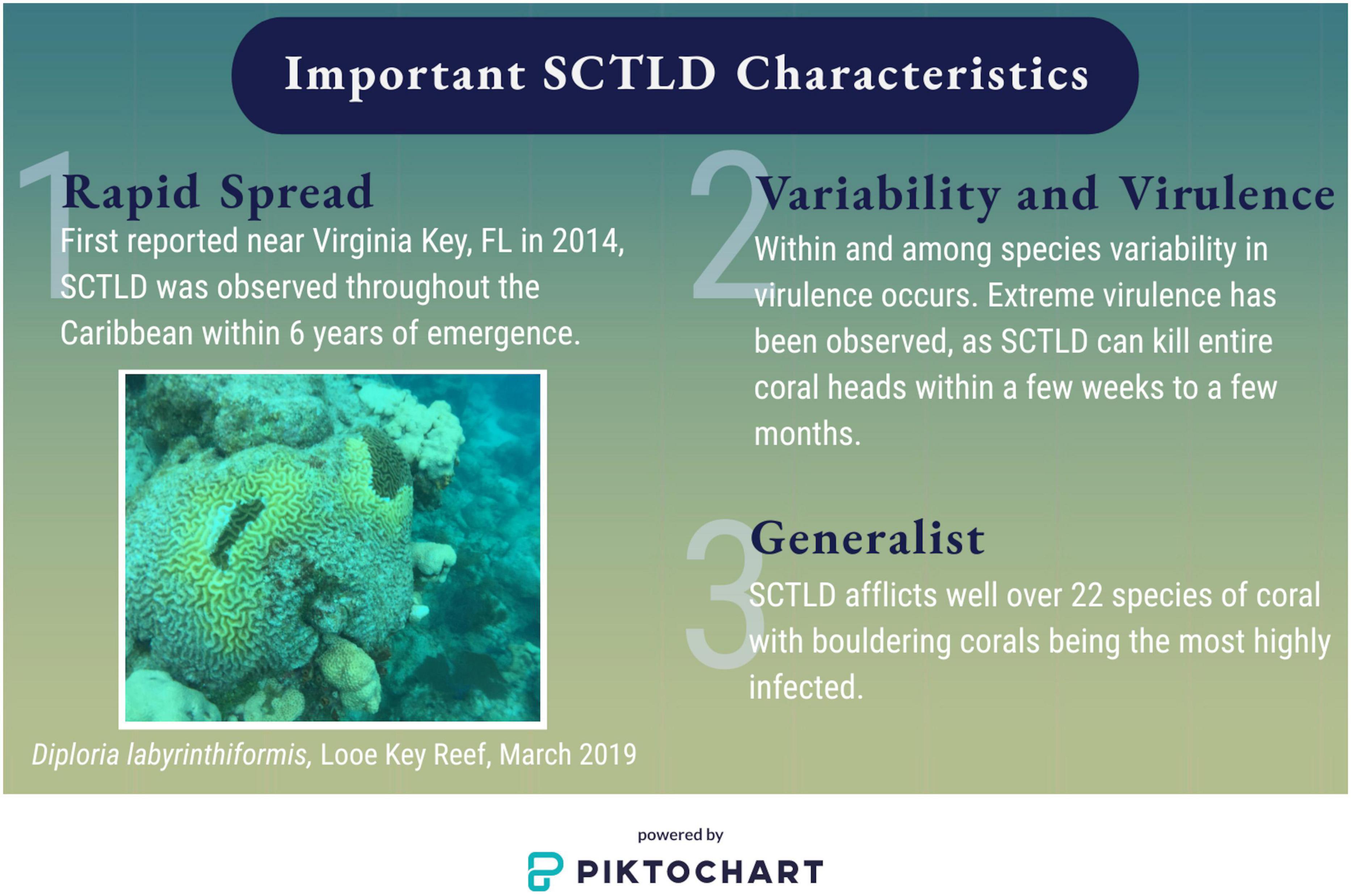
Stony coral tissue loss disease continues to severely impact corals in the Caribbean. Currently there are no direct mechanisms for prevention, and due to difficulty with scalability, treatment measures are also limited. I have outlined here the important characteristics of SCTLD to be: rapid spread, high levels of variance in virulence both within and among species, rapid disease progression resulting in high mortality, and infection of multiple host species (Figure 2).
At the forefront of marine science research, more “hands on deck” are needed to help answer questions rapidly. Sending samples to institutions that have undergraduate student researchers could increase the amount of throughput that is possible. Some institutions, even smaller ones, have access to molecular equipment and can be trained to perform protocols on other species first, such as Drosophila. One challenge is that large collaborations require relationship building among scientists in the field and undergraduate research professors. However, programs such as these have been developed and are successful despite the hurdles (Vidra et al., 2019). Another avenue that could be pursued are citizen science programs. One project already is underway for SCTLD in which divers and snorkelers can upload photos to a website to assist researchers in tracking already treated coral heads1. However, a more extensive photo uploading website could be developed to continue to track coral heads that are not tagged. General dive site locations could be used to give approximate geographic locations so that monitoring could be occurring even on reef sites that have not been visited by researchers. An already implemented example of this comes from the Sea Star Wasting Disease community that has partnered with iNaturalist to document disease patterns from general public photo uploads2.
Many remaining questions could be addressed with the assistance of student scientists engaged in faculty-led field sampling and laboratory analysis. For example, Meyer et al. (2019) found sequences from a number of different bacteria and opportunistic colonizers, but did not examine microbiota alterations over time sequence in stages of infection. Heterogeneity in resistance and variance in survival within species has been observed, but evaluation of heritability both within and between species is needed. The extent to which environmental factors play a role in the emergence and spread of SCTLD should also be addressed. The reality is that SCTLD is not likely to be the last highly virulent coral disease to strike a reef system and a better understanding of how physiologic stress impacts coral immunity might lead to predictors that could be used in monitoring projects. Quicker response times to a coral disease outbreak could assist with mitigating the impact and prevent spread (Precht, 2021). Utilizing more citizen science through uploading photos/video to a central database could help, but one issue is that experts in the field have limited time for analyzing this type of data. However, undergraduate students could be trained to flag photos/videos for further expert review to assist with reducing response time.
Finally, I would like to encourage the use of ongoing environmental events to engage students and utilize the opportunity to make a deeply transformative educational experience, despite the planning and foresight required. Many events can happen to foil even the best laid plans. During Spring 2020, I attempted to run my Marine Field Ecology course. We actually did travel to Key Largo, but got ripped from the reef after 2 days due to shut downs in the wake of the COVID-19 emergence in the United States. Despite the shortened time on the reef, my students were still able to see the effects of SCTLD. An immersive learning environment provides important outcomes because these experiences can significantly alter student career directions, lead to more environmentally-friendly personal decisions, and provide more informed citizens who can relate their experiences to others to create a more compelling narrative.
Data Availability Statement
The original contributions presented in the study are included in the article/Supplementary Material, further inquiries can be directed to the corresponding author.
Ethics Statement
Written informed consent was obtained from the individual(s) for the publication of any potentially identifiable images or data included in this article.
Author Contributions
The author confirms being the sole contributor of this work and has approved it for publication.
Funding
This study was supported by the Marcus Fund from University of Dallas.
Conflict of Interest
The author declares that the research was conducted in the absence of any commercial or financial relationships that could be construed as a potential conflict of interest.
Acknowledgments
I would like to thank all of my students for their hard work and dedication to the course and especially those who gave permission for me to share their work here. I am also appreciative of the feedback they passed along one really beautiful evening on Mallory Square. Bill and Bernie Becker provided housing for me during the lower keys portion of the trip. MarineLab in Key Largo and Mote’s Elizabeth Moore International Center for Coral Reef Research and Restoration in Summerland Key should be commended for their excellent educational programs and appreciation extended for housing and hosting the students. I am also grateful to the University of Dallas for all of their support during the planning and implementation process of my field courses. A special thank you to Ron Carvalho, Sandra Morgan, Kathy McGraw, Rebecca Davies, Scott Salzman, and the late Leonard Robertson for their execution support. I would also like to thank Laura Mydlarz, Lauren Fuess, Donna Hamilton, and Megan Wilson for their comments on previous drafts.
Supplementary Material
The Supplementary Material for this article can be found online at: https://www.frontiersin.org/articles/10.3389/fmars.2021.669472/full#supplementary-material
Supplementary Figure 1 | Syllabus from Spring 2019 Marine Field Ecology, University of Dallas.
Supplementary Figure 2 | Example student presentation slides. The students were asked to integrate what they learned into their perspectives coming into the course. Two students from different backgrounds and majors were paired to increase the level of discussion and contrast perspectives.
Supplementary Figure 3 | Rubric for student presentations after a week-long educational field trip to the Florida Keys.
Supplementary Figure 4 | Biodiversity laboratory report instructions. Students compared fish communities at three sites: one artificial reef (SS Benwood) and two natural reef locations (Dry Rocks and French Reef).
Supplementary Figure 5 | Diversity Index for fish at three locations. Two locations (Dry Rocks and French Reef) are natural reef habitats and one (Benwood) is an artificial reef located off the coast of Key Largo, FL. Pictures were taken during one dive at each location and students categorized fish into major fish groups and counted each fish that was able to be seen in each picture. The diversity index was calculated using the Shannon-Wiener Index. These data were used for educational purposes only. A more detailed study could be used in the future to better track and understand fish biodiversity alterations among sites over time.
Footnotes
References
Aeby, G., Ushijima, B., Campbell, J. E., Jones, S., Williams, G., Meyer, J. L., et al. (2019). Pathogenesis of a tissue loss disease affecting multiple species of corals along the Florida Reef Tract. Front. Mar. Sci. 6:678. doi: 10.3354/meps318103
Aeby, G. S., and Santavy, D. L. (2006). Factors affecting susceptibility of the coral Montastraea faveolata to black-band disease. Mar. Ecol. Prog. Ser. 318, 103–110.
Alvarez-Filip, L., Estrada-Saldívar, N., Pérez-Cervantes, E., Molina-Hernández, A., and González-Barrios, F. J. (2019). A rapid spread of the stony coral tissue loss disease outbreak in the Mexican Caribbean. PeerJ 7:e8069. doi: 10.7717/peerj.8069
Ballen, C. J., Aguillon, S. M., Awwad, A., Bjune, A. E., Challou, D., Drake, A. G., et al. (2019). Smaller classes promote equitable student participation in STEM. BioScience 69, 669–680. doi: 10.1093/biosci/biz069
Beltran, R. S., Marnocha, E., Race, A., Croll, D. A., Dayton, G. H., and Zavaleta, E. S. (2020). Field courses narrow demographic achievement gaps in ecology and evolutionary biology. Ecol. Evol. 10, 5184–5196. doi: 10.1002/ece3.6300
Borger, J. L. (2003). Three scleractinian coral diseases in Dominica, West Indies: distribution, infection patterns and contribution to coral tissue mortality. Rev. Biol. Trop. 51, 25–38.
Borger, J. L. (2005). Scleractinian coral diseases in south Florida: incidence, species susceptibility, and mortality. Dis. Aquat. Organ. 67, 249–258. doi: 10.3354/dao067249
Brandt, M. E., and McManus, J. W. (2009a). Disease incidence is related to bleaching extent in reef−building corals. Ecology 90, 2859–2867. doi: 10.1890/08-0445.1
Brandt, M. E., and McManus, J. W. (2009b). Dynamics and impact of the coral disease white plague: insights from a simulation model. Dis. Aquat. Organ. 87, 117–133. doi: 10.3354/dao02137
Bruno, J. F., Selig, E. R., Casey, K. S., Page, C. A., Willis, B. L., Harvell, C. D., et al. (2007). Thermal stress and coral cover as drivers of coral disease outbreaks. PLoS Biol. 5:e124. doi: 10.1371/journal.pbio.0050124
Dougan, K. E., Ladd, M. C., Fuchs, C., Vega Thurber, R., Burkepile, D. E., and Rodriguez-Lanetty, M. (2020). Nutrient Pollution and Predation Differentially Affect Innate Immune Pathways in the Coral Porites porites. Front. Mar. Sci. 7:563865. doi: 10.3389/fmars.2020.563865
Estrada-Saldívar, N., Molina-Hernández, A., Pérez-Cervantes, E., Medellín-Maldonado, F., González-Barrios, F. J., and Alvarez-Filip, L. (2020). Reef-scale impacts of the stony coral tissue loss disease outbreak. Coral Reefs 39, 861–866. doi: 10.1007/s00338-020-01949-z
Fleischner, T. L., Espinoza, R. E., Gerrish, G. A., Greene, H. W., Kimmerer, R. W., Lacey, E. A., et al. (2017). Teaching biology in the field: Importance, challenges, and solutions. BioScience 67, 558–567. doi: 10.1093/biosci/bix036
Fuess, L. E., Butler, C. C., Brandt, M. E., and Mydlarz, L. D. (2020). Investigating the roles of transforming growth factor-beta in immune response of Orbicella faveolata, a scleractinian coral. Dev. Comp. Immunol. 107:103639. doi: 10.1016/j.dci.2020.103639
Gintert, B. E., Manzello, D. P., Enochs, I. C., Kolodziej, G., Carlton, R., Gleason, A. C., et al. (2018). Marked annual coral bleaching resilience of an inshore patch reef in the Florida Keys: a nugget of hope, aberrance, or last man standing? Coral Reefs 37, 533–547. doi: 10.1007/s00338-018-1678-x
Gintert, B. E., Precht, W. F., Fura, R., Rogers, K., Rice, M., Precht, L. L., et al. (2019). Regional coral disease outbreak overwhelms impacts from a local dredge project. Environ. Monit. Assess. 191:630. doi: 10.1007/s10661-019-7767-7
Haslun, J. A., Hauff-Salas, B., Strychar, K. B., Ostrom, N. E., and Cervino, J. M. (2018). Biotic stress contributes to seawater temperature induced stress in a site-specific manner for Porites astreoides. Mar. Biol. 165:160. doi: 10.1007/s00227-018-3414-z
Kenkel, C. D., Goodbody−Gringley, G., Caillaud, D., Davies, S. W., Bartels, E., and Matz, M. V. (2013). Evidence for a host role in thermotolerance divergence between populations of the mustard hill coral (Porites astreoides) from different reef environments. Mol. Ecol. 22, 4335–4348. doi: 10.1111/mec.12391
Kenkel, C. D., and Matz, M. V. (2016). Gene expression plasticity as a mechanism of coral adaptation to a variable environment. Nat. Ecol. Evol. 1, 1–6. doi: 10.1038/s41559-016-0014
Kim, Y. K., and Sax, L. J. (2009). Student–faculty interaction in research universities: Differences by student gender, race, social class, and first-generation status. Res. High. Educ. 50, 437–459. doi: 10.1007/s11162-009-9127-x
Kramer, P. R., Roth, L., and Lang, L. (2019). Map of Stony Coral Tissue Loss Disease Outbreak in the Caribbean. Available online at: https://www.agrra.org/coral-disease-outbreak/ ArcGIS Online[accessed on May 24, 2020].
Landsberg, J. H., Kiryu, Y., Peters, E. C., Wilson, P. W., Perry, N., Waters, Y., et al. (2020). Stony Coral Tissue Loss Disease in Florida Is Associated With Disruption of Host–Zooxanthellae Physiology. Front. Mar. Sci. 7:24. doi: 10.3389/fmars.2020.576013
Lee, T. N., and Williams, E. (1999). Mean distribution and seasonal variability of coastal currents and temperature in the Florida Keys with implications for larval recruitment. Bull. Mar. Sci. 64, 35–56.
Manzello, D. P., Enochs, I. C., Kolodziej, G., and Carlton, R. (2015). Recent decade of growth and calcification of Orbicella faveolata in the Florida Keys: an inshore-offshore comparison. Mar. Ecol. Prog. Ser. 521, 81–89. doi: 10.3354/meps11085
Mayor, P. A., Rogers, C. S., and Hillis-Starr, Z. M. (2006). Distribution and abundance of elkhorn coral, Acropora palmata, and prevalence of white-band disease at Buck Island Reef National Monument, St. Croix, US Virgin Islands. Coral Reefs 25, 239–242. doi: 10.1007/s00338-006-0093-x
Meiling, S., Muller, E. M., Smith, T. B., and Brandt, M. E. (2020). 3D Photogrammetry Reveals Dynamics of Stony Coral Tissue Loss Disease (SCTLD) Lesion Progression Across a Thermal Stress Event. Front. Mar. Sci. 7:597643. doi: 10.3389/fmars.2020.597643
Meyer, J. L., Castellanos-Gell, J., Aeby, G. S., Häse, C. C., Ushijima, B., and Paul, V. J. (2019). Microbial community shifts associated with the ongoing stony coral tissue loss disease outbreak on the Florida Reef Tract. Front. Microbiol. 10:2244. doi: 10.3389/fmicb.2019.02244
Miller, M. W., Karazsia, J., Groves, C. E., Griffin, S., Moore, T., Wilber, P., et al. (2016). Detecting sedimentation impacts to coral reefs resulting from dredging the Port of Miami. Florida USA. PeerJ 4:e2711. doi: 10.7717/peerj.2711
Muller, E. M., Rogers, C. S., Spitzack, A. S., and van Woesik, R. (2008). Bleaching increases likelihood of disease on Acropora palmata (Lamarck) in Hawksnest Bay, St John, US virgin islands. Coral Reefs 27, 191–195. doi: 10.1007/s00338-007-0310-2
Muller, E. M., Sartor, C., Alcaraz, N. I., and van Woesik, R. (2020). Spatial epidemiology of the stony-coral-tissue-loss disease in Florida. Front. Mar. Sci. 7:163. doi: 10.3389/fmars.2020.00163
Muller, E. M., and van Woesik, R. (2012). Caribbean coral diseases: primary transmission or secondary infection? Glob. Chang. Biol. 18, 3529–3535. doi: 10.1111/gcb.12019
Neely, K. L., Macaulay, K. A., Hower, E. K., and Dobler, M. A. (2020). Effectiveness of topical antibiotics in treating corals affected by Stony Coral Tissue Loss Disease. PeerJ 8:e9289. doi: 10.7717/peerj.9289
Newman, C. B. (2011). Engineering success: The role of faculty relationships with African American undergraduates. J. Women. Minor. Sci. Eng. 17, 193–207. doi: 10.1615/JWomenMinorScienEng.2011001737
Noonan, K. R., and Childress, M. J. (2020). Association of butterflyfishes and stony coral tissue loss disease in the Florida Keys. Coral Reefs 39, 1581–1590. doi: 10.1007/s00338-020-01986-8
Nugues, M. M. (2002). Impact of a coral disease outbreak on coral communities in St. Lucia: What and how much has been lost? Mar. Ecol. Prog. Ser. 229, 61–71. doi: 10.3354/meps229061
Precht, W. F. (2021). Failure to respond to a coral disease outbreak: potential costs and consequences. Rethinking Ecol. 6, 1–47. doi: 10.3897/rethinkingecology.6.56285
Precht, W. F., Gintert, B. E., Robbart, M. L., Fura, R., and van Woesik, R. (2016). Unprecedented disease-related coral mortality in Southeastern Florida. Sci. Rep. 6:31374. doi: 10.1038/srep31374
Richardson, L. L., and Voss, J. D. (2005). Changes in a coral population on reefs of the northern Florida Keys following a coral disease epizootic. Mar. Ecol. Prog. Ser. 297, 147–156. doi: 10.3354/meps297147
Rippe, J. P., Kriefall, N. G., Davies, S. W., and Castillo, K. D. (2019). Differential disease incidence and mortality of inner and outer reef corals of the upper Florida Keys in association with a white syndrome outbreak. Bull. Mar. Sci. 95, 305–316. doi: 10.5343/bms.2018.0034
Rosales, S. M., Clark, A. S., Huebner, L. K., Ruzicka, R. R., and Muller, E. M. (2020). Rhodobacterales and Rhizobiales are associated with Stony Coral Tissue Loss Disease and its suspected sources of transmission. Front. Microbiol. 11:681. doi: 10.3389/fmicb.2020.00681
Rützler, K., Santavy, D. L., and Antonius, A. (1983). The black band disease of Atlantic reef corals, III: Distribution, ecology, and development. Mar. Ecol. 4, 329–358. doi: 10.1111/j.1439-0485.1983.tb00118.x
Sharp, W. C., Shea, C. P., Maxwell, K. E., Muller, E. M., and Hunt, J. H. (2020). Evaluating the small-scale epidemiology of the stony-coral-tissue-loss-disease in the middle Florida Keys. PLoS One 15:e0241871. doi: 10.1371/journal.pone.0241871
van Woesik, R., and Randall, C. J. (2017). Coral disease hotspots in the Caribbean. Ecosphere 8:e01814. doi: 10.1002/ecs2.1814
Vidra, R. L., Gallagher, D. R., and Wilson, V. (2019). Acknowledging the challenges of pedagogical innovation: the case of integrating research, teaching, and the practice of environmental leadership. J. Environ. Stud. Sci. 9:3. doi: 10.1007/s13412-019-00551-2
Voss, J. D., and Richardson, L. L. (2006). Coral diseases near Lee Stocking Island Bahamas: patterns and potential drivers. Dis. Aquat. Organ. 69, 33–40. doi: 10.3354/dao069033
Walton, C. J., Hayes, N. K., and Gilliam, D. S. (2018). Impacts of a regional, multi-year, multi-species coral disease outbreak in Southeast Florida. ‘. Mar. Sci. 5:323. doi: 10.3389/fmars.2018.00323
Weil, E. (2004). “Coral Reef diseases in the wider Caribbean,” in Coral Health and Disease. eds E. Rosenberg and Y. Loya,(Berlin: Springer), 35–68. doi: 10.1007/978-3-662-06414-6_2
Weil, E., and Cróquer, A. (2009). Spatial variability in distribution and prevalence of Caribbean scleractinian coral and octocoral diseases I. Community-level analysis. Dis. Aquat. Organ. 83, 195–208. doi: 10.3354/dao02011
Weil, E., Urreiztieta, I., and Garzón-Ferreira, J. (2002). “Geographic variability in the incidence of coral and octocoral diseases in the wider Caribbean,” in Proceedings of the Ninth International Coral Reef Symposium, Bali, 23-27 October 2000, (Berlin: Springer), 2, 1231–1237.
Keywords: marine education, field course, undergraduate research, SCTLD, coral, disease
Citation: Soper DM (2021) Education and Research: A Symbiosis to Better Understand a Novel Coral Disease. Front. Mar. Sci. 8:669472. doi: 10.3389/fmars.2021.669472
Received: 18 February 2021; Accepted: 27 April 2021;
Published: 24 May 2021.
Edited by:
William F. Precht, Dial Cordy and Associates Inc., United StatesReviewed by:
Mauricio Rodriguez-Lanetty, Florida International University, United StatesSalvatore Genovese, Boston University, United States
Copyright © 2021 Soper. This is an open-access article distributed under the terms of the Creative Commons Attribution License (CC BY). The use, distribution or reproduction in other forums is permitted, provided the original author(s) and the copyright owner(s) are credited and that the original publication in this journal is cited, in accordance with accepted academic practice. No use, distribution or reproduction is permitted which does not comply with these terms.
*Correspondence: Deanna M. Soper, ZHNvcGVyQHVkYWxsYXMuZWR1
 Deanna M. Soper