- 1Guangzhou Key Laboratory of Subtropical Biodiversity and Biomonitoring, Guangdong Provincial Key Laboratory of Healthy and Safe Aquaculture, School of Life Sciences, South China Normal University, Guangzhou, China
- 2The Fujian Provincial Key Laboratory for Coastal Ecology and Environmental Studies, Key Laboratory of the Coastal and Wetland Ecosystems, College of the Environment and Ecology, Xiamen University, Xiamen, China
- 3Department of Life Sciences, Natural History Museum, London, United Kingdom
Amphileptus is one of the largest genera of pleurostomatid ciliates and its species diversity has been reported in various habitats all over the world. In the present work, we review its biodiversity based on data with reliable morphological records. Our work confirms that there are 50 valid Amphileptus species, some of which have a wide range of salinity adaptability and diverse lifestyles. This genus has a high diversity in China but this might be because of the relatively intensive sampling. Phylogenetic analyses based on SSU rDNA sequence data verify the non-monophyly of the genus Amphileptus. Furthermore, two new and one poorly known Amphileptus species, namely A. shenzhenensis sp. n., A. cocous sp. n., and A. multinucleatus Wang, 1934, from coastal habitats of southern China were investigated using morphological and molecular phylogenetic methods. These three species are highly similar based on their contractile vacuoles and macronuclear nodules. However, they can be discriminated by details of their living morphology and somatic kineties. We also propose two new combinations, Amphileptus polymicronuclei (Li, 1990) comb. n. (original combination Hemiophrys polymicronuclei Li, 1990) and Amphileptus salimicus (Burkovsky, 1970b) comb. n. (original combination Hemiophrys salimica Burkovsky, 1970b).
Introduction
Ciliated protozoa (ciliates) are a highly differentiated and diverse group of eukaryotic unicellular organisms which are common in a wide range of habitats where is sufficient water for their survival (Carey, 1992; Foissner, 1999; Wilbert and Song, 2005; Lynn, 2008; Foissner and Hawksworth, 2009; Song et al., 2009; Vd’ačný and Foissner, 2012; Gao et al., 2016; Liu et al., 2017, 2019, Liu M.J. et al., 2020; Liu W.W. et al., 2020; Qu et al., 2018; Hu et al., 2019; Ma et al., 2019; Fan and Pan, 2020). Pleurostomatida Schewiakoff, 1896 are a large order within the class Litostomatea Small and Lynn, 1981 and recent studies have revealed that its species diversity is much higher than previously anticipated (Lin et al., 2009; Vd’ačný et al., 2011, 2014; Vd’ačný, 2015; Wu et al., 2017; Hu et al., 2019). In the last two decades, investigations in China have demonstrated that pleurostomatids have a high diversity in both marine and brackish habitats (Lin et al., 2005a, b, 2007a,b, 2008, 2009; Pan et al., 2010, 2013, 2014, 2015, 2020; Wu et al., 2013, 2014, 2015a,b, 2017). As a result of these findings, knowledge and understanding of the systematics of the pleurostomatids has greatly improved (Wu et al., 2015a, 2017).
Amphileptus Ehrenberg, 1830 is the oldest genus within the order Pleurostomatida and comprises over 60 nominal species reported from marine (Wang, 1934; Dragesco, 1965; Song, 1991; Carey, 1992; Lin et al., 2005a, b, 2007a), brackish waters (Pan et al., 2014; Wu et al., 2014, 2015b), and freshwater habitats (Wang and Nie, 1933; Wang, 1940; Curds, 1982; Song and Wilbert, 1989; Li, 1990) all over the world. Most are free-living but some live as parasites on the skin and gills of certain freshwater fishes and tadpoles (Wenrich, 1924; Chen, 1955; Mitchell and Smith, 1988; Masoumian et al., 2005). Amphileptus is generally defined by the following combination of characters: (1) a single anterior suture formed by the right somatic kineties; (2) the presence of two rows of perioral kineties [three rows were detected in a single species, A. yuianus, by Lin et al. (2005b); molecular data are needed to confirm its generic classification]; (3) extrusomes not distributed along the dorsal margin, and (4) the absence of a spoon-shaped apex in the anterior end of the body (Foissner, 1977, 1984; Song and Wilbert, 1989; Foissner and Leipe, 1995; Lin et al., 2007a). Species of Amphileptus have a high degree of morphological similarity in vivo, and many have not been studied using modern methods such as silver staining. This has resulted in numerous examples of misidentifications and/or synonyms and homonyms within the Amphileptus-Litonotus-Loxophyllum complex, especially among the many nominal species described before the 1960s (Kahl, 1931, 1933; Wang and Nie, 1932; Wang, 1934, 1940; Dragesco, 1960; Vuxanovici, 1960, 1961). Since the ciliary pattern as revealed by silver staining is of great importance for species identification, there is an urgent need to redescribe those that are currently known only from in vivo observation.
Amphileptus has long been considered to be monophyleticbased on morphological information (Fryd-Versavel et al., 1975; Foissner, 1977, 1984; Corliss, 1979; Song and Wilbert, 1989). However, recent studies based on the molecular data have indicated that the molecular and morphological data are not concordant and the molecular data suggest that the genus Amphileptus is non-monophyletic (Pan et al., 2014; Wu et al., 2015b). In addition, most congeners within this genus are very similar in terms of their body shape, the number and position of contractile vacuoles, and other aspects of their living morphology. Therefore, more detailed morphological information and molecular data obtained from expanded taxon sampling are necessary.
In this paper we: (1) briefly review previous studies of the species diversity of the genus Amphileptus; (2) provide a checklist of valid species including synonyms following analyses of nomenclatural problems, and (3) reconstruct the molecular phylogeny of the family Amphileptidae Ehrenberg, 1830 and the genus Amphileptus based on all reliable small subunit (SSU) rDNA sequences from the NCBI/GenBank database. In addition, we investigate three morphologically similar Amphileptus species from coastal waters of southern China. After detailed comparisons, they were identified as Amphileptus multinucleatus Wang, 1934, Amphileptus shenzhenensis sp. n. and Amphileptus cocous sp. n.
Materials and Methods
Sample Collection, Observation, and Identification
All samples were collected from coastal waters at two sites in southern China using 250 ml wide-mouth bottles after gently stirring the water. Amphileptus multinucleatus Wang, 1934 and A. cocous sp. n. were collected on 19 December 2011 and 27 October 2011, respectively, from Daya Bay mangrove wetland in Huizhou (22°41′ N, 114°23′ E). Amphileptus shenzhenensis sp. n. was isolated on 13 April 2011 from Futian mangrove wetland in Shenzhen (22°38′ N, 114°06′ E). Each species was cultivated at room temperature (∼25°C) in habitat water in Petri dishes with rice grains to enrich the growth of bacteria as a food source for the ciliates.
Observations of living cells were executed with bright field and differential interference contrast microscopy. The number, size and location of contractile vacuoles were recorded based on live observations. The protargol staining method according to Wilbert (1975) was used to reveal the ciliary pattern. Living cells were examined at 100–1,000 × magnifications. Measurements of stained specimens were performed at a magnification of 1,000×. Drawings of stained specimens were conducted with the help of a camera lucida at a magnification of 1,000×. Classification and terminology are according to Vd’ačný et al. (2015) and Wu et al. (2017).
DNA Extraction, Gene Amplification, and Gene Sequencing
For each species, one or several cells taken from cultures were isolated, repeatedly washed in filtered habitat water and transferred into 45 μl ATL buffer for DNA extraction. Genomic DNA was extracted using DNeasy Blood and Tissue Kit (Qiagen, Shanghai, China) according to the manufacturer’s protocol. SSU rDNA amplification and gene sequencing were conducted as described in Wu et al. (2013).
Phylogenetic Analyses
In total, 36 SSU rDNA sequences of the order Pleurostomatida, representing four families and including all available and reliable sequences of the family Amphileptidae, were used to conduct the phylogenetic analyses. Apart from the three new SSU rDNA sequences provided in the present study, all other sequences used in the phylogenetic analyses were obtained from the NCBI/GenBank database (see Figure 4 for GenBank accession numbers). Sequences were first aligned with CLUSTAL W and further modified manually using Bioedit v.7.0. The final alignment of 1625 characters and 40 taxa, including four haptorians as outgroup taxa, were used to construct phylogenetic trees using three different methods. Maximum likelihood (ML) analysis was carried out using RaxM-HPC2 v7.2.8 (Stamatakis et al., 2008) on CIPRES Science Gateway1. The reliability of internal branches came from a majority rule consensus tree by using a non-parametric bootstrap method with 1,000 replicates. Bayesian inference (BI) analysis was conducted in MrBayes 3.1.2 (Ronquist and Huelsenbeck, 2003) by using the Markov chain Monte Carlo algorithm under the GTR + G + I evolutionary model indicated by MrModeltest v.2 (Nylander, 2004), which was run for 1,500,000 generations with a sample frequency of 100 generations. The first 3,750 generations were discarded as burn-in. Maximum parsimony (MP) analysis was performed with PAUP 4.0b10 (Swofford, 2002) using the tree-bisection-reconnection algorithm and bootstrapping with 1000 replicates.
Statistical Tree Topology Test
The Kishino-Hasegawa (KH) test (Kishino and Hasegawa, 1989) was used to test the hypothesis that the genus Amphileptus is monophyletic. The ML tree was generated with a constraint block, enforcing the constraint of focal group monophyly in PAUP 4.0b10 under the GTR + I + G model. The site-wise likelihoods were calculated using PAUP 4.0b10 (Swofford, 2002) for the resulting constrained and non-constrained ML topologies. The scores were then subjected to the KH test as implemented in Consel (Shimodaira and Hasegawa, 2001).
Morphological Diversity Data Collection
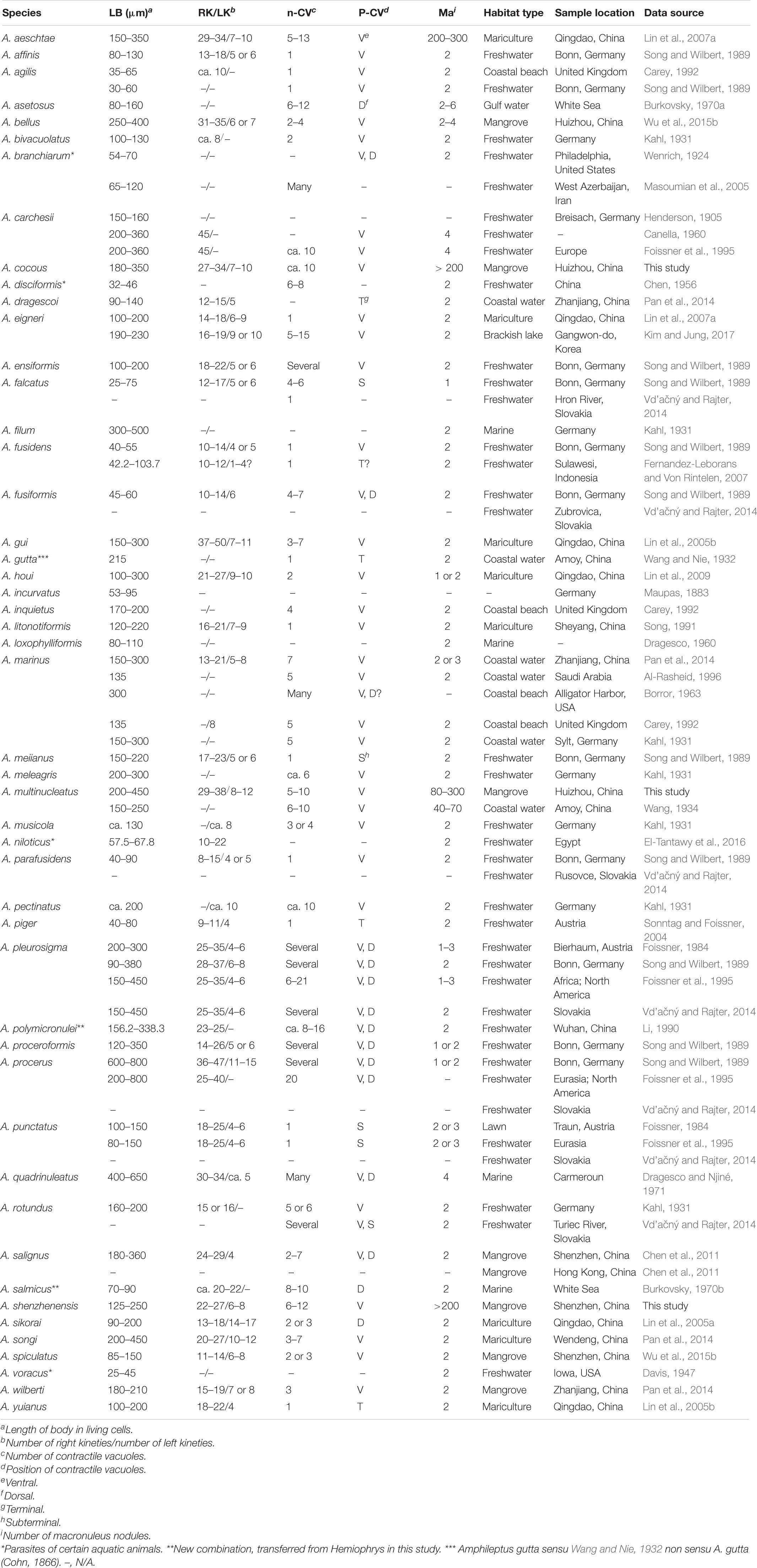
The species diversity of the genus Amphileptus was studied based on data from the present study and published sources, mainly monographs (Kahl, 1931; Song and Wilbert, 1989; Carey, 1992; Song et al., 2009; Vd’ačný and Foissner, 2012; Hu et al., 2019) and papers on the taxonomy and biodiversity of Amphileptus (see Tables 1, 2 for a complete list).
Results
Geographic Distribution of the Genus Amphileptus
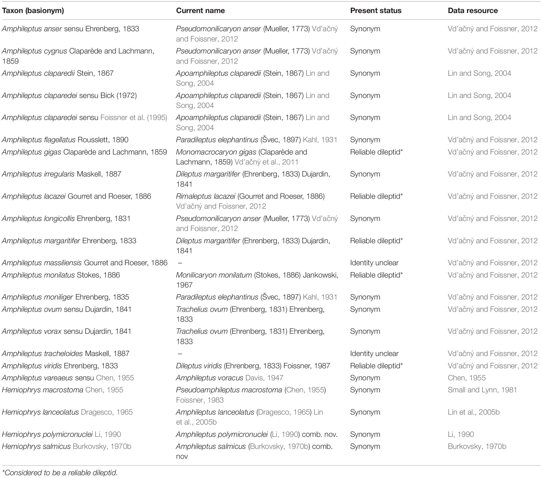
Amphileptus has been found in a wide variety of habitats worldwide. To date, 50 valid species of this genus have been reported from marine (Carey, 1992; Lin et al., 2005a, b, 2007a,b), brackish (Pan et al., 2010, 2014; Chen et al., 2011), freshwater (Wang and Nie, 1933; Wang, 1940; Song and Wilbert, 1989), and terrestrial (Foissner, 1984) habitats worldwide. In freshwater habitats, species of Amphileptus are most commonly reported from lakes (Song and Wilbert, 1989; Li, 1990), rivers (Stokes, 1884), wastewater treatment plants (Foissner, 1984), and as parasites on the body surface and gills of certain freshwater fishes and tadpoles in North America, Asia and Europe (Wenrich, 1924; Chen, 1955; Mitchell and Smith, 1988; Masoumian et al., 2005). In marine and brackish waterhabitats, species are most commonly reported from mangrove wetlands (Pan et al., 2010, 2013, 2014; Chen et al., 2011; Wu et al., 2013, 2014, 2015a,b, 2017; this study), mariculture ponds (Song, 1991; Lin et al., 2005a, b, 2007a; Song et al., 2009; Pan et al., 2014), the intertidal zones of beaches (Pan et al., 2014), and coastal marine waters (Kahl, 1931; Wang, 1934; Borror, 1963; Dragesco, 1965; Al-Rasheid, 1996). The vast majority of Amphileptus species are free-living although a few are reported as parasites on the skin and gills of fish (Chen, 1955; Masoumian et al., 2005), or tadpoles (Wenrich, 1924). Of the known Amphileptus species more than one-third have been found in the coastal waters of China (Song et al., 2009; Hu et al., 2019). These include eight species from mariculture ponds in the coastal waters of the Bohai and Yellow seas of northern China and 11 species (12 populations) from coastal waters of the South China Sea, seven of which were isolated from mangrove wetlands. We have listed the references to reliable morphological descriptions of Amphileptus in Table 1. Species no longer assigned to the genus Amphileptus, and species of Amphileptus originally assigned to other genera, are listed in Table 2 along with their current names and taxonomic status.
Morphology and Taxonomy of Three Amphileptus Species
Order Pleurostomatida Schewiakoff, 1896
Family Amphileptidae Bütschli, 1889
Genus Amphileptus Ehrenberg, 1830
Amphileptus multinucleatus Wang, 1934 (Tables 3, 4 and Figure 1)
Improved Diagnosis
Medium to large Amphileptus, 150–450 μm × 40–80 μm in vivo; posterior end constantly twisted from left to right in mid-body region; many (40–300) macronuclear nodules; 8–12 left and 29–38 right kineties; based on live observation, several (5–10) contractile vacuoles are located ventrally in posterior 2/3 of cell; extrusomes thick bar-shaped, densely arranged along oral slit; dot-like cortical granules; brackish or marine habitat.
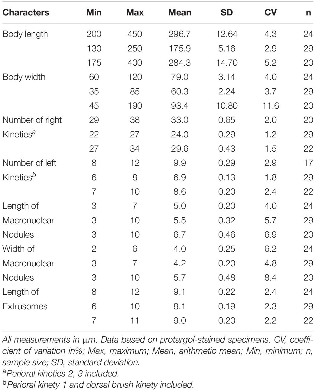
Table 3. Morphological characteristics of Amphileptus multinucleatus (1st line), A. shenzhenensis sp. n. (2nd line) and A. cocous sp. n. (3rd line).
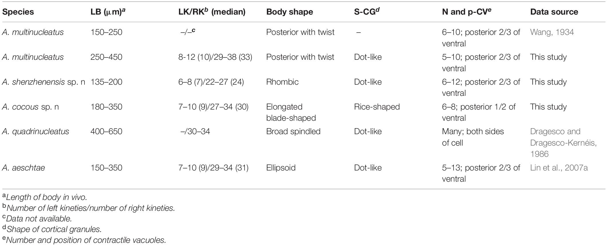
Table 4. List of all known Amphileptus spp. of multiple macronuclear nodules (≥4) and two new species in present study.
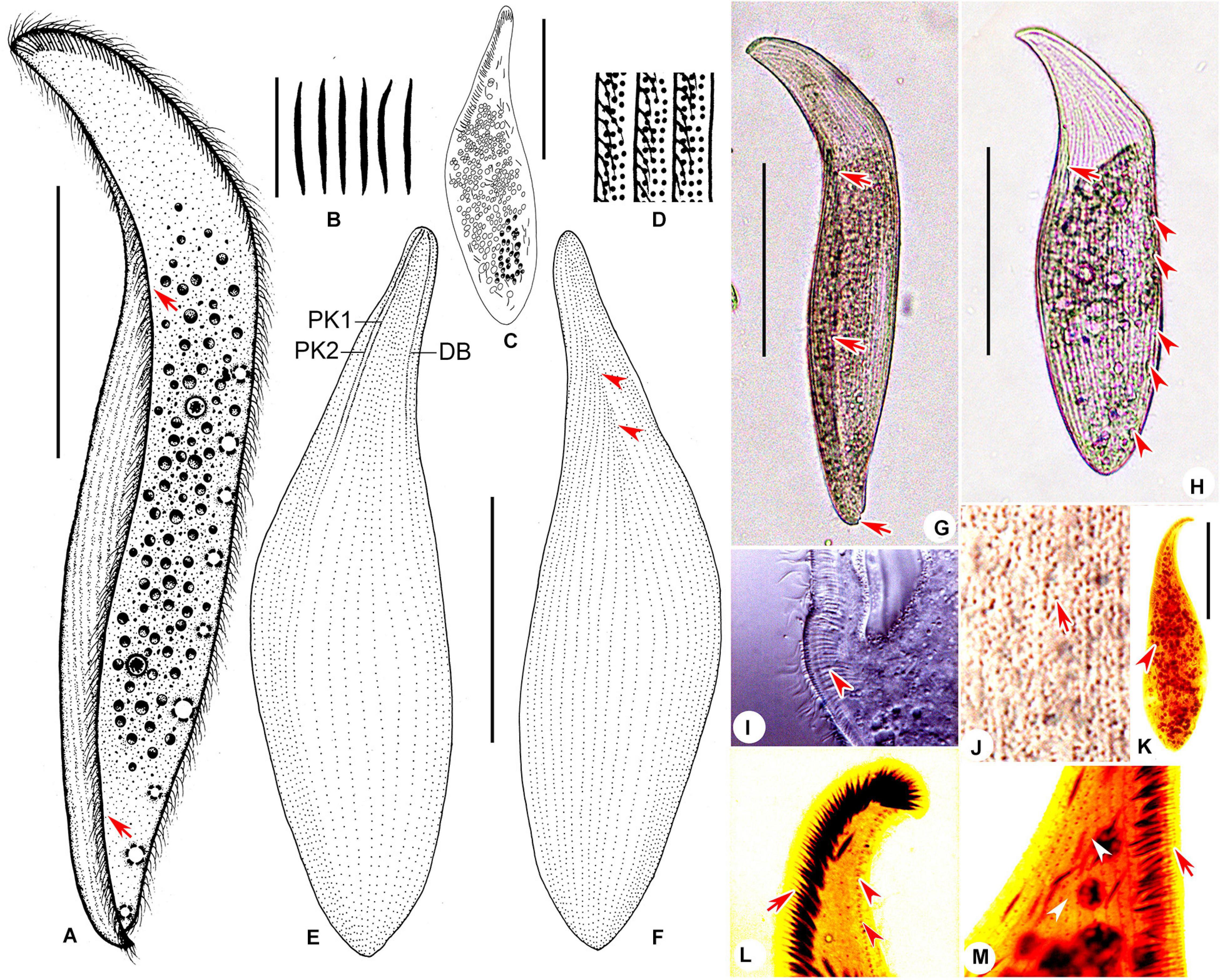
Figure 1. Amphileptus multinucleatus Wang, 1934; livingindividuals (A,B,D,G–J) and cells stained with protargol (C,E,F,K–L). (A) Right lateral view of a representative cell, arrowheads mark the twisted body. (B) Extrusomes. (C) Showing the distribution of extrusomes and macronuclear nodules. (D) The distribution of cortical granules. (E,F) Ciliary patterns of the left (E) and right (F) side; arrowheads point to the single-suture. (G,H) Right view of typical individual, arrows mark the twisted body, arrowheads show the distribution of contractile vacuoles. (I) The anterior part of right side, arrowhead shows the extrusomes. (J) The mid-region of right side, arrow marks the cortical granules. (K) To show the distribution of macronuclear nodules (arrowhead). (L) The anterior part of left side, to show perioral kinety 1 (arrow) and brush kinety (arrowheads). (M) The anterior part of right side, arrow marks perioral kinety 2, arrowheads show the suture. DB, dorsal brush; PK1, perioral kinety 1; PK2, perioral kinety 2. Scale bars: (A,G,H): 100 μm; (B): 10 μm; (C,E,F,K): 50 μm.
Ecological Features (Daya Bay Population)
Water temperature 19°C, salinity 24.5‰, pH 6.7.
Voucher Material
One voucher slide with protargol-stained specimens is deposited in the Laboratory of Protozoology, OUC, China, with registration number WL2011121901.
SSU rDNA Sequence
The SSU rDNA sequence of Amphileptus multinucleatus is deposited in the GenBank database with the accession number, length, and GC content as follows: MT653624, 1560 bp, 43.40%.
Morphological Description Based on Daya Bay Population
Body size highly variable in vivo, about 200–450 μm long; body shape fairly stable, generally elongate-pyriform with bluntly pointed; in all individuals (n > 20) observed in vivo, posterior portion perpetually twisted from left side to right side beginning at mid-dorsal region; conspicuous “neck” region (about 25% of cell length); pointed anterior end always bent toward dorsal side; laterally compressed about 3–4:1 (Figures 1A,G,H). Macronuclear nodules numerous (ca. 80–300), ovoid to elliptical in outline, about 3–7 μm × 2–6 μm in size after fixation, and scattered in cytoplasm although most are clustered in mid-region of cell (Figures 1C,K). Micronucleus not observed. Often with 5–10 contractile vacuoles, 4–8 μm in diameter, distributed along posterior 2/3 part of ventral margin (Figures 1A,H). Extrusomes thick bar-shaped, straight or slightly curved, about 10 μm long, densely arranged along anterior part of buccal area, some scattered in cytoplasm (Figures 1B,C,I). Pellicle thin with small (<0.5 μm across), densely spaced, grayish, dot-like cortical granules between ciliary rows on both sides of cell (Figures 1D,J). Cytoplasm colorless to pale yellow, often with numerous tiny, refringent globules (1–3 μm across) that render main part of body opaque (Figures 1G,H). Locomotion usually by gliding slowly on substrate, or swimming with a slow clockwise rotation about longitudinal axis.
Ciliary pattern as shown in Figures 1E,F,L,M. Eight to twelve left kineties (mean 9.9; median 10), including perioral kinety 1 and dorsal brush kinety (DB) which extends to about anterior 2/5 of cell-length and is composed of regularly spaced dikinetids (Figures 1E,L). Right side with 29–38 (mean 33.0; median 33) ciliated kineties including perioral kinety 2; intermediate somatic kineties are shortened forming a distinct anterior single-suture on right side (Figures 1F,M).
Two perioral kineties located along cytostome. Perioral kinety 1 (PK1) left of oral slit, composed of dikinetids in anterior 1/3 and monokinetids in posterior 2/3 (Figure 1E). Perioral kinety 2 (PK2) right of oral slit, consists of widely spaced dikinetids in anterior 1/3 part and continues posteriorly as a row of monokinetids (Figure 1E).
Amphileptus shenzhenensis sp. n. (Tables 3, 4 and Figure 2)
Zoobank Registration Number of Work
urn:lsid:zoobank.org:pub:DEE074BB-7B4F-46E7-8CA1-47820074ABBC
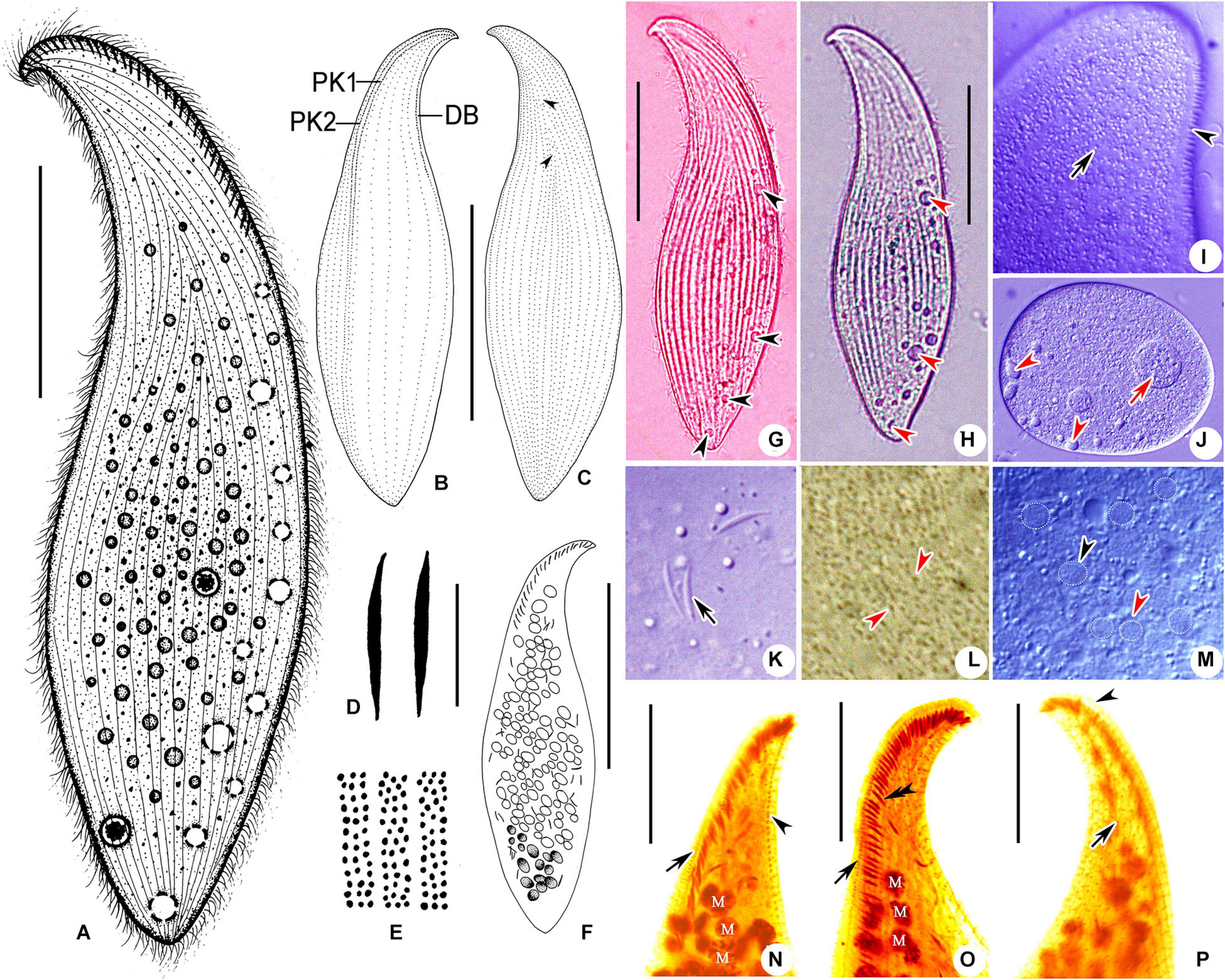
Figure 2. Amphileptus shenzhenensis sp. n.; living individuals (A,D,E,G–M) and cells stained with protargol (B,C,F,N–P). (A) Right lateral view of a representative cell. (B,C) Ciliary patterns of left (B) and right (C) side of the holotype, arrowheads show the suture. (D) Extrusomes. (E) Cortical granules. (F) Distribution of macronuclear nodules. (G,H) Right views of typical individuals, arrowheads show the distribution of contractile vacuoles. (I) The anterior part of left side, to show the cortical granules (arrow) and dorsal brush kinety (arrowhead). (J) A resistant cyst, arrowheads mark the contractile vacuoles, arrowhead shows food vacuole. (K) Extrusomes. (L) Cortical granules. (M) The mid-region of cell, to show the macronuclear nodules (arrowheads). (N,O) The anterior part of left side, arrow points to the left perioral kinety, arrowhead marks the dorsal brush kineties and double arrowheads show the extrusomes. (P) The anterior part of right side, to show perioral kinety 2 (arrowhead) and the suture (arrow). DB, dorsal brush; PK1, perioral kinety 1; PK2, perioral kinety 2. Scale bars: (A–C,F–H) 50 μm; (D) 5 μm; (N–P) 20 μm.
ZooBank Registration Number of A. shenzhenensis sp. n.
urn:lsid:zoobank.org:act:53746C79-64F6-46D3-9E04-4662951D7CED
Diagnosis
Body 125–250 μm × 40–50 μm in vivo; slightly contractile with an inconspicuous beak-like anterior body end; numerous (>200) macronuclear nodules; 6–8 left and 22–27 right kineties; several (6–12) contractile vacuoles along ventral side of posterior 2/3 of cell; extrusomes thick rod-shaped, densely arranged along oral slit; dot-like cortical granules; brackish habitat.
Etymology
Named after Shenzhen, where this species was first isolated.
Type Locality and Ecological Features
Futian mangrove wetland (22°38′N, 114°06′E), Shenzhen, China. Water temperature 26°C, salinity 19.1‰, and pH 7.3.
Type Slides
One protargol slide with the holotype specimen (circled in black ink) and several paratype specimens is deposited in the Laboratory of Protozoology, Ocean University of China (OUC), Qingdao, China, with registration number WL20110413-03.
SSU rDNA Sequence
The SSU rDNA sequence of Amphileptus shenzhenensis is deposited in the GenBank database with the accession number, length, and GC content as follows: MT653621, 1534 bp, 43.22%.
Morphological Description
Body 125–250 × 40–50 μmin vivo, usually 180–200 in length. Laterally compressed about 3:1, not flexible, with an inconspicuous, beak-like anterior end, a bluntly pointed posterior end, and a “neck” region about 1/5 of cell length (Figures 2A,G,H). Numerous macronuclear nodules (>200) scattered in cytoplasm but mostly clustered in central region of cell, about 5 μm in diameter in vivo, usually discernable in life as slightly transparent areas (Figures 2F,M–O). Micronucleus not observed. Six–12 contractile vacuoles, about 5–15 μm in diameter, located near ventral margin in posterior 2/3 of cell (Figures 2A,G,H,J). Extrusomes thick rod-shaped, straight to slightly curved, 6–8 μm long, some evenly arranged along oral slit, others scattered in cytoplasm (Figures 2D,K,N,O). Pellicle thin with numerous small (< 0.5 μm across), grayish, dot-like cortical granules, densely distributed between ciliary rows on both sides of cell (Figures 2E,I,L). Right side densely ciliated with cilia about 8 μm long arranged in rows located within conspicuous longitudinal furrows, and forms a distinct anterior single-suture that is detectable in vivo (Figures 2G,H). Left somatic cilia generally sparsely distributed, dorsal brush cilia detectable at high magnifications, about 2–3 μm long (Figure 2I). Cytoplasm slightly grayish, often with several food vacuoles, about 5–20 μm in diameter (Figure 2J). Generally sensitive to disturbance, tending to form a resistant cyst after being placed on glass slide (Figure 2J). Locomotion by gliding moderately fast on substrate or by swimming while rotating clockwise about longitudinal axis.
Ciliary pattern as shown in Figures 2B,C,N–P. Left side with 6–8 (mean 6.9; median 7) ciliated kineties including perioral kinety 1 (PK1) and dorsal brush kinety (DB) which extends to about 2/5 of cell length and is composed of widely spaced dikinetids (Figures 2B,N,O). Twenty-two–27 (mean 24.0; median 24) right kineties including perioral kinety 2 (PK2); intermediate somatic kineties shortened forming a distinct suture in anterior part of body (Figures 2C,P).
Two perioral kineties along cytostome. Perioral kinety 1 (PK1) left of oral slit, comprises dikinetids in anterior 2/5 and continues posteriorly as a row of monokinetids (Figures 2B,N,O). Perioral kinety 2 (PK2) right of oral slit, comprises regularly spaced dikinetids in anterior 1/3 and monokinetids in posterior 2/3 (Figures 2B,P).
Amphileptus cocous sp. n. (Tables 3, 4 and Figure 3)
ZooBank Registration Number of Amphileptus cocous sp. n.
urn:lsid:zoobank.org:act:96115FDB-E205-4FB0-ADE8-96DFC7767F60

Figure 3. Amphileptus cocous sp. n.; living individuals (A–D,H–N) and cells stained with protargol (E–G,O,P). (A,H) Right lateral view of a representative cell, arrowheads mark the contractile vacuoles. (B,I,M) To show the distribution of cortical granules (arrows). (D) To show the distribution of contractile vacuoles. (E) To show the distribution of macronuclear nodules. (F,G) Ciliary patterns of right (G) and left (F) side of the holotype. (J) The ventral part of right side, arrowheads point to extrusomes. (K) Contracted individual, arrowheads show the food vacuoles. (L) The ventral part of right side, arrow shows a macronuclear nodule, arrowhead shows a contractile vacuole. (N) The mid-region of cell, arrowheads show the extrusomes. (O) The anterior part of left side of cell, to show perioral kinety 1 (arrow) and dorsal brush kinety (arrowheads). (P) The anterior part of right side of cell, arrow marks perioral kinety 2, arrowheads show the suture. DB, dorsal brush; PK1, perioral kinety 1; PK2, perioral kinety 2. Scale bars: (A,H) 100 μm; (C) 10 μm; (D–G,K,O,P) 50 μm.
Diagnosis
Medium to large Amphileptus, 180–350 × 35–45 μm in vivo; body strongly contractile, elongated blade-shaped; numerous (> 200) macronuclear nodules; 7–10 left and 27–34 right kineties; several (6–8) contractile vacuoles along ventral side of posterior half of cell; extrusomes thick bar-shaped, densely arranged along oral slit; rice-shaped cortical granules; brackish habitat.
Etymology
The Latin adjective “cocous” (rice-shaped) refers to the shape of cortical granules.
Type Locality and Ecological Features
Daya Bay mangrove wetland (22°41′N, 114°23′E), Huizhou, China. Water temperature 26.8°C, salinity 15.9‰, and pH 7.0.
Type Slides
One protargol slide with the holotype specimen (circled in black ink) and several paratype specimens is deposited in the Laboratory of Protozoology, Ocean University of China (OUC), Qingdao, China, with registration number WL20111027-01.
SSU rDNA Sequence
The SSU rDNA sequence of Amphileptus cocous is deposited in the GenBank database with the accession number, length, and GC content as follows: MT653622, 1533 bp, 43.25%.
Morphological Description
Body size highly variable, about 180–350 × 30–45 μm in vivo, usually 200–250 μm in length. Elongated blade-shaped, flexible, and strongly contractile, with widely pointed posterior end and an inconspicuous neck region about 15–20% of cell length and usually curved slightly to dorsal side, laterally compressed about 2–3:1 (Figures 3A,H). Numerous (>200) macronuclear nodules, ovoid to elliptical in outline, about 5–10 μm × 5–10 μm in size in vivo, scattered in cytoplasm, usually discernable in life as slightly transparent areas (Figure 3L). Micronucleus not observed. Six–eight contractile vacuoles, about 8–13 μm in diameter, distributed along ventral margin in posterior half of cell (Figures 3A,D,H,L,K). Extrusomes spindle-shaped, about 10 μm long, some arranged in oral area, others some scattered in cytoplasm (Figures 3C,E,J,N,O). Pellicle thin with numerous short (about 1.0 μm in length), rice-shaped, colorless cortical granules densely packed between ciliary rows on both sides of cell (Figures 3B,I,M). Right side flat and densely ciliated, cilia about 8 μm long; left side sparsely ciliated, cilia difficult to detect in life. Cytoplasm colorless to grayish, often with numerous tiny, refringent globules (2–5 μm across) and numerous food vacuoles (3–10 μm across) that render main part of body opaque (Figures 3H,K). Locomotion moderately fast, usually gliding on substrate or swimming with a slow clockwise rotation about longitudinal axis.
Ciliary pattern as shown in Figures 3F,G,O,P. About 7–10 (mean 8.6; median 9) widely spaced left kineties, including perioral kinety 1 (PK1) and dorsal brush (DB) kinety which extends to 2/3 cell-length and is composed of regularly spaced dikinetids (Figures 3F,O). Right side densely ciliated, about 27–34 (mean 29.6; median 29) kineties including perioral kinety 2, intermediate somatic kineties shortened anteriorly forming a distinct anterior suture (Figures 3G,P).
Two perioral kineties around cytostome: PK1 left of oral slit, composed of closely spaced dikinetids in anterior 2/5 and continues posteriorly as a row of closely spaced monokinetids (Figures 3F,O); PK2 right of oral slit, formed of closely spaced dikinetids in anterior 2/5 and continues posteriorly as a row of closely spaced monokinetids (Figures 3G,P).
Molecular Phylogenetic Analysis of Amphileptus
Phylogenetic trees conducted using Bayesian inference (BI) and maximum likelihood (ML) had identical topologies so the two trees were combined (Figure 4A). The topology of the MP tree differed slightly from that of the ML/BI tree as shown in Figure 4B. The genus Amphileptus forms a polytomy with two clades in the ML/BI tree and three clades in the MP tree, and the three newly sequenced species form a clade with another four Amphileptus spp. (clade a in Figure 4A; clade 1 in Figure 4B) with poor to moderate support (60% ML, 0.71 BI, 79% MP). Within clade a/clade I, Amphileptus cocous groups with A. spiculatus which together group with A. shenzhenensis with high to maximum support (94% ML, 99% MP, 1.00 BI). These three species cluster with A. multinucleatus with poor to moderate support (86% ML, 0.77 BI, 59% MP), and this subclade groups with A. aeschtae with high support (100% ML, 1.00 BI, 99% MP), forming a clade that is sister to A. litonotiformis with high support (97% ML, 1.00 BI, 98% MP). In the second clade in ML/BI tree (clade b in Figure 4A), the remaining three Amphileptus spp. (Amphileptus sp., A. dragescoi and A. procerus) group with Pseudoamphileptus macrostoma with weak support (0.69 BI, 65% ML). In the MP tree, however, A. procerus clusters with A. dragescoi (81% MP) to form the second clade (clade II in Figure 4B), and the remaining two species (Pseudoamphileptus macrostoma and Amphileptus sp.) form the third clade (clade III in Figure 4B) with high support (99% MP).
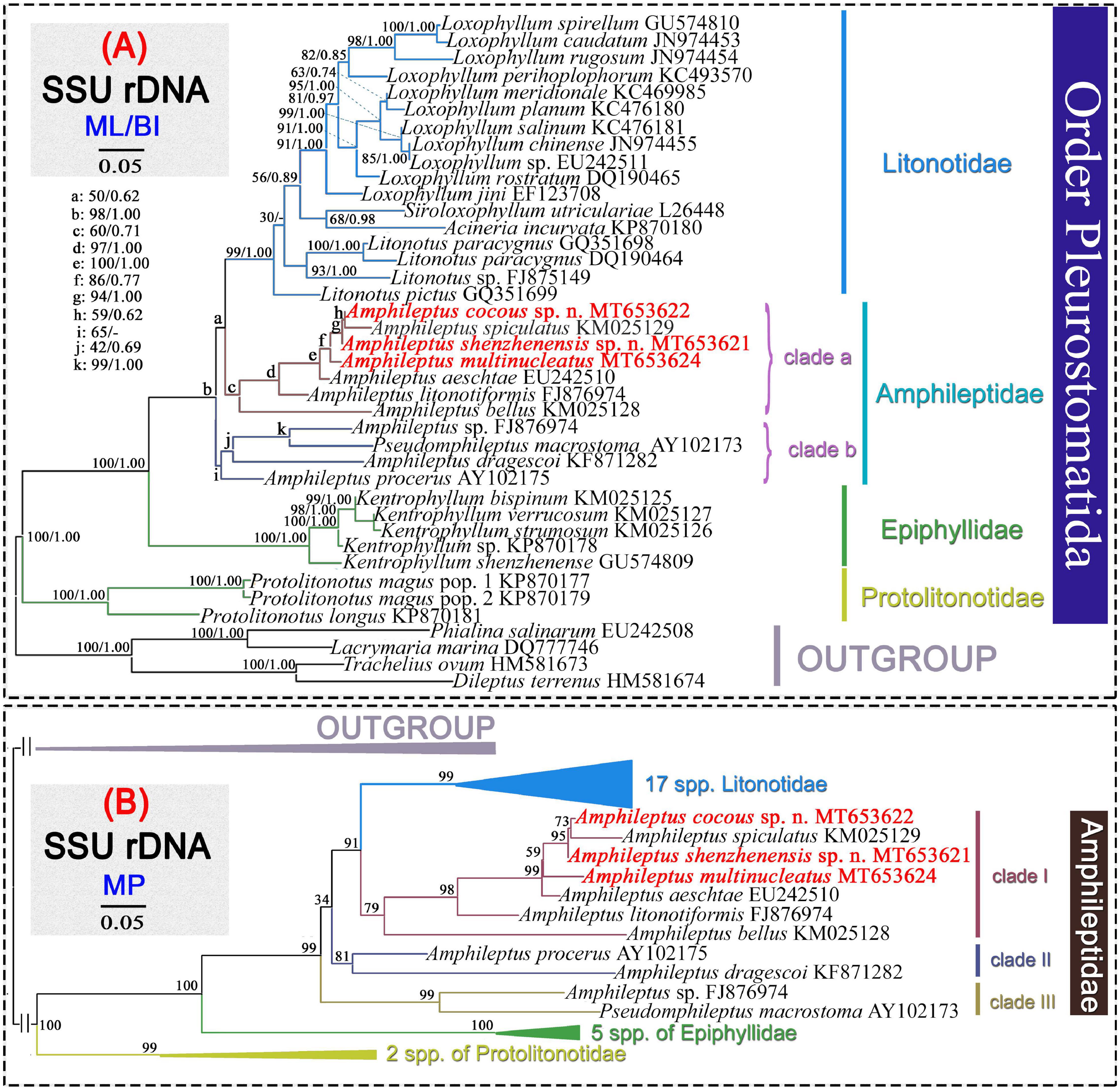
Figure 4. Phylogenetic trees inferred from SSU rDNA sequences performed with maximum likelihood (ML) (A) and maximum parsimony (MP) (B). The numbers at the nodes represent the posterior probabilities and the percentage of times the group occurred out of 1,000 trees, respectively. A dash indicates a different topology in the BI tree from ML tree. Newly sequenced species are in bold. GenBank accession numbers are given after the names of species. The scale bar corresponds to 5 substitutions per 100 nucleotide positions.
Discussion
A Brief Summary of the Genus Amphileptus Ehrenberg, 1830
The genus Amphileptus was established by Ehrenberg (1830) and another pleurostomatid genus, Hemiophrys, was established by Wrzesniowski (1870). Until the mid-twentieth century, descriptions of Amphileptus-Hemiophrys species were exclusively based on observations of live species (Ehrenberg, 1830; Kahl, 1931). Canella (1960) carried out the first detailed investigation of the ciliary pattern of these two genera using silver staining and revealed that the somatic kineties on the right side form a suture in the mid-to-anterior region of the cell in both genera. Furthermore, other morphological differences were regarded as species-level rather than genus-level characters, suggesting that Hemiophrys is a junior synonym of Amphileptus (Canella, 1960). This recommendation was accepted by Fryd-Versavel et al. (1975). Consequently, the genus Hemiophrys was merged into the genus Amphileptus, and the diagnosis of genus Amphileptus was emended, i.e., the formation of a single suture by the somatic kineties of the right side was considered to be the key diagnostic character. The emended diagnosis of the genus Amphileptus and the submersion of Hemiophrys were accepted by subsequent investigators (Foissner, 1984; Aescht, 2001; Lynn, 2008).
Species of the genus Amphileptus are easily identified by the pattern of right somatic kineties, i.e., the somatic kineties shortened to form a single suture in the median area, and this is the main differentiating feature for certain pleurostomatid genera, e.g., Amphileptus and Apoamphileptus within the order Pleurostomatida. Therefore, the presence of a single suture is thought to be an important genus-level, and possibly even a family-level character (Lin et al., 2005b; Wu et al., 2017).
The ciliature of Amphileptus consists of perioral kineties, right somatic kineties, left somatic kineties and dorsal brush kineties, the number and pattern of which are important for species delimitation. The key characteristics for species determination are: (1) body shape and size; (2) number of right and/or left kineties; (3) number and position of contractile vacuoles; (4) number of macronuclear nodules;, and (5) presence vs. absence and shape of cortical granules (Canella, 1960; Foissner, 1984; Song, 1991; Lin et al., 2007a; Wu et al., 2015b).
The first molecular phylogenetic study of Amphileptus was that of Gao et al. (2008) who sequenced the SSU rDNA of A. procerus (Penard, 1922) Song and Wilbert, 1989 and A. aeschtae Lin et al., 2007a. Pan et al. (2014) added another new sequence and reported the monophyly of the family Amphileptidae and the paraphyly of the genus Amphileptus with Pseudoamphileptus nested within it. These findings have been both confirmed and rejected in subsequent studies (Wu et al., 2015b, 2017; Pan et al., 2015) (see molecular analyses below). Including the three species described in the present study, there are nine identified and one unidentified SSU rDNA sequences in the NCBI/GenBank database.
Comments on Amphileptus multinucleatus Wang, 1934
Amphileptus multinucleatus was originally described by Wang (1934) who gave a good description based on live observations, although the ciliary pattern was not mentioned. It was characterized mainly as follows: body 150–250 μm in length, with 6–10 contractile vacuoles lying in ventral posterior half, numerous (40–70) macronuclear nodules scattered in cytoplasm, and posterior end twisted to one side. In particular, it was noted that “the twisted posterior portion is very constant in all observed individuals and may be considered as one of the specific characteristics” (Wang, 1934). This specific feature was also observed in all observed individuals of the Daya Bay population (n ¿ 20) and has not been recorded in any other species of Amphileptus. We conclude that the twisted posterior portion of the body should be considered as a diagnostic character of this species. In addition, our form was identified as A. multinucleatus based on the distribution of extrusomes along the oral slit and with some scattered in the cytoplasm. One significant difference between the original description and the Daya Bay population of A. multinucleatus is the number of macronuclear nodules, the former having 40–70 and the latter 80–300. It should be noted, however, that the number of macronuclear nodules in the original description was based on observations of specimens fixed in Schaudinn’s fluid and stained with iron-alum-hematoxylin which may not show the outline of the macronuclear nodules as clearly as the protargol stain. In addition, the body size of the Daya Bay population is considerably larger than that of the original population (200–400 vs. 150–250 μm in length), which may be another reason for the higher number of macronuclear nodules. We therefore conclude the Daya Bay population is conspecific with the original population of A. multinucleatus described by Wang (1934).
Comments on Amphileptus shenzhenensis sp. n. and A. cocous sp. n.
The most important characters for species identification and circumscription in the genus Amphileptus include the number of kineties, the number and positions of contractile vacuoles, the number of macronuclear nodules, the shape and distribution of extrusomes, the presence or absence and the shape of cortical granules, and the body shape in vivo (Foissneret al., 1995; Lin and Song, 2004; this study).
Among all the nominal species of Amphileptus, only three congeners are reported to have contractile vacuoles arranged along the ventral margin of the cell and four or more macronuclear nodules, i.e., A. multinucleatus, A. quadrinucleatus and A. aeschtae (Wang, 1934; Dragesco and Dragesco-Kernéis, 1986; Lin et al., 2007a; Table 4). The two new forms and the three described species strongly resemble each other in body size, position and number of macronuclear nodules, shape of cortical granules, and the number of kineties. However, A. multinucleatus is the only species in this genus with a posterior end constantly twisted to one side (Wang, 1934), therefore it can be clearly distinguished from the other species.
Amphileptus shenzhenensis sp. n. resembles A. multinucleatus, A. quadrinucleatus, and A. aeschtae in having dot-like cortical granules. However, A. shenzhenensis sp. n. differs: from A. multinucleatus by having fewer kineties on both sides of the cell (6–8 vs. 8–12 on left; 22–27 vs. 29–38 on right), and the posterior portion of the body not twisted (Wang, 1934); from A. quadrinucleatus by having fewer right kineties (22–27 vs. 30–34), the distribution of contractile vacuoles (in posterior 2/3 of cell on ventral side vs. down the length of both sides of body) and its significantly smaller body length (135–200 vs. 400–650 μm in vivo) (Dragesco and Dragesco-Kernéis, 1986); from A. aeschtae by having fewer kineties on both sides of cell, i.e., 6–8, mean 7 vs. 7–10, mean 9 on left; 22–27 vs. 29–34 on right (Lin et al., 2007a; Table 4).
Amphileptus cocous sp. n. resembles A. aeschtae in having the same number of left kineties (7–9) and almost the same number of right kineties (27–34, mean 30 vs. 29–34, mean 31). However, the former can be separated from the latter by the different shape of cortical granules (rice-shaped vs. dot-like), the distribution of contractile vacuoles (in the posterior half of the body vs. in the posterior 2/3 of the body) and the elongated blade-shaped (vs. ellipsoidal) body (Lin et al., 2007a; Table 4).
Amphileptus shenzhenensis sp. n. can be distinguished from A. cocous sp. n. by having fewer right kineties (22–27, mean 24 vs. 27–34, mean 30) and dot-like (vs. rice-shaped) cortical granules (Table 4).
The phylogenetic analyses based on SSU rDNA sequence data show that Amphileptus cocous sp. n. groups with A. spiculatus in the core clade. However, the former can be distinguished from the later by the following combination of morphological characters: (1) the different body shape in vivo (elongated blade-shaped vs. pyriform); (2) the rice-shaped (vs. dot-like) cortical granules; (3) the number of right kineties (27–34 vs. 11–14); (4) the larger body size (180–350 vs. 85–145 μm long in vivo), and (5) having significantly more (>300 vs. 2) macronuclear nodules (Wu et al., 2015b). In addition, the validity of the new species is supported by the SSU rDNA sequence data: Amphileptus cocous sp. n. differs in one, two, and five nucleotides from A. shenzhenensis sp. n., A. multinucleatus and A. aeschtae, respectively; and A. shenzhenensis sp. n. differs in six and three nucleotides from A. aeschtae and A. multinucleatus, respectively.
Two New Combinations of the Genus Amphileptus
Li (1990) reported a new species from Donghu Lake, Hubei Province, China, under the name Hemiophrys polymicronuclei. However, because Hemiophrys is a junior synonym of Amphileptus, this species should be transferred to the latter (Canella, 1960). Therefore, we propose a new combination, Amphileptus polymicronuclei (Li, 1990) comb. n. (original combination Hemiophrys polymicronuclei Li, 1990; Tables 1, 2). In addition, another species, Hemiophrys salimica, was reported by Burkovsky (1970b) from Kandalaksha Gulf, White Sea, which should be transferred to Amphileptus (Canella, 1960; Curds, 1982). Hence, a new combination, Amphileptus salimicus (Burkovsky, 1970b) comb. n. (original combination Hemiophrys salimica Burkovsky, 1970b), is suggested (Tables 1, 2).
Comments on the Phylogeny of the Genus Amphileptus
The family Amphileptidae is characterized by the presence of a single anterior suture on the right side, thereby differentiating it from the other three families within the order Pleurostomatida (Vd’ačný et al., 2015; Wu et al., 2017). The Amphileptidae, comprises five genera, namely Amphileptus (the type genus), Pseudoamphileptus, Amphileptiscus, Apoamphileptus, and Opisthodon, but molecular data are available only the former two genera. Traditionally, the genus Amphileptus has been considered to be monophyletic based on morphological (Foissner, 1977, 1983, 1984; Corliss, 1979; Lin et al., 2005a, b, 2007a; Lynn, 2008) and some molecular studies (Pan et al., 2010, 2013; Zhang et al., 2012; Vd’ačný and Foissner, 2013; Wu et al., 2013, 2015b, 2017; Vd’ačný, 2015; Vd’ačný et al., 2015). However, the monophyly of this Amphileptus has been questioned by several other molecular phylogenetic studies (Gao et al., 2008; Vd’ačný et al., 2011, 2015; Pan et al., 2014, 2015; Wu et al., 2014, 2015b). In our SSU rDNA trees (Figure 4) which include three new sequences of Amphileptus, the non-monophyly of the genus Amphileptus was supported, since it was divided into two and three groups in ML/BI and MP tree, respectively. Furthermore, the possibility of the monophyly of Amphileptus was also rejected (p = 0.004 < 0.05) by the KH test (Table 5). Although some previous studies have shown members of the genus Amphileptus to group together in phylogenetic trees (Pan et al., 2010, 2013; Zhang et al., 2012; Vd’ačný and Foissner, 2013; Wu et al., 2013, 2015a, 2017; Vd’ačný, 2015; Vd’ačný et al., 2015), these convergent topologies are based on insufficient taxon sampling and have only low nodal support (e.g., 76% ML, 0.65 BI in Pan et al., 2013; 17% ML, 20% MP in Wu et al., 2015a; 50% ML, 0.64 BI, 57% MP in Vd’ačný et al., 2015; 51% ML, 0.78 BI, 67% MP in Vd’ačný, 2015). Therefore, the molecular data strongly question the morphology-based relationship of the genus Amphileptus, and even the family Amphileptidae, and indicate that this group is paraphyletic. To date, however, only two genera of the family Amphileptidae have molecular data, namely Amphileptus and Pseudoamphileptus. It is noteworthy that Pseudoamphileptus is represented by a single sequence that clusters with sequences of Amphileptus (Figure 4). Furthermore, no morphological characters can be identified as plesiomorphic or apomorphic, so the question of whether the genus Amphileptus and/or the family Amphileptidae is monophyletic will remain unresolved pending the availability of more morphological and molecular data with expanded taxon sampling.
Data Availability Statement
The datasets presented in this study can be found in online repositories. The names of the repository/repositories and accession number(s) can be found below: NCBI GenBank (accession: MT653621, MT653622, and MT653624).
Author Contributions
LW and XL conceived and designed the manuscript. LW carried out the live observation and protargol staining. LW, JL, AW, and XL wrote and revised the manuscript. All authors contributed to the article and approved the submitted version.
Funding
This work was supported by the National Natural Science Foundation of China (project numbers: 42076113, 31761133001, and 415761486), Guangdong MEPP Fund [No. GDOE (2019) A23], Guangdong Basic and Applied Basic Research Foundation (project number: 2020A1515111125), and the China Postdoctoral Science Foundation (project number: 2018M640796).
Conflict of Interest
The authors declare that the research was conducted in the absence of any commercial or financial relationships that could be construed as a potential conflict of interest.
Acknowledgments
Many thanks to Ms. Wenping Chen, Mr. Chunxu Hu, and Mr. Xudong Cui, the graduates of our laboratory, SCNU, for their help in sampling. We are also grateful to the two reviewers and associate editor HP for their constructive comments and helpful suggestions for improving our manuscript.
Footnotes
References
Aescht, E. (2001). Catalogue of the generic names of ciliates (Protozoa, Ciliophora). Denisia 1, 1–350. doi: 10.1007/978-3-319-23534-9_1
Al-Rasheid, K. A. S. (1996). Records of free-living ciliates in Saudi Arabia, II. freshwater benthic ciliates of AI-Hassa Oasis, Eastern region. Arab. Gulf J. Sci. Res. 15, 187–205.
Borror, A. (1963). Morphology and ecology of the benthic ciliated protozoa of Alligator harbor, florida. Arch. Protistenk. 106, 465–534.
Burkovsky, I. V. (1970a). The ciliates of the mesopsammon of the Kandalaksha Gulf (white sea) I. Acta Protozool. 7, 475–489.
Burkovsky, I. V. (1970b). The ciliates of the mesopsammon of the Kandalaksha Gulf (white sea) II. Acta Protozool. 7, 47–65.
Canella, M. F. (1960). Contributo ad una revisione dei generi Amphileptus, Hemiophrys e Litonotus (Ciliata, Holotricha, Gymnostomata). Ann. Univ. Ferrara 2, 47–95.
Chen, C. L. (1955). The protozoan parasites from four species of Chinese pond fishes: Ctenopharyngodon idellus, Mylopharyngodon aethiops, Aristichthys nobilis and Hypophthalmichthys molitrix. I. the protozoan parasites of Ctenopharyngodon idellus. Acta Hydrobiol. Sinica 2, 123–164.
Chen, C. L. (1956). The protozoanparasites from four species of Chinese pond fishes: Ctenopharyngodon idellus, Mylopharyngodon aethiops, Aristichthys nobilis and Hypophthalmichthys molitrix. III. the protozoan parasites of Aristichthys nobilis and Hypophthalmichthys molitrix. Acta Hydrobiol. Sinica 2, 279–299.
Chen, R., Lin, X., and Warren, A. (2011). A new pleurostomatid ciliate, Amphileptus salignus n. sp. (Protozoa, Ciliophora), from mangrove wetlands in southern China. Zootaxa 3048, 62–68. doi: 10.11646/zootaxa.3048.1.4
Corliss, J. O. (1979). The Ciliated Protozoa: Characterization, Classification and Guide to the Literature. Second Edition. New York: Pergamon Press.
Curds, R. C. (1982). British and Other Freshwater Ciliated Protozoa. Part 1. Ciliophora: Kinetofragminophora. Cambridge: Cambridge University Press.
Davis, H. S. (1947). Studies of the protozoan parasites of freshwater fishes. U. S. Fisheries Wildlife Serv. Fishery Bull. 51, 1–29.
Dragesco, J. (1960). Ciliés mésopsammiques littoraux, systématique,morphologie, écologie. Trav. Stat. Biol. Roscoff 12, 1–356.
Dragesco, J., and Dragesco-Kernéis, A. (1986). Ciliés libres de l’Afrique intertropicale: introduction à la connaissance et à l’étude des Ciliés. Faune Trop. 26, 1–559. doi: 10.2307/j.ctv18phcb3.3
Dragesco, J., and Njiné, T. (1971). Compléments à la connaissance des ciliés libres du Cameroun. Annls. Fac. Sci. Univ. féd Cameroun 7– 8, 97–140.
Ehrenberg, C. G. (1830). Beiträge zur Kenntnis der Organisation der Infusorien und ihrer geographischen Verbreitung, besonders in Sibitien. Abh. dt. Akad. Wiss. Berl. 1832, 1–88. doi: 10.1159/000404845
El-Tantawy, S. A. M., Abdel-Aziz, A., Ei-Nour, M. F. A., Samn, A., Shaldoum, F., and Rady, I. (2016). Ectoparasitic protozoa scyphidians, amphileptids and tetrahymenid from the Nile perch, Lates niloticus (Linnaeus, 1758) in the Dakahlia Province, Egypt. Egypt. J. Exp. Biol. 12, 51–61.
Fan, X. P., and Pan, X. M. (2020). Scuticociliates and Peniculine Ciliates in China. Beijing: Science Press.
Fernandez-Leborans, G., and Von Rintelen, K. (2007). Epibiontic communities on the freshwater shrimp Caridina ensifera (Crustacea, Decapoda, Atyidae) from Lake Poso (Sulawesi, Indonesia). J. Nat. Hist. 41, 2891–2917. doi: 10.1080/00222930701787871
Foissner, W. (1977). Taxonomische studien ü ber die ciliaten der GroBglocknergebietes, 2. Familie Amphileptidae. Ber. Haus. Natur. Salzburg 8, 87–93.
Foissner, W. (1983). Morphologie und Infraciliatur zweier ectocommensaler Ciliaten (Protozoa: Ciliophora) von Cyprinuscarpio L. (Pisces: Cypriniformes): Heteropolaria lwoffi(Fauré-Fremiet, 1943) (Peritrichida: Epistylididae) und ihrPredator Pseudoamphileptus macrostoma (Chen, 1955) nov. gen. (Pleurostomatida: Amphileptidae). Zool. Jb. Syst. 110, 399–418.
Foissner, W. (1984). Taxonomie und Ökologie einiger Ciliaten (Protozoa, Ciliophora) des Saprobiensystems. I. Genera Litonotus, Amphileptus, Opisthodon. Hydrobiologia 119, 193–208. doi: 10.1007/bf00015210
Foissner, W. (1999). Protist diversity: estimates of the near-imponderable. Protist 150, 363–368. doi: 10.1016/s1434-4610(99)70037-4
Foissner, W., Berger, H., Blatterer, H. and Kohmann, F. (1995). Taxonomische und ökologische revision der Ciliaten desSaprobiensystems-Band IV: Gymnostomatea, Loxodes, Suctoria. Landesamtes für Wasserwirtsch. 1, 1–540.
Foissner, W., and Hawksworth, D. L. (2009). Protist Diversity and Geographical Distribution. Dordrecht: Springer.
Foissner, W., and Leipe, D. (1995). Morphology and ecology of Siroloxophyllum utriculariae (Penard, 1922) n. g., n. comb. (Ciliphora, Pleurostomatida) and an improved classification of pleurostomatid ciliates. J. Eukaryot. Microbiol. 42, 476–490. doi: 10.1111/j.1550-7408.1995.tb05894.x
Fryd-Versavel, G., Lftode, F., and Dragesco, J. (1975). Contribution à la connaissance de quepques ciliés gymnostomes. II. Prostomiens, pleurostomiens: morphologie, stomatogènésc. Protistologica 6, 509–530.
Gao, F., Warren, A., Zhang, Q. Q., Gong, J., Miao, M., Sun, P., et al. (2016). The all-data-based evolutionary hypothesis of ciliated protists with a revised classification of the phylum Ciliophora (Eukaryota, Alveolata). Sci. Rep. 6:24874.
Gao, S., Song, W. B., Ma, H. W., Clamp, J. C., Yi, Z. Z., Al-Rasheid, K. A. S., et al. (2008). Phylogeny of six genera of the subclass Haptoria (Ciliophora, Litostomatea) inferred from sequences of the gene coding for small subunit ribosomal RNA. J. Eukaryot. Microbiol. 55, 562–566. doi: 10.1111/j.1550-7408.2008.00360.x
Henderson, W. D. (1905). “Notes on the infusoria of freiburg in breigau,” in Zoologischer Anzeiger, ed. E. Korschelt (Leipzig: Wilhelm Engelmann), 1–24.
Hu, X. Z., Lin, X. F., and Song, W. B. (2019). Ciliates Atlas: Species Found in the South China Sea. Beijing: Science Press.
Kahl, A. (1931). Urtiere oder protozoa i: wimpertiere oder ciliata (Infusoria) 2. Holotricha außer den im 1. Teil behandelten Prostomata. Tierwelt Dtl. 21, 181–398.
Kahl, A. (1933). “Ciliata libera et ectocommensalia,” in Die Tierwelt der Nord-und Ostsee, Lief 23 (Teil P, c3), eds G. Grimpe and E. Wagler (Leipzig: Geest & Portig), 29–146.
Kim, J. H., and Jung, J. H. (2017). Brief descriptions of 12 ciliate species previously unrecorded (Protozoa: Ciliophora) in Korea. J. Species Res. 6, 15–25.
Kishino, H., and Hasegawa, M. (1989). Evaluation of the maximum likelihood estimate of the evolutionary tree topologies from DNA sequence data, and the branching order in Hominoidea. J. Mol. Evol. 29, 170–179. doi: 10.1007/bf02100115
Li, L. X. (1990). A new species of ciliates, Hemiophrys polymicronuclei sp. nov. from Donghu lake, Hubei province. Chin. J. Oceanol. Limnol. 8, 97–100. doi: 10.1007/bf02846456
Lin, X. F., Li, J. Q., Gong, J., Warren, A., and Song, W. (2008). Taxonomic studies on three marine pleurostomatid ciliates, Litonotus bergeri nov. spec., L. blattereri nov. spec. and L. petzi nov. spec. (Ciliophora, Pleurostomatida) from North China sea. Eur. J. Protistol. 44, 91–102. doi: 10.1016/j.ejop.2007.08.005
Lin, X. F., and Song, W. B. (2004). Establishment of a new amphileptid genus, Apoamphileptus nov. gen. (Ciliophora, Litostomatea, Pleurostomatida), with description of a new marine species, Apoamphileptus robertsi nov. spec. from Qingdao, China. J. Eukaryot. Microbiol. 51, 618–625. doi: 10.1111/j.1550-7408.2004.tb00595.x
Lin, X. F., Song, W. B., and Li, J. Q. (2007a). Amphileptus aeschtae nov. spec. and Amphileptus eigneri nov. spec. (Ciliophora, Pleurostomatida), two new marine pleurostomatid ciliates from China. Eur. J. Protistol. 43, 77–86. doi: 10.1016/j.ejop.2006.10.002
Lin, X. F., Song, W., and Li, J. Q. (2007b). Description of two new marine pleurostomatid ciliates, Loxophyllum choii nov. spec. and L. shini nov. spec. (Ciliophora, Pleurostomatida) from China. Eur. J. Protistol. 43, 131–139. doi: 10.1016/j.ejop.2006.12.004
Lin, X. F., Song, W. B., and Warren, A. (2005a). Taxonomic studies on three marine pleurostomatid ciliates: Kentrophyllum verrucosum (Stokes, 1893) Petz, Song et Wilbert, 1995, Epiphyllum soliforme (Fauré-Frémiet, 1908) gen. n., comb. n. and Amphileptus sikorai sp. n., with the establishment of a new genus Epiphyllum (Ciliophora: Pleurostomatida). Acta Protozool. 44, 129–145.
Lin, X. F., Song, W. B., and Warren, A. (2005b). Two new marine pleurostomatid ciliates from China, Amphileptus gui nov. spec. and Amphileptus yuianus nov. spec. (Ciliophora, Pleurostomatida). Eur. J. Protistol. 41, 163–173. doi: 10.1016/j.ejop.2005.01.002
Lin, X. F., Song, W. B., and Warren, A. (2009). “Pleurostomatids,” in Free-living Ciliates in the Bohai and Yellow Seas, eds W. B. Song, X. Z. Hu, and A. Warren (Beijing: Science Press), 93–134.
Liu, M. J., Wang, C. D., Hu, X. Z., Qu, Z. S., Jiang, L. M., Al-Farraj, S. A., et al. (2020). Taxonomy and molecular phylogeny of three species of scuticociliates from China: Citrithrix smalli gen. nov., sp. nov., Homalogastra binucleata sp. nov. and Uronema orientalis Pan et al., 2015 (Protozoa, Ciliophora, Oligohymenophorea), with the proposal of a new family, Citrithrixidae fam. nov. Front. Mar. Sci. 7:604704.
Liu, W. W., Jiang, J. M., Tan, Y. H., and Lin, X. F. (2020). Novel contributions to the taxonomy of the ciliates genus Euplotes (Ciliophora, Euplotida): redescription of two poorly known species, with a brief note on the distributions of this genus in coastal waters of southern China. Front. Mar. Sci. 7:615413.
Liu, W. W., Jiang, J. M., Xu, Y., Pan, X. M., Qu, Z. S., Luo, X. T., et al. (2017). Great diversity in marine ciliates: fauna studies in China seas during the years 2011–2016. Eur. J. Protistol. 61, 424–438. doi: 10.1016/j.ejop.2017.04.007
Liu, W. W., Zhang, K. X., Chen, C. Z., Li, J. Q., Tan, Y. H., Warren, A., et al. (2019). Overview of the biodiversity and geographic distribution of aloricate oligotrich ciliates (Protozoa, Ciliophora, Spirotrichea) in coastal waters of southern China. Syst. Biodivers. 17, 787–800. doi: 10.1080/14772000.2019.1691081
Lynn, D. (2008). The Ciliated Protozoa, Characterization, Classification, and Guide to the Literature. Dordrecht: Springer.
Ma, Z. H., Dong, T. Y., Liao, W. Y., Fan, X. P., Xu, Y., and Gu, F. K. (2019). Ciliates in the marsh wetlands of Chongming island: taxonomy of five species and a species list. J. Ocean Univ. China 18, 441–454. doi: 10.1007/s11802-019-3804-y
Masoumian, M., Pazouki, J., Yahyazadeh, M., and Teymornezhad, A. (2005). Protozoan from freshwater fishes from north west of Iran. Iran. J. Fish. Sci. 4, 31–42. doi: 10.1201/b18534-6
Maupas, E. (1883). Contribution à l’étude morphologique et anatomique des infusoires ciliés. Arch. Zool. Exp. 2. Ser. T. 1, 513–516.
Mitchell, A. J., and Smith, C. E. (1988). Amphileptus branchiarum (Protozoa: Amphileptidae) in pond reared fish in Arkansas. J. Wildl. Dis. 24, 642–646. doi: 10.7589/0090-3558-24.4.642
Nylander, J. (2004). MrModeltest version 2. Distributed by the Author. Uppsala: Evolutionary Biology Centre, Uppsala University.
Pan, H. B., Gao, F., Lin, X. F., Warren, A., and Song, W. B. (2013). Three new Loxophyllum species (Ciliophora: Pleurostomatida) from China with a brief review of the marine and brackish Loxophyllum species. J. Eukaryot. Microbiol. 60, 44–56. doi: 10.1111/jeu.12005
Pan, H. B., Li, L. F., Lin, X. F., Li, J. Q., Al-Farraj, S. A., and Al-Rasheid, K. A. S. (2014). Morphology of three species of Amphileptus (Protozoa, Ciliophora, Pleurostomatida) from the South China sea, with note on phylogeny of a. dragescoi sp. n. J. Eukaryot. Microbiol. 61, 644–654. doi: 10.1111/jeu.12146
Pan, H. B., Li, L. F., Wu, L., Miao, M., Al-Rasheid, K. A. S., and Song, W. B. (2015). Morphology of three Litonotus species (Ciliophora: Pleurostomatida) from China seas, with brief notes on their SSU rDNA-based phylogeny. Eur. J. Protistol. 51, 494–506. doi: 10.1016/j.ejop.2015.08.003
Pan, H. B., Zhang, Q. Q., Dong, J. Y., and Jiang, J. M. (2020). Morphology and phylogeny of two novel pleurostomatids (Ciliophora, Litostomatea), establishing a new genus. J. Eukaryot. Microbiol. 67, 252–262. doi: 10.1111/jeu.12779
Pan, H., Gao, F., Li, J. Q., Lin, X. F., Al-Farraj, S. A., and Al-Rasheid, K. A. S. (2010). Morphology and phylogeny of two new pleurostomatid ciliates, Epiphyllum shenzhenense n. sp. and Loxophyllum spirellum n. sp. (Protozoa, Ciliophora) from a mangrove wetland, south China. J. Eukaryot. Microbiol. 57, 421–428. doi: 10.1111/j.1550-7408.2010.00492.x
Qu, Z. S., Li, L. F., Lin, X. F., Stoeck, T., Pan, H. B., Al-Rasheid, K. A. S., et al. (2018). Diversity of the cyrtophorid genus Chlamydodon (Protista, Ciliophora): its systematics and geographic distribution, with taxonomic descriptions of three species. Syst. Biodivers. 16, 497–511. doi: 10.1080/14772000.2018.1456493
Ronquist, F., and Huelsenbeck, J. (2003). MrBayes 3: bayesian phylogenetic inference under mixed models. Bioinformatics 19, 1572–1574. doi: 10.1093/bioinformatics/btg180
Shimodaira, H., and Hasegawa, M. (2001). CONSEL: for assessing the confidence of phylogenetic tree selection. Bioinformatics 17, 1246–1247. doi: 10.1093/bioinformatics/17.12.1246
Small, E. B., and Lynn, D. H. (1981). A new macrosystem for the Phylum Ciliophora Doflein, 1901. BioSystems 14, 387–401.
Song, W. B. (1991). A new marine ciliate, Amphileptus litonotiformis nov. sp. (Protozoa Ciliophora). Chin. J. Oceanol. Limnol. 9, 300–305. doi: 10.1007/bf02850645
Song, W. B., and Wilbert, N. (1989). Taxonomische untersuchungen an aufwuchsciliaten (Protozoa, Ciliophora) im Poppelsdorfer Weiher, Bonn. Lauterbornia 3, 2–221.
Song, W. B., Warren, A., and Hu, X. Z. (2009). Free-living Ciliates in the Bohai and Yellow Seas. Beijing: Science Press.
Sonntag, B., and Foissner, W. (2004). Urotricha psenneri n. sp. and Amphileptus piger (Vuxanovici, 1962) n. comb., two planktonic ciliates (Protozoa, Ciliophora) from an oligotrophic lake in Austria. J. Eukaryot. Microbiol. 51, 670–677. doi: 10.1111/j.1550-7408.2004.tb00607.x
Stamatakis, A., Hoover, P., and Rougemont, J. (2008). A fast boostrap algorithm for the RAxML web-servers. Syst. Biol. 57, 758–771. doi: 10.1080/10635150802429642
Swofford, D. (2002). PAUP∗. Phylogenetic Analysis using Parsimony (∗and other Methods). Version 4. Sunderland, MA: Sinauer Associates.
Vd’ačný, P., and Rajter, L. (2014). An annotated and revised checklist of pleurostome ciliates (Protista: Ciliophora: Litostomatea) from Slovakia, central Europe. Zootaxa 3760, 501–521. doi: 10.11646/zootaxa.3760.4.1
Vd’ačný, P., Bourland, W. A., Orsi, W., Epstein, S. S., and Foissner, W. (2011). Phylogeny and classification of the Litostomatea (Protista, Ciliophora), with emphasis on free-living taxa and the 18S rRNA gene. Mol. Phylogenet. Evol. 59, 510–522. doi: 10.1016/j.ympev.2011.02.016
Vd’ačný, P., Rajter, L., Shazib, S. U. A., Jang, S. W., Kim, J. H., and Shin, M. K. (2015). Reconstruction of evolutionary history of pleurostomatid ciliates (Ciliophora, Litostomatea, Haptoria): interplay of morphology and molecules. Acta Protozool. 54, 9–29.
Vd’ačný, P. (2015). Estimation of divergence times in litostomatean ciliates (Ciliophora: Intramacronucleata), using Bayesian relaxed clock and 18S rRNA gene. Eur. J. Protistol. 51, 321–334. doi: 10.1016/j.ejop.2015.06.008
Vd’ačný, P., and Foissner, W. (2012). Monograph of the dileptids (Protista, Ciliophora, Rhynchostomatia). Denisia 31, 1–529.
Vd’ačný, P., and Foissner, W. (2013). Synergistic effects of combining morphological and molecular data in resolving the phylogenetic position of Semispathidium (Ciliophora, Haptoria) with description of Semispathidium breviarmatum sp. n. from tropical Africa. Zool. Scr. 42, 529–549. doi: 10.1111/zsc.12023
Vd’ačný, P., Breiner, H. W., Yashchenko, V., Dunthorn, M., Stoeck, T., and Foissner, W. (2014). The chaos prevails: molecular phylogeny of the Haptoria (Ciliophora, Litostomatea). Protist 165, 93–111. doi: 10.1016/j.protis.2013.11.001
Vuxanovici, A. (1960). Contribitii la studiul grupei subgenurilor litonotus-hemiophrys (Ciliata).Studii Cerc. Biol. 12, 125–139.
Vuxanovici, A. (1961). Noi contributii la studiul ciliatelor dulcicole din Republica Polularia Romina (Nota I). Studii Cerc. Biol. 12, 353–381.
Wang, C. C., and Nie, D. S. (1932). A survey of the marine protozoa of Amoy. Contr. Biol. Lab Sci. Soc. China Zoological Series 8, 285–385.
Wang, C. C., and Nie, D. S. (1933). Report on the rare and new species of fresh-water infusoria, part I. Contr. Biol. Lab Sci. Soc. China Zoological Series 10, 1–99.
Wenrich, D. H. (1924). A new protozoan parasite, Amphileptus branchiarum n. sp., on the gills of tadpoles. Trans. Am. Microsc. Soc. 43, 191–199. doi: 10.2307/3221736
Wilbert, N. (1975). Eine Verbesserte technik der protargolimprägnation für ciliaten. Mikrokosmos 64, 171–179.
Wilbert, N., and Song, W. (2005). New contribution to the marine benthic ciliates from the Antarctic area, including description of seven new species (Protozoa, Ciliophora). J. Nat. Hist. 39, 935–973. doi: 10.1080/00222930400001509
Wu, L., Chen, R., Yi, Z. Z., Li, J. Q., Warren, A., and Lin, X. F. (2013). Morphology and phylogeny of three new Loxophyllum species (Ciliophora, Pleurostomatida) from mangrove wetlands of southern China. J. Eukaryot. Microbiol. 60, 267–281. doi: 10.1111/jeu.12032
Wu, L., Chen, R., Yi, Z. Z., Li, J. Q., Warren, A., and Lin, X. F. (2014). The morphology of three Loxophyllum species (Ciliophora, Pleurostomatida) from southern China, L. lembum sp. n., L. vesiculosum sp. n. and L. perihoplophorum Buddenbrock, 1920, with notes on the molecular phylogeny of Loxophyllum. J. Eukaryot. Microbiol. 61, 115–125. doi: 10.1111/jeu.12089
Wu, L., Clamp, J. C., Yi, Z. Z., Li, J. Q., and Lin, X. F. (2015a). Phylogenetic and taxonomic revision of an enigmatic group of haptorian ciliates, with establishment of the Kentrophyllidae fam. n. (Protozoa, Ciliophora, Litostomatea, Pleurostomatida). PloS One 10:e0123720. doi: 10.1371/journal.pone.0123720
Wu, L., Jiao, X. X., Shen, Z., Yi, Z. Z., Li, J. Q., Warren, A., et al. (2017). New taxa refresh the phylogeny and classification of pleurostomatid ciliates (Ciliophora, Litostomatea). Zool. Scr. 46, 245–253. doi: 10.1111/zsc.12193
Wu, L., Yi, Z. Z., Li, J. Q., Warren, A., Xu, H. L., and Lin, X. F. (2015b). Two new brackish ciliates, Amphileptus spiculatus sp. n. and A. bellus sp. n. from mangrove wetlands in southern China, with notes on the molecular phylogeny of the family Amphileptidae (Protozoa, Ciliophora, Pleurostomatida). J. Eukaryot. Microbiol. 62, 662–669. doi: 10.1111/jeu.12225
Keywords: biodiversity, Hemiophrys, Litostomatea, new species, phylogeny, SSU rDNA
Citation: Wu L, Li J, Warren A and Lin X (2021) Species Diversity of the Pleurostomatid Ciliate Genus Amphileptus (Ciliophora, Haptoria), With Notes on the Taxonomy and Molecular Phylogeny of Three Species. Front. Mar. Sci. 8:642767. doi: 10.3389/fmars.2021.642767
Received: 16 December 2020; Accepted: 11 January 2021;
Published: 09 February 2021.
Edited by:
Hongbo Pan, Shanghai Ocean University, ChinaReviewed by:
Peter Vd’ačný, Comenius University, SlovakiaZhishuai Qu, University of Kaiserslautern, Germany
Copyright © 2021 Wu, Li, Warren and Lin. This is an open-access article distributed under the terms of the Creative Commons Attribution License (CC BY). The use, distribution or reproduction in other forums is permitted, provided the original author(s) and the copyright owner(s) are credited and that the original publication in this journal is cited, in accordance with accepted academic practice. No use, distribution or reproduction is permitted which does not comply with these terms.
*Correspondence: Xiaofeng Lin, bGlueGZAeG11LmVkdS5jbg==
 Lei Wu
 Jiqiu Li2
 Xiaofeng Lin