 Fatemeh Saheb Sharif-Askari
 Jacob Raber
 Karem H. Alzoubi
95% of researchers rate our articles as excellent or good
Learn more about the work of our research integrity team to safeguard the quality of each article we publish.
Find out more
EDITORIAL article
Front. Immunol. , 17 March 2025
Sec. Inflammation
Volume 16 - 2025 | https://doi.org/10.3389/fimmu.2025.1575669
This article is part of the Research Topic Long COVID and Brain Inflammation: Unravelling Mechanisms and Potential Therapies View all 10 articles
Editorial on the Research Topic
Long COVID and brain inflammation: unravelling mechanisms and potential therapies
Patients with Long COVID often experience persistent brain-related symptoms, including brain fog, mood changes, and dizziness (1–3), likely driven by neuroinflammation even after the virus is cleared. Imaging studies have shown structural and functional changes that indicate ongoing inflammation in the Long COVID brain. The biological mechanisms underlying these symptoms are still not fully understood, and research continues to identify effective therapies to improve both the physical and mental health of those affected (2).
In this Research Topic, we explore research on Long COVID and its investigation through preclinical animal models.
Missailidis et al. conducted RNA-Seq analysis of peripheral blood mononuclear cells (PBMCs) from individuals with Long COVID and those who had fully recovered COVID-19. Their findings revealed upregulation of ICOS and S1PR1, suggesting a persistent pro-inflammatory state (Figure 1A), as these genes are involved in immune cell survival and signaling. They also observed downregulation of LILRB1 and LILRB2, highlighting immune dysregulation as a distinguishing feature of Long COVID.
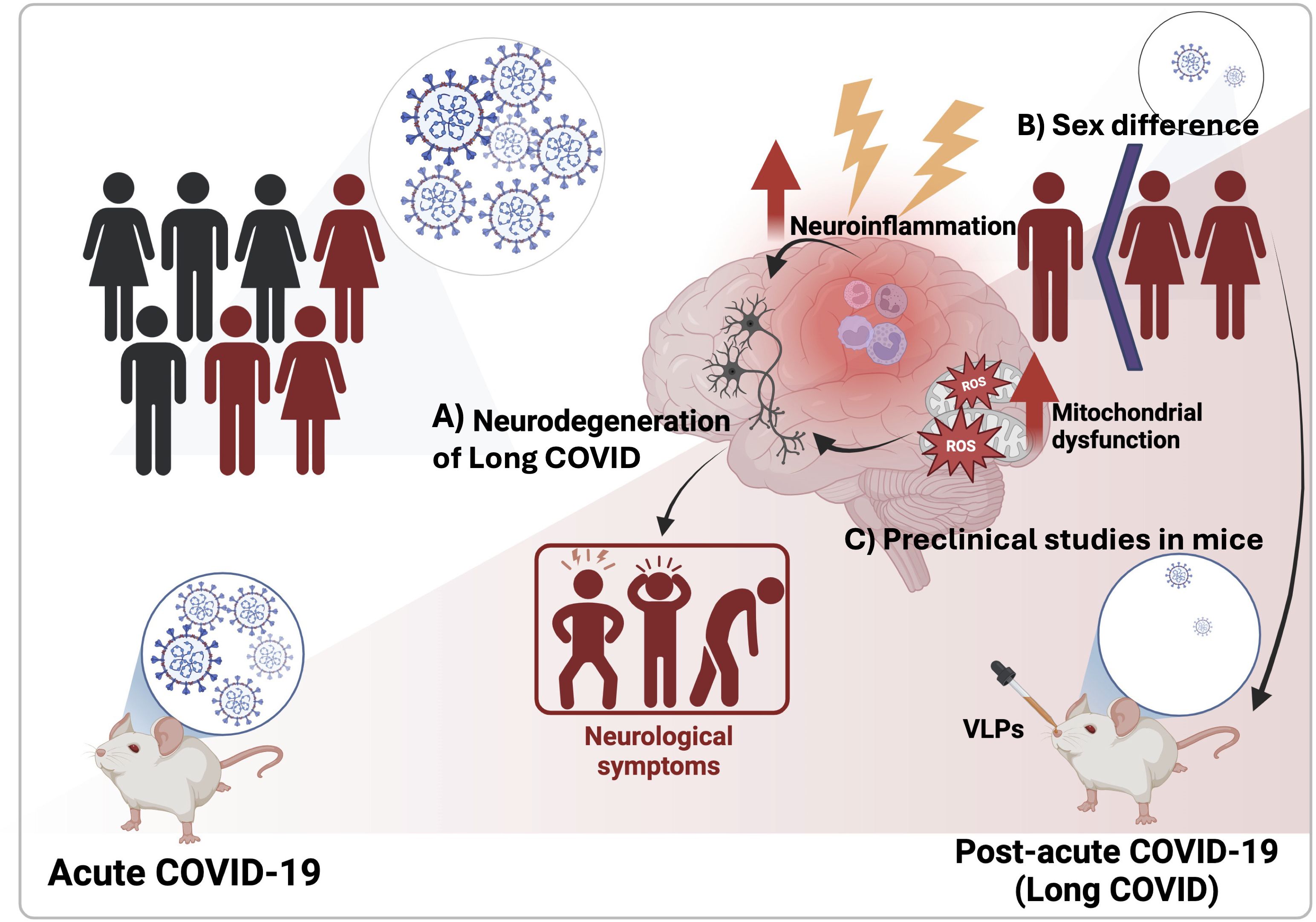
Figure 1. Neurological consequences of Long COVID. (A) In Long COVID, persistent neuroinflammation and mitochondrial dysfunction are implicated in ongoing neurodegeneration. (B) Certain populations, particularly females, are more susceptible due to hormonal and immune factors. (C) Preclinical models, including mice exposed to virus-like particles (VLPs), are being used to investigate the effects of Long COVID on the nervous system. These studies have linked neuroinflammation to behavioral disruptions, highlighting the need for further research into the neurological impacts of viral infections, particularly in Long COVID.
Lee et al. analyzed the expression of long noncoding RNAs (lncRNAs) in the brains of COVID-19 patients, identifying hundreds of differentially expressed lncRNAs compared to age- and sex-matched uninfected controls. Many of these lncRNAs correlated with cognitive decline and increased inflammation, aligning with cognitive dysfunction observed in Long COVID. These findings suggest a potential role for lncRNAs in the neurological effects of COVID-19, highlighting a key area for further research.
Noonong et al. proposed that mitochondrial dysfunction may play a crucial role in Long COVID, linking it to diabetes and oxidative stress (Figure 1A). Their hypothesis highlights mitochondria’s role in inflammation and metabolic homeostasis, suggesting broader systemic implications for viral recovery and chronic symptoms.
Hanafy and Jovin emphasized the role of chronic inflammation in exacerbating neurological symptoms commonly seen in patients with Long COVID, such as brain fog, extreme fatigue (asthenia), and depression- a condition they refer as “Brain FADE syndrome” (Figure 1A). They advocate for an integrated treatment approach that targets both inflammatory pathways and associated mental health challenges.
Gu et al. investigated sex differences in COVID-induced autoimmunity and neurological effects, highlighting how female sex hormones and X-chromosome factors may increase women’s susceptibility to Long COVID (Figure 1B). They proposed that COVID-19 may disturb immune tolerance, leading to autoantibodies infiltration into the central nervous system. They also suggested that COVID-19 could disrupt the female microbiome, contributing to neural damage, including demyelination, neuroinflammation, and neurodegeneration. Understanding these sex differences could help develop strategies to mitigate COVID-related neurological injuries.
Bustamante et al. explored risk factors for developing Long COVID, including a history of severe illness and intensive care. Using preclinical animal models, they examined the involvement of the nervous system in inflammation through the psychoneuroimmunoendocrine axes. They proposed that Long COVID involves peripheral and central sensitization, leading to dysregulation and chronic inflammation, and discussed therapeutic strategies to modulate these inflammation responses.
Dai et al. reviewed the utility of preclinical animal models in understanding Long COVID. They highlighted key features replicated in these models, including lung fibrosis, hyperglycemia, and neurological sequelae, while acknowledging limitations such as restricted genetic diversity and challenges in modeling Long COVID pathology. To improve translational relevance, they proposed incorporating genetically diverse populations, conducting longitudinal studies, and aligning animal findings with clinical data.
Singh et al. investigated the Long COVID implications of the Delta variant using K18-hACE2 mice. They found robust inflammatory responses linked to neuropsychiatric symptoms and motor behavior changes during acute infections, with persistent immune activation post-infection. Post-acute infection, the brain showed no detectable viral RNA and minimal residential immune cell activation in surviving mice. However, transcriptome analysis revealed persistent activation of immune pathways, including humoral responses, complement, phagocytosis, along with gene expression linked to ataxia telangiectasia, impaired cognitive function, and neuronal dysfunction. Surviving mice exhibited strong neutralizing antibodies against both Delta and Omicron variants, months after the infection.
O’Niel et al. used virus-like particles (VLPs) expressing SARS-CoV-2 structural proteins (nucleocapsid (N), membrane (M), envelope (E) and spike (S), in human apolipoprotein E (apoE)-targeted replacement mice (Figure 1C). The study found apoE isoform-dependent effects on behavioral measures, with E2 mice more affected than E3 or E4 mice, despite E2 being linked to a lower Alzheimer’s disease risk. VLPs also caused behavioral and circadian disruptions independent of apoE isoform, even in the absence of viral replication. Increased susceptibility in E2 mice was associated with elevated hippocampal CCL11, similar to CCL11 elevations seen in humans with cognitive symptoms after COVID-19 exposure. The authors emphasize the need for further research to better understand and treat neurological conditions associated with viral infections, especially as many continue to struggle with Long COVID.
The ongoing research discussed in this Research Topic reveals significant progress toward understanding the biological mechanisms of Long COVID. As we continue to uncover the complexities of the condition, further preclinical and clinical studies are critical for improving the well-being and brain function of those affected by Long COVID and other related neurological conditions.
FS: Writing – original draft, Writing – review & editing. JR: Writing – original draft, Writing – review & editing. KA: Writing – review & editing.
The author(s) declare that financial support was received for the research and/or publication of this article. FSSA is supported by the Seed Research Projects (No. 2401110399) from the University of Sharjah. JR is partially supported by R21 AG079158-01A1.
The guest editors would like to express their gratitude to all the authors and reviewers for their invaluable contributions to this Research Topic.
The authors declare that the research was conducted in the absence of any commercial or financial relationships that could be construed as a potential conflict of interest.
The author(s) declared that they were an editorial board member of Frontiers, at the time of submission. This had no impact on the peer review process and the final decision.
The author(s) declare that no Generative AI was used in the creation of this manuscript.
All claims expressed in this article are solely those of the authors and do not necessarily represent those of their affiliated organizations, or those of the publisher, the editors and the reviewers. Any product that may be evaluated in this article, or claim that may be made by its manufacturer, is not guaranteed or endorsed by the publisher.
1. Bobak L, Dorney I, Kovacevich A, Barnett B, Kaelber D. Preexisting psychiatric conditions as risk factors for diagnosed long COVID-19 syndrome within aggregated electronic health record data. Psychomat Med. (2024) 86:132–6. doi: 10.1097/PSY.0000000000001280
2. Saheb-Sharif-Askari F, Ali Hussain Alsayed H, Saheb-Sharif-Askari N, Saddik B, Al Sayed Hussain A, Halwani R. Risk factors and early preventive measures for long COVID in non-hospitalized patients: analysis of a large cohort in the United Arab Emirates. Public Health. (2024) 230:198–206. doi: 10.1016/j.puhe.2024.02.031
Keywords: long Covid, neuroinflammation, immune dysregulation, cognitive dysfunction, preclinical mice models
Citation: Saheb Sharif-Askari F, Raber J and Alzoubi KH (2025) Editorial: Long COVID and brain inflammation: unravelling mechanisms and potential therapies. Front. Immunol. 16:1575669. doi: 10.3389/fimmu.2025.1575669
Received: 12 February 2025; Accepted: 04 March 2025;
Published: 17 March 2025.
Edited and Reviewed by:
Pietro Ghezzi, Brighton and Sussex Medical School, United KingdomCopyright © 2025 Saheb Sharif-Askari, Raber and Alzoubi. This is an open-access article distributed under the terms of the Creative Commons Attribution License (CC BY). The use, distribution or reproduction in other forums is permitted, provided the original author(s) and the copyright owner(s) are credited and that the original publication in this journal is cited, in accordance with accepted academic practice. No use, distribution or reproduction is permitted which does not comply with these terms.
*Correspondence: Fatemeh Saheb Sharif-Askari, ZnNoYXJpZmFza2FyaUBzaGFyamFoLmFjLmFl; Jacob Raber, cmFiZXJqQG9oc3UuZWR1
†These authors have contributed equally to this work
Disclaimer: All claims expressed in this article are solely those of the authors and do not necessarily represent those of their affiliated organizations, or those of the publisher, the editors and the reviewers. Any product that may be evaluated in this article or claim that may be made by its manufacturer is not guaranteed or endorsed by the publisher.
Research integrity at Frontiers
Learn more about the work of our research integrity team to safeguard the quality of each article we publish.