 Maria Carolina Santos Guedes1,2
 Henrique Fernando Lopes-Araujo1,2
 Kleyverson Feliciano dos Santos2Esaú Simões2
 Wlisses Henrique Veloso Carvalho-Silva3,4*†Rafael Lima Guimarães1,2
- 1Department of Genetics, Federal University of Pernambuco (UFPE), Recife, Pernambuco, Brazil
- 2Keizo Asami Institute (iLIKA), Federal University of Pernambuco (UFPE), Recife, Pernambuco, Brazil
- 3Department of Immunology, Aggeu Magalhães Institute (IAM/FIOCRUZ), Recife, Pernambuco, Brazil
- 4Life Sciences Nucleus, Agreste Academic Center (CAA), Federal University of Pernambuco (UFPE), Caruaru, Pernambuco, Brazil
In recent decades, significant progress has been made in understanding the mechanisms underlying human immunodeficiency virus (HIV) infection and its treatment. Antiretroviral therapy (ART) has notable improved the life expectancy and quality of life for people living with HIV (PLHIV) by suppressing viral replication and promoting CD4+ T-cell recovery. However, despite its efficacy, approximately 10-40% of ART-treated PLHIV with virological suppression (<50 RNA copies/mL) do not achieve adequate immunological reconstitution. These PLHIV, classified as immunological non-responders (INR), experience higher morbidity and mortality rates compared to those with satisfactory immune reconstitution, known as immunological responders (IR). Various studies have explored the mechanisms contributing to immunological nonresponse, yet a major challenge remains: the lack of a standardized definition of immunological response and nonresponse across studies. Currently, definitions are inconsistent, limiting comparability between studies. This review proposes a clear and adequate classification for IR and INR PLHIV to support future advancements in understanding immunological recovery and improving the quality of life for ART-treated PLHIV.
Introduction
Since the identification of human immunodeficiency virus (HIV) as the causative agent of acquired immunodeficiency syndrome (AIDS), significant progress has been made in treatment of people living with HIV (PLHIV) (1). Advances in understanding the virus’s pathogenesis have enabled the development of new drugs and therapeutic strategies (2, 3). Consequently, many PLHIV have experienced an increase in life expectancy, with HIV infection now being managed as a chronic condition rather than a life-threatening disease (4). As a result, the life expectancy of PLHIV has increased to over 50 years (5). Currently, global data indicate that approximately 29 million PLHIV (76% of all PLHIV) are on antiretroviral therapy (ART), and 93% of them have achieved a reduction in plasma viral load to undetectable levels (<50 RNA copies/mL) (6).
Despite substantial advancements in understanding HIV infection, certain aspects such as the immune reconstitution of PLHIV on ART remain unclear (1). Normally, reducing viremia leads to a gradual recovery of CD4+ T-cell count over time (7). However, 10-40% of ART-treated PLHIV experience impaired immune reconstitution, characterized by limited CD4+ T-cell recovery even after virological suppression (8, 9). This condition is associated with an increased risk of HIV-related complications and death, and these PLHIV are defined as virological responders but immunological non-responders (INR) (9).
Several studies have suggested various mechanisms to explain the deficiency in immune reconstitution among ART-treated PLHIV (8–10). However, the lack of consensus among researchers on defining criteria for classifying immunological nonresponse has become a significant obstacle to fully understanding this condition. Thus, the present study aimed to propose a classification to define immunological nonresponse in ART-treated PLHIV, establishing a fundamental framework for future studies focused on elucidating the mechanisms involved in unsatisfactory immune reconstitution.
Immunological recovery in ART-treated PLHIV
In the first decade following the development of ART, there were different indications for its initiation, particularly regarding CD4+ T-cell count (11). Primarily, the World Health Organization (WHO) recommended initiating treatment in PLHIV with a CD4+ T-cell count ≤500 cells/mm3 (12, 13). However, in the early 2000s, the criteria were revised to a threshold of ≤200 cells/mm3 due to concerns about antiretrovirals toxicity (13). In 2006, the WHO adjusted its recommendation to initiate treatment in PLHIV with a CD4+ T-cell count of 350 cells/mm3, which was later revised back to ≤500 cells/mm3 (14). Currently, studies have demonstrated that early initiation of ART, regardless of CD4+ T-cell count, leads to a more rapid reduction in viral load, better immune reconstitution, and decrease in mortality (15). Thus, ART is now recommended to be initiated within 7 days after diagnosis, following the “treat-all” approach established by the WHO in 2017 (16, 17).
Successful therapy is expected to be followed by a gradual restoration of immunological function (Figure 1A) (18). Initially, treatment reduces viremia, which in turn decreases immune activation induced by HIV infection (19). In most PLHIV, plasma viral load reaches undetectable levels within six months (20, 21). This reduction downregulates the expression of intercellular adhesion molecule-1 (ICAM-1) and vascular cell adhesion molecule-1 (VCAM-1) on the surface of CD4+ T lymphocytes, the primary molecules responsible for retaining these cells within lymphoid organs (22). As a result, memory CD4+ T-cells are redistributed from lymphoid tissues to peripheral blood, increasing the circulating T-cell count (Figure 1B). This redistribution characterizes the initial phase of immunological recovery, usually resulting in a gain of 20-30 cells/µL per month and lasts for approximately 6 months (Figure 1A) (7).

Figure 1. Immunological recovery in PLHIV during ART. (A) Average CD4+ T-cell count reconstitution in ART-treated PLHIV. CD4+ T-cell count reconstitution in PLHIV undergoing ART is divided into three phases, each with distinct rates of cell gain. In the first phase, spanning 1 to 6 months of ART, CD4+ T-cell counts increase by approximately 20-30 cells/µL per month. The second phase, from 6 months to the second year of ART, shows a gain of 5-10 cells/µL per month. The third phase, extending beyond the second year and for at least 7 years, is characterized by a slower increase of 2-5 cells/µL per month. (B) Mechanisms responsible for CD4+ T-cell count increase during ART. The initial increase in CD4+ T-cell count following ART initiation results from reduced immune activation, allowing memory CD4+ T-cells to migrate from lymphoid organs into the peripheral circulation. Over time, additional mechanisms – such as thymic production, peripheral proliferation of CD4+ T-cells, and prolonged cell lifespan – support the gradual rise in T-lymphocytes throughout treatment.
During the second phase, CD4+ T-cell reconstitution is predominantly driven by thymic production, with an average gain of 5-10 cells/µL per month (7, 23). In this phase, larger thymic size is associated with higher percentages of T-cell receptor excision circles (TRECs), recent thymic emigrants (RTEs) and naïve CD4+ T-cell (23). By the end of the second year of ART, PLHIV typically exhibit a gain of approximately 200 cells/µL (Figure 1A) (7, 24). During this period, CD4+ T-cells also gradually begin to recover their antigen-specific response to various antigens, except HIV (9). Notably, the first two years of ART represent the most significant increase in cell count, with subsequent years showing minimal gains. Some studies suggest a tendency to initiate a plateau in CD4+ T-cell recovery after this period (23, 25, 26).
In addition, two other mechanisms contribute during the second and third phases: the homeostatic proliferation of residual CD4+ T-cells and an increased lifespan of these cells (Figure 1B) (27). The third phase, spanning from the third to the seventh year of ART, is responsible for a gain of 2-5 cells/µL per month (Figure 1A) (7). Although these mechanisms contribute to the increase in CD4+ T-cell count, they are not qualitatively equivalent. Only thymic production has the potential to partially restore the patient’s antigenic repertoire (7).
Although most processes involved in immune reconstitution have been described, ongoing debate remains regarding the optimal CD4+ T-cell count threshold indicative of therapeutic immunological success in PLHIV under ART. In healthy individuals not infected with HIV, the typical CD4+ T-cell count ranges between 500 and 4000 cells/µL (28). Consequently, many studies have reached a consensus that PLHIV treated with ART who achieve a CD4+ T-cell count above 500 cells/µL exhibit morbidity and mortality rates like those of HIV-negative individuals (29). At this cell count level, these PLHIV demonstrate reduced susceptibility to non-AIDS events and are considered to have restored immunocompetence (8, 30–32).
Understanding the immunological response capacity of PLHIV is essential, as it has been demonstrated that different CD4+ T-cell counts can affect their susceptibility to various coinfections and neoplasms (33). For instance, PLHIV with a CD4+ T-cell count below 100 cells/µL (in AIDS stage) are more susceptible to developing esophageal candidiasis, toxoplasmic encephalitis, and primary central nervous system lymphoma (PCNSL) (33). Meanwhile, in PLHIV with 200-500 cells/µL, conditions such as oral hairy leukoplakia, mucocutaneous Kaposi sarcoma, and cervical or anal neoplasia are more common (34). In contrast, ART-treated PLHIV with a CD4+ T-cell count above 500 cells/µL manifest similar infections to those seen in HIV-negative individuals (33). Hence, this threshold has garnered widespread acceptance as a marker of satisfactory immune reconstitution in PLHIV undergoing ART (8). Conversely, inadequate CD4+ T-cell gain directly impacts the infection’s prognosis and indicates immunological nonresponse (35).
Immunological nonresponse to ART
Therapeutic success in ART-treated PLHIV has traditionally focused on suppressing viral load. Nevertheless, it can also be characterized by distinct stages of CD4+ T-cell recovery (36). Consequently, the objective of ART has expanded beyond merely controlling viral replication to foster an environment conducive to immune reconstitution (10). However, a significant portion of ART-treated PLHIV, ranging from 10% to 40%, exhibit persistently low CD4+ T-cell counts despite achieving virological suppression, categorizing them as immunological non-responders (INR, Figure 2B) (37). In contrast, PLHIV who successfully achieve immune reconstitution are classified as immunological responders (IR, Figure 2A) (8). Approximately two decades ago, initial studies first identified this condition (38, 39), emerging two years after the introduction of combination therapy for treating PLHIV (40). Since then, further research has provided new insights into the complexities of immunological nonresponse (8, 10, 41)

Figure 2. Immunological response to ART. Following the initiation of ART, plasma viral load markedly decreases to undetectable levels, creating conditions favorable for CD4+ T-cell recovery. (A) PLHIV who experience satisfactory immune reconstitution are defined as immunological responders (IR). (B) Despite achieving virological success, some PLHIV exhibit reduced CD4+ T-cell reconstitution and are thus classified as immunological non-responders (INR).
Immunological nonresponse in PLHIV presents significant risks for infection prognosis, including progression to AIDS-defining events and increased mortality (42). Studies have shown that the mortality rate is 100 times higher in PLHIV with a CD4+ T-cell count below 50 cells/µL compared to those with a count above 500 cells/µL (30). Moreover, the immune dysfunction observed in INRs is also associated with the development of non-AIDS events, such as metabolic syndrome, cardiovascular diseases, and nephropathy (43, 44).
Immunological nonresponse is considered a multifactorial condition, with several factors identified as risk contributors, including male sex, advanced age, coinfections, exacerbated immune activation, thymic exhaustion, genetic alterations, and others (Figure 3) (45, 46). Additionally, reduced T-lymphocytes production and increased destruction of these cells are recognized as the primary processes associated with immunological nonresponse, given that T-cell homeostasis is crucial for effective immune reconstitution (47). Previous studies have demonstrated that INRs experience a reduction in the output of recent thymic emigrants (RTE), indicating thymic insufficiency, with these cells exhibiting an increased rate of cell death via pyroptosis – one of the main types of cell death observed in PLHIV (48, 49). Other immune dysfunctions observed in INRs include an altered cytokine secretion profile, disruptions in regulatory components such as T-reg and Th17 cells, mitochondrial dysfunction and dysregulated hematopoiesis, which results in the abnormal proliferation of myeloid-derived suppressor cells (MDSCs), T-cell exhaustion and senescence (50).

Figure 3. Factors related to immunological nonresponse. The main factors associated with immunological nonresponse include HIV reservoirs, cytokine dysregulation, coinfections, microbial translocation, mitochondrial damage, and dysregulated hematopoiesis. These factors can lead to exacerbated immune activation and increased proliferation of MDSCs, which contribute to increased CD4+ T-cell exhaustion and destruction, resulting in immunological nonresponse in ART-treated PLHIV. Reduced thymic output can contribute to decreased CD4+ T-cell production, which also result in immunological nonresponse Additionally, other factors such as drugs effects, genetic alterations, older age, persistent viral replication, male sex, extracellular vesicles, metabolic conditions, and late ART initiation may also be associated with the INR group. ART, antiretroviral therapy; INR, immunological non-responders; MDSCs, myeloid-derived suppressor cells; PLHIV, people living with human immunodeficiency virus.
Another crucial element influencing immune reconstitution is the pre-ART CD4+ T-cell count (51). Previous research conducted by our research group revealed that most INRs had a CD4+ T-cell count of ≤200 cells/µL at the initiation of ART (52, 53). Furthermore, it has been suggested that starting ART with a baseline CD4+ T-cell count of ≥500 cells/µL significantly enhances the likelihood of satisfactory immunological recovery (15). This underscores the importance of initiating ART regardless of CD4+ T-cell count, as PLHIV who delayed treatment until their count dropped below 200 cells/µL did not achieve immune reconstitution even after 10 years of viral suppression (54). This issue is particularly pronounced in older PLHIV (55). Additionally, a low pre-ART CD4+ T-cell count is associated with the emergence of X4 strains, co-infections, and microbial translocation (9, 56). Other contributors to immunological nonresponse include persistent residual viral replication within reservoirs, issues related to drug efficacy and delayed initiation of ART (41)
Classifications of immunological responders and non-responders
Although studies on immunological recovery are increasingly abundant, there remains no consensus among authors on the definition of immunological nonresponse. Criteria such as absolute CD4+ T-cell count, CD4+ T-cell increase, CD4/CD8 ratio, or combinations of them have been adopted in various classifications (Table 1). Furthermore, differences in the time frames required for these classifications and other factors contribute to a lack of standardization (41, 120). This diversity highlights the need to compile these classifications and recommend a more standardized approach.
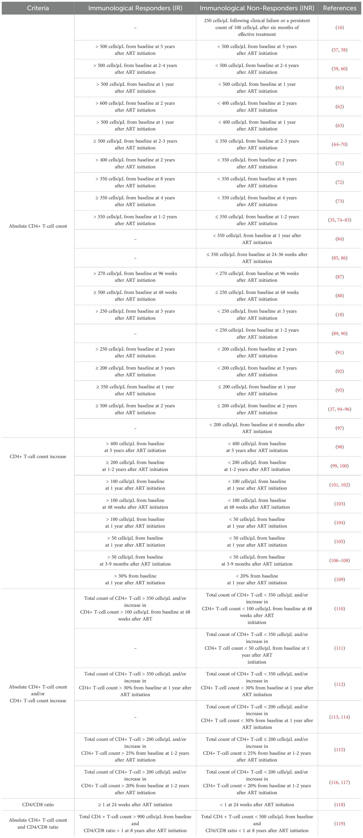
Table 1. Definitions of immunological responders and immunological non-responders.
Initially, it is important to note that some classifications were excluded from the study due to discrepancies in the threshold for defining an undetectable viral load (Supplementary Material). In the compiled data, these thresholds ranged from <20 to <1000 copies/mL. Plasma viral load is a critical factor influencing immune reconstitution, since elevated viremia is associated with increased levels of chronic inflammation and immune activation, which negatively affect CD4+ T-cell recovery (7, 27). Therefore, comparing PLHIV with significantly different viral loads is not meaningful. Consequently, this study only included classifications that considered an undetectable viral load <50 copies/mL, in alignment with WHO recommendations (16).
Another important point to highlight is that some studies provided classification only for INRs, while the IR status was not as clearly defined (84, 85, 97, 111). In our view, this circumstance leads to an insufficiently clear classification of IRs, resulting in doubts and questions about immune reconstitution in this group. In this context, the classification adopted by the WHO is noteworthy. The WHO currently characterizes INRs as PLHIV with an absolute CD4+ T-cell count of 250 cells/µL following clinical failure or a persistent count of 100 cells/µL after six months of effective treatment (16). However, the absence of a precise definition for IR within this classification leaves uncertainties regarding what constitutes adequate immune reconstitution, which is essential to provide benefits for PLHIV. We believe that having a clear and objective definition of both groups enhances fidelity and reproducibility in studies on immunological recovery, as well as having a real impact on the prognosis of the HIV infection. Rather than merely suggesting a definition, it is increasingly important to determine what would constitute the ideal immune response for PLHIV in order to achieve a quality of life comparable to that of HIV-negative individuals.
Regarding other classifications, as mentioned previously, various authors use different criteria to characterize immune nonresponse. The most used criterion across studies is the absolute CD4+ T-cell count. However, it is important to emphasize that even within this criterion, there are discrepancies, particularly concerning the threshold of CD4+ T-cells used to classify the INR group. In addition to the CD4+ T-cell count recommended by the WHO (16), other thresholds have been suggested over the years (<200, <350, <400, <500). While all studies provide justifications for their chosen counts and classifications, it is important to discuss some of these points.
Some studies recommend an absolute CD4+ T-cell count of less than 100 cells/µL or 200 cells/µL to classify an INR (see Table 1) (7, 17, 95, 96). When a patient exhibits a CD4+ T-cell count below 200 cells/µL, he is in the AIDS stage, the most advanced phase of HIV infection (121). Additionally, when a PLHIV reaches a CD4+ T-cell count < 100 cells/µL, there is an observed increase in mortality rates, even if the patient maintains viral suppression. At this point, the patient has reached the most severe level of immunodepression (122, 123). For instance, according to the WHO classification, an PLHIV with 100 to 250 cells/µL, without clinical complications, is not classified as having immunological nonresponse (16). Nevertheless, as previously mentioned, within this CD4+ T-cell range, PLHIV are at a significantly higher risk of acquiring a wide variety of coinfections compared to PLHIV with higher CD4+ T-cell counts (33). In addition, there are also PLHIV with CD4+ T-cell counts above 200 cells/µL who, despite not experiencing HIV-related complications, may face challenges in immunological recovery and should also be considered as INRs (53). Therefore, using only an absolute CD4+ T-cell count threshold of <100 cells/µL or <200 cells/µL may not be optimal for representing immunological nonresponse. It is crucial to address the following question: “What CD4+ T-cell count corresponds to a satisfactory immune reconstitution in these PLHIV?”
Several authors have identified 500 cells/µL as the absolute minimum CD4+ T-cell count necessary to indicate satisfactory immunological recovery in ART-treated PLHIV (30, 37, 124). Any count below this threshold increases the individual’s susceptibility to opportunistic infections and neoplasms (33). The same concern applies to the other counts mentioned, as they fall below the desired threshold for classifying a patient as IR. Therefore, a CD4+ T-cell count of ≥500 cells/µL is considered a strong indicator of a satisfactory immunological recovery, since at this stage PLHIV and HIV-negative individuals are closely susceptible to similar types of infections (33, 125).
In addition, various classifications consider only the absolute CD4+ T-cell count (see Table 1) (62, 70, 71). Although this remains the most used criterion, it is challenging to define immune reconstitution without accounting for the gain in cells during ART. A significant proportion of existing classifications do not include CD4+ T-cell count increase, potentially overlooking a critical aspect of immunological recovery, since this factor directly reflects the mechanisms responsible for CD4+ T-cell reconstitution (7). Thus, disregarding cell gain throughout ART could lead to misinterpretations in the classification process, especially when the increase in CD4+ T-cell count from the initiation of ART to the point of classification is not considered.
For example, by not considering CD4+ T-cell gain, the WHO’s criteria (16) may classify a PLHIV with a CD4+ T-cell count of 130 cells/µL and no clinical complications after 6 months of effective treatment as not exhibiting immunological nonresponse. However, if this patient started ART with 100 cells/µL and gained only 30 cells/µL cells after 6 months, this would not indicate satisfactory immune reconstitution, given that a PLHIV with virological success is expected to gain 20-30 cells/µL per month during this period (Figure 1A). In the classification recommended by the WHO, this aspect is not thoroughly assessed, which might occasionally lead to individuals at the AIDS stage, already experiencing significant immunodepression, being classified inaccurately. Thus, we suggest that CD4+ T-cell gain should be strongly considered alongside absolute cell count when differentiating between INR and IR.
Recently, another criterion with significant potential for immunological classification has emerged: the CD4/CD8 ratio. A low CD4/CD8 ratio in ART-treated PLHIV indicates exacerbated immune activation and an elevated risk of morbidity and mortality (126). However, the CD4/CD8 ratio also serves as an indicator of inflammation and other age-related immunological changes, such as arterial stiffness and sarcopenia (127, 128). This suggests that other factors, particularly age, can significantly influence this criterion without necessarily reflecting the process of immune reconstitution, especially when evaluated in isolation (126). Furthermore, following virological suppression, absolute CD4+ T-cell counts increases more slowly than CD8+ T-cell count decrease, which remains significantly elevated for an extended period, resulting in persistently reduced CD4/CD8 ratios even after an increase in CD4+ T-cell count (129).
In addition to determining which criterion to use, there are also divergences regarding the timeframes required to assess immune reconstitution, with suggested durations ranging from six months to seven years (56, 64, 100, 130). However, when evaluating the profile of a patient’s immune reconstitution under ART, it is evident that the most substantial cell gain occurs within the first two years of treatment (Figure 1A). After this period, the increase in CD4+ T-cells may reach a plateau (7, 26). Although a gradual rise in CD4+ T-cell count continues over the first seven years of ART, the increase is minimal and unlikely to be decisive in distinguishing between IR and INR (131). This highlights the importance of establishing classifications that incorporate a clearly defined time frame.
The classification proposed by Rb-Silva (132), used two distinct criteria to define IR and INR groups. The choice of criterion depends on the initial CD4+ T-cell count at the initiation of ART, with a specific cutoff of <200 cells/µL. A gain of <50 CD4+ T-cells after a short period of ART (e.g., 6 months) identifies the patient as a potential INR when evaluated over time. Alternatively, an absolute count of <350 cells/µL after a prolonged period of ART (≥24 months) defines PLHIV as INR, as they have likely reached their maximum potential for immunological recovery with persistently low CD4+ T-cell levels. Providing a specific time range for classifying immunological status is essential, as leaving the classification period undefined overlooks that the mechanisms driving CD4+ T-cell increases and the number of cells recovered vary across these periods. Moreover, applying of two different criteria introduces variability to the classification process, hindering standardization and affecting the reproducibility of studies.
It is also important to note that some studies have gaps in group classification, such as defining IR as CD4+ T-cell count ≥500 cells/µL and INR as ≤200 cells/µL (30). In our analysis, this omission leads to significant shortcomings, as it overlooks a substantial subset of PLHIV who do not fit into these predefined groups. To address this issue, some classifications, like that suggested by Cenderello (37), introduce a new group: the partial responders. This group includes PLHIV with a CD4+ T-cell count between 200 and 500 cells/µL within a period of 18 to 36 months, falling between the IR and INR classifications. The introduction of this new group allows for the classification of PLHIV who were uncategorized as either IR or INR. However, proposing another category may hinder the classification process, making it more challenging to reproduce and effectively apply. Furthermore, this classification shares the same limitation as the previously mentioned one: It lacks a precise time frame for patient classification.
All these various divergences in classification complicate both the understanding of immunological recovery and progress in research on this condition. This highlights the need for a classification system that accounts for all relevant aspects and accurately represents patient status. Our proposed system addresses this challenge by consolidating PLHIV into two comprehensive categories, ensuring that all individuals are properly classified.
Proposed immunological classification
In light of the above, we propose a classification that combines the two most representative criteria: the absolute CD4+ T- cell count and the gain in these cells over time (Figure 4).

Figure 4. Proposed classification for immunological recovery in ART-treated PLHIV. The initial assessment involves measuring the CD4+ T-cell count at baseline. PLHIV with ≥ 500 cells/µL and maintaining after 24 months are classified as immunological responders (IR) and are considered to have an immunocompetent system. Those with a lower CD4+ T-cells count at ART initiation undergo reassessment after 24 months. If the PLHIV achieve a count of ≥ 500 cells/µL within this period, they are also classified as IR. Those who remain below this threshold are further evaluated based on their CD4+ T-cell increase: a gain of ≥ 200 cells/µL results in an IR classification albeit with an immunocompromised system, while a gain of <200 cells/µL classified them as immunological non-responders (INR).
The classification is initially based on the absolute baseline CD4+ T-cell count for PLHIV who have achieved virological suppression (<50 RNA copies/mL). PLHIV initiating ART with a baseline CD4+ T-cell count of ≥500 cells/µL and maintained after 24 months will be defined as IR. Conversely, PLHIV with counts below this level will have their absolute CD4+ T-cell count evaluated after 24 months of ART. If, after this period, their CD4+ T-cell count achieves ≥500 cells/µL, they will also be classified as IR, signifying immunocompetence in both situations (Figure 4). Essentially, their immune system can effectively develop response and control infections caused by other pathogens (133). PLHIV not reaching this threshold will be further analyzed based on cell gain over the same period. Those gaining ≥200 CD4+ T-cells/µL will be classified as IR but immunocompromised, while those gaining <200 CD4+ T-cells/µL will be classified as INR (Figure 4).
As mentioned earlier, only PLHIV with an absolute CD4+ T-cell count of ≥500 cells/µL are immunologically equivalent to HIV-negative individuals. Even though PLHIV who gain ≥200 cells/µL after 24 months are considered IR, it is important to highlight that if they do not achieve a CD4+ T-cell count of ≥500 cells/µL, they remain immunocompromised, with a reduced capacity to combat and control infection by other pathogens (33). These PLHIV exhibit higher morbidity rates compared to those who reach the ≥500 cells/µL threshold (37, 134). Given the scarcity of classifications that incorporate this factor, assessing immunocompromised status is essential when distinguishing between IR and INR, as it directly impacts immune response competence and the PLHIV’s quality of life.
Conclusion
The lack of consensus among authors regarding criteria for immunological classification in ART-treated PLHIV significantly impacts advancements in this area. Establishing an objective and precise classification to define IR and INR PLHIV is crucial for the scientific community, as it not only addresses a critical gap in current research but also has implications for clinical practice. Additionally, it can be employed for risk stratification in the development of both AIDS-related and non-AIDS-related complications, as well as for providing a more effective prognosis of the immune response to treatment. Furthermore, our aim extends beyond defining immunological nonresponse; we seek to delineate what constitutes satisfactory immune reconstitution, ultimately aiming to enhance the quality of life of PLHIV. We believe that a simplified and standardized classification system will facilitate reproducibility across studies, preventing classification gaps, and ensure that all PLHIV are categorized into one of the defined groups. Moreover, given its objectivity and practicality, the classification has the potential to be incorporated into clinical practice, aiding in the management and monitoring of PLHIV. Thus, this study proposes a comprehensive classification that integrates relevant factors in immune reconstitution assessment, laying a foundation for future studies on immunological nonresponse in ART-treated PLHIV. In addition, we encouraged future studies to directly compare the performance of our classification system with existing ones, particularly in predicting clinical outcomes and guiding therapeutic decisions.
Author contributions
MG: Conceptualization, Data curation, Writing – original draft, Writing – review & editing. HL-A: Writing – original draft, Writing – review & editing. KS: Writing – original draft, Writing – review & editing. ES: Writing – original draft, Writing – review & editing. WC-S: Writing – original draft, Writing – review & editing. RG: Writing – original draft, Writing – review & editing.
Funding
The author(s) declare that financial support was received for the research and/or publication of this article. This work was partially supported by CNPq (403462/2023-1 to R.L.G.), FACEPE (APQ-0599-2.02/14 to R.L.G.) and UFPE (PROPG and PROPESQI to R.L.G.) grants.
Conflict of interest
The authors declare that the research was conducted in the absence of any commercial or financial relationships that could be construed as a potential conflict of interest.
Generative AI statement
The author(s) declare that no Generative AI was used in the creation of this manuscript.
Publisher’s note
All claims expressed in this article are solely those of the authors and do not necessarily represent those of their affiliated organizations, or those of the publisher, the editors and the reviewers. Any product that may be evaluated in this article, or claim that may be made by its manufacturer, is not guaranteed or endorsed by the publisher.
Supplementary material
The Supplementary Material for this article can be found online at: https://www.frontiersin.org/articles/10.3389/fimmu.2025.1535565/full#supplementary-material
References
1. Fauci AS, Lane HC. Four decades of HIV/AIDS. New Engl J Med. (2020) 383:1969–73. doi: 10.1056/NEJMp1916753
2. De Clercq E, Li G. Approved antiviral drugs over the past 50 years. Clin Microbiol Rev. (2016) 29:695–747. doi: 10.1080/17843286.2020.1770413
3. Maeda K, Das D, Kobayakawa T, Tamamura H, Takeuchi H. Discovery and development of anti-HIV therapeutic agents: progress towards improved HIV medication. Curr Top Med Chem. (2019) 19:1621–49. doi: 10.2174/1568026619666190712204603
4. Hsue PY, Waters DD. HIV infection and coronary heart disease: mechanisms and management. Nat Rev Cardiol. (2019) 16:745–59. doi: 10.1038/s41569-019-0219-9
5. Marcus JL, Leyden WA, Alexeeff SE, Anderson AN, Hechter RC, Hu H, et al. Comparison of overall and comorbidity-free life expectancy between insured adults with and without HIV infection, 2000-2016. JAMA Netw Open. (2020) 3:E207954. doi: 10.1001/jamanetworkopen.2020.7954
6. UNAIDS. Fact Sheet - Global HIV statistics (2023). Available online at: https://www.unaids.org/sites/default/files/media_asset/UNAIDS_FactSheet_en.pdf (Accessed June 5, 2024).
7. Corbeau P, Reynes J. Review article Immune reconstitution under antiretroviral therapy: the new challenge in HIV-1 infection. Therapy. (2011) 117:5582–90. doi: 10.1182/blood-2010-12-322453
8. Yang X, Zhang T, Su B, Zhang X, Liu Y, Wu H. Incomplete immune reconstitution in HIV/AIDS patients on antiretroviral therapy: Challenges of immunological non-responders. J Leukoc Biol. (2020) 7:1–16. doi: 10.1002/JLB.4MR1019-189R
9. Zhang W, Ruan L. Recent advances in poor HIV immune reconstitution: what will the future look like? Front Microbiol. (2023) 14. doi: 10.3389/fmicb.2023.1236460
10. Yan L, Xu K, Xiao Q, Tuo L, Luo T, Wang S, et al. Cellular and molecular insights into incomplete immune recovery in HIV/AIDS patients. Front Immunol. (2023) 14:1–15. doi: 10.3389/fimmu.2023.1152951
11. Stanley K, Lora M, Merjavy S, Chang J, Arora S, Menchine M, et al. HIV prevention and treatment: the evolving role of the emergency department. Ann Emergency Med. (2017) 70:562–572.e3. doi: 10.1016/j.annemergmed.2017.01.018
12. Vitoria M, Vella S, Ford N. Scaling up antiretroviral therapy in resource-limited settings: Adapting guidance to meet the challenges. Curr Opin HIV AIDS. (2013) 8:12–8. doi: 10.1097/COH.0b013e32835b8123
13. Yeni PG, Hammer Sm J, Carpenter CC, Cooper DA, Fischl MA, Gatell JM, et al. Antiretroviral treatment for adult HIV infection in 2002 updated recommendations of the international AIDS society-USA panel(2002). Available online at: http://jama.jamanetwork.com/ (Accessed June 14, 2024).
14. Anglemyer A, Rutherford GW, Easterbrook PJ, Horvath T, Vitória M, Jan M, et al. Early initiation of antiretroviral therapy in HIV-infected adults and adolescents: A systematic review. AIDS. (2014) 28:105–18. doi: 10.1097/QAD.0000000000000232
15. Song A, Liu X, Huang X, Meyers K, Oh DY, Hou J, et al. From CD4-based initiation to treating all HIV-infected adults immediately: An evidence-based meta-analysis. Front Immunol. (2018) 9:1–9. doi: 10.3389/fimmu.2018.00212
16. World Health Organization. Consolidated guidelines on HIV prevention, testing, treatment, service delivery and monitoring : recommendations for a public health approach. Geneva: World Health Organization (2021). 548 p.
17. World Health Organization. Managing advanced HIV disease and rapid initiation of antiretroviral therapy. Geneva: World Health Organization (2017). 548 p.
18. Rodríguez-Gallego E, Gómez J, Pacheco YM, Peraire J, Viladés C, Beltrán-Debón R, et al. A baseline metabolomic signature is associated with immunological CD4 + T-cell recovery after 36 months of antiretroviral therapy in HIV-infected patients. AIDS. (2018) 32:565–73. doi: 10.1097/QAD.0000000000001730
19. Gutierrez MDM, Mateo MG, Vidal F, Domingo P. Does choice of antiretroviral drugs matter for inflammation? Expert Rev Clin Pharmacol. (2019) 12:389–96. doi: 10.1080/17512433.2019.1605902
20. Qin Y, Song T, Su B, Jiao Y, Liu L, Liu Z, et al. Comparison of HIV DNA decay and immune recovery between early and chronic HIV-infected individuals 96 weeks after ART. HIV Med. (2022) 23:6–13. doi: 10.1111/hiv.v23.s1
21. Ministério da Saúde. Protocolo Clínico E Diretrizes Terapêuticas Para Manejo Da Infecção Pelo Hiv Em Adultos (2024). Available online at: https://www.gov.br/aids (Accessed July 8, 2024).
22. Bosch RJ, Wang R, Vaida F, Lederman MM, Albrecht MA, Squibb BM, et al. Changes in the slope of the CD4 cell count increase after initiation of potent antiretroviral treatment. J Acquir Immune Defic Syndr. (2006) 43:433–5. doi: 10.1097/01.qai.0000243106.88017.ea
24. Battegay M, Nüesch R, Hirschel B, Kaufmann GR. Immunological recovery and antiretroviral therapy in HIV-1 infection. Lancet Infect Dis. (2006) 6:280–7. http://www.thelancet.com/article/S1473309906704637/fulltext (Accessed July 21, 2024).
25. Kaufmann GR, Furrer H, Ledergerber B, Perrin L, Opravil M, Vernazza P, et al. Characteristics, determinants, and clinical relevance of CD4 T cell recovery to <500 cells/L in HIV type 1–infected individuals receiving potent antiretroviral therapy. Clin Infect Dis. (2005) 41:361–72. doi: 10.1086/431484
26. Kaufmann GR, Perrin L, Pantaleo G, Opravil M, Furrer H, Telenti A, et al. CD4 T-lymphocyte recovery in individuals with advanced HIV-1 infection receiving potent antiretroviral therapy for 4 years the swiss HIV cohort study(2003). Available online at: http://archinte.jamanetwork.com/ (Accessed July 21, 2024).
27. Pinzone MR, Di Rosa M, Cacopardo B, Nunnari G. HIV RNA suppression and immune restoration: Can we do better? Clin Dev Immunol. (2012) 2012:1–12. doi: 10.1155/2012/515962
28. Jia Z, Ren Z, Ye D, Li J, Xu Y, Liu H, et al. Immune-ageing evaluation of peripheral T and NK lymphocyte subsets in chinese healthy adults. Phenomics. (2023) 3:360–74. doi: 10.1007/s43657-023-00106-0
29. Lewden C, Bouteloup V, De Wit S, Sabin C, Mocroft A, Wasmuth JC, et al. All-cause mortality in treated HIV-infected adults with CD4 ≥500/mm3 compared with the general population: Evidence from a large European observational cohort collaboration. Int J Epidemiol. (2012) 41:433–45. doi: 10.1093/ije/dyr164
30. Gaardbo JC, Hartling HJ, Gerstoft J, Nielsen SD. Incomplete immune recovery in HIV infection: Mechanisms, relevance for clinical care, and possible solutions. Clin Dev Immunol. (2012) 2012:670957. doi: 10.1155/2012/670957
31. Kroeze S, Ondoa P, Kityo CM, Siwale M, Akanmu S, Wellington M, et al. Suboptimal immune recovery during antiretroviral therapy with sustained HIV suppression in sub-Saharan Africa. AIDS. (2018) 32:1043–51. doi: 10.1097/QAD.0000000000001801
32. Lembas A, Załęski A, Mikuła T, Dyda T, Stańczak W, Wiercińska-Drapało A. Evaluation of clinical biomarkers related to CD4 recovery in HIV-infected patients—5-year observation. Viruses. (2022) 14:2287. doi: 10.3390/v14102287
33. McGrath B, Broadhurst M, Roman C. Infectious disease considerations in immunocompromised patients. J Am Acad Physician Assist. (2020) 33:16–25. doi: 10.1097/01.JAA.0000694948.01963.f4
34. Lomelí-Martínez SM, González-Hernández LA, Ruiz-Anaya ADJ, Lomelí-Martínez MA, Martínez-Salazar SY, Mercado González AE, et al. Oral manifestations associated with HIV/AIDS patients. Medicina (Lithuania). (2022) 58. doi: 10.3390/medicina58091214
35. Fan L, Li P, Yu A, Liu D, Wang Z, Wu Y, et al. Prevalence of and prognosis for poor immunological recovery by virally suppressed and aged HIV-infected patients. Front Med (Lausanne). (2023) 10. doi: 10.3389/fmed.2023.1259871
36. Taramasso L, Labate L, Briano F, Brucci G, Mora S, Blanchi S, et al. CD4+ T lymphocyte recovery in the modern antiretroviral therapy era: Toward a new threshold for defining immunological non-responders. Front Virol. (2022) 2. doi: 10.3389/fviro.2022.822153
37. Cenderello G, De Maria A. Discordant responses to cART in HIV-1 patients in the era of high potency antiretroviral drugs: Clinical evaluation, classification, management prospects. Expert Rev Anti Infect Ther. (2016) 14:29–40. doi: 10.1586/14787210.2016.1106937
38. Autran B, Carcelaint G, Li TS, Gorochov G, Blanc C, Renaud M, et al. Restoration of the immune system with anti-retroviral therapy. Immunol Lett. (1999) 66:207–11. doi: 10.1016/S0165-2478(98)00159-X
39. Piketty C, Castiel P, Belec L, Batisse D, Mohamed AS, Gilquin J, et al. Discrepant responses to triple combination antiretroviral therapy in advanced HIV disease. AIDS. (1998) 12:745–50. doi: 10.1097/00002030-199807000-00011
40. Arts EJ, Hazuda DJ. HIV-1 antiretroviral drug therapy. Cold Spring Harb Perspect Med. (2012) 2:a007161. doi: 10.1101/cshperspect.a007161
41. Bono V, Augello M, Tincati C, Marchetti G. Failure of CD4+ T-cell Recovery upon Virally-Effective cART: an Enduring Gap in the Understanding of HIV+ Immunological non-Responders. New Microbiologica. (2022) 45:155–72.
42. Xiao Q, Yu F, Yan L, Zhao H, Zhang F. Alterations in circulating markers in HIV/AIDS patients with poor immune reconstitution: Novel insights from microbial translocation and innate immunity. Front Immunol. (2022) 13. doi: 10.3389/fimmu.2022.1026070
43. Lapadula G, Cozzi-Lepri A, Marchetti G, Antinori A, Chiodera A, Nicastri E, et al. Risk of clinical progression among patients with immunological nonresponse despite virological suppression after combination antiretroviral treatment. AIDS. (2013) 27:769–79. doi: 10.1097/QAD.0b013e32835cb747
44. Pacheco YM, Jarrin I, Rosado I, Campins AA, Berenguer J, Iribarren JA, et al. Increased risk of non-AIDS-related events in HIV subjects with persistent low CD4 counts despite cART in the CoRIS cohort. Antiviral Res. (2015) 117:69–74. doi: 10.1016/j.antiviral.2015.03.002
45. Carvalho-Silva WHV, Andrade-Santos JL, Dos Santos Guedes MC, Guimarães RL. Genetics and immunological recovery with antiretroviral treatment for HIV. Pharmacogenomics. (2020) 21:979–83. doi: 10.2217/pgs-2020-0083
46. Zhang Y, Jiang T, Li A, Li Z, Hou J, Gao M, et al. Adjunct therapy for CD4+ T-cell recovery, inflammation and immune activation in people living with HIV: A systematic review and meta-analysis. Front Immunol. (2021) 12. doi: 10.3389/fimmu.2021.632119
47. Okoye AA, Picker LJ. CD4+ T-cell depletion in hiv infection: mechanisms of immunological failure. Immunol Rev. (2013) 254:54–64. doi: 10.1111/imr.2013.254.issue-1
48. Carvalho-Silva WHV, Andrade-Santos JL, Souto FO, Coelho AVC, Crovella S, Guimarães RL. Immunological recovery failure in cART-treated HIV-positive patients is associated with reduced thymic output and RTE CD4+ T cell death by pyroptosis. J Leukoc Biol. (2020) 107:85–94. doi: 10.1002/JLB.4A0919-235R
49. Doitsh G, Greene WC. Dissecting how CD4 T cells are lost during HIV infection. Cell Host Microbe. (2016) 19:280–91. doi: 10.1016/j.chom.2016.02.012
50. Espineira S, Flores-Piñas M, Chafino S, Viladés C, Negredo E, Fernández-Arroyo S, et al. Multi-omics in HIV: searching insights to understand immunological non-response in PLHIV. Front Immunol. (2023) 14:1–12. doi: 10.3389/fimmu.2023.1228795
51. Darraj M, Shafer LA, Chan S, Kasper K, Keynan Y. Rapid CD4 decline prior to antiretroviral therapy predicts subsequent failure to reconstitute despite HIV viral suppression. J Infect Public Health. (2018) 11:265–9. doi: 10.1016/j.jiph.2017.08.001
52. Carvalho-Silva WHV, Andrade-Santos JL, Guedes MCDS, Crovella S, Guimarães RL. CCR5 genotype and pre-treatment CD4+ T-cell count influence immunological recovery of HIV-positive patients during antiretroviral therapy. Gene. (2020) 741:144568. doi: 10.1016/j.gene.2020.144568
53. Dos Santos Guedes MC, Carvalho-Silva WHV, Andrade-Santos JL, Brelaz-de-Castro MCA, Souto FO, Guimarães RL. Thymic exhaustion and increased immune activation are the main mechanisms involved in impaired immunological recovery of HIV-positive patients under ART. Viruses. (2023) 15:440. doi: 10.3390/v15020440
54. Kelley CF, Kitchen CMR, Hunt PW, Rodriguez B, Hecht FM, Kitahata M, et al. Incomplete peripheral CD4+ cell count restoration in HIV-infected patients receiving long-term antiretroviral treatment. Clin Infect Diseases. (2009) 48:787–94. doi: 10.1086/597093
55. Molina JM, Grund B, Gordin F, Williams I, Schechter M, Losso M, et al. Which HIV-infected adults with high CD4 T-cell counts benefit most from immediate initiation of antiretroviral therapy? A post-hoc subgroup analysis of the START trial. Lancet HIV. (2018) 5:e172–80. doi: 10.1016/S2352-3018(18)30003-1
56. Delobel P, Nugeyre MT, Cazabat M, Sandres-Sauné K, Pasquier C, Cuzin L, et al. Naïve T-cell depletion related to infection by X4 human immunodeficiency virus type 1 in poor immunological responders to highly active antiretroviral therapy. J Virol. (2006) 80:10229–36. doi: 10.1128/JVI.00965-06
57. Horta A, Nobrega C, Amorim-MaChado P, Coutinho-Teixeira V, Barreira-Silva P, Boavida S, et al. Poor immune reconstitution in HIV-infected patients associates with high percentage of regulatory CD4+ T cells. PLoS One. (2013) 8:e57336. doi: 10.1371/journal.pone.0057336
58. Tasca KI, Correa CR, Caleffi JT, Mendes MB, Gatto M, Manfio VM, et al. Asymptomatic HIV People Present Different Profiles of sCD14, sRAGE, DNA Damage, and Vitamins, according to the Use of cART and CD4+ T Cell Restoration. J Immunol Res. (2018) 2018:1–11. doi: 10.1155/2018/7531718
59. Kim KH, Yi J, Lee SH. The CD4 slope can be a predictor of immunologic recovery in advanced HIV patients: A case-control study. Korean J Internal Med. (2015) 30:705–13. doi: 10.3904/kjim.2015.30.5.705
60. Girard A, Vergnon-Miszczycha D, Depincé-Berger AE, Roblin X, Lutch F, Lambert C, et al. A High Rate of b7 + Gut-Homing Lymphocytes in HIV-Infected Immunological Nonresponders is Associated With Poor CD4 T-Cell Recovery During Suppressive HAART(2016). Available online at: http://links.lww.com/QAI/A787 (Accessed August 15, 2024).
61. Saison J, Ferry T, Demaret J, Maucort Boulch D, Venet F, Perpoint T, et al. Association between discordant immunological response to highly active anti-retroviral therapy, regulatory T cell percentage, immune cell activation and very low-level viraemia in HIV-infected patients. Clin Exp Immunol. (2014) 176:401–9. doi: 10.1111/cei.12278
62. Stiksrud B, Aass HCD, Lorvik KB, Ueland T, Trøseid M, Dyrhol-Riise AM. Activated dendritic cells and monocytes in HIV immunological nonresponders: HIV-induced interferon-inducible protein-10 correlates with low future CD4+ recovery. AIDS. (2019) 33:1117–29. doi: 10.1097/QAD.0000000000002173
63. Thiebaut R, Jarne A, Routy JP, Sereti I, Fischl M, Ive P, et al. Repeated cycles of recombinant human interleukin 7 in HIV-infected patients with low CD4 T-cell reconstitution on antiretroviral therapy: results of 2 phase II multicenter studies. Clin Infect Diseases. (2016) 62:1178–85. doi: 10.1093/cid/ciw065
64. Guo XY, Qu MM, Wang X, Wang ZR, Song JW, Yang BP, et al. Characteristics of blood immune cell profile and their correlation with disease progression in patients infected with HIV-1. BMC Infect Dis. (2023) 23:847. doi: 10.1186/s12879-023-08847-z
65. Shive CL, Freeman ML, Younes SA, Kowal CM, Canaday DH, Rodriguez B, et al. Markers of T cell exhaustion and senescence and their relationship to plasma TGF-β Levels in treated HIV+ Immune non-responders. Front Immunol. (2021) 12. doi: 10.3389/fimmu.2021.638010
66. Lee SC, Chua LL, Yap SH, Khang TF, Leng CY, Raja Azwa RI, et al. Enrichment of gut-derived Fusobacterium is associated with suboptimal immune recovery in HIV-infected individuals. Sci Rep. (2018) 8:14277. doi: 10.1038/s41598-018-32585-x
67. De Benedetto I, Masetti M, Fabbiani M, Biasin M, Muscatello A, Squillace N, et al. Higher levels of peripheral th17 T CD4 + Cells are associated with immunological non response in HIV-infected patients under effective ART. J Acquired Immune Deficiency Syndromes. (2018) 77:e45–7. doi: 10.1097/QAI.0000000000001627
68. Giuliani E, Vassena L, Di Cesare S, Malagnino V, Desimio MG, Andreoni M, et al. NK cells of HIV-1-infected patients with poor CD4+ T-cell reconstitution despite suppressive HAART show reduced IFN-γ production and high frequency of autoreactive CD56bright cells. Immunol Lett. (2017) 190:185–93. doi: 10.1016/j.imlet.2017.08.014
69. Wójcik-Cichy K, Piekarska A, Jabłonowska E. Intestinal barrier impairment and immune activation in HIV-infected advanced late presenters are not dependent on CD4 recovery. Arch Immunol Ther Exp (Warsz). (2018) 66:321–7. doi: 10.1007/s00005-018-0508-8
70. Nguyen TP, Shukla S, Asaad R, Freeman ML, Lederman MM, Harding CV, et al. Responsiveness to IL-7 but not to IFN-α is diminished in CD4+ T cells from treated HIV infected patients who experience poor CD4+ T-cell recovery. AIDS. (2016) 30:2033–42. doi: 10.1097/QAD.0000000000001161
71. Negredo E, Massanella M, Puig J, Pérez-Álvarez N, Gallego-Escuredo JM, Villarroya J, et al. Nadir CD4 T cell count as predictor and high CD4 T cell intrinsic apoptosis as final mechanism of poor CD4 T cell recovery in virologically suppressed HIV-infected patients: Clinical implications. Clin Infect Diseases. (2010) 50:1300–8. doi: 10.1086/651689
72. Luo Z, Health SL, Li M, Yang H, Wu Y, Collins M, et al. Variation in blood microbial lipopolysaccharide (LPS) contributes to immune reconstitution in response to suppressive antiretroviral therapy in HIV. JSES Open Access. (2017) 1:139–40. doi: 10.1016/j
73. Gunda DW, Kilonzo SB, Kamugisha E, Rauya EZ, Mpondo BC. Prevalence and risk factors of poor immune recovery among adult HIV patients attending care and treatment centre in northwestern Tanzania following the use of highly active antiretroviral therapy: A retrospective study. BMC Res Notes. (2017) 10:197. doi: 10.1186/s13104-017-2521-0
74. Zhu A, Real F, Zhu J, Greffe S, de Truchis P, Rouveix E, et al. HIV-sheltering platelets from immunological non-responders induce a dysfunctional glycolytic CD4+ T-cell profile. Front Immunol. (2022) 12. doi: 10.3389/fimmu.2021.781923
75. Lu W, Feng Y, Jing F, Han Y, Lyu N, Liu F, et al. Association between gut microbiota and CD4 recovery in HIV-1 infected patients. Front Microbiol. (2018) 9. doi: 10.3389/fmicb.2018.01451
76. Younes SA, Talla A, Ribeiro SP, Saidakova EV, Korolevskaya LB, Shmagel KV, et al. Cycling CD4+ T cells in HIV-infected immune nonresponders have mitochondrial dysfunction. J Clin Invest. (2018) 128:5083–94. doi: 10.1172/JCI120245
77. Saidakova EV, Korolevskaya LB, Shmagel NG, Shmagel KV, Chereshnev VA. The role of interleukin 7 and its cell receptor in a poor recovery of CD4+ T cells in HIV-infected patients receiving antiretroviral therapy. Doklady Biol Sci. (2014) 458:313–5. doi: 10.1134/S0012496614050068
78. Casotti JAS, Passos LN, Oliveira FJP, Cerutti C. Factors associated with paradoxical immune response to antiretroviral therapy in HIV infected patients: A case control study. BMC Infect Dis. (2011) 11:306. doi: 10.1186/1471-2334-11-306
79. Sennepin A, Baychelier F, Guihot A, Nel I, Fang RHT, Calin R, et al. NKp44L expression on CD4+ T cells is associated with impaired immunological recovery in HIV-infected patients under highly active antiretroviral therapy. AIDS. (2013) 27:1857–66. doi: 10.1097/QAD.0b013e328361a3fe
80. Geng ST, Zhang JB, Wang YX, Xu Y, Lu D, Zhang Z, et al. Pre-digested protein enteral nutritional supplementation enhances recovery of CD4+ T cells and repair of intestinal barrier in HIV-infected immunological non-responders. Front Immunol. (2021) 12. doi: 10.3389/fimmu.2021.757935
81. Rousseau RK, Szadkowski L, Kovacs CM, Saikali MF, Nadeem R, Malazogu F, et al. Activation and gut-homing of peripheral T cells in HIV immunologic non-responders despite long term viral suppression. PLoS One. (2021) 16:e0254149. doi: 10.1371/journal.pone.0254149
82. Malazogu F, Rousseau RK, Shivappa N, Huibner S, Walmsley SL, Kovacs CM, et al. The dietary inflammatory index is not associated with gut permeability or biomarkers of systemic inflammation in HIV immunologic non-responders. Front Nutr. (2021) 8. doi: 10.3389/fnut.2021.736816
83. Gómez-Mora E, Robert-Hebmann V, García E, Massanella M, Clotet B, Cabrera C, et al. Impaired CD4 T-cell response to autophagy in treated HIV-1-infected individuals(2016). Available online at: www.jaids.com (Accessed September 3, 2024).
84. Hunt PW, Shulman NS, Hayes TL, Dahl V, Somsouk M, Funderburg NT, et al. The immunologic effects of maraviroc intensification in treated HIV-infected individuals with incomplete CD41 T-cell recovery: A randomized trial. Blood. (2013) 121:4635–46. doi: 10.1182/blood-2012-06-436345
85. Minami R, Takahama S, Kaku Y, Yamamoto M. Addition of maraviroc to antiretroviral therapy decreased interferon-γ mRNA in the CD4+ T cells of patients with suboptimal CD4+ T-cell recovery. J Infection Chemotherapy. (2017) 23:29–34. doi: 10.1016/j.jiac.2016.09.003
86. Routy JP, Angel JB, Patel M, Kanagaratham C, Radzioch D, Kema I, et al. Assessment of chloroquine as a modulator of immune activation to improve CD4 recovery in immune nonresponding HIV-infected patients receiving antiretroviral therapy. HIV Med. (2015) 16:48–56. doi: 10.1111/hiv.2015.16.issue-1
87. Lisco A, Wong CS, Lage SL, Levy I, Brophy J, Lennox J, et al. Identification of rare HIV-1-infected patients with extreme CD4+ T cell decline despite ART-mediated viral suppression. JCI Insight. (2019) 4:e127113. doi: 10.1172/jci.insight.127113
88. Molina-Pinelo S, Vallejo A, Díaz L, Soriano-Sarabia N, Ferrando-Martínez S, Resino S, et al. Premature immunosenescence in HIV-infected patients on highly active antiretroviral therapy with low-level CD4 T cell repopulation. J Antimicrobial Chemotherapy. (2009) 64:579–88. doi: 10.1093/jac/dkp248
89. Stepanyuk O, Chiang TS, Dever LL, Paez SL, Smith SM, Perez G, et al. Impact of adding maraviroc to antiretroviral regimens in patients with full viral suppression but impaired CD4 recovery. AIDS. (2009) 23:1911–3. doi: 10.1097/QAD.0b013e32832f3c65
90. Méndez-Lagares G, Pozo-Balado MM, Genebat M, García-Pergañeda A, Leal M, Pacheco YM. Severe immune dysregulation affects CD4 +CD25 hiFoxP3 + regulatory T cells in HIV-infected patients with low-level CD4 T-Cell repopulation despite suppressive highly active antiretroviral therapy. J Infect Diseases. (2012) 205:1501–9. doi: 10.1093/jac/dkr594
91. Rosado-Sánchez I, Herrero-Fernández I, Álvarez-Ríos AI, Genebat M, Abad-Carrillo MA, Ruiz-Mateos E, et al. A lower baseline CD4/CD8 T-cell ratio is independently associated with immunodiscordant response to antiretroviral therapy in HIV-infected subjects. Antimicrob Agents Chemother. (2017) 61:e0660517. doi: 10.1128/AAC.00605-17
92. Engsig FN, Gerstoft J, Kronborg G, Larsen CS, Pedersen G, Røge B, et al. Long-term mortality in HIV patients virally suppressed for more than three years with incomplete CD4 recovery: A cohort study(2010). Available online at: http://www.biomedcentral.com/1471-2334/10/318 (Accessed September 19, 2024).
93. Soria A, Guerini FR, Bandera A, Bolognesi E, Uglietti A, Fusco C, et al. KIR-HLA genotypes in HIV-infected patients lacking immunological recovery despite effective antiretroviral therapy. PLoS One. (2011) 6:e27349. doi: 10.1371/journal.pone.0027349
94. Xie Y, Sun J, Hu C, Ruan B, Zhu B. Oral microbiota is associated with immune recovery in human immunodeficiency virus-infected individuals. Front Microbiol. (2021) 12. doi: 10.3389/fmicb.2021.794746
95. Marchetti G, Gazzola L, Trabattoni D, Bai F, Ancona G, Ferraris L, et al. Skewed T-cell maturation and function in HIV-infected patients failing CD4+ recovery upon long-term virologically suppressive HAART. AIDS. (2010) 24:1455–60. doi: 10.1097/QAD.0b013e328339cf40
96. Gaardbo JC, Hartling HJ, Ronit A, Springborg K, Gjerdrum LMR, Ralfkiær E, et al. Regulatory T cells in HIV-infected immunological nonresponders are increased in blood but depleted in lymphoid tissue and predict immunological reconstitution. J Acquir Immune Defic Syndr (1988). (2014) 66:349–57. doi: 10.1097/QAI.0000000000000173
97. Bellistri GM, Casabianca A, Merlini E, Orlandi C, Ferrario G, Meroni L, et al. Increased bone marrow interleukin-7 (IL-7)/IL-7R levels but reduced IL-7 responsiveness in HIV-positive patients lacking CD4+ gain on antiviral therapy. PLoS One. (2010) 5:e15663. doi: 10.1371/journal.pone.0015663
98. Bader J, Schöni-Affolter F, Böni J, Gorgievski-Hrisoho M, Martinetti G, Battegay M, et al. Correlating HIV tropism with immunological response under combination antiretroviral therapy. HIV Med. (2016) 17:615–22. doi: 10.1111/hiv.2016.17.issue-8
99. Woelk CH, Beliakova-Bethell N, Goicoechea M, Zhao Y, Du P, Rought SE, et al. Gene expression before haart initiation predicts hiv-infected individuals at risk of poor cd4+ t-cell recovery. AIDS. (2010) 24:217–22. doi: 10.1097/QAD.0b013e328334f1f0
100. García M, Jiménez-Sousa MA, Blanco J, Restrepo C, Pacheco YM, BroChado-Kith Ó, et al. CD4 recovery is associated with genetic variation in IFNγ and IL19 genes. Antiviral Res. (2019) 170:24–28. doi: 10.1016/j.antiviral.2019.104577
101. Su QJ, Li YZ, Liang FL, Xiao J, Deng X. Polyactin A increases CD4+ T-cell counts in HIV-infected individuals with insufficient immunologic response to highly active antiretroviral therapy. Int J STD AIDS. (2014) 25:24–8. doi: 10.1177/0956462413496771
102. Kayigamba FR, Franke MF, Bakker MI, Rodriguez CA, Bagiruwigize E, Wit FW, et al. Discordant treatment responses to combination antiretroviral therapy in Rwanda: A prospective cohort study. PLoS One. (2016) 11:e0159446. doi: 10.1371/journal.pone.0159446
103. Goicoechea M, Smith DM, Liu L, May S, Tenorio AR, Ignacio CC, et al. Determinants of Immune Recovery during ART. JID. (2006) 194:29–37. Available online at: https://academic.oup.com/jid/article/194/1/29/795972 (Accessed October 10, 2024).
104. Shete A, Dhayarkar S, Sangale S, Medhe U, Panchal N, Rahane G, et al. Incomplete functional T-cell reconstitution in immunological non-responders at one year after initiation of antiretroviral therapy possibly predisposes them to infectious diseases. Int J Infect Diseases. (2019) 81:114–22. doi: 10.1016/j.ijid.2019.01.017
105. El-Beeli M, Al-Mahrooqi SH, Youssef RM, Zadjali F, Balkhair A, Al-Balushi MS, et al. HLA-A68 and HLA-B15 alleles correlate with poor immune response among AIDS patients on combined antiretroviral therapy. Hum Immunol. (2016) 77:490–7. doi: 10.1016/j.humimm.2016.04.009
106. Tan R, Westfall AO, Willig JH, Mugavero MJ, Saag MS, Kaslow RA, et al. Clinical outcome of HIV-infected antiretroviral-naive patients with discordant immunologic and virologic responses to highly active antiretroviral therapy. J Acquir Immune Defic Syndr (1988). (2006) 47:553–58. doi: 10.1097/QAI.0b013e31816856c5
107. Tuboi SH, Pacheco AG, Harrison LH, Stone RA, May M, Brinkhof MWG, et al. Mortality associated with discordant responses to antiretroviral therapy in resource-constrained settings. J Acquir Immune Defic Syndr (1988). (2010) 53:70–7. doi: 10.1097/QAI.0b013e3181c22d19
108. Batista G, Buvé A, Ngom Gueye NF, Manga NM, Diop MN, Ndiaye K, et al. Initial suboptimal CD4 reconstitution with antiretroviral therapy despite full viral suppression in a cohort of HIV-infected patients in Senegal. Med Mal Infect. (2015) 45:199–206. doi: 10.1016/j.medmal.2015.03.009
109. Lv JN, Li JQ, Cui YB, Ren YY, Fu YJ, Jiang YJ, et al. Plasma microRNA signature panel predicts the immune response after antiretroviral therapy in HIV-infected patients. Front Immunol. (2021) 12. doi: 10.3389/fimmu.2021.753044
110. Asmelash A, Zheng Y, Kaloustian KW, Shaffer D, Sawe F, Ogwu A, et al. Predictors of suboptimal CD4 response among women achieving virologic suppression in a randomized antiretroviral treatment trial, Africa. BMC Infect Dis. (2014) 14:331. doi: 10.1186/1471-2334-14-331
111. Byakwaga H, Kelly M, Purcell DFJ, French MA, Amin J, Lewin SR, et al. Intensification of antiretroviral therapy with raltegravir or addition of hyperimmune bovine colostrum in HIV-infected patients with suboptimal CD4 + T-cell response: A randomized controlled trial. J Infect Diseases. (2011) 204:1532–40. doi: 10.1093/infdis/jir559
112. Tincati C, Merlini E, Braidotti P, Ancona G, Savi F, Tosi D, et al. Impaired gut junctional complexes feature late-treated individuals with suboptimal CD4 + T-cell recovery upon virologically suppressive combination antiretroviral therapy. AIDS. (2016) 30:991–1003. doi: 10.1097/QAD.0000000000001015
113. Mingbunjerdsuk P, Asdamongkol N, Sungkanuparph S. Factors associated with immunological discordance in HIV-infected patients receiving antiretroviral therapy with complete viral suppression in a resource-limited setting. Jpn J Infect Dis. (2015) 68:301–4. doi: 10.7883/yoken.JJID.2014.062
114. Asdamongkol N, Phanachet P, Sungkanuparph S. Low plasma zinc levels and immunological responses to zinc supplementation in HIV-infected patients with immunological discordance after antiretroviral therapy. Jpn J Infect Dis. (2013) 66:469–74. doi: 10.7883/yoken.66.469
115. Rusconi S, Vitiello P, Adorni F, Colella E, Focà E, Capetti A, et al. Maraviroc as intensification strategy in HIV-1 positive patients with deficient immunological response: An Italian randomized clinical trial. PLoS One. (2013) 8:e80157. doi: 10.1371/journal.pone.0080157
116. Marziali M, De Santis W, Carello R, Leti W, Esposito A, Isgrò A, et al. T-cell homeostasis alteration in HIV-1 infected subjects with low CD4 T-cell count despite undetectable virus load during HAART. AIDS. (2006) 20:2033–41. doi: 10.1097/01.aids.0000247588.69438.fd
117. Isgrò A, Leti W, De Santis W, Marziali M, Esposito A, Fimiani C, et al. Altered clonogenic capability and stromal cell function characterize bone marrow of HIV-infected subjects with low CD4+ T cell counts despite viral suppression during HAART. Clin Infect Diseases. (2008) 46:1902–10. doi: 10.1086/588480
118. Russo E, Nannini G, Sterrantino G, Kiros ST, Di Pilato V, Coppi M, et al. Effects of viremia and CD4 recovery on gut “microbiome-immunity” axis in treatment-naïve HIV-1-infected patients undergoing antiretroviral therapy. World J Gastroenterol. (2022) 28:635–52. doi: 10.3748/wjg.v28.i6.635
119. Menkova-Garnier I, Hocini H, Foucat E, Tisserand P, Bourdery L, Delaugerre C, et al. P2X7 receptor inhibition improves CD34 T-cell differentiation in HIV-infected immunological nonresponders on c-ART. PLoS Pathog. (2016) 12:e1005571. doi: 10.1371/journal.ppat.1005571
120. Shuai Y, Peng H, Wang X, Peng X. A method for the definition of immunological non-response to antiretroviral therapy based on review analysis and supervised classification model. J Antivir Antiretrovir. (2022) 14:1–14. doi: 10.35248/1948-5964-22.14.005
121. Naif HM. Pathogenesis of HIV infection. Infect Dis Rep. (2013) 5:26–30. doi: 10.4081/idr.2013.s1.e6
122. Gilks CF, Walker AS, Munderi P, Kityo C, Reid A, Katabira E, et al. A single CD4 test with 250 cells/mm3 threshold predicts viral suppression in HIV-infected adults failing first-line therapy by clinical criteria. PLoS One. (2013) 8:e57580. doi: 10.1371/journal.pone.0057580
123. Ghosn J, Taiwo B, Seedat S, Autran B, Katlama C. HIV. Lancet. (2018) 392:685–97. https://linkinghub.elsevier.com/retrieve/pii/S0140673618313114 (Accessed October 10, 2024).
124. Falster K, Petoumenos K, Chuah J, Mijch A, Mulhall B, Kelly M, et al. Poor baseline immune function predicts an incomplete immune response to combination antiretroviral treatment despite sustained viral suppression. J Acquir Immune Defic Syndr (1988). (2009) 50:307–13. doi: 10.1097/QAI.0b013e3181945ed4
125. Maman D, Pujades-Rodriguez M, Nicholas S, McGuire M, Szumilin E, Ecochard R, et al. Response to antiretroviral therapy: Improved survival associated with CD4 above 500 cells/μl. AIDS. (2012) 26:1393–8. doi: 10.1097/QAD.0b013e328352d054
126. Serrano-Villar S, Moreno S, Fuentes-Ferrer M, Sánchez-Marcos C, Ávila M, Sainz T, et al. The CD4: CD8 ratio is associated with markers of age-associated disease in virally suppressed HIV-infected patients with immunological recovery. HIV Med. (2014) 15:40–9. doi: 10.3851/IMP3354
127. Mussini C, Lorenzini P, Cozzi-Lepri A, Lapadula G, Marchetti G, Nicastri E, et al. CD4/CD8 ratio normalisation and non-AIDS-related events in individuals with HIV who achieve viral load suppression with antiretroviral therapy: An observational cohort study. Lancet HIV. (2015) 2:e98–106. doi: 10.1016/S2352-3018(15)00006-5
128. Ma J, Wang G, Zhu X, Li L, Wang L, Hao L, et al. Combining CD4 count, CD8 count and CD4/CD8 ratio to predict risk of mortality among HIV-positive adults after therapy: a group-based multi-trajectory analysis. Front Immunol. (2023) 14. doi: 10.3389/fimmu.2023.1269650
129. Mounzer K, Brunet L, Fusco JS, McNicholl IR, Dunbar M, Sension M, et al. Immune response to ART initiation in advanced HIV infection. HIV Med. (2023) 24:716–26. doi: 10.1111/hiv.13467
130. Jarrin I, Pantazis N, Dalmau J, Phillips AN, Olson A, Mussini C, et al. Does rapid HIV disease progression prior to combination antiretroviral therapy hinder optimal CD4 + T-cell recovery once HIV-1 suppression is achieved? AIDS. (2015) 29:2323–33. doi: 10.1097/QAD.0000000000000805
131. Lok JJ, Bosch RJ, Benson CA, Collier AC, Robbins GK, Shafer RW, et al. Long-term increase in CD4+ T-cell counts during combination antiretroviral therapy for HIV-1 infection. AIDS. (2010) 24:1867–76. doi: 10.1097/QAD.0b013e32833adbcf
132. Rb-Silva R, Goios A, Kelly C, Teixeira P, João C, Horta A, et al. Definition of immunological nonresponse to antiretroviral therapy: A systematic review. J Acquir Immune Defic Syndr (1988). (2019) 82:452–61. doi: 10.1097/QAI.0000000000002157
133. Chatzileontiadou DSM, Sloane H, Nguyen AT, Gras S, Grant EJ. Molecular sciences the many faces of CD4 + T cells: immunological and structural characteristics. Int J Mol Sci. (2020) 22:73. doi: 10.3390/ijms
Keywords: AIDS, ART, immunological non-responders, CD4+ T-cell reconstitution, immunological classification
Citation: Guedes MCS, Lopes-Araujo HF, dos Santos KF, Simões E, Carvalho-Silva WHV and Guimarães RL (2025) How to properly define immunological nonresponse to antiretroviral therapy in people living with HIV? an integrative review. Front. Immunol. 16:1535565. doi: 10.3389/fimmu.2025.1535565
Received: 27 November 2024; Accepted: 20 March 2025;
Published: 07 April 2025.
Edited by:
Massimiliano Lanzafame, Santa Chiara Hospital, ItalyReviewed by:
Nina Stoyanova Yancheva-Petrova, Medical University Sofia, BulgariaMaria Mazzitelli, University Hospital of Padua, Italy
Copyright © 2025 Guedes, Lopes-Araujo, dos Santos, Simões, Carvalho-Silva and Guimarães. This is an open-access article distributed under the terms of the Creative Commons Attribution License (CC BY). The use, distribution or reproduction in other forums is permitted, provided the original author(s) and the copyright owner(s) are credited and that the original publication in this journal is cited, in accordance with accepted academic practice. No use, distribution or reproduction is permitted which does not comply with these terms.
*Correspondence: Wlisses Henrique Veloso Carvalho-Silva, d2xpc3Nlcy52ZWxvc29AdWZwZS5icg==
†ORCID: Wlisses Henrique Veloso Carvalho-Silva, orcid.org/0000-0001-9798-8438