 Randa AlYafie
 Dinesh Velayutham1†‡
 Nicholas van Panhuys
 Puthen Veettil Jithesh
94% of researchers rate our articles as excellent or good
Learn more about the work of our research integrity team to safeguard the quality of each article we publish.
Find out more
REVIEW article
Front. Immunol., 18 February 2025
Sec. Primary Immunodeficiencies
Volume 16 - 2025 | https://doi.org/10.3389/fimmu.2025.1516068
This article is part of the Research TopicDecoding Syndromic Immunodeficiencies: Bridging Genetics and Immune DysfunctionsView all 3 articles
Hyper IgE syndromes (HIES) form a rare group of primary immunodeficiency disorders (PIDs) distinguished by persistent skin abscesses, dermatitis, allergies, and infections, in addition to their characteristic high serum IgE levels. Autosomal dominant (AD) and autosomal recessive (AR) genetic defects have been reported in HIES. From a clinical perspective, AD-HIES cases generally exhibit several non-immunologic features, including connective tissue, dental and skeletal abnormalities, whilst AR-HIES conditions have a higher incidence of neurologic complications and cutaneous viral infections. Genetic defects associated with HIES lead to impaired immune signaling, affecting pathways crucial for immune cell development, function, and immune response to pathogens/allergens. As a result, HIES patients are predisposed to recurrent bacterial and/or fungal infections, as well as atopic allergic responses. In many cases, the exact biological mechanisms responsible for the variations observed in the clinical phenotypes between the two inherited forms of HIES are still unclear. In this review, we describe the genetic basis of HIES with a distinction between the AR-HIES and AD-HIES forms, to better comprehend the different underlying molecular mechanisms, a distinction which is imperative for the accurate diagnosis, management, and development of targeted therapies for HIES patients.
Hyper IgE syndromes (HIES) are a category of primary immunodeficiency disorders (PIDs) broadly characterized by three major features: hyper-elevated serum IgE levels, enhanced susceptibility to infection, and eczema/atopic dermatitis, which together form the HIES symptomatic triad (1). Typically, eczema appears in early infancy coincident with bacterial infections, of which Staphylococcus infection is especially common, mostly in the respiratory system and skin. HIES-associated eczema manifests as “cold skin abscesses” which lack obvious indications of inflammation, such as redness and warmth. These abscesses can also develop in the joints (arthritis), sinuses (sinusitis), bones (osteomyelitis), bronchi (bronchitis), and lungs (resulting in pneumonia) (2). Dental, skeletal, and connective tissue abnormalities, as well as susceptibility to viral skin infections, are also common (3). The normal adult total serum IgE is commonly considered as below 140 UI/mL, though this range may vary based on ethnicity and age. In HIES, serum IgE levels often observed to be ten times greater than those seen at the top of the range for unaffected individuals (4).
HIES results from either spontaneous de novo genetic changes or can be inherited in either an AR or AD manner (5). The majority of AD-HIES cases currently diagnosed, have been linked to mutations in the STAT3 gene, while the most common cause of AR-HIES is mutations in DOCK8 (6). In 2006, Minegishi et al. identified the first genetic cause of HIES and described an autosomal recessive TYK2 deficiency in a single patient (7). This case was atypical, as subsequent reports of TYK2 deficiency did not consistently exhibit HIES. However, the documented impairment of IL-6 and IL-10 responses in this patient helped lead to the identification of autosomal dominant STAT3 deficiency in individuals with severe HIES (8), encouraging researchers to investigate additional JAK/STAT genes as potential contributors to autosomal dominant HIES.
In 2007, the first genetic cause of HIES was revealed as a monoallelic dominant negative (DN) missense mutation of STAT3, which was subsequently shown to account for over 90% of both sporadic and familial HIES cases (8). The most recent classification by the International Union of Immunological Societies (IUIS) describes eleven genetic defects associated with HIES, resulting from mutations in ten different genes. In addition to the AD loss-of-function (LOF) STAT3 deficiency, HIES encompasses a range of conditions including AR LOF IL6ST partial deficiency, AD IL6ST partial deficiency, AR IL6ST complete deficiency, AR PGM3 deficiency, AD ERBIN deficiency, AR ZNF341 deficiency, AR IL6R deficiency, AR SPINK5 deficiency, AD TGFBR deficiency (TGFBR1 and TGFBR2) deficiency, and AD CARD11 deficiency (9). Recent findings suggest that gain-of-function (GOF) mutations in STAT6 can also cause classic hyper-IgE phenotypes (10, 11). Figure 1 illustrates the genetic defects associated with abnormalities in T-cells in HIES. In addition to the HIES conditions reviewed here, several other PIDs associated with the onset of atopy and allergic symptoms have been identified, which fall outside of the current IUIS or Online Mendelian Inheritance in Man (OMIM) definitions for HIES (1) and detailed reviews of these conditions can be found here (12, 13).
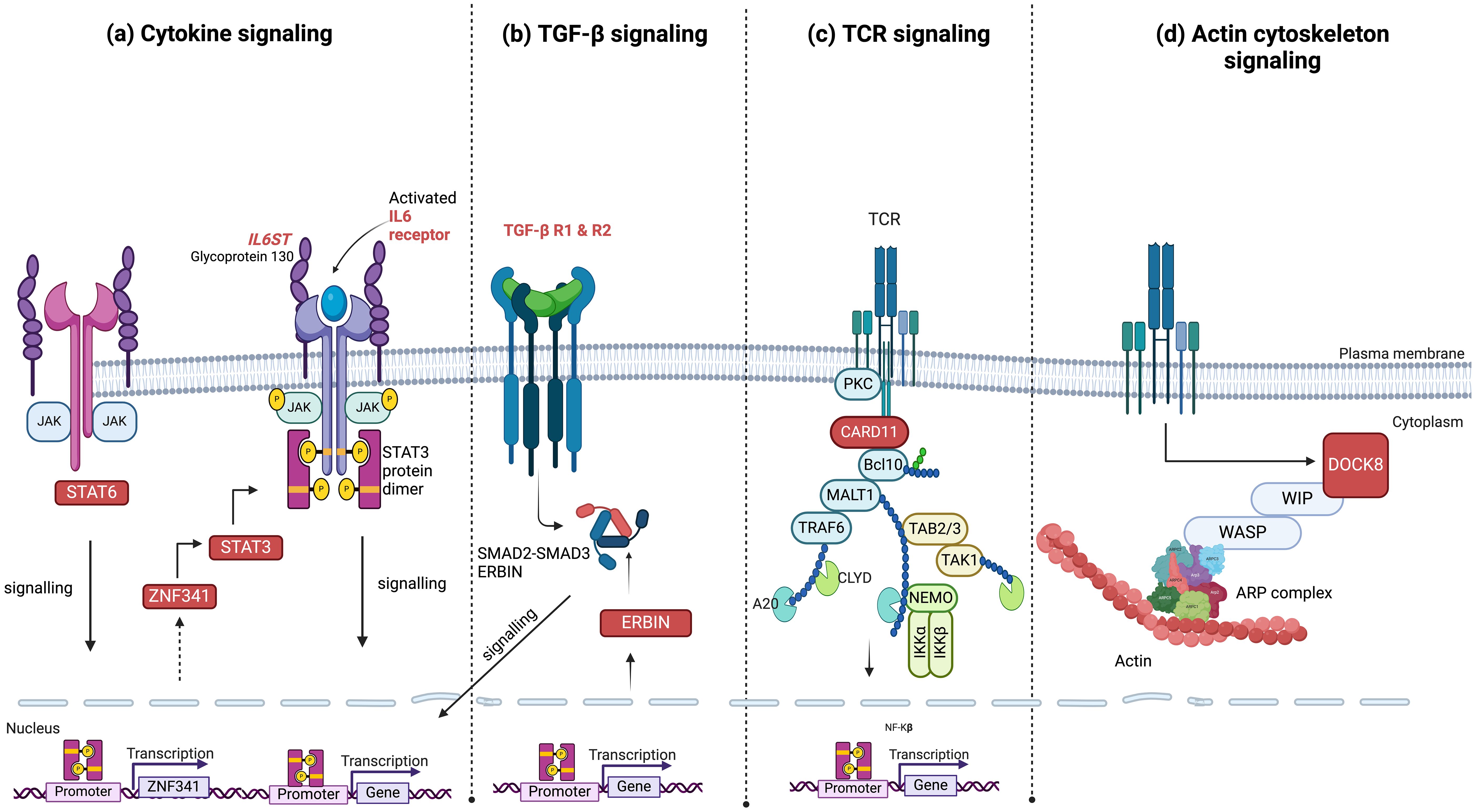
Figure 1. Signaling pathways and associated genes with mutations linked to hyper-IgE syndrome. (A) Components of cytokine signaling such as STAT3, ZNF341, STAT6, IL6R, and IL6ST; (B) TGF-β signaling pathway genes, including TGFBR1, TGFBR2, and ERBIN; (C) the antigen receptor signaling molecule CARD11; and (D) the actin cytoskeleton signaling molecule DOCK8. These pathways highlight the major molecular mechanisms currently identified which contribute to the clinical phenotypes of hyper-IgE syndrome. HIES genes are highlighted in red boxes or in red font and phosphorylation is indicated by yellow circles. JAK, Janus Kinase; STAT, Signal Transducer and Activator of Transcription; ZNF, zinc-finger; IL, Interleukin; ST, Signal Transducer; TGFBR, Transforming Growh Factor Beta Receptor; NF-κB, Nuclear Factor kappa-light-chain-enhancer of activated B cells; SMAD, Suppressor of Mothers Against Decapentaplegic; ERBIN, Erbb2 Interacting Protein; A20, Tumor Necrosis Factor; Alpha-Induced Protein 3; TCR, T Cell Receptor; PKC, Protien Kinase C; CARD11, Caspase recruitment domain-containing protein 11; Bcl10, B-cell lymphoma/leukemia 11; MALT1, Mucosa-associated lymphoid tissue lymphoma translocation protein 1; TRAF6, TNF receptor associated factor 6; TAB2/3, TGFb activated kinase 1 binding protein 2; TAK1, TGFb-activated kinase 1; CYLD, Cylindromatosis deubiquitinase; IKK, Inhibitor of nuclear factor kappa-B kinase; DOCK8, Dedicator of cytokinesis protein 8; WIP, WAS/WASL-interacting protein; WASP, Wiskott–Aldrich syndrome protein; ARP, actin-related protein.
In 2007, it was discovered that DN mutations in Signal transducer and activator of transcription 3 (STAT3) were present in the majority of AD-HIES cases, thus establishing a mechanistic explanation for both the infectious and connective tissue abnormalities observed (8, 14). By 2023, around 384 patients with mutations in the STAT3 gene (OMIM: #147060) (15) had been described in the literature (16). Most of the skeletal abnormalities in patients with STAT3 deficiency, such as scoliosis and osteoporosis, are due to poor responses to leukemia inhibitory factor (LIF) (17). Asano and colleagues evaluated more than 100 known STAT3 variants, and found that 95% of the in-frame variants (128 out of 135) demonstrated dominant negative (DN) effects (18). In the context of the immune system, STAT3 mutations impair the capacity of human CD8+ T cells to develop into granzyme B-expressing effector cells in response to IL-21 (19). STAT3 is selectively activated by certain gamma-chain (γc) family cytokines (20), particularly IL-21 (6, 21–23), whereas others mainly trigger STAT5 activation with limited involvement of STAT3 (24, 25). STAT3 is also crucial for the development of Th17 cells, a specific subset of T helper cells engaged in host defense against intracellular bacteria and anti-fungal responses. The ligands IL-6, IL-10, IL-11, IL-21, IL-22, and IL-23 transduce their signals through STAT3 (26) and as such AD LOF STAT3-HIES patients exhibit impaired cytokine signaling and defective differentiation of Th17 cells. STAT3 deficiency results in a Th1/Th2 imbalance, with a dominance of Th2 activity (27). While STAT3 is critical for IL-10 and IL-6 signaling, which regulate anti-inflammatory and pro-inflammatory pathways, respectively, STAT3-DN HIES patients exhibit defective responses to IFNγ signaling (14, 28), resulting in compromised Th1-driven immunity and a predisposition to chronic infections and inflammation. Critically, Th17 lymphocytes mediate effector responses via the release of antimicrobial peptides and production of IL-17 and IL-22 in response to bacteria, particularly S aureus, as well as fungi such as Aspergillus and Candida. Therefore, a deficiency in Th17 differentiation accounts for much of the increased frequency of infections in these patients (29, 30). STAT3 LOF mutations have been observed to be closely associated with a reduced proportion of Th17 cells (less than 0.3% of CD4+ T cells), in contrast to atopic dermatitis patients with unaffected STAT3 who generally exhibit significantly elevated levels of Th17 cells (31). Furthermore, IL-17 signaling plays a role in the regulation of neutrophil proliferation and chemotaxis. Consequently, the impaired neutrophil activity and their reduced recruitment to the lungs and skin could explain the recurrent staphylococcal infections observed in these areas (32–34). Additionally, STAT3 is crucial for the development, differentiation, and maintenance of memory T cell, and individuals with HIES show a reduced number of CD4 and CD8 central memory T cells (35, 36). STAT3-deficient patients have been shown to exhibit a diminished capacity of CD8+T cells to manage herpesvirus infections (37), which may be explained by the observation that in STAT3 deficient patients, there are increased levels of naïve CD8 T cells, and a concomitant decrease in central memory, effector memory, and terminally differentiated effector memory cytotoxic CD8 T cells (19).
Additionally, STAT3 plays a key role in naïve B cell activation, affinity maturation and class switching during the induction of humoral immune response (38, 39). In B cells, STAT3 signaling governs the structure and recycling of maturing cells through the germinal center light and dark zones, as well as the exit of mature cells from germinal centers, providing a mechanistic explanation of how STAT3 defects adversely affect production of memory B cells (40), leading to a reduction in the maintenance of circulating memory B cells. Although, STAT3 LOF mutations lead to a dysregulation of B cell responses, pateints typically have normal levels of total serum IgM, IgG, and IgA, whilst exhibiting hyper-elevated levels of serum IgE, impaired antigen-specific antibody responses, and decreased memory B cell levels (3, 41–45). Considering the importance of STAT3 for both plasma cell differentiation (IL-21 signaling) and CD4+ T-follicular helper cell (Tfh) (IL-6 signaling), IgA, IgG and IgM levels are found to be surprisingly within the normal range in many cases (46). However, there is a notable reduction in vaccine responses, linked to the noted deficiencies in B-cell maturation and as seen by a decrease in CD27+ switched memory B cells (44). Immunoglobulin replacement therapy (IgRt) has shown a positive impact on the improvement of S. aureus infections in individuals with STAT3-HIES (3, 31, 44). Studies indicate that IgRT can enhance the levels of S. aureus–specific IgG levels in STAT3-HIES patients (47). Studies reported the association of STAT3 mutations and impaired memory B cell function (48, 49). The National Institutes of Health (NIH) scoring system, assesses the presence and severity of 21 clinical and laboratory measures and was created for the purpose of identifying AD-HIES (50). 96% of the 48 AD-HIES patients with STAT3 mutations present in an observational study achieved a score of ≥40, which indicates the diagnostic utillity of this scoring system (31). In young patients, the score may be lower due to the absence of certain clinical features (51), such as scoliosis, fractures, and facial dysmorphic traits, which typically appear and become more pronounced as they grow and the full phenotype emerges over time.
Clinically having a normal IgE level or one below 2000 kU/I is not necessarily exclusive of the diagnosis (46). Eosinophilia is found in over 90% of STAT3-DN patients and is linked to impaired neutrophil function (52), and the induction of neutropenia with circulating neutrophils decreasing to approximately 700 cells per microliter. Interestingly, IgE levels and eosinophilia do not neccesarily predict the severity of the disease (50, 53).
Syndromic connective tissue features linked to STAT3-HIES exhibit variability: the characteristic facies, include a high-arched mouth, expanded nasal bridge, deep-set eyes, and large forehead, which typically manifest during adolescence, may not appear in early childhood (54). Most children need to have their primary teeth extracted, due to a lack of root resorption in the primary teeth, which is required for eruption (55). One-third of patients have joint hyperextensibility and degenerative joint disease manifesting in adulthood. Moreover, there is a predisposition to early-onset long bone fractures with minimal trauma. In 79% of cases, there is a decrease in bone mineral density (BMD), which does not correlate with the rate of fractures. While lower BMD is shown in the femoral neck and spine in classical osteoporosis (56), in STAT3-HIES, only radial BMD is associated with the risk of fractures (57). Vasculopathy in STAT3-HIES presents significant challenges for both understanding its pathogenesis and managing it clinically (58). Predominantly, medium-sized arterial abnormalities are observed, particularly affecting the coronary and intracranial vessels. In a prospective study, 50% of patients showed coronary artery abnormalities with ectasia and aneurysms being the most prevalent findings, alongside radiographic evidence of small infarcts in some cases (53). Mutations in the STAT3 gene have additionally been linked to various malignancies (16). Patients with HIES face a markedly elevated risk of developing aggressive B cell lymphomas, potentially due to defects in the STAT3/IL-21 pathway influencing B cell differentiation into plasma cells, with potential involvement of T follicular helper cells (45, 59). A study of 65 AD-HIES patients (including 35 with DNA-binding mutations and 30 with SH2 mutations) examined clinical characteristics using the NIH HIES scoring system alongside findings such as brain and coronary artery abnormalities. While patients with SH2 mutations exhibited a higher occurrence of certain features, such as a high palate, wider inter-alar distance, upper respiratory tract infections, and scoliosis (particularly in pediatric cases), individuals with DNA-binding mutations showed a higher risk of mortality (60). Recent cases, including an 18-year follow-up and seven additional patients, have shown improved survival and significant alleviation of disease-related complications (61). Allogeneic hematopoietic stem cell transplantation (HSCT) is an option to treat STAT3 deficiency, especially the immunological elements of the condition and stabilize even severe lung complications and abscesses and recurring skin infections. Notably, Th17 cells from donor cells may generate comparable amounts of IL-17A to those of healthy controls. However, long-term monitoring is necessary for determining the extent of syndromic reversal due to the broader functions controlled by STAT3 (61, 62).
IL6ST encodes Glycoprotein 130 (GP130), the common subunit of the signaling receptors for all ten IL-6 family cytokines (2, 63). Stimulation of these cell-membrane receptors triggers JAK/STAT pathway, essential for transmitting signals to the nucleus across multiple cell types and plays a major role in the regulation of T helper differentiation (64). A study involving 12 patients from 8 unrelated kindreds with AD-HIES caused by DN IL6ST mutations (OMIM: #619752) revealed that natural killer (NK) cell counts, and differentiation were normal in all patients, except for a few who exhibited low NK cell counts. CD4+ and CD8+ T cell counts were generally normal, however, one patient had low CD8+ T cell counts, while another had high CD4+ T cell counts. In the CD4+ and CD8+ T cell compartments, patients showed high naive T cell frequencies, but there were low frequencies of central memory T cells observed in the CD8+ compartment. Additionally, the CD8+ T cells contained a low proportion of effector memory T cells. The proportions of γδ T cells, invariant natural killer T (iNKT) cells, and regulatory T (T reg) cells remained normal, whereas the proportion of mucosal-associated invariant T (MAIT) cells was low. Within the memory CD4+ T cell compartment, the frequency of T follicular helper (Tfh) cells was low, whereas T helper type 1 (Th1), and Th17 cell frequencies were normal, with very high frequencies of Th2 cells (65).
During adaptive immune response, IL-6 plays a significant role in stimulating both antibody production and the development of effector T-cells (66). Pro-inflammatory cascades and the activation and differentiation of both B and T cells are disrupted in the absence of functional IL-6 signaling (67). IL-10, synthesized by regulatory T cells and B cells, has been linked to changes in production of IgG4 rather than IgE (68, 69), whereas IL-21 regulates both the induction and inhibition of IgE production (70, 71). Individuals with IL6ST deficiency often exhibit elevated levels of IgE due to the involvement of IL-6, IL-11, and IL-27 and their influence on other cytokine signaling pathways such as IL-10 and IL-21 (67, 72). IL-6 is believed to regulate IgE production through acting directly on B cells and indirectly on CD4+ T cells (73). Patients with autosomal recessive IL-10 or IL-10R deficiency maintain IgE levels within normal reference ranges (74, 75), suggesting that defective IL-10/STAT3 signaling does not impact class switch recombination (CSR) to IgE (73). Individuals with IL-11RA deficiency present with craniosynostosis and dental anomalies, but do not exhibit significant immunodeficiency (76). Therefore, IL-6 is considered the principal cytokine activating STAT3 that inhibits IgE production in human B cells (73). The hyper-IgE phenotype and clinical features of atopic disease, like dermatitis, food allergy, and eosinophilia, seen in STAT3 deficiency and ZNF341 deficiency are replicated by pathogenic variants in IL6R or IL6ST (17, 67, 77–80).
In mouse models, a complete deficiency of gp130 is lethal at the embryonic stage, while complete IL6ST LOF in humans results in the lethal Stüve-Wiedemann syndrome, defined by neonatal lung dysfunction, defective acute-phase response, skeletal dysplasia, renal abnormalities, atopic dermatitis, and congenital thrombocytopenia (81). Heterozygous DN IL6ST mutations were identified in 12 patients belonging to eight unrelated families, who exhibited clinical characteristics strikingly similar to STAT3-DN HIES, involving pneumatocele. Individuals with STAT3 and IL6ST mutations exhibit infectious and allergic symptoms characteristic of IL-6R deficiency, as well as some of the skeletal deformities present in IL-11R insufficiency. Thus, DN STAT3 and IL6ST mutations result in similar clinical phenocopies due to the disruption of the IL-6 and IL-11 response pathways (17). Patients who lack IL6ST suffer from impaired innate immune response to bacterial infections, which can cause acute-phase reactions to be delayed or completely absent (82). IL6ST-DN HIES patients also exhibit non-immunological characteristics such as scoliosis, osteoporosis, and retention of primary teeth. In contrast to individuals with partial LOF IL6ST mutations, complete IL11RA deficiency, or STAT3-DN mutations, individuals with IL6ST-DN do not exhibit craniosynostosis, most likely because of remaining functionality in IL-11 signaling (76). The skeletal phenotype is probably influenced by impaired IL-11 signaling, given that individuals with IL11RA mutations manifest with craniosynostosis and dental malformations (76, 83). Patients with DN mutations in IL6ST underwent treatment with subcutaneous immunoglobulin treatment (17). The analysis of IL6ST mutations brought to light that IL6ST/IL6R cytokine receptor complex formation is essential for the signal transduction upstream of STAT3 in HIES pathogenesis. After JAKs are phosphorylated by the hexameric IL6-IL6R-IL6ST complex, tyrosine residues within the cytoplasmic domain of GP130 are phosphorylated by JAKs (Figure 1), which leads to the attraction and activation of STAT1, STAT3, and STAT5. Subsequently, the phosphorylated STATs form dimers and migrate into the nucleus to interact with the promoters of acute-phase protein genes that contain IL-6-responsive regions. Hence, mutations in the IL6R, IL6ST, and STAT3 genes all lead to disruption of IL-6 family signaling pathways, but with distinct differences in their clinical manifestations (1).
TGFBR1 and TGFBR2, GOF mutations (OMIM: #609192) and (OMIM: #610168) (84), were the first documented genetic causes of Loeys–Dietz syndrome (LDS) (85–88). LDS was first described in 2005 as an autosomal-dominant connective tissue disorder associated with aortic aneurysms, hypertelorism, widespread arterial tortuosity, and bifid/broad uvula or cleft palate (84, 89). As of now, 53 patients with Loeys-Dietz syndrome have been identified in published studies (90–92).
TGF-β signaling can influence the activation and differentiation of T cells, and the process of immunoglobulin class switching by B cells following allergen exposure (93–95). The ability of regulatory CD4+ T cells (Treg) to produce TGF-β plays a significant role in their ability to maintain homeostasis and prevent allergic disease and autoimmune responses (96).
Dysregulated TGF-β signaling in LDS patients increases serum IgE concentration through dysregulated CD4+ T cell differentiation and elevation in Th2 cytokine levels, leading to the induction of allergic reactions similar to those seen in STAT3-HIES (1, 2). LDS mutations promote TH2 skewing in naïve lymphocytes through a cell-autonomous mechanism. Naïve CD4(+) T cells from LDS patients, unlike those from controls, exhibited an accumulation of TH2 cytokine-producing cells following stimulation with TGF-β (97). In agreement with the role of TGF-β in promoting the generation of Treg cells (98), but inconsistent with the observed loss of tolerance, LDS patients exhibited an increased frequency of peripheral FOXP3+ Treg cells with intact ability to suppress effector T cell proliferation (97). Additionally, TGF-β can exert its effects on mast cells during an acute allergy episode to regulate the severity of the reaction. This is particularly relevant in hyper-IgE syndromes, which are marked by dysregulated mast cell function and intensified allergic responses. TGF-β also plays a role in promoting tissue remodeling following the damage resulting from an allergic attack (95). TGF-β signaling serves as a crucial connection between the function of vascular smooth muscle cells (SMCs) and the composition of the extracellular matrix (ECM). LOF mutations resulting in TGF-β signaling pathway genes have been linked to alterations in aortic SMC phenotypes, causing structural alterations in the aorta and contributing to hereditary connective tissue disorders (99). Variants in TGF-β pathway have been associated with atopic conditions. Haque et al. investigated the role of TGF-β signaling in IgE production in allergic diseases by studying both patients with Loeys-Dietz Syndrome (LDS) and mice with specific gene defects causing partially reduced canonical TGF-β signaling. CD4+ T cells from both LDS patients and affected mice demonstrated impaired TGF-β signaling, which was correlated with atopy. The mutations seen in LDS led to an increase in T follicular helper 2 cells, heightened humoral immune responses, and the production of allergen-specific IgE. Additionally, LDS variants caused dysregulation PI3Kγ/mTOR signaling, which was able to be corrected through pharmacological inhibition (100). Immunological tissues from HIES individuals with TGFBR1/2 mutations display an increased accumulation of phosphorylated suppressor of mothers against decapentaplegic homolog 2 (SMAD2) in the thymus (1). This disrupted signaling affects downstream pathways, which helps to explain the increased allergic reactions and connective tissue issues observed in these patients.
LDS is characterized by rapidly progressing aortic aneurysmal disease, making close surveillance crucial (84, 89). Regular echocardiography is essential to evaluate he aortic root, ascending aorta, and heart valves in affected individuals (101). In LDS patients, sinusitis and otitis may occur together with mucus buildup due to the heightened allergic reactivity and malformed craniofacial structures (2). An instance of febrile status epilepticus linked to COVID-19 in an infant with Loeys-Dietz syndrome (LDS) has been reported (102). A case has also been reported of a patient with LDS who developed annular aortic valve abscess and ascending aortic dissection caused by Staphylococcus aureus endocarditis (103). Additionally, LDS is associated with several atopic conditions including eosinophilic gastrointestinal disease, allergic rhinitis, atopic dermatitis, food allergy, and asthma (1, 2).
ERBIN2IP encodes ERBIN (OMIM: *606944), which plays a crucial role in regulating TGF-β associated activities. ERBIN is essential in facilitating the crosstalk between STAT3 and TGF-β signaling. ERBIN is activated via STAT3 whereupon they form a complex that inhibits SMAD2/3 nuclear localization and TGFBR signal propagation (104).
Unlike TGFBR deficiency, which alters TGF-β receptor signaling directly, STAT3 activation of ERBIN in individuals with STAT3 or ERBIN2IP LOF mutations may induce an increased level of TGF-β activity. This results in increased IgE and Th2 cytokine expression as well as excessive activation of IL-4/IL4R (2). ERBIN defective memory CD45RO+ CD4+ cells produce high levels of Th2 associated cytokines (IL-4, IL-5, and IL-13) ex vivo, which accounts for the allergic symptoms (105). Under homeostatic conditions, TGF-β signaling is essential in preventing the spontaneous development of allergic disorders such as asthma, eosinophilic esophagitis, and atopy in mouse models (106). Another important result of this heightened signaling is the elevated expression of IL-4 and IL4Rα (107).
So far, only 3 cases of ERBIN deficiency have been reported (108, 109). Patients with ERBIN or STAT3 mutations commonly exhibit severe eosinophilic gastrointestinal illness (EGID), bacterial infections, allergen-specific reactivity, hyper-IgE, and connective tissue anomalies, including aneurysms and joint hypermobility (108). Scoliosis, a significant connective tissue anomaly observed in individuals with LDS and those with STAT3-DN mutations, is frequently associated with genetic variations in the ERBIN gene (110). Despite similarities in connective tissue symptoms between STAT3 DN, TGFBR LOF, and ERBIN LOF mutations, further insights into the syndrome specific effects may be provided by a deeper analysis in the differences induced in the TGF-β signaling pathway (107). Further, ERBIN mutations could also play a role in specific epithelial disease types like atopic diathesis (104).
Notably, administration of IL-4Rα blockade (using biologic therapeutics like dupilumab) to patients with STAT3 DN and ERBIN LOF conditions shows considerable improvement in otherwise unresponsive cutaneous and gastrointestinal inflammation in these patients (105, 111–116). Targeting type 2 signaling through dupilumab treatment in a patient with ERBIN deficiency effectively controlled co-existing allergic inflammation, suggesting a possible treatment strategy for comorbid atopy in other individuals with disrupted TGF-β signaling pathways (105).
Heterozygous variations can either enhance or significantly impair CARD11 functional efficiency, given that CARD11 oligomerization is necessary for downstream signaling (117). Germline CARD11 mutations lead to at least three differentially characterised PIDs, including severe atopic disease (dominant interfering, heterozygous and loss-of-function mutations), B cell expansion with NF-κB and T cell anergy (BENTA; heterozygous, gain-of-function mutations), and severe combined immunodeficiency (biallelic null mutations) (118–120). In 2017, it was shown that a form of AD-HIES with atopy and hypogammaglobulinemia is driven by heterozygous LOF DN mutations in the CARD11 gene, also referred to as immunodeficiency 11B with atopic dermatitis, (OMIM: #617638) (121). CARD11-associated atopy with dominant interference of NF-κB signaling (CADINS), was first identified in 2017, and is clinically defined by the presence of atopy and a variety of autoimmune and/or infectious symptoms (122). Also, it has been reported that heterozygous LOF CARD11 mutations, which function as strong DN alleles, are the cause of this condition (123).
To date, 62 patients have been identified with 17 distinct dominant-negative mutations in the CARD11 gene, with most mutations located in the N-terminal CARD and CC domains (118, 121, 122, 124–128). CADINS symptoms, can include asthma and other allergic diseases, autoimmune diseases, and infections including respiratory tract and viral skin infections. Significant variations in the incidence of symptoms have been noted between kindreds and even within family members (126). Due to the accompanying B and/or T cell defects, CADINS patients may experience a more severe infectious phenotype than those with classical atopic dermatitis (125). Clinical symptoms observed have been linked to specific immunologic defects in patients including neutropenia, hypogammaglobulinemia, aberrant T-cell proliferation and differentiation (122). Further, CARD11 mutations may additionally predispose patients to an increase incidence of malignancies (16). Although IgE levels are high in all CADINS patients, these are generally lower than those seen in STAT3 deficiency (46, 118). The presence of eosinophilia is a recurrent finding (46) and a study involving 15 CADINS patients revealed that the majority exhibited consistent eosinophilia (80%) (125). Quantitative immunoglobulin levels for IgM, IgG and IgA are generally within normal ranges, with only sporadic observations of slightly reduced IgG levels and elevated IgA levels. Vaccine-induced antibody response may exhibit variability, notably response to carbohydrate antigens are consistently suppressed (46). Patient B- and T-cell counts are within the normal range, while phytohemagglutinin (PHA) and anti-CD3-induced cell proliferation has been shown to be reduced. Whilst, AD-CARD11 individuals share a lot of similarities with DOCK8-deficient patients in terms of clinical and immunological characteristics, mucocutaneous Candidiasis and cutaneous viral infections occur less frequently, and only one affected patient has presented with neurologic sequelae (seizures and nystagmus) (46). According to reports, individuals with CARD11 deficiency may experience improvements when treated with intravenous immunoglobulin (IVIG) or Subcutaneous immunoglobulin (SCIG) because it would lower their infection rates (46).
STAT6 is critical for propagation of signaling from the IL-4 and IL-13 receptors to the nucleus in order to drive a multitude of effects, chiefly directing Th2 differentiation in naïve cells and inducing IgE class switching in B cells (129). As such, there is a close connection between STAT6 and the biology that induces allergic inflammation. The primary and most well researched function of STAT6 involves facilitating the biological impacts of IL-4, an essential cytokine required for various processes such as M2 macrophage polarization (10, 130), Th2 cell differentiation, as well as B cell survival, proliferation, and class switching to produce IgE (131, 132). STAT6 activation in T cells leads to the induction of GATA3 expression, the master regulator of Th2 differentiation. Increased Th2 differentiation subsequently lead to increases in production of IL-4, IL-5, and IL-13, cytokines essential for inducing allergic reactions by stimulating downstream innate immune responses in the form of mast cells, eosinophils, basophils and innate lymphoid type-2 cells (ILC2) (133, 134). For both acute and chronic allergic disease symptoms, elevated IgE in conjunction with increased levels of mast cells and basophils which express high affinity immunoglobulin E receptor (FcER) is a critical component, which may additionally contribute to the allergic predisposition and the atopic march (135). Elevated TH2 cell populations or TH2 cells that secrete large amounts of IL-4, IL-5, and IL-13 may underlie the allergic phenotype observed in the STAT6 GOF patients (10). Overactive STAT6 signaling in airway epithelial cells and dendritic cells can foster conditions that support asthma and chronic lung diseases by driving the production of chemokines that attract TH2 cells and eosinophils (6, 136, 137). This highlights how dysregulated STAT6 activity contributes to the development of allergic and chronic inflammatory diseases, underscoring its potential as a therapeutic target for managing these symptoms.
Recently, 21 individuals with severe allergic disease from birth were diagnosed with STAT6-GOF disease (OMIM: #620532) (130). While the complete spectrum of characteristics exhibited by individuals with GOF STAT6 variants remains to be fully elucidated pending identification of more affected individuals, current clinical indicators include early onset of elevated levels of serum IgE, increased eosinophils in peripheral blood. Patients may also present with persistent and widespread atopic dermatitis resistant to treatment, recurring infections affecting the skin and respiratory system, multiple allergies to both food and drugs. Severe cases of anaphylaxis (sometimes fatal), eosinophilic gastrointestinal disorders, allergic rhinoconjunctivitis, asthma are frequently reported. Other notable features may include shorter stature and potentially cerebral vascular anomalies, although further research is needed to confirm these findings (10, 11). A case has been reported of a family with STAT6 GOF disease presenting with severe early-onset atopy and follicular lymphoma and Linked to a newly identified germline heterozygous STAT6 mutation, c.1255G>C p.D419H, residing in the DNA binding domain of STAT6 and co-segregating with the clinical phenotype (138). Dupilumab and tofacitinib treatments have demonstrated efficacy in the treatment of STAT6 GOF patients (10).
In 2009, mutations in DOCK8 (OMIM: #243700) were identified as the predominant cause of AR-HIES (139, 140). Based on the update of IUIS phenotypical classification in 2022, AR DOCK8 deficiency was reclassified as a B cell low subgroup of combined immunodeficiency (CID) (141). DOCK8 belongs to a protein family containing 11 members having major roles in control of cell adhesion, migration, and morphology. DOCK8 has a central role in regulating the reorganization of the actin cytoskeleton during migration and synapse formation, and immune abnormalities associated with DOCK8 deficiency are linked to these functions (2). Around 230 patients of DOCK8 deficiency have been reported by 2017 (142).
Studies assessing immune function in mice, lacking DOCK8, found abnormalities in dendritic cell (DC) functionality, showing DCs were unable to accumulate in draining lymph nodes, thereby reducing T-cell priming (143). Additionally, DOCK8 serves as an intermediary protein in B cells, functioning upstream of STAT3 and downstream of toll-like receptor 9 (TLR9), facilitating the proliferation of B cells and the generation of immunoglobulins (144). Studies of both humans and mice with DOCK8 inactivating mutations have demonstrated that DOCK8 is involved in the survival of naive CD8 T cells, the polarization of lymphocyte function–associated antigen 1 (LFA-1) towards the immune synapse, and the memory and recall responses of CD8 T cells following viral infection (145). Lack of DOCK8 may affect long-term B cell memory (145, 146). DOCK8 mutant B cells were unable to develop into marginal zone B cells or sustain their presence in germinal centers during affinity maturation. DOCK8 mutation disrupted the concentration of Intercellular Adhesion Molecule 1 (ICAM-1) in the B cell immune synapse, but did not change other features of B cell antigen receptor signaling. The development of a humoral immunodeficiency caused by a DOCK8 mutation suggests that the arrangement of an immune synapse is essential for signaling and the survival of those B cell subsets needed for sustaining long-lasting immunity (147). As CD8+ T-cells have essential activity in antiviral defense, in case of DOCK8 deficiency, CD8+ T-cells exhibit significant decreases in IFN-γ and TNF-α production as well as CD8+ T cell proliferation (2). DOCK8 deficiency may affect survival of memory CD8+ T cells specific to viruses, which potentially explains the increased susceptibility to bacterial and chronic viral infections (145, 146). Furthermore, leukocyte migration to infected skin is impaired (2). LFA-1 is a key adhesion molecule that plays an essential role in T cell migration and retention within lymph nodes (148, 149). Moreover, the interplay between LFA-1 and ICAM-1/2 on B cells is essential for the positive Tfh–B cell contacts that result in B cell clonal growth (150). DOCK8 expression in T cells is crucial for LFA1-dependent positioning within germinal centers (GCs), GC B cell production, and IgG antibody responses to T cell-dependent antigens (151). Therefore, the effectiveness of adaptive immunity is significantly diminished in individuals with DOCK8 deficiency because the accumulation of adhesion molecules and cytotoxic granules at immunologic sites is hindered (2). In DOCK8 deficient individuals, T cells exhibit a skewing towards Th2 differentiation, leading to subsequent increases in IgE levels (152). In vivo studies have revealed cell death as a crucial signal leading to the Th2-skewed CD4+ T cell response in DOCK8-deficient mice. Here, lung-infiltrating DOCK8−/− mononuclear phagocytes expressing the fractalkine receptor CX3CR1 exhibit heightened susceptibility to migration-induced shattering (cytothripsis), which releases IL-1β and causes CD4+ T cells to produce granulocyte-macrophage colony-stimulating factor (GM-CSF). Caspase-dependent cell death, in conjunction with IL-1β, is also a crucial driver for the Th2 bias (152).
In addition to the systemic manifestations shared by many combined immunodeficiencies, a key characteristic of DOCK8 deficiency is the elevated incidence of skin infections and eczema (153). From a clinical perspective, patients with DOCK8 deficiency have symptoms resembling those of AD-HIES, such as recurrent sinopulmonary infections, mucocutaneous Candidiasis, Staphylococcal skin abscesses, heightened IgE levels, eczema, eosinophilia, and an increased likelihood of developing malignancies (154). Compared to other PIDs (even those with significantly elevated IgE levels, such as DN mutations in STAT3), DOCK8 deficiency leads to an extremely high frequency of severe food allergies which is a distinctive aspect of DOCK8 deficiency (140, 144, 155–157). DOCK8 deficient individuals have in contrast been found to exhibit far lower rates of skeletal and connective tissue anomalies, specifically the characteristic facies, retained dentition, and minimal trauma fractures commonly observed in AD-HIES (143). Patients with DOCK8 deficiency are also vulnerable to viral infections such as human papilloma virus (2), which are thought to play a significant role in the increased frequency at which they develop malignancies, with squamous cell carcinoma and lymphoma being the primary types, affecting around 17% of patients (158). Other types of cancers, including micro-cystic adnexal carcinomas and cutaneous T cell carcinomas, are unrelated to viral infections (159). While STAT3-HIES patients frequently live into their sixth decade, the median lifespan in DOCK8 patients, despite the use of preventive anti-microbial measures and immunoglobulin replacement therapy, is typically 20 years (4). Studies have shown that HSCT is effective as a cure for most patients with DOCK8 deficiency, establishing this method as the treatment of choice (160, 161). Also, dupilumab was effectively employed to achieve remission of chronic widespread eczema herpeticum in a patient with hyper-IgE syndrome due to DOCK8 deficiency (162).
Although NIH scores exceeding 40 are sometimes obtained, such occurrences are less common compared to individuals with AD-STAT3 deficiency (46). While both conditions exhibit increased serum IgE levels and eosinophilia, primarily because of decreases in T cells, patients with DOCK8 deficiency display decreased serum IgM levels and lymphopenia (154). Normally, IgG and IgA levels are within the normal range or increased (163), and a reduction in B-cells is less common (46), poor vaccination reactions are a commonly observed problem (155, 163).
Two studies published in 2018 described biallelic LOF variants of zinc finger protein 341 (ZNF341) (OMIM: #618282), which encodes a protein with a previously unknown function and linking it to a new AR-HIES. The truncated forms of ZNF341 were found to be responsible for inducing a STAT3-like hyper-IgE phenotype (164). These studies derived the role for ZNF341 as a transcription factor responsible for regulating both the inducible and baseline levels of STAT3 expression (164, 165).
Compared to those with STAT3-DN HIES, ZNF341 patients exhibited fewer non-immunological symptoms and more robust inflammatory reactions (1). Here, IL-6 family member receptors such as the IL-11 receptor, which contributes to the joint, skeletal, tooth, and vascular symptoms of STAT3-HIES, may not exert the same level of influence as they present in STAT3 deficiencies, suggesting that ZNF341 has additional activities which are independent of the STAT3 signaling pathway (2). Similar to STAT3 LOF mutations in HIES patients, the disruption of ZNF341 signaling leads to an inhibition of Th17 differentiation and results in less IL17 and IL-23 production by CD4+ memory and effector T cells, due to defects in STAT3 dependent signaling (164).
Whilst individuals affected by AR-ZNF341 deficiency exhibit several clinical and immunological features similar to those with AD-STAT3, their clinical symptoms tend to be milder. However, NK cell numbers are notably lower in this patient population (166). Currently, there is a lack of correlation between ZNF341 deficiency and common atopic symptoms such as asthma, dermatitis, rhinitis, and food allergies. A potential explanation for this observation is that ZNF341 with its 12 zinc fingers functions as the activator of cytokine-mediated STAT3 production. This suggests that zinc finger transcription factors can recruit transcriptional activators and repressors without functional domains (2). An adult patient with ZNF341 deficiency and severe atopic dermatitis showed significant clinical improvement and a reduction in IgE levels after treatment with dupilumab (167). ZNF341 also serves as an enhancer of STAT3 expression, and homozygous nonsense mutations in ZNF341 result in inadequate STAT3 expression. This insufficiency is likely a contributing factor to the symptoms of HIES. However, further research is needed to understand the precise role of ZNF341 in the pathogenesis of ZNF341 LOF HIES, as there is not conclusive evidence showing that reintroducing normal ZNF341 restores STAT3 expression and signaling. Additionally, the STAT3-independent functions of the ZNF341 may also play a role in the syndromes’ manifestation (1). To date, 20 patients with ZNF341 deficiency have been reported (166). The overall number of reported patients is very limited, which constrains the understanding of both the clinical features and immunophenotype associated with this condition.
Hypomorphic autosomal-recessive mutations in PGM3 (OMIM: #615816) can result in a glycosylation disorder that exhibits a variety of clinical manifestations with varied penetrance and expressivity. Additional reported issues include, connective tissue abnormalities, immune dysregulation, and impairments in neurodevelopment (104). Clinical manifestations in patients with PGM3 deficiency include recurrent infections (particularly Staphylococcus aureus), Candidiasis, or viral etiologies in the skin, respiratory tract, or gastrointestinal tract, atopy, neurologic symptoms, enteropathy, bone marrow failure, skeletal dysplasia (168–170), as well as hyper-elevated serum IgE (168). Severe combined immunodeficiency (SCID), neutropenia, and skeletal anomalies can also be present in infants (170). Neurocognitive impairments and likely hypomyelination, which are not observed in cases of STAT3 or DOCK8 deficiency, are distinct features of PGM3 deficiency (171). Vascular abnormalities have been observed in PGM3-deficient patients, although they are uncommon (168). By 2014, a total of 40 cases from 18 families with PGM3 deficiency had been documented in the literature (168–170, 172–178). Due to the significant overlap with AD-HIES, a large percentage (87%) of these patients have an NIH score of ≥40, higher than generally observed in DOCK8 deficiency (168, 169).
Patients with PGM3 deficiency commonly experience neutropenia, reduced memory B-cell, and T-cells counts. Nonetheless, they also exhibit hypergammaglobulinemia, while having intact antibody responses to protein and carbohydrate antigens (168, 169). Antibody levels against carbohydrate and protein antigens are generally sufficient for protection. T-cell lymphopenia is a common finding affecting predominantly the CD4+ subset, leading to an inverted CD4:CD8 ratio (169). While the PGM3 protein levels may appear normal, analysis of activity in PGM3-deficient fibroblasts can show a distinct reduction, and serves as an additional relevant diagnostic criterion (168). Individuals with PGM3 deficiency may potentially improve the catalytic activity of PGM3 when an enzymatic route is blocked by supplements such as N-acetyl-glucosamine (GlcNAc), which leads to a reduction in pathologic phenotypes (108). Additionally, reports on the outcomes of HSCT in PGM3 deficiency, though limited, highlight that hematopoietic stem cell transplantation with cord blood or bone marrow from matched related donors led to successful engraftment and resolution of both neutropenia and lymphopenia (170). These findings underscore the potential of HSCT as a therapeutic option for this rare disorder. Along with HSCT, immunoglobulin replacement therapy and antimicrobial prophylaxis are key treatment approaches for PGM3 deficiency patients (170, 174, 179).
LOF mutations in IL6R (OMIM: #618944) were first observed in two unrelated patients, who exhibited atopic dermatitis, recurrent skin and lung infections, and reduced inflammatory responses. These mutations were found to be responsible for encoding an IL-6R ligand binding component. The patients experienced repeated skin abscesses caused by S. aureus, upper and lower respiratory tract infections, though intriguingly their levels of C-reactive protein (CRP) were only slightly elevated during acute infection (78). Four additionally identified patients exhibited impaired B-cell class-switching and antibody production, a tendency toward severe invasive bacterial infections, severe eczema, absence of acute-phase reactant responses, and potential elevation in IL-6 production (79). The impairment of cellular responses to IL-6 alone may account for most of the characteristics of the primary HIES (180). Immunoglobulin replacement therapy is a cornerstone treatment for patients with bacterial infections and hypogammaglobulinemia, along with antimicrobial prophylaxis (181). In severe cases involving pronounced immunodeficiency and recurrent life-threatening infections, hematopoietic stem cell transplantation (HSCT) might be considered (182). However, contrary evidence exists as patients with neutralizing autoantibodies against IL-6 have symptoms comparable to those of IL-6R LOF, specifically reduced inflammatory response but not atopic manifestations. Around 10 patients with IL-6R deficiency have been documented in the literature (183). Further research is required, especially to determine the exact molecular mechanism by which IL-6 and/or other signaling molecules control atopic features (1).
N404Y and P498L were the first to be identified as novel homozygous missense mutations in IL6ST (OMIM: #618523) from two patients of South Asian and Turkish heritage respectively. To date, a partial loss-of-function mutation in the IL6ST gene have been identified across 20 patients (184). Validation of the putative defects were confirmed as partial signaling defects after evaluation of the functionality of reconstituted IL-6 receptor complexes in HEK 293 cells lacking intrinsic GP130, following transfection with patient IL6ST sequences (67, 80). When patient-derived fibroblasts were stimulated with IL-6 and IL-11, the response of STAT3 was significantly diminished and the deficiency was repaired when wild-type GP130 was expressed. Both patients exhibited eczema, eosinophilia, elevated serum IgE levels, scoliosis, recurring lung infections, decreased acute-phase responses, retained deciduous teeth as well as craniosynostosis (1).
Patient P498L was treated with monthly IVIG (80). These results imply that IL6ST plays a predominant role as cytokine receptor upstream in the pathophysiology of STAT3-DN HIES (1).
Comel-Netherton syndrome is a result of AR mutations in SPINK5 (OMIM: #256500) and is an extremely rare condition with an incidence of 1/200,000 (185). SPINK5 produces the lymphoepithelial Kazal-type-5 related inhibitor, which plays a role in regulating the process of desquamation. Symptoms typically include an ichthyosiform erythroderma, presenting early in life, with many patients going on to acquire distinctive syndromic features such as ichthyosis linearis circumflexa and anomalies of the hair shaft (easily broken hairs or bamboo hair). As of 2021, more than 200 cases of Netherton syndrome have been reported (186–191). Comel-Netherton patients are also likely to experience atopic symptoms, including anaphylaxis, severe atopic dermatitis, hay fever, and food allergies (46).
Comel-Netherton syndrome patients were shown to have a reduced proportion of both non-switched and class-switched memory B cells, deficiencies in NK cell cytotoxicity, and to exhibit a limited response to the T-cell-dependent neoantigen, bacteriophage phiX174. In addition, patients have an impaired skin barrier, which increases the likelihood of cutaneous infections and bloodstream bacterial infections (most commonly with S. aureus) (192).
Clinically, the HIES symptomatic triad of atopic dermatitis and recurrent infections, eosinophilia, as well as elevated IgE is present in Comel-Netherton patients. The definitive non-genetic diagnostic signs for Comel-Netherton syndrome are the abnormalities in the hair shaft in combination with the other syndromic features. Table 1 shows the summary of Hyper IgE syndromes describing the primary causative gene, their mode of inheritance and further details. Multiple case reports have documented significant clinical improvement in both adults and children received dupilumab treatment (193–196). Treatment with omalizumab reduced skin and mucosal symptoms in a 20-year-old Comel-Netherton patient (197). A Notable skin improvement was shown in a young adult with Comel-Netherton after omalizumab therapy (198). Promising results were achieved using monoclonal antibodies targeting IL-17, such as ixekizumab and secukinumab, along with TNF-α inhibitors like infliximab and anakinra, for managing inflammatory skin lesions in Comel-Netherton (199–203).
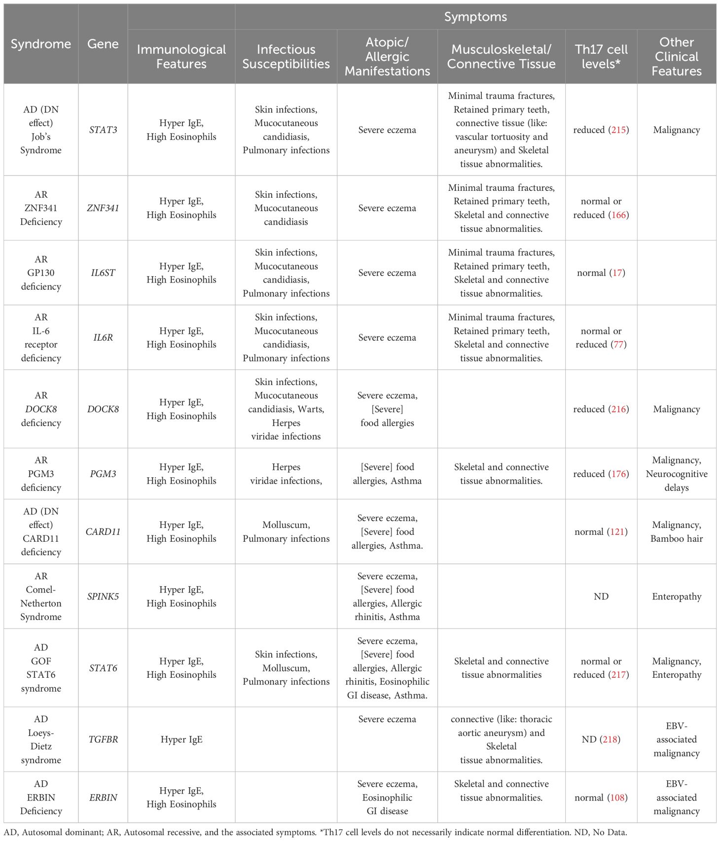
Table 1. Summary of Hyper IgE syndromes describing the primary causative gene, their mode of inheritance.
High IgE levels are a feature of numerous other PIDs falling under the combined immunodeficiency category. These conditions encompass Omenn syndrome, DiGeorge syndrome, and Wiskott-Aldrich syndrome, all distinguished by significant decreases in T-cell counts and profound deficiencies in T-cell functionality (54, 204–206). The clonal expansion of specific T and B cells, reduced TCR affinity and signaling, or an imbalance in the development of Th1/Th2 cells, could all contribute to elevated levels of serum IgE (207). Immune dysregulation, polyendocrinopathy, enteropathy X-linked (IPEX) syndrome, caused by FOXP3 LOF mutations, also feature elevated serum IgE levels and an absence of Treg cells, indicating that the absence of Treg cells by itself is adequate to cause increased serum IgE levels in affected patients. Further, additional skin barrier syndromes, which may or may not lead to IgE elevation, and may or may not lead to increased infection (certainly not beyond the skin) include SAM syndrome, complete filaggrin deficiency, epidermolysis bullosa. However, in these cases the elevation in IgE levels is thought to occur due to a standard atopic response caused by an increased degree of environmental allergen exposure, as opposed to a direct genetic deficiency in the immune tolerogenic pathway that underlies HIES cases. To effectively diagnose HIES, it is also essential to rule out these alternative conditions which can be carried out through genetic screening approaches (1) or with clinical diagnosis as in the cases of alternative IEIs with high IgE such as Omenn syndrome and IPEX which are very distinct and do not overlap with the traditional HIES.
HIES is defined as a complex PID, distinguished by hyper-elevated serum IgE levels, recurrent infections, and atopic dermatitis (50). Genetic categorization reveals that HIES can be inherited in either an AR or AD manner or may arise from de novo spontaneous genetic changes. While the majority of patients with AD-HIES are linked to mutations in the STAT3 gene, most instances of AR-HIES are connected to mutations in DOCK8 (6). However, recent research has identified other genetic disorders linked to HIES, including mutations in IL6ST, PGM3, ERBIN, ZNF341, IL6R, SPINK5, TGFBR1, TGFBR2, STAT6 and CARD11, expanding our knowledge of the genetic basis for this syndrome (133, 141, 208).
Atopic conditions, such as food allergies, are a significant feature of hyper-IgE syndromes (HIES), with their presentation varying based on the underlying genetic mutation.Patients with DOCK8, STAT3, or atopic dermatitis exhibit distinct sensitization patterns (209), with allergic symptoms and skin prick test outcomes strongly aligning with specific IgE levels in DOCK8 and atopic dermatitis cases, aiding dietary management decisions (210). A global study of 136 DOCK8 cases revealed that more than 70% experienced allergy-related symptoms, including food-induced anaphylaxis in 16%, as well as drug allergies, asthma, and rhinitis (163) Food allergies in DOCK8-HIES patients can persist even after HSCT, despite achieving full donor chimerism (211) PGM3-HIES patients often have multiple allergies (168), whilst WAS patients and murine models frequently develop IgE-mediated reactions to environmental and food allergens (212). STAT3-HIES patients have specific IgE levels and skin prick test outcomes similar to those of healthy individuals, despite their extremely high total IgE levels (209), and they experience higher rates of lifetime prevalence and severity of food allergies than healthy controls but lower rates compared to atopic controls with similar IgE levels. This phenomenon may be due to the severe impairment of mast cell degranulation associated with STAT3 loss-of-function mutations (213). Understanding these genetic distinctions is crucial for developing tailored management strategies for food allergies in HIES patients.
Understanding the distinct molecular mechanisms of AD-HIES and AR-HIES is essential for precise diagnosis, management, and the establishment of targeted therapeutic strategies. Although HSCT has demonstrated promise in restoring immune function and decreasing IgE levels in affected individuals, additional research is required to clarify the specific biological mechanisms driving the clinical variations observed between the AD and AR inherited forms of HIES. HSCT presents challenges, since donors may not always be readily available, including the reactivation of viral infections, the potential for graft-versus-host disease, and donor chimerism. Hence, innovative and promising approaches to treating DOCK8 deficiency include adaptive cellular therapies and gene therapy. However, these methods are still being investigated (46, 61, 214). Ultimately, continued investigation into the genetic and molecular basis of HIES will facilitate the development of more efficient therapeutic interventions and enhance the outcomes for individuals affected by these rare immunodeficiency disorders.
RA: Data curation, Formal analysis, Investigation, Methodology, Writing – original draft, Writing – review & editing. DV: Investigation, Resources, Supervision, Writing – original draft, Writing – review & editing. Nv: Conceptualization, Resources, Supervision, Writing – original draft, Writing – review & editing. PJ: Conceptualization, Funding acquisition, Resources, Supervision, Writing – original draft, Writing – review & editing.
The author(s) declare financial support was received for the research, authorship, and/or publication of this article. Thanks to HBKU for the faculty funding for PJ and scholarship for RA.
The authors declare that the research was conducted in the absence of any commercial or financial relationships that could be construed as a potential conflict of interest.
The author(s) declare that no Generative AI was used in the creation of this manuscript.
All claims expressed in this article are solely those of the authors and do not necessarily represent those of their affiliated organizations, or those of the publisher, the editors and the reviewers. Any product that may be evaluated in this article, or claim that may be made by its manufacturer, is not guaranteed or endorsed by the publisher.
1. Minegishi Y. Hyper-IgE syndrome, 2021 update. Allergol Int. (2021) 70:407–14. doi: 10.1016/j.alit.2021.07.007
2. Gharehzadehshirazi A, Amini A, Rezaei N. Hyper IgE syndromes: A clinical approach. Clin Immunol. (2022) 237:108988. doi: 10.1016/j.clim.2022.108988
3. Chandesris MO, Melki I, Natividad A, Puel A, Fieschi C, Yun L, et al. Autosomal dominant STAT3 deficiency and hyper-IgE syndrome: molecular, cellular, and clinical features from a French national survey. Med (Baltimore). (2012) 91:e1–e19. doi: 10.1097/MD.0b013e31825f95b9
4. Ponsford MJ, Klocperk A, Pulvirenti F, Dalm V, Milota T, Cinetto F, et al. Hyper-IgE in the allergy clinic–when is it primary immunodeficiency? Allergy. (2018) 73:2122–36. doi: 10.1111/all.2018.73.issue-11
5. Ishizaka K, Ishizaka T. Physicochemical properties of reaginic antibody. 1. Association of reaginic activity with an immunoglobulin other than gammaA- or gammaG-globulin. J Allergy. (1966) 37:169–85. doi: 10.1016/0021-8707(66)90091-8
6. Yong PF, Freeman AF, Engelhardt KR, Holland S, Puck JM, Grimbacher B. An update on the hyper-IgE syndromes. Arthritis Res Ther. (2012) 14:228. doi: 10.1186/ar4069
7. Minegishi Y, et al. Human tyrosine kinase 2 deficiency reveals its requisite roles in multiple cytokine signals involved in innate and acquired immunity. Immunity. (2006) 25:745–55. doi: 10.1016/j.immuni.2006.09.009
8. Minegishi Y, et al. Dominant-negative mutations in the DNA-binding domain of STAT3 cause hyper-IgE syndrome. Nature. (2007) 448:1058–62. doi: 10.1038/nature06096
9. Tangye SG, Al-Herz W, Bousfiha A, Cunningham-Rundles C, Franco JL, Holland SM, et al. Human inborn errors of immunity: 2022 update on the classification from the international union of immunological societies expert committee. J Clin Immunol. (2022) 42:1473–507. doi: 10.1007/s10875-022-01289-3
10. Sharma M, Leung D, Momenilandi M, Jones LCW, Pacillo L, James AE, et al. Human germline heterozygous gain-of-function STAT6 variants cause severe allergic disease. J Exp Med. (2023) 220(5). doi: 10.1016/j.clim.2023.109364
11. Baris S, Benamar M, Chen Q, Catak MC, Martinez-Blanco M, Wang M, et al. Severe allergic dysregulation due to a gain of function mutation in the transcription factor STAT6. J Allergy Clin Immunol. (2023) 152:182–194 e7. doi: 10.1016/j.jaci.2023.01.023
12. James AE, Abdalgani M, Khoury P, Freeman AF, Milner JD. T(H)2-driven manifestations of inborn errors of immunity. J Allergy Clin Immunol. (2024) 154:245–54. doi: 10.1016/j.jaci.2024.05.007
13. Nelson RW, Geha RS, McDonald DR. Inborn errors of the immune system associated with atopy. Front Immunol. (2022) 13:860821. doi: 10.3389/fimmu.2022.860821
14. Holland SM, DeLeo FR, Elloumi HZ, Hsu AP, Uzel G, Brodsky N, et al. STAT3 mutations in the hyper-IgE syndrome. N Engl J Med. (2007) 357:1608–19. doi: 10.1056/NEJMoa073687
15. McKusick-Nathans Institute of Genetic Medicine, J.H.U. Online Mendelian Inheritance in Man, OMIM®. Baltimore, USA: Johns Hopkins University (2025).
16. Mohammadi T, et al. A systematic review regarding the prevalence of Malignancy in patients with the hyper-IgE syndrome. Clin Exp Med. (2023) 23:4835–59. doi: 10.1007/s10238-023-01228-5
17. Beziat V, Tavernier SJ, Chen YH, Ma CS, Materna M, Laurence A, et al. Dominant-negative mutations in human IL6ST underlie hyper-IgE syndrome. J Exp Med. (2020) 217(6). doi: 10.1084/jem.20191804
18. Asano T, Khourieh J, Zhang P, Rapaport F, Spaan AN, Li J, et al. Human STAT3 variants underlie autosomal dominant hyper-IgE syndrome by negative dominance. J Exp Med. (2021) 218(8):e20202592. doi: 10.1084/jem.20202592
19. Ives ML, Ma CS, Palendira U, Chan A, Bustamante J, Boisson-Dupuis S, et al. Signal transducer and activator of transcription 3 (STAT3) mutations underlying autosomal dominant hyper-IgE syndrome impair human CD8(+) T-cell memory formation and function. J Allergy Clin Immunol. (2013) 132:400–11 e9. doi: 10.1016/j.jaci.2013.05.029
20. Leonard WJ, Lin JX, O’Shea JJ. The gamma(c) family of cytokines: basic biology to therapeutic ramifications. Immunity. (2019) 50:832–50. doi: 10.1016/j.immuni.2019.03.028
21. Wan CK, Andraski AB, Spolski R, Li P, Kazemian M, Oh J, et al. Opposing roles of STAT1 and STAT3 in IL-21 function in CD4+ T cells. Proc Natl Acad Sci U.S.A. (2015) 112:9394–9. doi: 10.1073/pnas.1511711112
22. Wan CK, Oh J, Li P, West EE, Wong EA, Andraski AB, et al. The cytokines IL-21 and GM-CSF have opposing regulatory roles in the apoptosis of conventional dendritic cells. Immunity. (2013) 38:514–27. doi: 10.1016/j.immuni.2013.02.011
23. Zeng R, Spolski R, Casas E, Zhu W, Levy DE, Leonard WJ. The molecular basis of IL-21-mediated proliferation. Blood. (2007) 109:4135–42. doi: 10.1182/blood-2006-10-054973
24. Demoulin JB, Uyttenhove C, Van Roost E, DeLestre B, Donckers D, Van Snick J, et al. A single tyrosine of the interleukin-9 (IL-9) receptor is required for STAT activation, antiapoptotic activity, and growth regulation by IL-9. Mol Cell Biol. (1996) 16:4710–6. doi: 10.1128/MCB.16.9.4710
25. Lin JX, Mlgone T-S, Tsang M, Friedmann M, Weatherbee JA, Zhou L, et al. The role of shared receptor motifs and common Stat proteins in the generation of cytokine pleiotropy and redundancy by IL-2, IL-4, IL-7, IL-13, and IL-15. Immunity. (1995) 2:331–9. doi: 10.1016/1074-7613(95)90141-8
26. O’Shea JJ, Holland SM, Staudt LM. JAKs and STATs in immunity, immunodeficiency, and cancer. N Engl J Med. (2013) 368:161–70. doi: 10.1056/NEJMra1202117
27. Bachus H, McLaughlin E, Lewis C, Papillion AM, Benveniste EN, Hill DD, et al. IL-6 prevents Th2 cell polarization by promoting SOCS3-dependent suppression of IL-2 signaling. Cell Mol Immunol. (2023) 20:651–65. doi: 10.1038/s41423-023-01012-1
28. Welte T, Zhang SS, Wang T, Zhang Z, Hesslein DG, Yin Z, et al. STAT3 deletion during hematopoiesis causes Crohn’s disease-like pathogenesis and lethality: a critical role of STAT3 in innate immunity. Proc Natl Acad Sci U.S.A. (2003) 100:1879–84. doi: 10.1073/pnas.0237137100
29. O’Shea JJ, Murray PJ. Cytokine signaling modules in inflammatory responses. Immunity. (2008) 28:477–87. doi: 10.1016/j.immuni.2008.03.002
30. Speckmann C, Enders A, Woellner C, Thiel D, Rensing-Ehl A, Schlesier M, et al. Reduced memory B cells in patients with hyper IgE syndrome. Clin Immunol. (2008) 129:448–54. doi: 10.1016/j.clim.2008.08.002
31. Schimke LF, Sawalle-Belohradsky J, Roesler J, Wollenberg A, Rack A, Borte M, et al. Diagnostic approach to the hyper-IgE syndromes: immunologic and clinical key findings to differentiate hyper-IgE syndromes from atopic dermatitis. J Allergy Clin Immunol. (2010) 126:611–7 e1. doi: 10.1016/j.jaci.2010.06.029
32. Laan M, Cui Z-H, Hoshino H, Lo tvall J, Sjöstrand M, Gruenert DC, et al. Neutrophil recruitment by human IL-17 via C-X-C chemokine release in the airways. J Immunol. (1999) 162:2347–52. doi: 10.4049/jimmunol.162.4.2347
33. Hill HR, Quiea PG, Pabsta HF, Ochsa HD, Clarka RA, Klebanoffa SJ, et al. Defect in neutrophil granulocyte chemotaxis in Job’s syndrome of recurrent “cold” staphylococcal abscesses. Lancet. (1974) 2:617–9. doi: 10.1016/S0140-6736(74)91942-4
34. Buckley RH, Becker WG. Abnormalities in the regulation of human IgE synthesis. Immunol Rev. (1978) 41:288–314. doi: 10.1111/j.1600-065X.1978.tb01469.x
35. Siegel AM, Heimall J, Freeman AF, Hsu AP, Brittain E, Brenchley JM, et al. A critical role for STAT3 transcription factor signaling in the development and maintenance of human T cell memory. Immunity. (2011) 35:806–18. doi: 10.1016/j.immuni.2011.09.016
36. Deenick EK, Pelham SJ, Kane A, Ma CS. Signal transducer and activator of transcription 3 control of human T and B cell responses. Front Immunol. (2018) 9:168. doi: 10.3389/fimmu.2018.00168
37. Kane A, Deenick EK, Ma CS, Cook MC, Uzel G, Tangye SG. STAT3 is a central regulator of lymphocyte differentiation and function. Curr Opin Immunol. (2014) 28:49–57. doi: 10.1016/j.coi.2014.01.015
38. McGinnis CS, Murrow LM, Gartner ZJ. DoubletFinder: doublet detection in single-cell RNA sequencing data using artificial nearest neighbors. Cell Syst. (2019) 8:329–337 e4. doi: 10.1016/j.cels.2019.03.003
39. Borger JG, Zamoyska R, Gakamsky DM. Proximity of TCR and its CD8 coreceptor controls sensitivity of T cells. Immunol Lett. (2014) 157:16–22. doi: 10.1016/j.imlet.2013.11.005
40. Fike AJ, Chodisetti SB, Wright NE, Bricker KN, Domeier PP, Maienschein-Cline M, et al. STAT3 signaling in B cells controls germinal center zone organization and recycling. Cell Rep. (2023) 42:112512. doi: 10.1016/j.celrep.2023.112512
41. Dreskin SC, Goldsmith PK, Gallin JI. Immunoglobulins in the hyperimmunoglobulin E and recurrent infection (Job’s) syndrome. Deficiency of anti-Staphylococcus aureus immunoglobulin A. J Clin Invest. (1985) 75:26–34. doi: 10.1172/JCI111683
42. Leung DY, Ambrosino DM, Arbeit RD, Newton JL, Geha RS. Impaired antibody responses in the hyperimmunoglobulin E syndrome. J Allergy Clin Immunol. (1988) 81:1082–7. doi: 10.1016/0091-6749(88)90873-1
43. Sheerin KA, Buckley RH. Antibody responses to protein, polysaccharide, and phi X174 antigens in the hyperimmunoglobulinemia E (hyper-IgE) syndrome. J Allergy Clin Immunol. (1991) 87:803–11. doi: 10.1016/0091-6749(91)90126-9
44. Meyer-Bahlburg A, Renner ED, Rylaarsdam S, Reichenbach J, Schimke LF, Marks A, et al. Heterozygous signal transducer and activator of transcription 3 mutations in hyper-IgE syndrome result in altered B-cell maturation. J Allergy Clin Immunol. (2012) 129:559–62, 562 e1-2. doi: 10.1016/j.jaci.2011.09.017
45. Avery DT, Deenick EK, Ma CS, Suryani S, Simpson N, Chew GY, et al. B cell-intrinsic signaling through IL-21 receptor and STAT3 is required for establishing long-lived antibody responses in humans. J Exp Med. (2010) 207:155–71. doi: 10.1084/jem.20091706
46. Al-Shaikhly T, Ochs HD. Hyper IgE syndromes: clinical and molecular characteristics. Immunol Cell Biol. (2019) 97:368–79. doi: 10.1111/imcb.2019.97.issue-4
47. Stentzel S, Hagl B, Abel F, Kahl BC, Rack-Hoch A, Broker BM, et al. Reduced immunoglobulin (Ig) G response to Staphylococcus aureus in STAT3 hyper-IgE syndrome. Clin Infect Dis. (2017) 64:1279–82. doi: 10.1093/cid/cix140
48. Wu J, Chen J, Tian ZQ, Zhang H, Gong RL, Chen TX, et al. Clinical manifestations and genetic analysis of 17 patients with autosomal dominant Hyper-IgE syndrome in Mainland China: new reports and a literature review. J Clin Immunol. (2017) 37:166–79. doi: 10.1007/s10875-017-0369-7
49. Zhang LY, Tian W, Shu L, Jiang LP, Zhan YZ, Liu W, et al. Clinical features, STAT3 gene mutations and Th17 cell analysis in nine children with hyper-IgE syndrome in mainland China. Scand J Immunol. (2013) 78:258–65. doi: 10.1111/sji.2013.78.issue-3
50. Grimbacher B, Holland SM, Gallin JI, Greenberg F, Hill SC, Malech HL, et al. Hyper-IgE syndrome with recurrent infections–an autosomal dominant multisystem disorder. N Engl J Med. (1999) 340:692–702. doi: 10.1056/NEJM199903043400904
51. Alyasin S, Esmaeilzadeh H, Ebrahimi N, Nabavizadeh SH, Kashef S, Esmaeilzadeh E, et al. Phenotyping and long-term follow up of patients with hyper IgE syndrome. Allergol Immunopathol (Madr). (2019) 47:152–8. doi: 10.1016/j.aller.2018.07.009
52. Farmand S, Kremer B, Haffner M, Putsep K, Bergman P, Sundin M, et al. Eosinophilia and reduced STAT3 signaling affect neutrophil cell death in autosomal-dominant Hyper-IgE syndrome. Eur J Immunol. (2018) 48:1975–88. doi: 10.1002/eji.201847650
53. Chandesris MO, Azarine A, Ong KT, Taleb S, Boutouyrie P, Mousseaux E, et al. Frequent and widespread vascular abnormalities in human signal transducer and activator of transcription 3 deficiency. Circ Cardiovasc Genet. (2012) 5:25–34. doi: 10.1161/CIRCGENETICS.111.961235
54. Grimbacher B, Schaffer AA, Holland SM, Davis J, Gallin JI, Malech HL, et al. Genetic linkage of hyper-IgE syndrome to chromosome 4. Am J Hum Genet. (1999) 65:735–44. doi: 10.1086/302547
55. Meixner I, Hagl B, Kroner CI, Spielberger BD, Paschos E, Duckers G, et al. Retained primary teeth in STAT3 hyper-IgE syndrome: early intervention in childhood is essential. Orphanet J Rare Dis. (2020) 15:244. doi: 10.1186/s13023-020-01516-3
56. Ross PD, Santora A, Yates AJ. Epidemiology and consequences of osteoporotic fractures. In: The aging skeleton. Academic Press; San Diego: Elsevier (1999). p. 339–47.
57. Sowerwine KJ, Shaw PA, Gu W, Ling JC, Collins MT, Darnell DN, et al. Bone density and fractures in autosomal dominant hyper IgE syndrome. J Clin Immunol. (2014) 34:260–4. doi: 10.1007/s10875-013-9982-2
58. Tsilifis C, Freeman AF, Gennery AR. STAT3 hyper-IgE syndrome-an update and unanswered questions. J Clin Immunol. (2021) 41:864–80. doi: 10.1007/s10875-021-01051-1
59. Casanova JL, Holland SM, Notarangelo LD. Inborn errors of human JAKs and STATs. Immunity. (2012) 36:515–28. doi: 10.1016/j.immuni.2012.03.016
60. Heimall J, Davis J, Shaw PA, Hsu AP, Gu W, Welch P, et al. Paucity of genotype-phenotype correlations in STAT3 mutation positive Hyper IgE Syndrome (HIES). Clin Immunol. (2011) 139:75–84. doi: 10.1016/j.clim.2011.01.001
61. Harrison SC, Tsilifis C, Slatter MA, Nademi Z, Worth A, Veys P, et al. Hematopoietic stem cell transplantation resolves the immune deficit associated with STAT3-dominant-negative hyper-IgE syndrome. J Clin Immunol. (2021) 41:934–43. doi: 10.1007/s10875-021-00971-2
62. Dimitrova D FA, Holland SM, Gea-Banacloche J, Kanakry and KJCG. Allogeneic bone marrow transplantation for STAT3 deficiency. Biol Blood Marrow Transplant. (2017) 23:S267–8. doi: 10.1016/j.bbmt.2016.12.168
63. Rose-John S. Interleukin-6 family cytokines. Cold Spring Harb Perspect Biol. (2018) 10. doi: 10.1101/cshperspect.a028415
64. Seif F, Khoshmirsafa M, Aazami H, Mohsenzadegan M, Sedighi G, Bahar M. The role of JAK-STAT signaling pathway and its regulators in the fate of T helper cells. Cell Commun Signal. (2017) 15:23. doi: 10.1186/s12964-017-0177-y
65. Beziat V, Tavernier SJ, Chen YH, Ma CS, Materna M, Laurence A, et al. Correction: Dominant-negative mutations in human IL6ST underlie hyper-IgE syndrome. J Exp Med. (2020) 217(7):e20191804. doi: 10.1084/jem.2019180405272020c
66. Tanaka T, Narazaki M, Kishimoto T. IL-6 in inflammation, immunity, and disease. Cold Spring Harb Perspect Biol. (2014) 6:a016295. doi: 10.1101/cshperspect.a016295
67. Schwerd T, Twigg SRF, Aschenbrenner D, Manrique S, Miller KA, Taylor IB, et al. A biallelic mutation in IL6ST encoding the GP130 co-receptor causes immunodeficiency and craniosynostosis. J Exp Med. (2017) 214:2547–62. doi: 10.1084/jem.20161810
68. Akdis M, Akdis CA. Mechanisms of allergen-specific immunotherapy: multiple suppressor factors at work in immune tolerance to allergens. J Allergy Clin Immunol. (2014) 133:621–31. doi: 10.1016/j.jaci.2013.12.1088
69. Jeannin P, Lecoanet S, Delneste Y, Gauchat JF, Bonnefoy JY. IgE versus IgG4 production can be differentially regulated by IL-10. J Immunol. (1998) 160:3555–61. doi: 10.4049/jimmunol.160.7.3555
70. Kasaian MT, Whitters MJ, Carter LL, Lowe LD, Jussif JM, Deng B, et al. IL-21 limits NK cell responses and promotes antigen-specific T cell activation: a mediator of the transition from innate to adaptive immunity. Immunity. (2002) 16:559–69. doi: 10.1016/S1074-7613(02)00295-9
71. Ozaki K, Spolski R, Feng CG, Qi CF, Cheng J, Sher A, et al. A critical role for IL-21 in regulating immunoglobulin production. Science. (2002) 298:1630–4. doi: 10.1126/science.1077002
72. Avery DT, Ma CS, Bryant VL, Santner-Nanan B, Nanan R, Wong M, et al. STAT3 is required for IL-21-induced secretion of IgE from human naive B cells. Blood. (2008) 112:1784–93. doi: 10.1182/blood-2008-02-142745
73. Tangye SG, Pathmanandavel K, Ma CS. Cytokine-mediated STAT-dependent pathways underpinning human B-cell differentiation and function. Curr Opin Immunol. (2023) 81:102286. doi: 10.1016/j.coi.2023.102286
74. Kotlarz D, Beier R, Murugan D, Diestelhorst J, Jensen O, Boztug K, et al. Loss of interleukin-10 signaling and infantile inflammatory bowel disease: implications for diagnosis and therapy. Gastroenterology. (2012) 143:347–55. doi: 10.1053/j.gastro.2012.04.045
75. Sharifinejad N, Zaki-Dizaji M, Sepahvandi R, Fayyaz F, Dos Santos Vilela MM, ElGhazali G, et al. The clinical, molecular, and therapeutic features of patients with IL10/IL10R deficiency: a systematic review. Clin Exp Immunol. (2022) 208:281–91. doi: 10.1093/cei/uxac040
76. Nieminen P, Morgan NV, Fenwick AL, Parmanen S, Veistinen L, Mikkola ML, et al. Inactivation of IL11 signaling causes craniosynostosis, delayed tooth eruption, and supernumerary teeth. Am J Hum Genet. (2011) 89:67–81. doi: 10.1016/j.ajhg.2011.05.024
77. Chen YH, Spencer S, Laurence A, Thaventhiran JE, Uhlig HH. Inborn errors of IL-6 family cytokine responses. Curr Opin Immunol. (2021) 72:135–45. doi: 10.1016/j.coi.2021.04.007
78. Spencer S, Kostel Bal S, Egner W, Lango Allen H, Raza SI, Ma CA, et al. Loss of the interleukin-6 receptor causes immunodeficiency, atopy, and abnormal inflammatory responses. J Exp Med. (2019) 216:1986–98. doi: 10.1084/jem.20190344
79. Nahum A, Sharfe N, Broides A, Dadi H, Naghdi Z, Mandola AB, et al. Defining the biological responses of IL-6 by the study of a novel IL-6 receptor chain immunodeficiency. J Allergy Clin Immunol. (2020) 145:1011–1015 e6. doi: 10.1016/j.jaci.2019.11.015
80. Shahin T, Aschenbrenner D, Cagdas D, Bal SK, Conde CD, Garncarz W, et al. Selective loss of function variants in IL6ST cause Hyper-IgE syndrome with distinct impairments of T-cell phenotype and function. Haematologica. (2019) 104:609–21. doi: 10.3324/haematol.2018.194233
81. Chen YH, Grigelioniene G, Newton PT, Gullander J, Elfving M, Hammarsjo A, et al. Absence of GP130 cytokine receptor signaling causes extended Stuve-Wiedemann syndrome. J Exp Med. (2020) 217(3):e20191306. doi: 10.1084/jem.20191306
82. Hunter CA, Jones SA. IL-6 as a keystone cytokine in health and disease. Nat Immunol. (2015) 16:448–57. doi: 10.1038/ni.3153
83. Keupp K, Li Y, Vargel I, Hoischen A, Richardson R, Neveling K, et al. Mutations in the interleukin receptor IL11RA cause autosomal recessive Crouzon-like craniosynostosis. Mol Genet Genomic Med. (2013) 1:223–37. doi: 10.1002/mgg3.2013.1.issue-4
84. Loeys BL, Schwarze U, Holm T, Callewaert BL, Thomas GH, Pannu H, et al. Aneurysm syndromes caused by mutations in the TGF-beta receptor. N Engl J Med. (2006) 355:788–98. doi: 10.1056/NEJMoa055695
85. van de Laar IM, Oldenburg RA, Pals G, Roos-Hesselink JW, de Graaf BM, Verhagen JM, et al. Mutations in SMAD3 cause a syndromic form of aortic aneurysms and dissections with early-onset osteoarthritis. Nat Genet. (2011) 43:121–6. doi: 10.1038/ng.744
86. Regalado ES, Guo DC, Villamizar C, Avidan N, Gilchrist D, McGillivray B, et al. Exome sequencing identifies SMAD3 mutations as a cause of familial thoracic aortic aneurysm and dissection with intracranial and other arterial aneurysms. Circ Res. (2011) 109:680–6. doi: 10.1161/CIRCRESAHA.111.248161
87. Lindsay ME, Schepers D, Bolar NA, Doyle JJ, Gallo E, Fert-Bober J, et al. Loss-of-function mutations in TGFB2 cause a syndromic presentation of thoracic aortic aneurysm. Nat Genet. (2012) 44:922–7. doi: 10.1038/ng.2349
88. Boileau C, Guo DC, Hanna N, Regalado ES, Detaint D, Gong L, et al. TGFB2 mutations cause familial thoracic aortic aneurysms and dissections associated with mild systemic features of Marfan syndrome. Nat Genet. (2012) 44:916–21. doi: 10.1038/ng.2348
89. Loeys BL, Chen J, Neptune ER, Judge DP, Podowski M, Holm T, et al. A syndrome of altered cardiovascular, craniofacial, neurocognitive and skeletal development caused by mutations in TGFBR1 or TGFBR2. Nat Genet. (2005) 37:275–81. doi: 10.1038/ng1511
90. Nakajima T, Iba Y, Shibata T, Hosaka I, Nakazawa J, Kawaharada N. Ten-year follow-up study of a young woman with loeys-dietz syndrome: a case report. J Cardiothorac Surg. (2023) 18:209. doi: 10.1186/s13019-023-02322-1
91. Pees C, Michel-Behnke I, Hagl M, Laccone F. Detection of 15 novel mutations in 52 children from 40 families with the Marfan or Loeys-Dietz syndrome and phenotype-genotype correlations. Clin Genet. (2014) 86:552–7. doi: 10.1111/cge.2014.86.issue-6
92. Yamada S, Tsushima K, Kinoshita M, Sakashita H, Kajikawa T, Fujihara C, et al. Mouse model of Loeys-Dietz syndrome shows elevated susceptibility to periodontitis via alterations in transforming growth factor-beta signaling. Front Physiol. (2021) 12:715687. doi: 10.3389/fphys.2021.715687
93. Dedobbeleer O, Stockis J, van der Woning B, Coulie PG, Lucas S. Cutting edge: active TGF-beta1 released from GARP/TGF-beta1 complexes on the surface of stimulated human B lymphocytes increases class-switch recombination and production of IgA. J Immunol. (2017) 199:391–6. doi: 10.4049/jimmunol.1601882
94. Gros MJ, Naquet P, Guinamard RR. Cell intrinsic TGF-beta 1 regulation of B cells. J Immunol. (2008) 180:8153–8. doi: 10.4049/jimmunol.180.12.8153
95. Weissler KA, Frischmeyer-Guerrerio PA. Genetic evidence for the role of transforming growth factor-beta in atopic phenotypes. Curr Opin Immunol. (2019) 60:54–62. doi: 10.1016/j.coi.2019.05.002
96. Marie JC, Liggitt D, Rudensky AY. Cellular mechanisms of fatal early-onset autoimmunity in mice with the T cell-specific targeting of transforming growth factor-beta receptor. Immunity. (2006) 25:441–54. doi: 10.1016/j.immuni.2006.07.012
97. Frischmeyer-Guerrerio PA, Guerrerio AL, Oswald G, Chichester K, Myers L, Halushka MK, et al. TGFbeta receptor mutations impose a strong predisposition for human allergic disease. Sci Transl Med. (2013) 5:195ra94. doi: 10.1126/scitranslmed.3006448
98. Konkel JE, Jin W, Abbatiello B, Grainger JR, Chen W. Thymocyte apoptosis drives the intrathymic generation of regulatory T cells. Proc Natl Acad Sci U.S.A. (2014) 111:E465–73. doi: 10.1073/pnas.1320319111
99. Chen PY, Qin L, Simons M. TGFbeta signaling pathways in human health and disease. Front Mol Biosci. (2023) 10:1113061. doi: 10.3389/fmolb.2023.1113061
100. Haque TT, Weissler KA, Schmiechen Z, Laky K, Schwartz DM, Li J, et al. TGFbeta prevents IgE-mediated allergic disease by restraining T follicular helper 2 differentiation. Sci Immunol. (2024) 9:eadg8691. doi: 10.1126/sciimmunol.adg8691
101. Arslan-Kirchner M, Epplen JT, Faivre L, Jondeau G, Schmidtke J, De Paepe A, et al. Clinical utility gene card for: Loeys-Dietz syndrome (TGFBR1/2) and related phenotypes. Eur J Hum Genet. (2011) 19(10). doi: 10.1038/ejhg.2011.68
102. Akbar A, Ahmad S, Creeden S, Huynh H. Infant with Loeys-Dietz syndrome treated for febrile status epilepticus with COVID-19 infection: first reported case of febrile status epilepticus and focal seizures in a patient with Loeys-Dietz syndrome and review of literature. BMJ Case Rep. (2022) 15(11):e250587. doi: 10.1136/bcr-2022-250587
103. Caza TN, Mercedes A, Stoppacher R, Catanese CA. A complex case of Loeys-Dietz syndrome: A case report and review of the literature. Acad Forensic Pathol. (2016) 6:731–8. doi: 10.23907/2016.069
104. Milner JD. ERBIN and phosphoglucomutase 3 deficiency. Curr Opin Immunol. (2023) 84:102353. doi: 10.1016/j.coi.2023.102353
105. Droghini HR, Abonia JP, Collins MH, Milner JD, Lyons JJ, Freeman AF, et al. Targeted IL-4Ralpha blockade ameliorates refractory allergic eosinophilic inflammation in a patient with dysregulated TGF-beta signaling due to ERBIN deficiency. J Allergy Clin Immunol Pract. (2022) 10:1903–6. doi: 10.1016/j.jaip.2022.01.012
106. Duong LD, Rawson R, Bezryadina A, Manresa MC, Newbury RO, Dohil R, et al. TGFbeta1 single-nucleotide polymorphism C-509T alters mucosal cell function in pediatric eosinophilic esophagitis. Mucosal Immunol. (2020) 13:110–7. doi: 10.1038/s41385-019-0214-9
107. Chen C, Akiyama K, Wang D, Xu X, Li B, Moshaverinia A, et al. mTOR inhibition rescues osteopenia in mice with systemic sclerosis. J Exp Med. (2015) 212:73–91. doi: 10.1084/jem.20140643
108. Lyons JJ, Liu Y, Ma CA, Yu X, O'Connell MP, Lawrence MG, et al. ERBIN deficiency links STAT3 and TGF-beta pathway defects with atopy in humans. J Exp Med. (2017) 214:669–80. doi: 10.1084/jem.20161435
109. Redmond MT, Scherzer R, Prince BT. Novel genetic discoveries in primary immunodeficiency disorders. Clin Rev Allergy Immunol. (2022) 63:55–74. doi: 10.1007/s12016-021-08881-2
110. Liu J, Zhou Y, Liu S, Song X, Yang XZ, Fan Y, et al. The coexistence of copy number variations (CNVs) and single nucleotide polymorphisms (SNPs) at a locus can result in distorted calculations of the significance in associating SNPs to disease. Hum Genet. (2018) 137:553–67. doi: 10.1007/s00439-018-1910-3
111. James AE, West L, Schloss K, Nataraj P, Urban A, Hirsch A, et al. Treatment of STAT3-deficient hyper-immunoglobulin E syndrome with monoclonal antibodies targeting allergic inflammation. J Allergy Clin Immunol Pract. (2022) 10:1367–1370 e1. doi: 10.1016/j.jaip.2022.01.011
112. Lu CW, Lee WI, Chung WH. Dupilumab for STAT3-hyper-IgE syndrome with refractory intestinal complication. Pediatrics. (2021) 148. doi: 10.1542/peds.2021-050351
113. Matucci-Cerinic C, Viglizzo G, Pastorino C, Corcione A, Prigione I, Bocca P, et al. Remission of eczema and recovery of Th1 polarization following treatment with Dupilumab in STAT3 hyper IgE syndrome. Pediatr Allergy Immunol. (2022) 33:e13770. doi: 10.1111/pai.13770
114. Sogkas G, Hirsch S, Jablonka A, Witte T, Schmidt RE, Atschekzei F. Dupilumab to treat severe atopic dermatitis in autosomal dominant hyper-IgE syndrome. Clin Immunol. (2020) 215:108452. doi: 10.1016/j.clim.2020.108452
115. Staudacher O, Kruger R, Kolsch U, Thee S, Gratopp A, Wahn V, et al. Relieving job: Dupilumab in autosomal dominant STAT3 hyper-IgE syndrome. J Allergy Clin Immunol Pract. (2022) 10:349–351 e1. doi: 10.1016/j.jaip.2021.08.042
116. Wang HJ, Yang TT, Lan CE. Dupilumab treatment of eczema in a child with STAT3 hyper-immunoglobulin E syndrome. J Eur Acad Dermatol Venereol. (2022) 36:e367–9. doi: 10.1111/jdv.17889
117. Hara H, Yokosuka T, Hirakawa H, Ishihara C, Yasukawa S, Yamazaki M, et al. Clustering of CARMA1 through SH3-GUK domain interactions is required for its activation of NF-kappaB signalling. Nat Commun. (2015) 6:5555. doi: 10.1038/ncomms6555
118. Dorjbal B, Stinson JR, Ma CA, Weinreich MA, Miraghazadeh B, Hartberger JM, et al. Hypomorphic caspase activation and recruitment domain 11 (CARD11) mutations associated with diverse immunologic phenotypes with or without atopic disease. J Allergy Clin Immunol. (2019) 143:1482–95. doi: 10.1016/j.jaci.2018.08.013
119. Brohl AS, Stinson JR, Su HC, Badgett T, Jennings CD, Sukumar G, et al. Germline CARD11 mutation in a patient with severe congenital B cell lymphocytosis. J Clin Immunol. (2015) 35:32–46. doi: 10.1007/s10875-014-0106-4
120. Greil J, Rausch T, Giese T, Bandapalli OR, Daniel V, Bekeredjian-Ding I, et al. Whole-exome sequencing links caspase recruitment domain 11 (CARD11) inactivation to severe combined immunodeficiency. J Allergy Clin Immunol. (2013) 131:1376–83 e3. doi: 10.1016/j.jaci.2013.02.012
121. Ma CA, Stinson JR, Zhang Y, Abbott JK, Weinreich MA, Hauk PJ, et al. Germline hypomorphic CARD11 mutations in severe atopic disease. Nat Genet. (2017) 49:1192–201. doi: 10.1038/ng.3898
122. Dadi H, Jones TA, Merico D, Sharfe N, Ovadia A, Schejter Y, et al. Combined immunodeficiency and atopy caused by a dominant negative mutation in caspase activation and recruitment domain family member 11 (CARD11). J Allergy Clin Immunol. (2018) 141:1818–1830 e2. doi: 10.1016/j.jaci.2017.06.047
123. Lu HY, Biggs CM, Blanchard-Rohner G, Fung SY, Sharma M, Turvey SE. Germline CBM-opathies: From immunodeficiency to atopy. J Allergy Clin Immunol. (2019) 143:1661–73. doi: 10.1016/j.jaci.2019.03.009
124. Charvet E, Bourrat E, Hickman G, Donadieu J, Bellanne-Chantelot C, Jachiet M, et al. Efficacy of dupilumab for controlling severe atopic dermatitis with dominant-negative CARD11 variant. Clin Exp Dermatol. (2021) 46:1334–5. doi: 10.1111/ced.14686
125. Urdinez L, Erra L, Palma AM, Mercogliano MF, Fernandez JB, Prieto E, et al. Expanding spectrum, intrafamilial diversity, and therapeutic challenges from 15 patients with heterozygous CARD11-associated diseases: A single center experience. Front Immunol. (2022) 13:1020927. doi: 10.3389/fimmu.2022.1020927
126. Pietzsch L, Korholz J, Boschann F, Sergon M, Dorjbal B, Yee D, et al. Hyper-IgE and carcinoma in CADINS disease. Front Immunol. (2022) 13:878989. doi: 10.3389/fimmu.2022.878989
127. Hu W, Ye Z, Li P, Wang Y, Huang Y. Very early onset inflammatory bowel disease caused by a novel dominant negative mutation of caspase recruitment domain 11 (CARD11). J Clin Immunol. (2023) 43:528–31. doi: 10.1007/s10875-022-01387-2
128. Zhao P, Meng Q, Wu Y, Zhang L, Zhang X, Tan L, et al. A new-disease-causing dominant-negative variant in CARD11 gene in a Chinese case with recurrent fever. Sci Rep. (2024) 14:24247. doi: 10.1038/s41598-024-71673-z
129. Forbes E, van Panhuys N, Min B, Le Gros G. Differential requirements for IL-4/STAT6 signalling in CD4 T-cell fate determination and Th2-immune effector responses. Immunol Cell Biol. (2010) 88:240–3. doi: 10.1038/icb.2009.101
130.c3R1cnZleUBiY2Noci5jYQ==SG-o-FICEa, Consortium SG-o-FI. Human germline gain-of-function in STAT6: from severe allergic disease to lymphoma and beyond. Trends Immunol. (2024) 45:138–53. doi: 10.1016/j.it.2023.12.003
131. Elo LL, Jarvenpaa H, Tuomela S, Raghav S, Ahlfors H, Laurila K, et al. Genome-wide profiling of interleukin-4 and STAT6 transcription factor regulation of human Th2 cell programming. Immunity. (2010) 32:852–62. doi: 10.1016/j.immuni.2010.06.011
132. Takeda K, Tanaka T, Shi W, Matsumoto M, Minami M, Kashiwamura S, et al. Essential role of Stat6 in IL-4 signalling. Nature. (1996) 380:627–30. doi: 10.1038/380627a0
133. Sloka S, Silva C, Wang J, Yong VW. Predominance of Th2 polarization by vitamin D through a STAT6-dependent mechanism. J Neuroinflamm. (2011) 8:56. doi: 10.1186/1742-2094-8-56
134. Veres TZ, Kopcsanyi T, van Panhuys N, Gerner MY, Liu Z, Rantakari P, et al. Allergen-induced CD4+ T cell cytokine production within airway mucosal dendritic cell-T cell clusters drives the local recruitment of myeloid effector cells. J Immunol. (2017) 198:895–907. doi: 10.4049/jimmunol.1601448
135. Galli SJ, Tsai M. IgE and mast cells in allergic disease. Nat Med. (2012) 18:693–704. doi: 10.1038/nm.2755
136. Matsukura S, Stellato C, Georas SN, Casolaro V, Plitt JR, Miura K, et al. Interleukin-13 upregulates eotaxin expression in airway epithelial cells by a STAT6-dependent mechanism. Am J Respir Cell Mol Biol. (2001) 24:755–61. doi: 10.1165/ajrcmb.24.6.4351
137. Medoff BD, Seung E, Hong S, Thomas SY, Sandall BP, Duffield JS, et al. CD11b+ myeloid cells are the key mediators of Th2 cell homing into the airway in allergic inflammation. J Immunol. (2009) 182:623–35. doi: 10.4049/jimmunol.182.1.623
138. Minskaia E, Maimaris J, Jenkins P, Albuquerque AS, Hong Y, Eleftheriou D, et al. Autosomal dominant STAT6 gain of function causes severe atopy associated with lymphoma. J Clin Immunol. (2023) 43:1611–22. doi: 10.1007/s10875-023-01530-7
139. Zhang Q, Davis JC, Dove CG, Su HC. Genetic, clinical, and laboratory markers for DOCK8 immunodeficiency syndrome. Dis Markers. (2010) 29:131–9. doi: 10.1155/2010/972591
140. Engelhardt KR, McGhee S, Winkler S, Sassi A, Woellner C, Lopez-Herrera G, et al. Large deletions and point mutations involving the dedicator of cytokinesis 8 (DOCK8) in the autosomal-recessive form of hyper-IgE syndrome. J Allergy Clin Immunol. (2009) 124:1289–302 e4. doi: 10.1016/j.jaci.2009.10.038
141. Bousfiha A, Moundir A, Tangye SG, Picard C, Jeddane L, Al-Herz W, et al. The 2022 update of IUIS phenotypical classification for human inborn errors of immunity. J Clin Immunol. (2022) 42:1508–20. doi: 10.1007/s10875-022-01352-z
142. Opoka-Winiarska V, Winiarska N, Lejman M, Gdak M, Gosik K, Lewandowski F, et al. DOCK8 mutation in patient with juvenile idiopathic arthritis and Sjogren’s syndrome. Int J Mol Sci. (2024) 25. doi: 10.3390/ijms25042259
143. Harada Y, Tanaka Y, Terasawa M, Pieczyk M, Habiro K, Katakai T, et al. DOCK8 is a Cdc42 activator critical for interstitial dendritic cell migration during immune responses. Blood. (2012) 119:4451–61. doi: 10.1182/blood-2012-01-407098
144. Jabara HH, McDonald DR, Janssen E, Massaad MJ, Ramesh N, Borzutzky A, et al. DOCK8 functions as an adaptor that links TLR-MyD88 signaling to B cell activation. Nat Immunol. (2012) 13:612–20. doi: 10.1038/ni.2305
145. Randall KL, Chan SS, Ma CS, Fung I, Mei Y, Yabas M, et al. DOCK8 deficiency impairs CD8 T cell survival and function in humans and mice. J Exp Med. (2011) 208:2305–20. doi: 10.1084/jem.20110345
146. Randall KL, Lambe T, Goodnow CC, Cornall RJ. The essential role of DOCK8 in humoral immunity. Dis Markers. (2010) 29:141–50. doi: 10.1155/2010/143612
147. Randall KL, Lambe T, Johnson AL, Treanor B, Kucharska E, Domaschenz H, et al. Dock8 mutations cripple B cell immunological synapses, germinal centers and long-lived antibody production. Nat Immunol. (2009) 10:1283–91. doi: 10.1038/ni.1820
148. Schriever F, Korinth D, Salahi A, Lefterova P, Schmidt-Wolf IG, Behr SI. Human T lymphocytes bind to germinal centers of human tonsils via integrin alpha4/VCAM-1 and LFA-1/ICAM-1 and -2. Eur J Immunol. (1997) 27:35–9. doi: 10.1002/eji.1830270106
149. Munoz MA, Biro M, Weninger W. T cell migration in intact lymph nodes in vivo. Curr Opin Cell Biol. (2014) 30:17–24. doi: 10.1016/j.ceb.2014.05.002
150. Zaretsky I, Atrakchi O, Mazor RD, Stoler-Barak L, Biram A, Feigelson SW, et al. ICAMs support B cell interactions with T follicular helper cells and promote clonal selection. J Exp Med. (2017) 214:3435–48. doi: 10.1084/jem.20171129
151. Janssen E, Tohme M, Butts J, Giguere S, Sage PT, Velazquez FE, et al. DOCK8 is essential for LFA-1-dependent positioning of T follicular helper cells in germinal centers. JCI Insight. (2020) 5(15). doi: 10.1172/jci.insight.134508
152. Schneider C, Shen C, Gopal AA, Douglas T, Forestell B, Kauffman KD, et al. Migration-induced cell shattering due to DOCK8 deficiency causes a type 2-biased helper T cell response. Nat Immunol. (2020) 21:1528–39. doi: 10.1038/s41590-020-0795-1
153. Fusaro M, Dupre L. Mechanisms underlying skin inflammation of DOCK8 deficiency. J Allergy Clin Immunol. (2024) 154:88–90. doi: 10.1016/j.jaci.2024.04.026
154. Sanal O, Jing H, Ozgur T, Ayvaz D, Strauss-Albee DM, Ersoy-Evans S, et al. Additional diverse findings expand the clinical presentation of DOCK8 deficiency. J Clin Immunol. (2012) 32:698–708. doi: 10.1007/s10875-012-9664-5
155. Zhang Q, Davis JC, Lamborn IT, Freeman AF, Jing H, Favreau AJ, et al. Combined immunodeficiency associated with DOCK8 mutations. N Engl J Med. (2009) 361:2046–55. doi: 10.1056/NEJMoa0905506
156. Su HC, Jing H, Zhang Q. DOCK8 deficiency. Ann N Y Acad Sci. (2011) 1246:26–33. doi: 10.1111/j.1749-6632.2011.06295.x
157. Dasouki M, Okonkwo KC, Ray A, Folmsbeel CK, Gozales D, Keles S, et al. Deficient T Cell Receptor Excision Circles (TRECs) in autosomal recessive hyper IgE syndrome caused by DOCK8 mutation: implications for pathogenesis and potential detection by newborn screening. Clin Immunol. (2011) 141:128–32. doi: 10.1016/j.clim.2011.06.003
158. Biggs CM, Keles S, Chatila TA. DOCK8 deficiency: Insights into pathophysiology, clinical features and management. Clin Immunol. (2017) 181:75–82. doi: 10.1016/j.clim.2017.06.003
159. Engelhardt KR, Gertz ME, Keles S, Schaffer AA, Sigmund EC, Glocker C, et al. The extended clinical phenotype of 64 patients with dedicator of cytokinesis 8 deficiency. J Allergy Clin Immunol. (2015) 136:402–12. doi: 10.1016/j.jaci.2014.12.1945
160. Aydin SE, Freeman AF, Al-Herz W, Al-Mousa HA, Arnaout RK, Aydin RC, et al. Hematopoietic stem cell transplantation as treatment for patients with DOCK8 deficiency. J Allergy Clin Immunol Pract. (2019) 7:848–55. doi: 10.1016/j.jaip.2018.10.035
161. Aydin S, Freeman AF, Su H, Hickstein D, Pai S-Y, Geha R, et al. HSCT for DOCK8 deficiency - an international study on 74 patients. Biol Blood Marrow Transplant. (2016) 22:S103–4. doi: 10.1016/j.bbmt.2015.11.406
162. Johar RA, Hasanain A, Khouqeer Y. Efficacy of dupilumab in treating atopic dermatitis with recurrent eczema herpeticum in a patient with DOCK8-deficiency hyper-IgE syndrome: A case report. Cureus. (2023) 15:e43360. doi: 10.7759/cureus.43360
163. Aydin SE, Kilic SS, Aytekin C, Kumar A, Porras O, Kainulainen L, et al. DOCK8 deficiency: clinical and immunological phenotype and treatment options - a review of 136 patients. J Clin Immunol. (2015) 35:189–98. doi: 10.1007/s10875-014-0126-0
164. Frey-Jakobs S, Hartberger JM, Fliegauf M, Bossen C, Wehmeyer ML, Neubauer JC, et al. ZNF341 controls STAT3 expression and thereby immunocompetence. Sci Immunol. (2018) 3. doi: 10.1126/sciimmunol.aat4941
165. Beziat V, Li J, Lin JX, Ma CS, Li P, Bousfiha A, et al. A recessive form of hyper-IgE syndrome by disruption of ZNF341-dependent STAT3 transcription and activity. Sci Immunol. (2018) 3(24):aat4941. doi: 10.1126/sciimmunol.aat4956
166. Beziat V, Fieschi C, Momenilandi M, Migaud M, Belaid B, Djidjik R, et al. Inherited human ZNF341 deficiency. Curr Opin Immunol. (2023) 82:102326. doi: 10.1016/j.coi.2023.102326
167. Levy R, Beziat V, Barbieux C, Puel A, Bourrat E, Casanova JL, et al. Efficacy of dupilumab for controlling severe atopic dermatitis in a patient with hyper-IgE syndrome. J Clin Immunol. (2020) 40:418–20. doi: 10.1007/s10875-020-00751-4
168. Zhang Y, Yu X, Ichikawa M, Lyons JJ, Datta S, Lamborn IT, et al. Autosomal recessive phosphoglucomutase 3 (PGM3) mutations link glycosylation defects to atopy, immune deficiency, autoimmunity, and neurocognitive impairment. J Allergy Clin Immunol. (2014) 133:1400–9, 1409 e1-5. doi: 10.1016/j.jaci.2014.02.013
169. Sassi A, Lazaroski S, Wu G, Haslam SM, Fliegauf M, Mellouli F, et al. Hypomorphic homozygous mutations in phosphoglucomutase 3 (PGM3) impair immunity and increase serum IgE levels. J Allergy Clin Immunol. (2014) 133:1410–9, 1419 e1-13. doi: 10.1016/j.jaci.2014.02.025
170. Stray-Pedersen A, Backe PH, Sorte HS, Morkrid L, Chokshi NY, Erichsen HC, et al. PGM3 mutations cause a congenital disorder of glycosylation with severe immunodeficiency and skeletal dysplasia. Am J Hum Genet. (2014) 95:96–107. doi: 10.1016/j.ajhg.2014.05.007
171. Jacob M, Masood A, Abdel Rahman AM. Multi-omics profiling in PGM3 and STAT3 deficiencies: A tale of two patients. Int J Mol Sci. (2023) 24(3):24032406. doi: 10.3390/ijms24032406
172. Bernth-Jensen JM, Holm M, Christiansen M. Neonatal-onset T(-)B(-)NK(+) severe combined immunodeficiency and neutropenia caused by mutated phosphoglucomutase 3. J Allergy Clin Immunol. (2016) 137:321–4. doi: 10.1016/j.jaci.2015.07.047
173. Garcia-Garcia A, Buendia Arellano M, Deya-Martinez A, Lozano Blasco J, Serrano M, Van Den Rym A, et al. Novel PGM3 compound heterozygous variants with IgE-related dermatitis, lymphopenia, without syndromic features. Pediatr Allergy Immunol. (2021) 32:566–75. doi: 10.1111/pai.13398
174. Ittiwut C, Manuyakorn W, Tongkobpetch S, Benjaponpitak S, Fisher MR, Milner D, et al. Compound heterozygous PGM3 mutations in a Thai patient with a specific antibody deficiency requiring monthly IVIG infusions. J Clin Immunol. (2020) 40:227–31. doi: 10.1007/s10875-019-00693-6
175. Lundin KE, Hamasy A, Backe PH, Moens LN, Falk-Sorqvist E, Elgstoen KB, et al. Susceptibility to infections, without concomitant hyper-IgE, reported in 1976, is caused by hypomorphic mutation in the phosphoglucomutase 3 (PGM3) gene. Clin Immunol. (2015) 161:366–72. doi: 10.1016/j.clim.2015.10.002
176. Lundin KE, Wang Q, Hamasy A, Marits P, Uzunel M, Wirta V, et al. Eleven percent intact PGM3 in a severely immunodeficient patient with a novel splice-site mutation, a case report. BMC Pediatr. (2018) 18:285. doi: 10.1186/s12887-018-1258-9
177. Pacheco-Cuellar G, Gauthier J, Desilets V, Lachance C, Lemire-Girard M, Rypens F, et al. A novel PGM3 mutation is associated with a severe phenotype of bone marrow failure, severe combined immunodeficiency, skeletal dysplasia, and congenital malformations. J Bone Miner Res. (2017) 32:1853–9. doi: 10.1002/jbmr.3173
178. Fusaro M, Vincent A, Castelle M, Rosain J, Fournier B, Veiga-da-Cunha M, et al. Two novel homozygous mutations in phosphoglucomutase 3 leading to severe combined immunodeficiency, skeletal dysplasia, and malformations. J Clin Immunol. (2021) 41:958–66. doi: 10.1007/s10875-021-00985-w
179. Fallahi M, Jamee M, Enayat J, Abdollahimajd F, Mesdaghi M, Khoddami M, et al. Novel PGM3 mutation in two siblings with combined immunodeficiency and childhood bullous pemphigoid: a case report and review of the literature. Allergy Asthma Clin Immunol. (2022) 18:111. doi: 10.1186/s13223-022-00749-0
180. Puel A, Casanova JL. The nature of human IL-6. J Exp Med. (2019) 216:1969–71. doi: 10.1084/jem.20191002
181. Orange JS, Hossny EM, Weiler CR, Ballow M, Berger M, Bonilla FA, et al. Use of intravenous immunoglobulin in human disease: a review of evidence by members of the Primary Immunodeficiency Committee of the American Academy of Allergy, Asthma and Immunology. J Allergy Clin Immunol. (2006) 117:S525–53. doi: 10.1016/j.jaci.2006.01.015
182. Tie R, Li H, Cai S, Liang Z, Shan W, Wang B, et al. Interleukin-6 signaling regulates hematopoietic stem cell emergence. Exp Mol Med. (2019) 51:1–12. doi: 10.1038/s12276-019-0320-5
183. Barman P, Chawla S, Sharma J, Tyagi R, Karim A, Rawat A, et al. Cold abscess and high IgE beyond Job’s syndrome: Four cases of IL-6R deficiency. Pediatr Allergy Immunol. (2024) 35:e14244. doi: 10.1111/pai.14244
184. Chen YH, van Zon S, Adams A, Schmidt-Arras D, Laurence ADJ, Uhlig HH. The human GP130 cytokine receptor and its expression-an atlas and functional taxonomy of genetic variants. J Clin Immunol. (2023) 44:30. doi: 10.1007/s10875-023-01603-7
185. Orphanet. Comel Netherton Syndrome. Paris, France: French National Institute of Health and Medical Research (INSERM) (2008).
186. Mintoff D, Borg I, Vornweg J, Mercieca L, Merdzanic R, Numrich J, et al. A novel SPINK5 donor splice site variant in a child with Netherton syndrome. Mol Genet Genomic Med. (2021) 9:e1611. doi: 10.1002/mgg3.1611
187. Komatsu N, Takata M, Otsuki N, Ohka R, Amano O, Takehara K, et al. Elevated stratum corneum hydrolytic activity in Netherton syndrome suggests an inhibitory regulation of desquamation by SPINK5-derived peptides. J Invest Dermatol. (2002) 118:436–43. doi: 10.1046/j.0022-202x.2001.01663.x
188. Herz-Ruelas ME, Chavez-Alvarez S, Garza-Chapa JI, Ocampo-Candiani J, Cab- Morales VA, Kubelis-Lopez DE. Netherton syndrome: case report and review of the literature. Skin Appendage Disord. (2021) 7:346–50. doi: 10.1159/000514699
189. Sarri CA, Roussaki-Schulze A, Vasilopoulos Y, Zafiriou E, Patsatsi A, Stamatis C, et al. Netherton syndrome: A genotype-phenotype review. Mol Diagn Ther. (2017) 21:137–52. doi: 10.1007/s40291-016-0243-y
190. Schepis C, Siragusa M, Centofanti A, Vinci M, Cali F. Two siblings affected by Netherton/Comel syndrome. Diagnostic pathology and description of a new SPINK5 variant. Dermatol Online J. (2019) 25(7). doi: 10.5070/D3257044805
191. Erden E, Ceylan AC, Emre S. A new splice-site mutation of SPINK5 gene in the netherton syndrome with different clinical features: A case report. Balkan J Med Genet. (2020) 23:91–4. doi: 10.2478/bjmg-2020-0012
192. Renner ED, Hartl D, Rylaarsdam S, Young ML, Monaco-Shawver L, Kleiner G, et al. Comel-Netherton syndrome defined as primary immunodeficiency. J Allergy Clin Immunol. (2009) 124:536–43. doi: 10.1016/j.jaci.2009.06.009
193. Sussmuth K, Traupe H, Loser K, Stander S, Kessel C, Wittkowski H, et al. Response to dupilumab in two children with Netherton syndrome: Improvement of pruritus and scaling. J Eur Acad Dermatol Venereol. (2021) 35:e152–5. doi: 10.1111/jdv.16883
194. Murase C, Takeichi T, Taki T, Yoshikawa T, Suzuki A, Ogi T, et al. Successful dupilumab treatment for ichthyotic and atopic features of Netherton syndrome. J Dermatol Sci. (2021) 102:126–9. doi: 10.1016/j.jdermsci.2021.03.003
195. Steuer AB, Cohen DE. Treatment of Netherton syndrome with dupilumab. JAMA Dermatol. (2020) 156:350–1. doi: 10.1001/jamadermatol.2019.4608
196. Andreasen TH, Karstensen HG, Duno M, Lei U, Zachariae C, Thyssen JP. Successful treatment with dupilumab of an adult with Netherton syndrome. Clin Exp Dermatol. (2020) 45:915–7. doi: 10.1111/ced.14317
197. Yalcin AD. A case of netherton syndrome: successful treatment with omalizumab and pulse prednisolone and its effects on cytokines and immunoglobulin levels. Immunopharmacol Immunotoxicol. (2016) 38:162–6. doi: 10.3109/08923973.2015.1115518
198. Volc S, Maier L, Gritsch A, Aichelburg MC, Volc-Platzer B. Successful treatment of Netherton syndrome with ustekinumab in a 15-year-old girl. Br J Dermatol. (2020) 183:165–7. doi: 10.1111/bjd.v183.1
199. Ragamin A, Nouwen AEM, Dalm V, van Mierlo MMF, Lincke CR, Pasmans S. Treatment experiences with intravenous immunoglobulins, ixekizumab, dupilumab, and anakinra in Netherton syndrome: A case series. Dermatology. (2023) 239:72–80. doi: 10.1159/000525987
200. Roda A, Mendonca-Sanches M, Travassos AR, Soares-de-Almeida L, Metze D. Infliximab therapy for Netherton syndrome: A case report. JAAD Case Rep. (2017) 3:550–2. doi: 10.1016/j.jdcr.2017.07.019
201. Liu T, Tang M, Liu J, Kong T, Tian Y, Yang S, et al. A combination therapy with secukinumab and dupilumab in Netherton syndrome: A prospective pilot study. J Am Acad Dermatol. (2024) 91:1258–60. doi: 10.1016/j.jaad.2024.07.1513
202. Luchsinger I, Knopfel N, Theiler M, Bonnet des Claustres M, Barbieux C, Schwieger-Briel A, et al. Secukinumab therapy for Netherton syndrome. JAMA Dermatol. (2020) 156:907–11. doi: 10.1001/jamadermatol.2020.1019
203. Blanchard SK, Prose NS. Successful use of secukinumab in Netherton syndrome. JAAD Case Rep. (2020) 6:577–8. doi: 10.1016/j.jdcr.2020.04.025
204. Zhang Q, Boisson B, Beziat V, Puel A, Casanova JL. Human hyper-IgE syndrome: singular or plural? Mamm Genome. (2018) 29:603–17. doi: 10.1007/s00335-018-9767-2
205. Donabedian H, Gallin JI. The hyperimmunoglobulin E recurrent-infection (Job’s) syndrome. A review of the NIH experience and the literature. Med (Baltimore). (1983) 62:195–208. doi: 10.1097/00005792-198307000-00001
206. Grimbacher B, Holland SM, Puck JM. The interleukin-4 receptor variant Q576R in hyper-IgE syndrome. N Engl J Med. (1998) 338:1073–4. doi: 10.1056/NEJM199804093381516
207. Milner JD, Ward JM, Keane-Myers A, Paul WE. Lymphopenic mice reconstituted with limited repertoire T cells develop severe, multiorgan, Th2-associated inflammatory disease. Proc Natl Acad Sci U.S.A. (2007) 104:576–81. doi: 10.1073/pnas.0610289104
208. Goenka S, Kaplan MH. Transcriptional regulation by STAT6. Immunol Res. (2011) 50:87–96. doi: 10.1007/s12026-011-8205-2
209. Boos AC, Hagl B, Schlesinger A, Halm BE, Ballenberger N, Pinarci M, et al. Atopic dermatitis, STAT3- and DOCK8-hyper-IgE syndromes differ in IgE-based sensitization pattern. Allergy. (2014) 69:943–53. doi: 10.1111/all.2014.69.issue-7
210. Savova R, Arshinkova M, Houghton J, Konstantinova M, Gaydarova M, Georgieva E, et al. Clinical case of immune dysregulation, polyendocrinopaty, enteropathy, X-linked (IPEX) syndrome with severe immune deficiency and late onset of endocrinopathy and enteropathy. Case Rep Med. (2014) 2014:564926. doi: 10.1155/2014/564926
211. Happel CS, Stone KD, Freeman AF, Shah NN, Wang A, Lyons JJ, et al. Food allergies can persist after myeloablative hematopoietic stem cell transplantation in dedicator of cytokinesis 8-deficient patients. J Allergy Clin Immunol. (2016) 137:1895–1898 e5. doi: 10.1016/j.jaci.2015.11.017
212. Lexmond WS, Goettel JA, Sallis BF, McCann K, Rings E, Jensen-Jarolim E, et al. Spontaneous food allergy in Was(-/-) mice occurs independent of FcepsilonRI-mediated mast cell activation. Allergy. (2017) 72:1916–24. doi: 10.1111/all.2017.72.issue-12
213. Siegel AM, Stone KD, Cruse G, Lawrence MG, Olivera A, Jung MY, et al. Diminished allergic disease in patients with STAT3 mutations reveals a role for STAT3 signaling in mast cell degranulation. J Allergy Clin Immunol. (2013) 132:1388–96. doi: 10.1016/j.jaci.2013.08.045
214. Pillay BA, Fusaro M, Gray PE, Statham AL, Burnett L, Bezrodnik L, et al. Somatic reversion of pathogenic DOCK8 variants alters lymphocyte differentiation and function to effectively cure DOCK8 deficiency. J Clin Invest. (2021) 131(3). doi: 10.1172/JCI142434
215. Dureault A, Tcherakian C, Poiree S, Catherinot E, Danion F, Jouvion G, et al. Spectrum of pulmonary aspergillosis in hyper-IgE syndrome with autosomal-dominant STAT3 deficiency. J Allergy Clin Immunol Pract. (2019) 7:1986–1995 e3. doi: 10.1016/j.jaip.2019.02.041
216. Tangye SG, Pillay B, Randall KL, Avery DT, Phan TG, Gray P, et al. Dedicator of cytokinesis 8-deficient CD4(+) T cells are biased to a T(H)2 effector fate at the expense of T(H)1 and T(H)17 cells. J Allergy Clin Immunol. (2017) 139:933–49. doi: 10.1016/j.jaci.2016.07.016
217. Chen K, Ochs HD, Allenspach EJ. STAT6 joins the gain-of-function club. J Allergy Clin Immunol. (2023) 152:53–5. doi: 10.1016/j.jaci.2023.05.003
Keywords: Mendelian disorders, primary immunodeficiency disorders, HIES, elevated serum IgE, immune signaling, genetic etiology, recurrent infections, atopic dermatitis
Citation: AlYafie R, Velayutham D, van Panhuys N and Jithesh PV (2025) The genetics of hyper IgE syndromes. Front. Immunol. 16:1516068. doi: 10.3389/fimmu.2025.1516068
Received: 23 October 2024; Accepted: 29 January 2025;
Published: 18 February 2025.
Edited by:
Maaike Kusters, Great Ormond Street Hospital, United KingdomReviewed by:
Julian Joseph Bosco, The Alfred Hospital, AustraliaCopyright © 2025 AlYafie, Velayutham, van Panhuys and Jithesh. This is an open-access article distributed under the terms of the Creative Commons Attribution License (CC BY). The use, distribution or reproduction in other forums is permitted, provided the original author(s) and the copyright owner(s) are credited and that the original publication in this journal is cited, in accordance with accepted academic practice. No use, distribution or reproduction is permitted which does not comply with these terms.
*Correspondence: Nicholas van Panhuys, bmljaG9sYXMudmFuLnBhbmh1eXNAZ21haWwuY29t; Puthen Veettil Jithesh, anZlZXR0aWxAaGJrdS5lZHUucWE=
†These authors have contributed equally to this work
‡ORCID: Randa AlYafie, orcid.org/0000-0002-7337-4332
Dinesh Velayutham, orcid.org/0000-0002-9425-7266
Nicholas van Panhuys, orcid.org/0000-0003-2199-852X
Puthen Veettil Jithesh, orcid.org/000-0001-7747-0930
Disclaimer: All claims expressed in this article are solely those of the authors and do not necessarily represent those of their affiliated organizations, or those of the publisher, the editors and the reviewers. Any product that may be evaluated in this article or claim that may be made by its manufacturer is not guaranteed or endorsed by the publisher.
Research integrity at Frontiers
Learn more about the work of our research integrity team to safeguard the quality of each article we publish.