 Anand Patel
 Thiago Lima
 Stefano R. Bonissone
 Natalie Castellana
- Abterra Biosciences, Inc., San Diego, CA, United States
The rapid spread of SARS-CoV-2 and its continuing impact on human health has prompted the need for effective and rapid development of monoclonal antibody therapeutics. In this study, we investigate polyclonal antibodies in serum and B cells from the whole blood of three donors with SARS-CoV-2 immunity to find high-affinity anti-SARS-CoV-2 antibodies to escape variants. Serum IgG antibodies were selected by their affinity to the receptor-binding domain (RBD) and non-RBD sites on the spike protein of Omicron subvariant B.1.1.529 from each donor. Antibodies were analyzed by bottom-up mass spectrometry, and matched to single- and bulk-cell sequenced repertoires for each donor. The antibodies observed in serum were recombinantly expressed, and characterized to assess domain binding, cross-reactivity between different variants, and capacity to inhibit RBD binding to host protein. Donors infected with early Omicron subvariants had serum antibodies with subnanomolar affinity to RBD that also showed binding activity to a newer Omicron subvariant BQ.1.1. The donors also showed a convergent immune response. Serum antibodies and other single- and bulk-cell sequences were similar to publicly reported anti-SARS-CoV-2 antibodies, and the characterized serum antibodies had the same variant-binding and neutralization profiles as their reported public sequences. The serum antibodies analyzed were a subset of anti-SARS-CoV-2 antibodies in the B cell repertoire, which demonstrates significant dynamics between the B cells and circulating antibodies in peripheral blood.
1 Introduction
Severe acute respiratory syndrome coronavirus 2 (SARS-CoV-2), the virus that causes coronavirus disease 19 (COVID-19), continues to be a serious global health threat as new escape variants emerge. Early in the COVID-19 pandemic, researchers sought to develop high-affinity therapeutic monoclonal antibodies (mAbs) to rapidly clear SARS-CoV-2 from infected patients, and prevent onward transmission in high-risk environments (9). In late 2021, a number of antibodies (e.g., bamlanivimab, casirivimab, sotrovimab, bebtelovimab, and tixagevimab) received emergency use authorization from the United States Food and Drug Administration (FDA) and European Medicines Agency (EMA) for early infection and treatment of mild to moderate disease. However, with the global spread of SARS-CoV-2, new variants have emerged with enhancing mutations that escape immunity. Due to ineffectiveness against the currently dominant Omicron subvariants, the antibodies that were developed against the wild-type strain are no longer recommended for therapeutic use. In March 2024, pemivibart was approved for the prevention of COVID-19 for moderate-to-severely immune-compromised individuals. Derived from an existing mAb, adintervimab, that was developed to target the Delta subvariant, pemivibart is capable of neutralizing the JN.1 Omicron subvariant. While vaccine programs have significantly reduced the lethality of the disease, there is still a need for surveillance of new escape variants, and rapid discovery and development of therapeutics to treat infection.
Severe acute COVID is not the only health concern. An estimated 5% to 30% of individuals who have been infected with the virus have long COVID with persistent or recurrent symptoms, such as fatigue, weakness, and brain fog, lasting from 4 weeks to years. The etiology of long COVID is poorly understood, with multiple mechanisms potentially contributing to disease onset (10), including the persistence of dormant SARS-CoV-2 that initiates disease recurrence, or uncoordinated humoral and cellular immune responses. Similar to severe acute COVID (28), long COVID may require a coordinated anti-SARS-CoV-2-specific T and B cell response for protection from disease (36). There are conflicting reports as to whether elevated anti-SARS-CoV-2 antibody titers or slow neutralizing antibody response to infection are correlated with long COVID (5, 24, 30, 36). A deeper characterization of antibodies in serum may provide clearer insights into the causes of long COVID.
The humoral immune response has been primarily investigated by measuring antibody titers against various antigens, and more recently with single-cell technologies that sequence Ig transcripts from individual B cells (scBCR-seq). Measuring titers provides an overview of abundance and/or strength of antibody binding, but is incapable of distinguishing if the strength is from one antibody clone, many related clones, or many distinct clones. However, single-cell technologies provide a highly detailed view of the cellular component, showing diverse paired heavy and light chain sequences for thousands of cells, some showing clonal expansions. Many research groups have applied the single-cell approach to identify neutralizing antibodies by screening B-cell receptors (BCRs) from the peripheral blood of convalescent or vaccinated individuals for activity to viral antigens (15, 25, 37). To initiate infection, coronaviruses enter a host cell by relying on the engagement of the receptor-binding domains (RBDs) of spike proteins on the viral envelope with angiotensin-converting enzyme-2 (ACE2) receptors on epithelial cells. Both RBDs and spike proteins have been used as antigens to screen B cells for producers of highly potent therapeutic antibodies (25). However, the B cells accessible via peripheral blood are only a sub-sampling of the millions of naive and memory B cells that are present in the bone marrow. Serum antibodies, however, specifically antigen-specific antibodies observed in the same peripheral blood, represent a functional state of the humoral immune response with a highly selected and focused set of clones (17). Deep characterization of antibodies is now possible with proteogenomics methods that combine next-generation sequencing of Ig transcripts and mass spectrometry analysis of antibody proteins (2, 8, 18, 19). The discordance can be stark with <0.1% of peripheral B cell clones represented by serum antibodies at steady-state (18).
The broad adoption of proteogenomic methods has been challenging due to the required resources and expertise across genomics, proteomics, and bioinformatics (4). Despite the challenges, proteogenomics has been applied to more deeply characterize anti-SARS-CoV-2 serum antibodies. In one example, antibodies from four study subjects with mild COVID and varied virus-neutralization titers were further characterized by proteogenomics to show that 84% of clones in serum are directed to non-RBD epitopes of the spike protein (34). Additional efforts have confirmed the effects of immunological imprinting, where initial exposure to a virus or vaccine directs the type of immune response upon subsequent exposures (33). Individuals infected with wild-type SARS-CoV-2 are imprinted with a serum antibody response directed at non-RBD spike protein epitopes, whereas the response for vaccinated individuals tends to primarily be targeted against RBD epitopes (35).
We used proteogenomics in this study to profile serum antibodies from two donors who were vaccinated and later developed an Omicron breakthrough infection, and one donor who was naturally infected with SARS-CoV-2 and later vaccinated to investigate how imprinting may provide protection against escape variants. All three donors experienced mild symptoms after vaccination and infection, and whole blood was collected at least 4 months from the most recent boost or breakthrough infection (see Figure 1A). The collection timing captures when antigen-specific IgGs to the most recent exposure are declining, but still detectable in serum. With SARS-CoV-2 infection, the antigen-specific IgG peaks in abundance in serum within the first month after post-symptom onset, and then declines over the following 2 to 3 months (16). We compared the serum antibodies to the B cell repertoire generated by single-cell sorting and sequencing, revealing the presence of class-switched clones between the B cell compartment and secreted antibodies (see Figure 1B). Many of the cellular BCRs and serum antibodies observed showed sequence similarity to previously publicly reported anti-SARS-CoV-2 antibodies. We selected a panel of antibodies for recombinant expression and characterization, which revealed epitope coverage of both the RBD and N-terminal domain of the spike protein. We screened the antibodies for binding to the SARS-CoV-2 wild-type strain and two Omicron subvariants: B.1.1.529 and BQ.1.1. We found monoclonal antibodies that retained binding activity to a variant-of-concern BQ.1.1, despite reports of the variant first appearing more than 5 months after each donor’s infection.
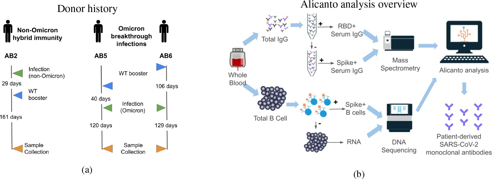
Figure 1. (A) Schedule of vaccination and infections for three donors prior to blood collection for this study. Blood was collected between 120 and 161 days from the most recent exposure. Donor AB2 was only exposed to wild-type vaccines and a non-Omicron natural infection. Donors AB5 and AB6 received wild-type vaccines and suspected Omicron natural infections. (B) Overview of antibody and B cell processing, data generation, and analysis. Total B cells and plasma were collected from peripheral blood. RBD- and spike-reactive antibodies were purified from plasma and analyzed by tandem mass spectrometry. Spike-reactive B cells were enriched from total B cells and processed for single B-cell V(D)J transcript sequencing. Alicanto software was used to analyze the mass spectrometry data and next-generation sequencing data to identify a set of patient-derived antibody candidates.
2 Materials and methods
2.1 Sample collection and processing
Samples were collected from three donors who had been vaccinated and infected with SARS-CoV-2. Whole blood was drawn by venipuncture from each donor between 120 and 161 days after exposure. In total, 150mL to 200mL of blood was collected from each donor and stored in Vacutainer tubes with EDTA (BD #366643). The study design was approved and materials were collected following IRB protocol #22-ABTE-101, and was reviewed by Pearl Pathways, LLC.
2.2 B cell enrichment and paired VH : VL sequencing
On the same day as collection, B cells were enriched using the RosetteSep Human B Cell Cocktail (StemCell #15064). The SepMate protocol was followed using Lymphoprep density gradient medium, followed by dilution with Dulbecco’s PBS with 2% FBS (StemCell #07905) as the medium in SepMate-50 tubes. Following separation by centrifugation, the top layer containing serum antibodies was extracted by serological pipette into a PETG media bottle, and the second layer containing B cells was poured out into separate collection tubes for further washing with PBS. Cells were counted by Countess 3 and then pelleted and cryopreserved in CryoStor CS10 (# 07930) buffer.
B cells were thawed at 37°C and washed twice with EasySep buffer (PBS, 2% fetal bovine serum, 1mM EDTA; StemCell). Cells were incubated with 3ug of biotinylated omicron spike trimer (Acro Biosystems # SPN-C82Ee) for 15 min before proceeding with the isolation (StemCell Human Biotin Positive Selection Kit #17663). Isolated cells were resuspended in EasySep buffer and loaded onto a 10X Chromium Next Gem Chip K. BCR libraries were constructed using the Chromium Next GEM Single Cell 5’ v2 V(D)J kit on a 10X Genomics Chromium Controller following the user guide protocol (11).
Antigen-specific BCR enrichment can miss B cells with low BCR expression and will not capture B cells secreting antigen-specific antibodies. Cells remaining after isolation, i.e., non-enriched cells, were used to generate a bulk IgG transcript repertoire. Bulk repertoires provide greater sensitivity and depth but at the cost of pairing heavy and light chain sequences. The non-enriched cells were added to 2mL of lysis buffer and 500uL was used for RNA extraction following the Zymo Quick RNA protocol (27). Extracted RNA was quantified by spectrophotometry (VarioSkan uDrop Duo plate). Bulk IgG transcript libraries were constructed using a SMARTer Human BCR IgG IgM H/K/L Profiling Kit (Takara #634466).
2.3 Ig and BCR transcript sequencing and repertoire construction
scBCR-seq data was generated on the Illumina HiSeq platform in a 2 x 150bp paired-end read format, generating 380M reads with ≈128M per donor. Data was analyzed using CellRanger 7.0.1 in VDJ mode. The consensus sequences in fasta file format were then annotated using an in-house pipeline and stored in AIRR-seq format (32).
Bulk Ig transcript sequencing data was generated on the Illumina NextSeq 1000 platform in a 2 x 300bp paired-end read format. Transcript sequencing data were processed starting from FASTQ files with an in-house pipeline for trimming adaptors, pair-stitching, and filtering low-quality reads. Briefly, raw paired-end reads were filtered to remove pairs with either a read containing an N base or a mean quality value lower than 20. Reads were stitched together using prefix-suffix pairwise alignment, and the maximum quality value in the overlap portion was retained. Stitched reads were then error-corrected using an in-house variant of a Hamming graph approach, similar to (29). All reads in a dense subgraph of the Hamming graph are error-corrected by consensus, with the new clustered read ascribed abundance equal to the number of raw reads in the cluster. The cluster size is used as the RNA abundance value for the error-corrected sequence. Any clusters with an abundance of less than two are removed as they likely contain errors and cannot be corrected.
BCR and Ig transcript repertoire sequences were V(D)J annotated using alignment (3) to human functional V, D, and J gene references from IMGT (22). Complementarity-determining regions (CDRs) and somatic hypermutation events are inferred from gene alignments. An AIRR-seq file containing the constructed repertoire, V(D)J labeling, CDR identification, and mutation calling was generated for each sample. AIRR-seq files were concatenated to use as a single database in searching mass spectra to estimate false discovery rates. Transcripts that share the same CDR3 are referred to as clonal. The CDR-H3 is defined as the residue after the conserved second cysteine in the V region and residue preceding the J region tryptophan prior to the conserved WGXG motif, which is equivalent to IMGT unique Lefranc numbering (21).
2.4 IgG and antigen-enrichment from plasma
IgG antibody was purified from 10mL of plasma from each donor by affinity purification with Protein G. Briefly, plasma was diluted to 20mL in PBS buffer and passed through a 0.2-micron filter. Filtrate was then loaded onto a 5mL HiTrap Protein G HP column (Cytiva #17040501), washed with 25mL of PBS, and eluted with 15mL of DEA at pH 11. The HiTrap column was regenerated with washes of 30mL DEA and 30mL PBS, and re-used for each sample.
IgG antibody was fractionated for affinity to SARS-CoV-2 Omicron (B.1.1.529) RBD (Acro #SPD-C522e) or (Acro #SPN-C5224) spike trimer. Affinity resin for purification was prepared by conjugating 0.5mL NHS agarose resin with 300ug of RBD or spike at 4°C overnight. Resin was split into three columns, one for each donor.
Two sets of purifications were performed using 6mL and 10mL of IgG from each donor (see Supplementary Figure 1 for a flow diagram). For the first set, 6mL of IgG from each donor was loaded onto RBD columns, and incubated for 30min at room temperature (RT). The flow-through was collected and the column was washed with 6mL PBS, and eluted with 0.25mL DEA, pH 11 in four fractions, and neutralized with 25uL 1M Tris pH 7. The flow-through was purified one more time to deplete anti-RBD IgGs with their respective columns. The flow-through was then loaded onto columns with coupled spike trimer, and purified as above, except spike+ IgG which was eluted in one fraction with 0.75mL DEA, pH 11. For the second purification set, 10mL of IgG from each donor was loaded onto regenerated RBD columns. The RBD-depleted flow-through was then loaded onto spike columns, and eluted in one fraction with 0.75mL DEA, pH 11.
In total, four fractions were analyzed per patient, two 6mL RBD+ IgG fractions, and 6mL and 10mL RBD-depleted spike+ IgGs. The quantity and purity of IgG were estimated using absorbance at A280 and SDS-PAGE.
2.5 Bottom-up mass spectrometry analysis
Antigen-purified IgG from each donor were separated into heavy and light chain by SDS-PAGE, and digested into peptides by four different proteases for tandem mass spectrometry (MS/MS) analysis. Briefly, IgG samples were reduced with NuPAGE Sample Reducing Agent (#NP0009 Thermo), and split into four wells with 5ug-10ug per lane of 10% Bis-Tris agarose gels (NuPAGE #NP0306BOX Thermo). Gels were run in MOPS buffer, and heavy and light chains were visualized with SimplyBlue SafeStain (#LC6060 Thermo). Heavy chain bands were cut from the gel, and sliced into approximately 1mm squares. Gel pieces were destained, reduced, alkylated, and normalized to digestion buffers. Briefly, gel pieces were washed with 500uL of 50% acetonitrile (ACN), dried, and resuspended in 300uL of 100mM ammonium bicarbonate (ABC) with 10mM dithiothreitol (DTT). The reduction of disulfide bonds was carried out for 30 min at 50°C. Iodoacetic acid (IAA) was added for a final concentration of 60mM, and incubation for 30 min at RT in the dark. The buffer containing ABC, DTT, and IAA was then removed, and the gel pieces were washed twice using 50% ACN, with a final dry-down by speed-vac for 15 min. The dried gel pieces were then prepared for digestion by chymotrypsin, elastase, pepsin, and trypsin by resuspending in 300uL of their respective protease buffers: 100mM Tris-HCl 10mM CaCl2, pH 8.0 for chymotrypsin; 50mM Tris-HCl, pH 9.0 for elastase; 100mM ABC, pH 1.5 for pepsin; and 100mM ABC, pH 8.5 for trypsin. Furthermore, 1 ug of each protease was added per digest, and the samples were incubated overnight (16 to 18 hours) at 37°C. Proteases were inactivated with 1% formic acid. Peptides were extracted from gel pieces with resuspension in 120 uL of 50% ACN, and 5% formic acid buffer, incubated for 45min at RT and sonicated for 5min, and extracted peptides were transferred to new glass vials. Extraction was repeated once more, and peptides were dried in a speed-vac and resuspended in 300uL of 100mM ABC, 0.5% trifluoroacetic acid (TFA). Peptide digests were further desalted with C18 solid-phase extraction (Empore 96-well #6030SD CDS Analytical). Eluted peptide samples were dried down and resuspended in 50uL of 2% ACN 0.1% formic acid, incubated for 5 min at RT, and then transferred to mass spec vials for analysis; injection volumes of 10 to 20 uL (corresponding to 20-40% of the sample digests) were used.
Peptide sample digests were analyzed using an Aurora Ultimate Column (1.7um, C18, 25cm x 75um ID #AUR3-15075C18 IonOpticks) on a Dionex Ultimate 3000 HPLC coupled to an Orbitrap Eclipse mass spectrometer. Buffer A was 2% ACN and 0.1% formic acid in Optima-grade water and Buffer B was 80% ACN and 0.1% formic acid in Optima-grade water. Peptides were eluted using the following 2-hour gradient (percent Buffer B) at 0.3 uL/min: 10% to 28% (100 min.), 28% to 55% (20 min.). MS1 scans were performed with a cycle time of 3s at 60K resolution, a scan range of 300-1600 m/z, an automatic gain control (AGC) target of 4e5, and a maximum injection time of 50ms. Precursors for MS2 were selected by passing the monoisotopic precursor selection (MIPS) filter, an intensity threshold of 50K, a charge of +2 to +8, and dynamic exclusion of 60s after two scans. Fragmentation of peptide precursors at an Orbitrap resolution of 30K with quadrupole isolation and an AGC target of 4e5 took place using a hybrid high-energy collision dissociation (HCD)/electron transfer dissociation with supplemental HCD activation (EThcD) method. All the peptide precursors were fragmented by HCD, with some of the same precursors undergoing EThcD. HCD fragmentation was done using a quadrupole isolation window of 1.6 m/z, and was stepped at 25%, 35%, and 50% normalized collision energies (NCE); the scan range was set to 101-3000 m/z, and the maximum injection time set at 54 ms. EThcD was performed using a quadrupole isolation window of 3.0 m/z, with calibrated charge-dependent electron transfer dissociation (ETD) parameters and 15% NCE supplemental activation; the scan range was set to 105-3000 m/z and the maximum injection time set at 250 ms.
2.6 Clone selection with Alicanto data analysis
Alicanto is a proteogenomic platform for identifying antigen-specific antibodies present in donor serum, previously described in (2, 29) and applied in (7, 12). Briefly, Alicanto matches tandem mass spectra generated from antigen-purified antibody protein from serum to an antibody repertoire generated from RNA found in B cells. Alicanto scores each antibody clone, defined by unique amino acid CDR-H3, in the repertoire based on supporting peptide-spectrum match evidence, and counts fragmentation support for each position of the clone’s CDR-H3 sequence. Additionally, a small number of clones with high degeneracy are removed by 1) marking antibody fixed-length sub-sequences that are repetitive, and 2) requiring each selected clone’s CDR-H3 to have at least two non-repetitive sub-sequences. More explicitly, all 12-mers (subsequences of length 12) that appear in greater than 1% of clones from a donor are marked as repetitive, and each CDR-H3 must have at least two unique 12-mers.
Alicanto was applied to the RBD+ and the spike trimer+ fractions matched with the Ig and BCR repertoire from three donors. Serum clones were identified from the six Alicanto runs, and a set of 17 antibodies from BCR repertoires were selected for recombinant expression and characterization. Candidates from all fractions were evaluated including one from AB2, 12 from AB5, and four from AB6. Furthermore, 12 of the candidates were found in RBD-purified proteomic fractions and 10 were found in spike-trimer+ proteomic fractions.
Repertoire amino acid sequences were identified as public clones by matching heavy chain V-gene annotations and requiring CDR-H3s to be at least 10 amino acids and align with ≥80% match to an antibody reported in CoV-AbDab (26). For each sequence with a clone match, a single entry in AbDab was chosen to propagate characterization information by aligning heavy variable chains, and selecting the entry with greatest percent match.
2.7 Recombinant antibody expression and purification
The variable region sequences were codon-optimized, synthesized with respective heavy and light chain signal peptides, and cloned into an expression vector containing a human IgG1/Kappa for Chinese hamster ovary (CHO) cell line expression as a recombinant monoclonal antibody (mAb). Codon optimization was performed by selecting for frequent codon and codon pairs used by the CHO-K1 cell line. A large-scale plasmid preparation was performed, followed by 3mL transient expression in CHO-K1 cells. Supernatant was collected for a one-step purification using Protein G. Quantity and purity was estimated using absorbance at A280 and SDS-PAGE.
2.8 ELISAs for domain mapping and neutralization
Expressed mAbs were tested for binding to a panel of targets from three SARS-CoV-2 variants. Each antibody was tested for binding to RBD, N-terminal domain (NTD), and spike trimer from wild-type virus, Omicron B.1.1.529, and a newer variant of concern, BQ.1.
MAbs were also tested for neutralization using a SARS-CoV-2 Variant Inhibitor Screening Assay (sVNT; #VANC00, R&D Systems). Briefly, 100uL/well of His-tagged RBD from Omicron B.1.1.529 at 6ng/uL (#SPD-C522e, Acro) was captured on a plate using anti-His capture antibody for 75min at RT. mAbs were normalized to 0.46mg/mL, and incubated on the plate to capture antibody binding to the immobilized RBD. Each mAb was 3-fold diluted serially into six wells with two replicates per dilution. Next, biotinylated ACE2 was added to the plate, and incubated for 90min at RT. Plates were washed four times, followed by detection with streptavidin-HRP and colorimetric substrate. The reduction of absorbance at 450nm with increasing concentrations of mAb indicated ACE2 blocking and were identified as neutralizers in the assay.
2.9 Biolayer inteferometry
Expressed antibodies with observe binding activity were selected for biolayer inteferometry (BLI) analysis on an Octet RED96 (Sartorius). Expressed monoclonal antibody was immobilized to the AHC sensor at 5ug/mL concentration. Recombinant His-tagged RBD Omicron B.1.1.529 was used as analyte with varying concentration in 3-fold and 2-fold dilution starting at 1uM and 50nM in PBS pH7.4 buffer, and 0.02% Tween-20. Kinetic analysis was performed with 120s capture, 120s association, and 180s or 600s dissociation. Sensor regeneration was performed with 10mM Glycine pH1.5 with three 5s cycles.
3 Results
3.1 Serum proteomics detected antibodies to Omicron virus variant months after exposure
B cells from each donor were sorted for cells expressing BCRs that bind to SARS-CoV-2 Omicron spike protein, and single-cell sequenced to obtain a repertoire of paired heavy and light V(D)J antibody sequences. The number of distinct heavy chain sequences range from 17,812 to 22,817 in the BCR repertoire (see Figure 2). As expected when sampling the antigen-experienced memory B cells, the number of clones, as defined by unique amino acid CDR3s, is similar to that of distinct sequences in the repertoire. On the other hand, the Ig repertoire, which is derived from bulk B cells and selected for IgG class transcripts, has a lower clone to sequence ratio and 33%-46% of sequences have somatic mutations differences from other sequences of the same clones. The Ig and BCR repertoires have distinct clones, with only 0.5%-2.0% of clones in common between the repertoires.
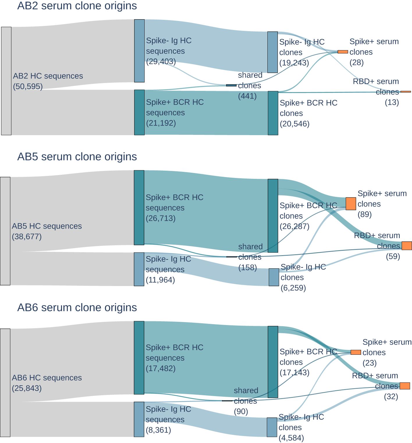
Figure 2. Sankey plots for donors AB2, AB5, and AB6 show heavy chain BCR (dark blue) and Ig (light blue) repertoire sequence counts collapsing to BCR, Ig, and shared repertoire clones (second blue boxes), and then serum selected anti-RBD and anti-spike clones (orange). Counts of sequences or clones are shown in parentheses. Few clones are shared between BCR and Ig repertoires, but are still present in serum.
The population of B cells and circulating antibodies in peripheral whole blood reflect the dynamics of the immune response. Even though BCRs and antibodies were both enriched for specificity to spike protein, 99.4%-99.8% of BCR repertoire clones are not observed as circulating IgG antibodies in serum. The highest number of clones in serum are from donors AB5 and AB6, both of whom were suspected to be exposed to an Omicron variant through natural infection. Despite having no exposure to the Omicron antigens, either by vaccination or natural infection, donor AB2 still had serum antibodies that are reactive to Omicron variant RBD and spike.
3.2 Serum IgG appeared in BCR repertoire as non-class switched IgM
The BCR repertoire is primarily dominated by IgM class clones with fewer than 3 amino acid mutations from a germline V-gene, and a low somatic hypermutation count (low SHM) is a signature of naive B cells (20). While the serum antibodies analyzed are all IgG as a result of Protein G purification prior to antigen purification, they can map to sequences of a different antibody class in the BCR repertoire due to the same B-cell clone having expanded into differentiated and class-switched cells. In the AB5 BCR repertoire, 84% of repertoire clones were IgMs compared to 6% being IgG (see Figure 3). Of the AB5 clones in serum, 67% of spike+ and 47% of RBD+ IgG clones were IgM class in the BCR repertoire, and most of these clones had low SHM. The Ig repertoire, which is focused on B cells expressing IgG class transcripts, is predominantly composed of IgG1 clones with 3 or more amino acid mutations(high SHM), which is a signature of affinity maturation. All spike+ and RBD+ serum clones originating from IgG1 clones in the AB5 donor Ig repertoire had high SHM. The same trend is observed with serum clones in donors AB2 and AB6 (data not shown). The observation of BCR IgM clones as circulating IgG clones in serum shows the clones have undergone class-switching from IgM to IgG.
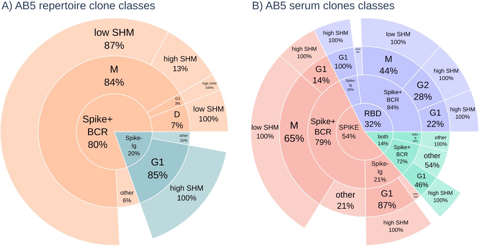
Figure 3. (A) The class distribution of all sequences in the repertoire for donor AB5. The majority of antibodies in the BCR repertoire (orange) were IgM class with most sequences having low somatic hypermutation count (low SHM) with < 3a.a.mutations in the V-region. The Ig repertoire (blue) are high mutation (high SHM) count IgG class sequences. (B) The antibody class distribution of serum clones (RBD in blue, spike in red, and both in green) shows enrichment of IgG class antibodies, but low mutation count IgM class antibodies from the BCR repertoire are still predominant.
3.3 Characterization of serum antibodies found diverse and high-affinity binding to SARS-CoV-2 domains and variants
Of the many antibodies observed in the BCR and Ig repertoires, antibodies circulating in serum are actively responding to infection. We selected 17 high SHM antibodies across the donors to express recombinantly as mAbs for further characterization (see Supplementary Data Sheet 1 for VH and VL sequences, and annotations). First, we tested reactivity in ELISA to Spike trimer, RBD, and NTD for the SARS-CoV-2 wild-type (WT) and Omicron subvariant B.1.1.529. To evaluate the potential of the antibodies to neutralize emerging variants, we further tested the antibodies in ELISA against the spike timer and RBD from Omicron subvariant BQ.1.1.
Two antibodies (AB5-5 and AB5-12) from AB5 show binding to the spike protein, but not to any subdomain. Another two antibodies (AB5-4 and AB5-7) from the same donor have binding to both the spike protein and the NTD across different Omicron variants (see Figure 4).
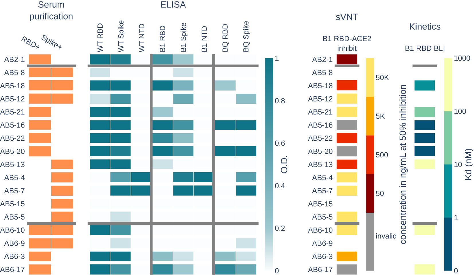
Figure 4. Serum RBD+ and spike+ clones from the BCR repertoire were selected and recombinantly expressed and tested as mAbs. Panels show the properties of each mAb. The serum purification panel shows which antigen purification the mAb is present in. ELISA panel shows binding activity as measured by optical density (O.D.) of each mAb to three SARS-CoV-2 variants and their RBD, spike, or NTD domains. The surrogate viral neutralization test (sVNT) shows the concentration of mAb needed to inhibit 50% of B.1.1.529 RBD-ACE2 interactions. The kinetics panel shows the dissociation constant (Kd) of eight mAbs tested (non-white) for B.1.1.529 RBD binding as measured by biolayer inteferometry (BLI).
Eleven antibodies show cross-reactivity to Omicron B.1.1.529, including antibody AB2-1 from donor AB2 who had never been exposed to an Omicron variant of the virus. In addition, seven antibodies show cross-reactivity to Omicron subvariant BQ.1.1, which evolved after the peripheral blood was collected for this study.
We assayed the antibodies using surrogate viral neutralization testing (sVNT). Specifically, a competition ELISA was performed where B.1.1.529 RBD was immobilized, and each mAb was bound to the RBD at concentrations spanning 50,000 ng/mL to 5ng/mL. The complexes were then incubated with ACE2 and washed to detect ACE2 binding to RBD. For each mAb, we tracked the lowest concentrations at which the mAb depleted 50% of the signal, which indicates that 50% of ACE2 was blocked from binding RBD. Six antibodies show potential for neutralization with 50% blocking at a concentration <5,000 ng/mL. Antibodies AB5-16, AB5-20, and AB6-17 result in inconclusive inhibition values with increasing ACE2 binding at increasing mAb concentrations, but show no binding to ACE2 in ELISA (data not shown). Strangely, these antibodies show binding activity to RBD and spike for all three SARS-CoV-2 variants, with strong affinity to B.1.1.529 RBD.
For antibodies with ELISA activity to RBD from donors AB5 and AB6, we measured the binding kinetics to B.1.1.529 RBD using biolayer interferometry. Antibodies AB5-13, AB6-10, and AB6-17 have dissociation constants (Kd) ranging from 171nM to 723nM, and antibodies AB5-18 and AB5-21 had low Kd of 3.4nM and 30nM, respectively. Antibodies AB5-16, AB5-20, and AB5-22 showed no dissociation from RBD, which is an indicator for sub-nanomolar affinity antibodies that are below the detection limits of the instrument.
3.4 Serum antibodies showed similarity to previously known public clones
Clones that are highly similar across donors, in terms of originating heavy chain germline V-gene and CDR3 sequences, suggest convergent immune responses and are referred to as public clones. Public clones of SARS-CoV-2 have been previously reported (26) and known anti-SARS-CoV-2 antibodies have been collected in the coronavirus antibody database (CoV-AbDab). We compared our Ig and BCR repertoires and serum-selected antibodies to previously reported sequences and found the characterization data provided in CoV-AbDab to agree with that of similar mAbs in our list of 17. For example, AB5-18, which binds to RBD and neutralizes B.1.1.529 matches antibody BD56-411 with 83% CDR-H3 similarity and 91% HC similarity (see Figure 5 and detailed alignments in Supplementary Table 1). BD56-411 is reported to bind RBD Omicron, including neutralization of subvariants B.1.1.529 to BA.2. All the serum clones with public clone matches originate from the BCR repertoire. Additionally, 76, 95, and 38 public clones in the BCR and Ig repertoires for donors AB2, AB5, and AB6, respectively, were not observed in serum. This provides alternate confirmation that the serological compartment retains a narrow population of antibodies that are distinct from the cellular repertoire.

Figure 5. Propagation of activity profiles from public CoV-AbDab sequences to sequences observed in donors AB5 and AB6. Each column shows a source of the donor-derived sequence, observed in serum binding to RBD and SPIKE, RBD-only, SPIKE-only, or present in BCR or Ig repertoire. The similarity of each donor-derived sequence is shown by the percentage CDR-H3 match on the y-axis and the percentage HC match on the x-axis to a CoV-AbDab sequence. Markers are colored by the most recent variant binding and styled as neutralizing (diamond) or non-neutralizing (circle) as reported in Cov-AbDab. Recombinant mAbs AB5-13, AB5-18, AB5-20, and AB6-10 matched public clones, and our characterization data showed the same activity profiles as reported public sequences.
4 Discussion
Of the more than 100,000 clones in the BCR and Ig repertoire across the three donors in this study, less than <0.1% are represented as anti-SARS-CoV-2 Omicron variant antibodies. The B-cell repertoire in peripheral blood is a weak proxy for representing the dynamics of active antibodies circulating in serum, as shown by other proteogenomic studies (12, 17, 31). In adult healthy human serum, approximately 75% of secreted antibodies are IgG class by mass (13). Conversely, the cellular repertoire accessible in peripheral blood is primarily mature-naive B cells and memory B cells expressing surface IgM. Predictably, a large proportion of serum-observed IgG clones are of class IgM in the BCR repertoire (46% to 76% between donors), which suggests that these clone lineages are undergoing expansion, accruing mutations, and are present in the donors with multiple cell types. Our study design did not specifically characterize plasma cells, which secrete massive amounts of antibodies, but are in very low cell counts in peripheral blood (<2% of B cell lymphocytes) (6). Plasma cells observed circulating in the blood are hypothesized to be in transit to other lymphoid organs, where their lifespan is dictated by exposure to environmental survival factors: short-lived plasma cells lasting for 3 to 5 days in lymph nodes and the spleen, and long-lived plasma cells lasting for many months in bone marrow (6, 23). Further experimentation is needed to show that anti-SARS-CoV-2 IgG in serum may originate from plasmablasts or plasma cells circulating in the blood many months after virus antigen exposure.
Peripheral blood was collected from each donor at least 4 months after their self-reported exposure to viral antigens through vaccination or infection to uncover a significant proportion of B cells and circulating antibodies still responding to SARS-CoV-2 antigens with our Alicanto platform. Two donors were exposed to an early Omicron variant by breakthrough infection and had significantly more spike+ (Omicron variant B.1.1.529) B cells and circulating antibodies matching RBD+ and spike+ clones than the donor who was only exposed to SARS-CoV-2 wild-type antigens via hybrid immunity infection followed by vaccination. Previous proteogenomic studies (33, 35) reported that the imprinting effects of natural SARS-CoV-2 infection versus vaccination directs the immune response to divergent epitopes, where vaccinated individuals have more circulating clones directed to RBD than non-RBD spike proteins. We did not find a compelling trend in our data to support the previous finding. The donor with initial exposure by natural infection had only slightly more spike+ serum clones than RBD+ and the donors who were vaccinated prior to infection had insignificant differences in RBD+ and spike+ serum clones counts. The difference could be explained by the effects of imprinting and proportions of epitope focus in serum may diminish over time since viral exposure.
Using our Alicanto platform, we selected the highly confident serum clones observed in the BCR repertoire to recombinantly express for further validation and characterization. Of the candidates selected, we found diverse binding modalities to the RBD, NTD, and non-RBD/NTD of the spike protein. Candidates from the donors exposed to an early Omicron variant were observed to be cross-reactive to the wild-type strain and other Omicron variants, including BQ.1.1, an escape variant discovered many months later. Two candidates from a donor with breakthrough infection bound to the tested RBD Omicron variants, but the dissociation kinetics were too slow to be reliably measured by BLI. These candidates are likely strongly neutralizing with sub-nanomolar dissociation constants.
To further characterize the clones observed in the BCR, Ig, and serum repertoires, we leveraged sequences and annotations in CoV-AbDab (26) to investigate convergent immune responses. Across the different donors, 209 clones matched to publicly reported sequences, i.e., public clones, with seven clones present in serum. Four of the serum clones had been recombinantly expressed and have strong agreement with neutralization activity and variant binding to the reported public clones. Public clones are also present in the BCR repertoire, corroborating evidence that the antigen-specific antibodies circulating in serum represent a distinct dynamic from the antigen-specific B cell repertoires.
Previous findings of SARS-CoV-2 reactive B cells have primarily been IgM class with few somatic mutations (15). Likewise, our spike+ BCR repertoires have fewer somatic mutations compared to the Ig repertoire. Circulating IgG in serum is a product of affinity maturation, and clones that match the Ig repertoire are all somatically mutated in the repertoire (>3 amino acid mutations). In contrast, most of the clones matching the BCR repertoire had low mutation counts. This suggests that the circulating antibodies in serum that match the BCR repertoire likely contain missed somatic mutations and are a potential boon for naturally evolved and higher affinity anti-SARS-CoV-2 antibodies. A limitation of the proteogenomic method is that the serum clone needs representation in the BCR repertoire to be observed and tested as a recombinant monoclonal. A promising direction is to directly sequence antibodies de novo without a repertoire. Existing efforts have involved a significant amount of sample preparation and mass spectrometry instrument time and labor but show potential for deriving a functioning antibody sequence from IgG protein alone (1, 14). Continual development of mass spectrometry and informatic methods is necessary for an in-depth characterization of the multitude of antibodies circulating in serum.
Data availability statement
The data presented in the study are deposited in the zenodo.org repository, accession number doi:10.5281/zenodo.14835053.
Ethics statement
IRB protocol #22-ABTE-101 was reviewed by Pearl Pathways, LLC. The studies were conducted in accordance with the local legislation and institutional requirements. The participants provided their written informed consent to participate in this study.
Author contributions
AP: Conceptualization, Data curation, Formal analysis, Funding acquisition, Investigation, Methodology, Project administration, Software, Supervision, Validation, Visualization, Writing – original draft, Writing – review & editing. TL: Conceptualization, Investigation, Methodology, Project administration, Resources, Supervision, Validation, Writing – review & editing. RC: Conceptualization, Data curation, Formal analysis, Investigation, Methodology, Writing – review & editing. QH: Data curation, Investigation, Writing – review & editing. SB: Conceptualization, Funding acquisition, Investigation, Resources, Software, Writing – review & editing. NC: Conceptualization, Data curation, Funding acquisition, Investigation, Methodology, Project administration, Resources, Software, Supervision, Visualization, Writing – original draft, Writing – review & editing.
Funding
The author(s) declare financial support was received for the research, authorship, and/or publication of this article. Research reported in this publication was supported by NIGMS of the National Institutes of Health under award number R44GM125485 and R44GM140607.
Acknowledgments
We thank Miin Lin for contributions in reviewing and editing the manuscript.
Conflict of interest
Authors AP, TL, RC, QH, SB, and NC were employed by the company Abterra Biosciences, Inc.
Generative AI statement
The author(s) declare that no Generative AI was used in the creation of this manuscript.
Publisher’s note
All claims expressed in this article are solely those of the authors and do not necessarily represent those of their affiliated organizations, or those of the publisher, the editors and the reviewers. Any product that may be evaluated in this article, or claim that may be made by its manufacturer, is not guaranteed or endorsed by the publisher.
Supplementary material
The Supplementary Material for this article can be found online at: https://www.frontiersin.org/articles/10.3389/fimmu.2025.1509888/full#supplementary-material
References
1. Bondt A, Hoek M, Tamara S, de Graaf B, Peng W, Schulte D, et al. Human plasma IgG1 repertoires are simple, unique, and dynamic. Cell Syst. (2021) 12:1131–43. doi: 10.1016/j.cels.2021.08.008
2. Bonissone SR, Lima T, Harris K, Davison L, Avanzino B, Trinklein N, et al. Serum proteomics expands on identifying more high-affinity antibodies in immunized rabbits than deep b-cell repertoire sequencing alone. bioRxiv. (2019). doi: 10.1101/833871
3. Bonissone SR, Pevzner PA. Immunoglobulin classification using the colored antibody graph. J Comput Biol. (2016) 23:483–94. doi: 10.1089/cmb.2016.0010
4. Boutz DR, Horton AP, Wine Y, Lavinder JJ, Georgiou G, Marcotte EM. Proteomic identification of monoclonal antibodies from serum. Analytical Chem. (2014) 86:4758–66. doi: 10.1021/ac4037679
5. Buck AM, Deitchman AN, Takahashi S, Lu S, Goldberg SA, Bodansky A, et al. The breadth of the neutralizing antibody response to original SARS-CoV-2 infection is linked to the presence of Long COVID symptoms. J Med Virol. (2023) 95:e29216. doi: 10.1002/jmv.v95.11
6. Caraux A, Klein B, Paiva B, Bret C, Schmitz A, Fuhler GM, et al. Circulating human b and plasma cells. Age-associated changes in counts and detailed characterization of circulating normal cd138- and cd138+ plasma cells. Haematologica. (2010) 95:1016–20. doi: 10.3324/haematol.2009.018689
7. Chen EC, Gilchuk P, Zost SJ, Ilinykh PA, Binshtein E, Huang K, et al. Systematic analysis of human antibody response to ebolavirus glycoprotein shows high prevalence of neutralizing public clonotypes. Cell Rep. (2023) 42:112370. doi: 10.1016/j.celrep.2023.112370
8. Cheung WC, Beausoleil SA, Zhang X, Sato S, Schieferl SM, Wieler JS, et al. A proteomics approach for the identification and cloning of monoclonal antibodies from serum. Nat Biotechnol. (2012) 30:447–52. doi: 10.1038/nbt.2167
9. Cox M, Peacock TP, Harvey WT, Hughes J, Wright DW, Willett BJ, et al. SARS-CoV-2 variant evasion of monoclonal antibodies based on in vitro studies. Nat Rev Microbiol. (2023) 21:112–24. doi: 10.1038/s41579-022-00809-7
10. Davis HE, McCorkell L, Vogel JM, Topol EJ. Long COVID: major findings, mechanisms and recommendations. Nat Rev Microbiol. (2023) 21:408. doi: 10.1038/s41579-023-00896-0
11. 10X Genomics. Chromium gem-x single cell 5’ v3 gene expression user guide . Available online at: https://www.10xgenomics.com/support/single-cell-immune-profiling/documentation/steps/libraryprep/chromium-gem-x-single-cell-5-v3-gene-expression-user-guide (Accessed November 20, 2019).
12. Gilchuk P, Guthals A, Bonissone SR, Shaw JB, Ilinykh PA, Huang K, et al. Proteo-genomic analysis identifies two major sites of vulnerability on ebolavirus glycoprotein for neutralizing antibodies in convalescent human plasma. Front Immunol. (2021) 12:706757. doi: 10.3389/fimmu.2021.706757
13. Gonzalez-Quintela A, Alende R, Gude F, Campos J, Rey J, Meijide LM, et al. Serum levels of immunoglobulins (igg, iga, igm) in a general adult population and their relationship with alcohol consumption, smoking and common metabolic abnormalities. Clin Exp Immunol. (2007) 151:42–50. doi: 10.1111/j.1365-2249.2007.03545.x
14. Guthals A, Gan Y, Murray L, Chen Y, Stinson J, Nakamura G, et al. De novo MS/MS sequencing of native human antibodies. J Proteome Res. (2017) 16:45–54. doi: 10.1021/acs.jproteome.6b00608
15. Hurtado J, Rogers TF, Jaffe DB, Adams BA, Bangaru S, Garcia E, et al. Deep repertoire mining uncovers ultra-broad coronavirus neutralizing antibodies targeting multiple spike epitopes. Cell Rep. (2024) 43:114307. doi: 10.1016/j.celrep.2024.114307
16. Isho B, Abe KT, Zuo M, Jamal AJ, Rathod B, Wang JH, et al. Persistence of serum and saliva antibody responses to sars-cov-2 spike antigens in covid-19 patients. Sci Immunol. (2020) 5:10. doi: 10.1126/sciimmunol.abe5511
17. Lavinder JJ, Horton AP, Georgiou G, Ippolito GC. Next-generation sequencing and protein mass spectrometry for the comprehensive analysis of human cellular and serum antibody repertoires. Curr Opin Chem Biol. (2015) 24:112–20. doi: 10.1016/j.cbpa.2014.11.007
18. Lavinder JJ, Wine Y, Giesecke C, Ippolito GC, Horton AP, Lungu OI, et al. Identification and characterization of the constituent human serum antibodies elicited by vaccination. Proc Natl Acad Sci. (2014) 111:2259–64. doi: 10.1073/pnas.1317793111
19. Le Quy K, Chernigovskaya M, Stensland M, Singh S, Leem J, Revale S, et al. Benchmarking and integrating human B-cell receptor genomic and antibody proteomic profiling. NPJ Syst Biol Appl. (2024) 10:73. doi: 10.1038/s41540-024-00402-z
20. LeBien TW, Tedder TF. B lymphocytes: how they develop and function. Blood. (2008) 112:1570–80. doi: 10.1182/blood-2008-02-078071
21. Lefranc MP. Unique database numbering system for immunogenetic analysis. Immunol Today. (1997) 18:509. doi: 10.1016/S0167-5699(97)01163-8
22. Lefranc M-P, Giudicelli V, Duroux P, Jabado-Michaloud J, Folch G, Aouinti S, et al. Imgt®, the international immunogenetics information system® 25 years on. Nucleic Acids Res. (2015) 43:D413–22. doi: 10.1093/nar/gku1056
23. Mei HE, Yoshida T, Sime W, Hiepe F, Thiele K, Manz RA, et al. Blood-borne human plasma cells in steady state are derived from mucosal immune responses. Blood. (2009) 113:2461–9. doi: 10.1182/blood-2008-04-153544
24. García-Abellán J, Fernández M, Padilla S, García JA, Agulló V, Lozano V, et al. Immunologic phenotype of patients with long-COVID syndrome of 1-year duration. Front Immunol. (2022) 13:920627. doi: 10.3389/fimmu.2022.920627
25. Pinto D, Park YJ, Beltramello M, Walls AC, Tortorici MA, Bianchi S, et al. Cross-neutralization of SARS-CoV-2 by a human monoclonal SARS-CoV antibody. Nature. (2020) 583:290–5. doi: 10.1038/s41586-020-2349-y
26. Raybould MIJ, Kovaltsuk A, Marks C, Deane CM. CoV-AbDab: the coronavirus antibody database. Bioinformatics. (2021) 37:734–5. doi: 10.1093/bioinformatics/btaa739
27. Zymo Research. Quick-RNA Miniprep Kit. Available at: https://files.zymoresearch.com/protocols/_r1054_r1055_quick-rna_miniprep_kit.pdf (Accessed July 1, 2019)
28. Rydyznski Moderbacher C, Ramirez SI, Dan JM, Grifoni A, Hastie KM, Weiskopf D, et al. Antigen-specific adaptive immunity to SARS-CoV-2 in acute COVID-19 and associations with age and disease severity. Cell. (2020) 183:996–1012. doi: 10.1016/j.cell.2020.09.038
29. Safonova Y, Bonissone S, Kurpilyansky E, Starostina E, Lapidus A, Stinson J, et al. Igrepertoireconstructor: a novel algorithm for antibody repertoire construction and immunoproteogenomics analysis. Bioinformatics. (2015) 31:i53–61. doi: 10.1093/bioinformatics/btv238
30. Sakurada Y, Sunada N, Honda H, Tokumasu K, Otsuka Y, Nakano Y, et al. Serial changes of long COVID symptoms and clinical utility of serum antibody titers for evaluation of long COVID. J Clin Med. (2022) 11(5):1309. doi: 10.3390/jcm11051309
31. Tomescu-Baciu A, Johansen JN, Holmøy T, Greiff V, Stensland M, de Souza GA, et al. Persistence of intrathecal oligoclonal B cells and IgG in multiple sclerosis. J Neuroimmunol. (2019) 333:576966. doi: 10.1016/j.jneuroim.2019.576966
32. Vander Heiden JA, Marquez S, Marthandan N, Bukhari SAC, Busse CE, Corrie B, et al. AIRR community standardized representations for annotated immune repertoires. Front Immunol. (2018) 9:2206. doi: 10.3389/fimmu.2018.02206
33. Voss W, Huang Y, Marchioni J, Seeger A, Paresi C, Kain J, et al. Differential serological immune imprinting following sars-cov-2 infection, vaccination, and breakthrough infection. J Immunol. (2022) 208:112–09. doi: 10.4049/jimmunol.208.Supp.112.09
34. Voss WN, Hou YJ, Johnson NV, Delidakis G, Kim JE, Javanmardi K, et al. Prevalent, protective, and convergent igg recognition of sars-cov-2 non-rbd spike epitopes. Science. (2021) 372:1108–12. doi: 10.1126/science.abg5268
35. Voss WN, Mallory MA, Byrne PO, Marchioni JM, Knudson SA, Powers JM, et al. Hybrid immunity to SARS-CoV-2 arises from serological recall of IgG antibodies distinctly imprinted by infection or vaccination. Cell Rep Med. (2024) 5:101668. doi: 10.1016/j.xcrm.2024.101668
36. Yin K, Peluso MJ, Luo X, Thomas R, Shin MG, Neidleman J, et al. Long COVID manifests with T cell dysregulation, inflammation and an uncoordinated adaptive immune response to SARS-CoV-2. Nat Immunol. (2024) 25:218–25. doi: 10.1038/s41590-023-01724-6
Keywords: SARS-CoV-2, mass spectrometry - LC-MS/MS, serum proteomics, monoclonal antibody, human-derived, next-generation sequencing (NGS), variants of concern (VOC), receptor binding domain (RBD)
Citation: Patel A, Lima T, Carson R, Huang Q, Bonissone SR and Castellana N (2025) Serum proteomics reveals high-affinity and convergent antibodies by tracking SARS-CoV-2 hybrid immunity to emerging variants of concern. Front. Immunol. 16:1509888. doi: 10.3389/fimmu.2025.1509888
Received: 11 October 2024; Accepted: 21 January 2025;
Published: 25 February 2025.
Edited by:
Anthony Rees, Consultant, Stockholm, SwedenReviewed by:
Jean-Philippe Bürckert, Ability Biologics, CanadaMatthew Raybould, University of Oxford, United Kingdom
Copyright © 2025 Patel, Lima, Carson, Huang, Bonissone and Castellana. This is an open-access article distributed under the terms of the Creative Commons Attribution License (CC BY). The use, distribution or reproduction in other forums is permitted, provided the original author(s) and the copyright owner(s) are credited and that the original publication in this journal is cited, in accordance with accepted academic practice. No use, distribution or reproduction is permitted which does not comply with these terms.
*Correspondence: Anand Patel, YW5hbmRAYWJ0ZXJyYWJpby5jb20=