 Xiang Li1
 Casey C. Heirman2
 Ashlyn G. Rickard3Gina Sotolongo4Rico Castillo3Temitayo Adanlawo3
 Jeffery I. Everitt4
 Jeffery B. Hodgin5
 Tammara L. Watts6Andrew Janowczyk7,8,9Yvonne M. Mowery3,6,10Laura Barisoni4,11*
 Kyle J. Lafata1,2,3,4,12*
- 1Department of Electrical and Computer Engineering, Duke University, Durham, NC, United States
- 2Medical Physics Graduate Program, Duke University, Durham, NC, United States
- 3Department of Radiation Oncology, Duke University, Durham, NC, United States
- 4Department of Pathology, Duke University, Durham, NC, United States
- 5Department of Pathology, University of Michigan, Ann Arbor, MI, United States
- 6Department of Head and Neck Surgery & Communication Sciences, Duke University, Durham, NC, United States
- 7Department of Biomedical Engineering, Emory University and Georgia Institute of Technology, Atlanta, GA, United States
- 8Department of Oncology, Division of Precision Oncology, Geneva University Hospitals, Geneva, Switzerland
- 9Department of Diagnostics, Division of Clinical Pathology, Geneva University Hospitals, Geneva, Switzerland
- 10Department of Radiation Oncology, UPMC Hillman Cancer Center/University of Pittsburgh, Pittsburgh, PA, United States
- 11Department of Medicine, Division of Nephrology, Duke University, Durham, NC, United States
- 12Department of Radiology, Duke University, Durham, NC, United States
Introduction: Immune dysregulation plays a major role in cancer progression. The quantification of lymphocytic spatial inflammation may enable spatial system biology, improve understanding of therapeutic resistance, and contribute to prognostic imaging biomarkers.
Methods: In this paper, we propose a knowledge-guided deep learning framework to measure the lymphocytic spatial architecture on human H&E tissue, where the fidelity of training labels is maximized through single-cell resolution image registration of H&E to IHC. We demonstrate that such an approach enables pixel-perfect ground-truth labeling of lymphocytes on H&E as measured by IHC. We then experimentally validate our technique in a genetically engineered, immune-compromised Rag2 mouse model, where Rag2 knockout mice lacking mature lymphocytes are used as a negative experimental control. Such experimental validation moves beyond the classical statistical testing of deep learning models and demonstrates feasibility of more rigorous validation strategies that integrate computational science and basic science.
Results: Using our developed approach, we automatically annotated more than 111,000 human nuclei (45,611 CD3/CD20 positive lymphocytes) on H&E images to develop our model, which achieved an AUC of 0.78 and 0.71 on internal hold-out testing data and external testing on an independent dataset, respectively. As a measure of the global spatial architecture of the lymphocytic microenvironment, the average structural similarity between predicted lymphocytic density maps and ground truth lymphocytic density maps was 0.86 ± 0.06 on testing data. On experimental mouse model validation, we measured a lymphocytic density of 96.5 ± %1% in a Rag2+/- control mouse, compared to an average of 16.2 ± %5% in Rag2-/- immune knockout mice (p<0.0001, ANOVA-test).
Discussion: These results demonstrate that CD3/CD20 positive lymphocytes can be accurately detected and characterized on H&E by deep learning and generalized across species. Collectively, these data suggest that our understanding of complex biological systems may benefit from computationally-derived spatial analysis, as well as integration of computational science and basic science.
1 Introduction
Inflammatory mechanisms and a well-regulated immune response are essential for protecting against pathogens, preventing chronic inflammatory conditions, and responding to tissue damage (1). In contrast, immune dysregulation can lead to a variety of health issues, including autoimmune diseases, chronic inflammation, susceptibility to infection, and the development and progression of cancer. Inflammatory processes mediated by B and T lymphocytes play a major role in both cancer immunity (e.g., immune surveillance, tumor infiltrating lymphocytes, stromal inflammation) and therapy (e.g., immune checkpoint inhibitors, CAR T-cell therapy, etc.). Precision and accuracy in quantifying lymphocytic inflammation are critical for diagnosis and classification of certain disease processes, enabling use of lymphocytic infiltration as a prognostic marker, and potentially developing targeted treatments that improve patient outcomes. Pathomics, i.e., high-throughput extraction and analysis of features from digital pathology images, represents a promising approach to derive a multi-scale mathematical representation of lymphocytic infiltration phenotypes for both clinical and research purposes.
Image-based quantification and characterization of lymphocytes on digitized tissue biopsies via deep learning enables spatial interrogation of the tumor immune microenvironment, thus providing a better description of the inflammatory spatial phenotype than other techniques, such as flow cytometry or RNA sequencing. For example, deep learning has been applied to count CD3 positive T lymphocytes stained by immunohistochemistry (IHC) on whole slide images (WSIs) and/or to quantify CD8 expression in prostate (2), colon (2, 3), neuroblastoma (4), gastric (5), breast (6), and lung (7) cancers. Deep learning has also been used to quantify other relevant inflammation-related protein markers on IHC such as Inducible T-cell COStimulator (ICOS), which is involved in T-cell activation and adaptive immune response (8).
Notably, IHC is expensive and often not feasible in routine pathology and exploratory retrospective research. Consequently, there is a growing need to develop deep learning algorithms that can accurately detect lymphocytes on WSIs from hematoxylin and eosin (H&E) stained tissue (i.e., the standard staining technique used to visualize tissue morphology and structure). Deep learning-derived digital staining (9) of H&E WSIs can facilitate efficient and unbiased detection and quantification of different cell types across various tissues and diseases, such as melanoma (10), breast cancer (11–13), colorectal cancer (13), and testicular cancer (14).
However, the application of deep learning to characterize the immune response on digital pathology presents several key challenges. First, a notable constraint is that these models generally require manual annotation of class labels on H&E during the training process. Obtaining accurate annotations for large numbers of lymphocytes is a labor-intensive, time-consuming process that is limited by both inter- and intra-observer variability. Second, while these models excel at pattern recognition, they lack the mechanistic understanding required to rigorously evaluate immune responses. That is, deep learning may not fully capture basic biological characteristics beyond surface-level image representation. This hinders the ability of these models to provide deeper insight into the underlying mechanisms of different immune responses, highlighting the need for complementary testing of deep learning solutions under controlled experimental conditions.
In this paper, we address these challenges via an integrated research design that combines computational (i.e., “dry lab”) techniques and experimental (i.e., “wet lab”) rigor (Figure 1). First, we developed a knowledge-guided deep learning framework to measure lymphocytes on H&E tissue, where the fidelity of training labels is maximized through single-cell resolution image registration of H&E to IHC. We demonstrate that such an approach enables pixel-perfect ground-truth labeling of lymphocytes on H&E as measured by IHC. Second, we move beyond conventional statistical testing of our deep learning model by subjecting it to rigorous testing in a genetically engineered mouse model, where Rag2 knockout mice lacking mature lymphocytes are used as a negative experimental control. Our results demonstrate that the immune microenvironment can be accurately captured on H&E by deep learning across species and tissue types, generalizing a model developed on kidney tissue from humans to various human cancers, as well as splenic and thymic tissues form mice. Collectively, these data suggest that our understanding of complex biological systems may benefit from combining data-driven insights with empirical biology.
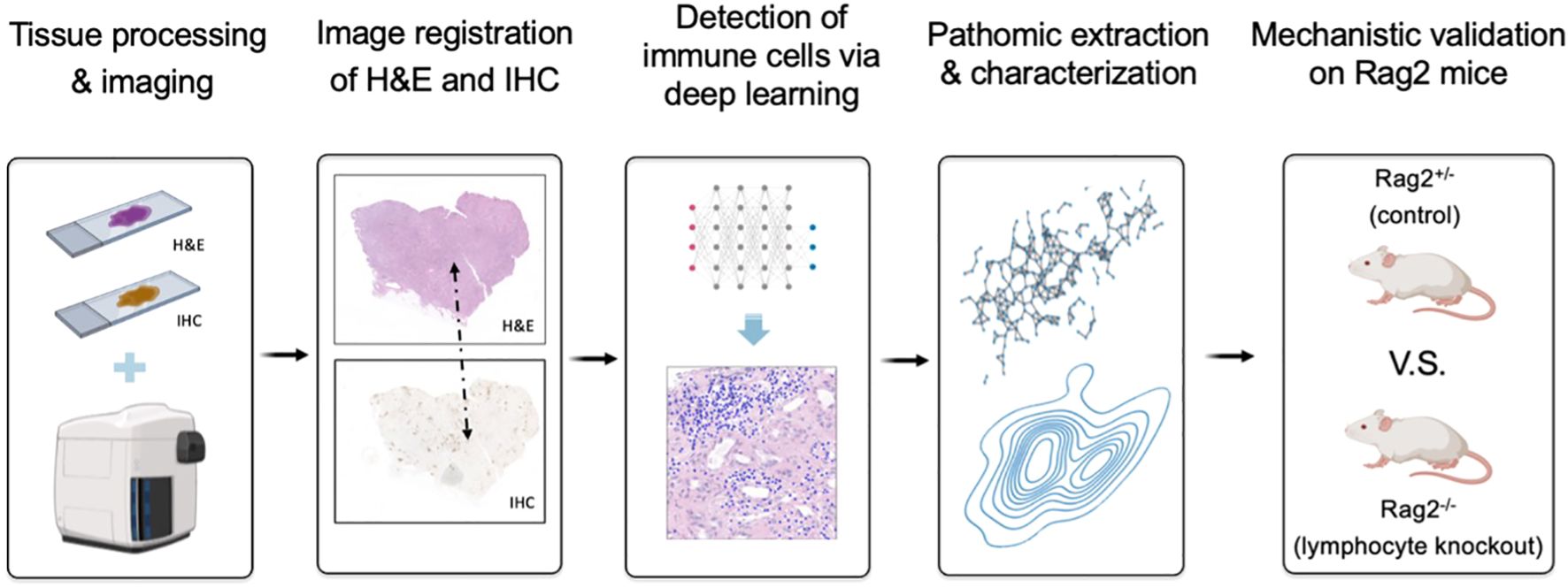
Figure 1. Overview of research design for lymphocyte deep learning model development and evaluation. We first developed a knowledge-guided deep learning framework to measure lymphocytes on H&E-stained kidney tissue, where the fidelity of training labels was maximized through single-cell resolution image registration of H&E to IHC staining with a cocktail of anti-CD3 and anti-CD20 antibodies to stain T and B lymphocytes, respectively. Following traditional statistical testing of the model based on receiver operating characteristic (ROC) curve analysis, we then characterized pattern-preserving features of the immune microenvironment based on pathomic feature extraction and calculated their error rates relative to the shift-invariant IHC antibody measurements. Finally, we performed a preclinical experiment to confirm that our deep learning model is able to identify lymphocytes in a genetically engineered mouse model, where Rag2 knockout mice without mature lymphocytes are used as a negative control.
2 Methods
2.1 Experimental tissue preparation and whole slide image acquisition
Formalin-fixed, paraffin-embedded tissue blocks from nephrectomies of patients (N=18) with kidney cancer and moderate-to-severe inflammation in renal tissue away from the cancer were obtained from the Duke University Pathology Paraffin Tissue Archives. Nephrectomies were chosen as the basis of generating scalable deep learning training examples because they often involve significant inflammation, thus providing a rich source of lymphocytes and diverse inflammatory microenvironments. Tissues were cut at 2 microns thick, stained with H&E (Figure 2A), and digitized into WSIs at 40X magnification (0.25 µm/pixel) using a Leica Aperio AT2 whole slide scanner (Figure 2B). Following H&E image acquisition, the same tissue specimens were re-stained with a CD3/CD20 IHC cocktail to detect T (CD3+) lymphocytes and B (CD20+) lymphocytes (Figure 2C). The IHC-stained specimens were then re-scanned at 40X magnification on the same Leica Aperio AT2 imaging system (Figure 2D), resulting in 2-channel, multi-contrast WSIs encoding both tissue morphology (on H&E) and tissue CD3/CD20 expression (on IHC) (Figure 2E).
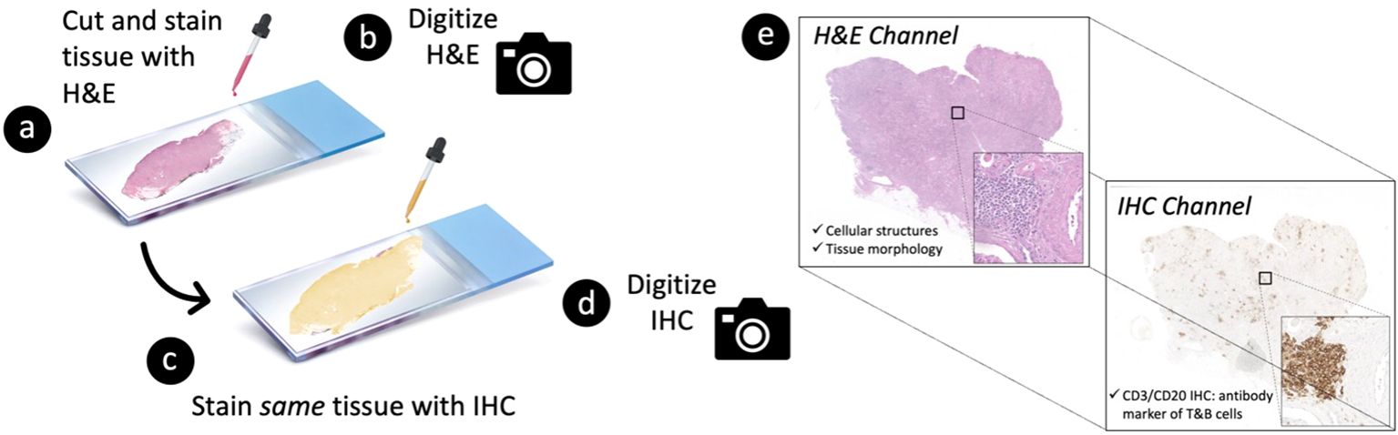
Figure 2. Tissue processing strategy to generate 2-channel, multi-contrast digital images of paired hematoxylin and eosin (H&E) vs. immunohistochemistry (IHC) stains at single-cell resolution. A 2-micron thick tissue specimen is first (A) stained with H&E and subsequently (B) digitized into a whole slide image (WSI) at 40X magnification. The specimen is then (C) re-stained with CD3/CD20 IHC (i.e., antibody markers of T and B cells, respectively) and (D) re-imaged on the same whole slide scanner. This process results in (E) a 2-channel, multi-contrast WSI encoding both (i) the morphological characteristics of the tissue on H&E and (ii) the corresponding CD3/CD20 expression profile of the tissue on IHC.
2.2 Image registration of H&E and IHC at single-cell resolution
To minimize shift artifacts, each H&E image was co-registered to its matched IHC image to map the corresponding location of CD3/CD20 antibody expression on IHC to individual nuclei on H&E. A multiscale, two-step rigid registration process (Figure 3) was implemented as follows. First, the H&E and IHC images were each downsampled to 25% of their native resolution to accommodate RAM constraints, and their center-of-mass (CoM) was calculated as a coarse tissue landmark. The IHC image was shifted to the H&E image based on a CoM alignment. Second, 1024x1024 tiles of pixels were stochastically sampled at full resolution from both H&E and IHC images at regions of high CD3/CD20 signal. The IHC tiles and H&E tiles were co-registered at single-cell resolution and stored as a tiled database for downstream deep learning model development. The accuracy of image registration was quantified based on Normalized Cross Correlation (NCC).
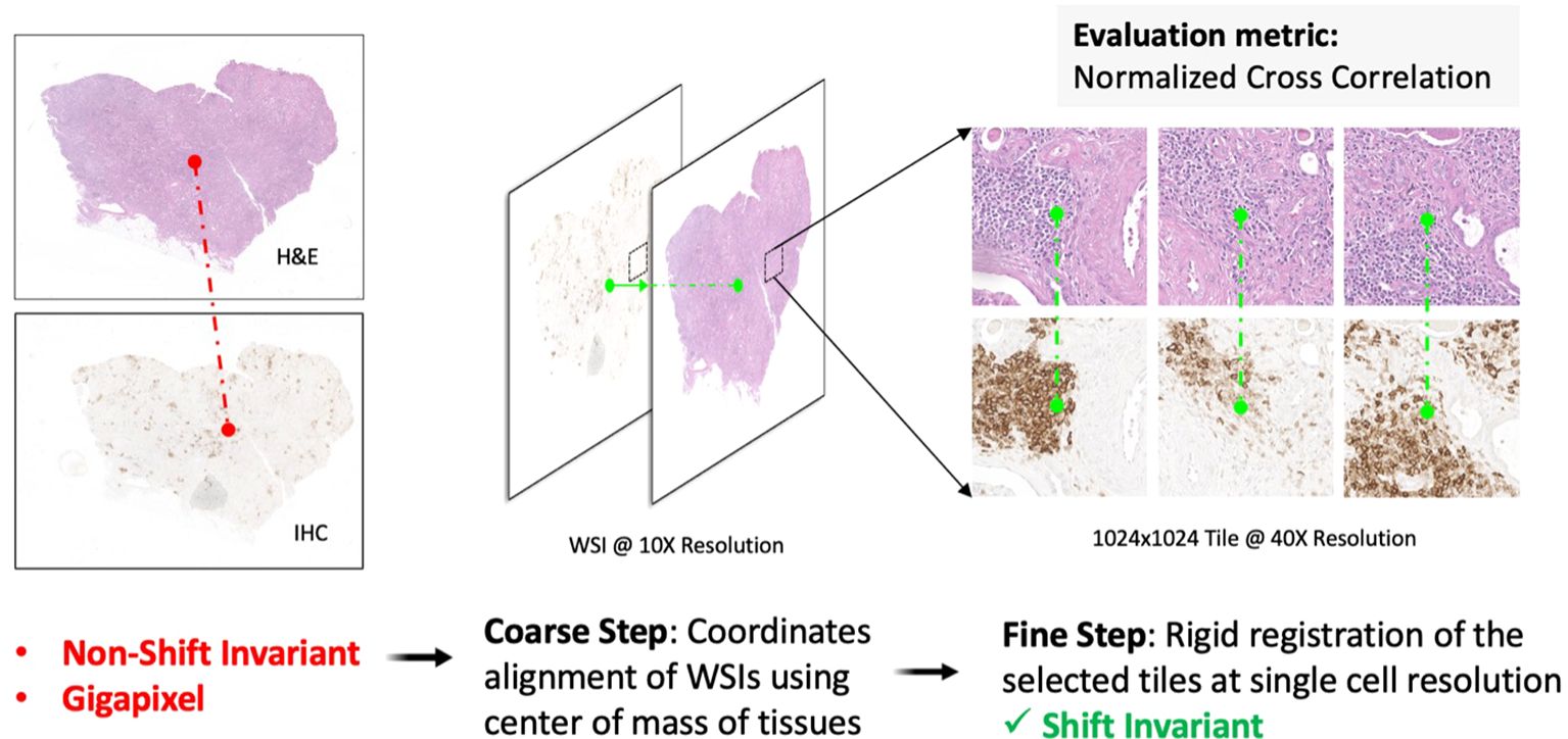
Figure 3. Multiscale H&E to IHC image registration. First, a coarse registration step provides an initial alignment of the entire tissue based on the center-of-mass shift between the H&E image and the IHC image. Second, a fine registration step aligns individual nuclei on tiled sections (1024x1024 pixels) of tissue, resulting in a one-to-one matching of nuclei across the H&E channel and the IHC channel. The 2-channel, shift-invariant tiled image data is stored for downstream deep learning model development.
2.3 Knowledge-based lymphocyte labeling to generate deep learning training data
The set of shift-invariant, co-registered H&E/IHC tile pairs (Figure 3) were used to curate single-cell deep learning training data labels. First, using a pre-trained StartDist deep learning model (15), all nuclei (irrespective of cell type) were detected and segmented on each H&E image tile (Figure 4A). Next, color deconvolution of the CD3/CD20 stain was used to isolate and threshold lymphocyte-positive regions on each IHC image tile (Figure 4B). Finally, nuclei on H&E with at least 70% of their pixels overlapping with the corresponding IHC signal were labeled as lymphocytes and denoted as the positive class label (Figure 4C). This 70% overlap threshold was empirically determined to balance precision and accuracy, as requiring 100% overlap was too restrictive given that IHC stains can leak beyond the nuclei, causing variability and potential errors. The nuclei on H&E that were non-overlapping with IHC were labeled as non-lymphocytes and denoted as the negative class label (Figure 4C).

Figure 4. Automatic lymphocyte labeling to generate deep learning training data at single-cell resolution. (A) Nuclei are first identified and segmented using a pretrained StarDist model. (B) The CD3/CD20 IHC signal (brown stain) is then thresholded and mapped onto the segmented nuclei to identify lymphocytes on H&E. (C) Deep learning training examples are curated by labeling nuclei as either lymphocytes (positive class: overlap between nuclear contour and IHC signal) or non-lymphocytes (negative class: non-overlap between nuclear contour and IHC signal).
2.4 Deep learning detection of lymphocytes on H&E based on prior-knowledge of IHC
Using the IHC-guided nuclear labels (Figure 4), a deep learning model was developed to detect lymphocytes on H&E in the absence of IHC (Figure 5). Our model was based on the HoverNet architecture (16), which is a multi-task deep learning framework to simultaneously detect, segment, and classify nuclei. The HoverNet architecture consists of one image encoding branch and three decoding branches, each with a specific task: (1) a nuclear segmentation branch, which is responsible for segmentation of nuclei; (2) a nuclear classification branch, which is responsible for classifying the segmented nuclei into types; and (3) an instance separation branch, which is responsible for separating overlapping nuclei based on distance vector fields (e.g., hover maps) from each pixel inside a nucleus to its center. By encoding directional information, this state-of-the-art architecture is designed to effectively handle the challenges of nuclear segmentation in complex tissue environments with dense regions of overlapping nuclei, such as in lymphocytic inflammation.
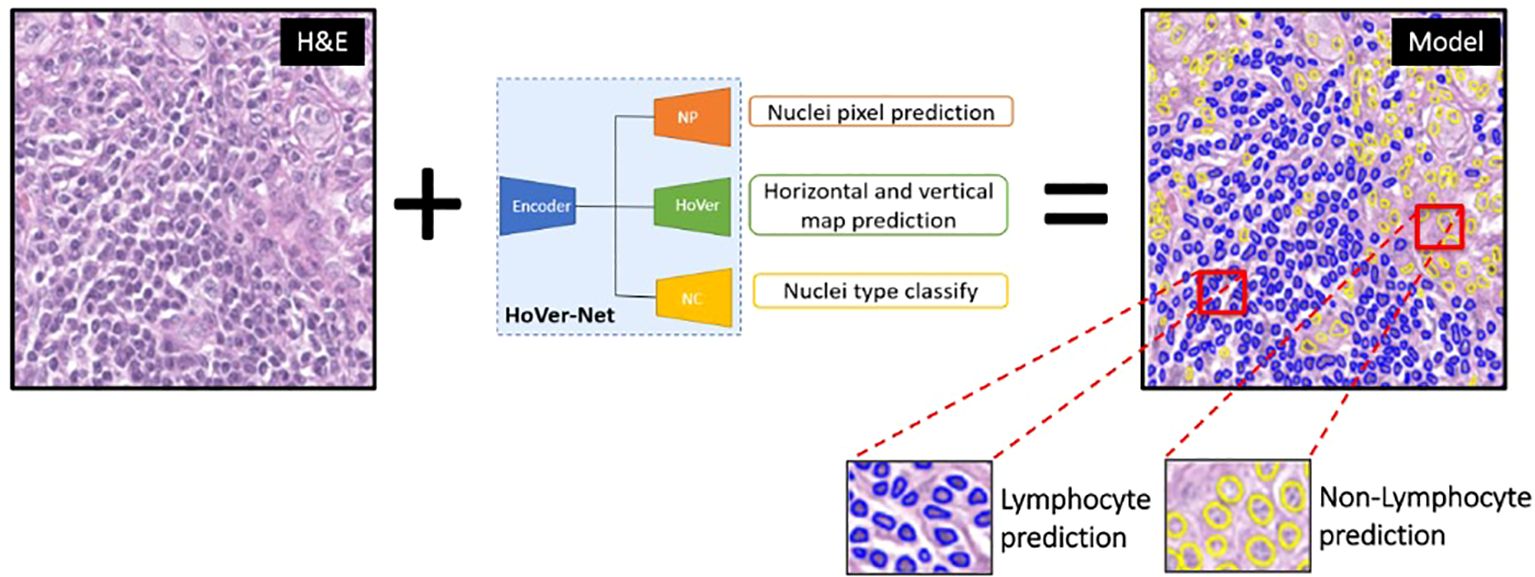
Figure 5. Hover-Net model architecture for lymphocyte identification. This multi-task deep learning framework was implemented to simultaneously detect, segment, and classify lymphocytes (blue predictions) and non-lymphocytes (yellow predictions) on H&E images.
The data were split at the patient level into a training/validation cohort (N=280 tiles from N=14 WSIs) and an internal testing cohort (N=40 tiles from N=4 WSIs). The deep learning input patch size was 270x270 pixels, which was randomly extracted 50 times from each 1024x1024 tile. During training, concurrent parameter optimization was based on Dice loss for nuclear segmentation, binary cross entropy for nuclear classification, and mean square error loss for distance prediction between each nuclear pixel and its center. Training utilized the Adam optimizer (100 epochs) with learning rate linear degradation every 30 epochs. Various data augmentation techniques were applied to increase the variability of the training data. Model parameters were fine-tuned during training with an 8:2 ratio of training and validation, and independently evaluated on the held out internal testing cohort. Specific hyperparameters are provided in Table 1.
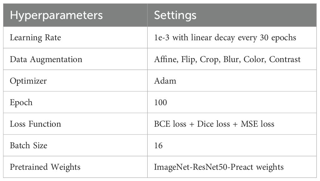
Table 1. Hyperparameter settings for Hover-Net.
2.5 Lymphocyte deep learning model evaluation and multi-scale performance metrics
Using the internal testing set, model performance was evaluated at two different length-scales relative to IHC measurements (Figure 6A): (1) a local length-scale to characterize the raw, cell-by-cell deep learning performance of detecting lymphocytes vs. non-lymphocytes; and (2) a global length-scale to characterize pattern-preserving pathomic features of the immune microenvironment derived from deep learning detection of lymphocytes. Local model performance was based on analysis of precision-recall curves and receiver operating characteristic (ROC) curves (Figure 6B). Global model performance was based on extraction of pathomic graph features and texture features, where graph nodes represent individual lymphocytes detected via the deep learning model, and graph edges characterize the spatial connections between different lymphocytes (Figure 6C). Graph features were compared between the predicted lymphocytic patterns (based on deep learning detection of lymphocytes as graph nodes) and the measured lymphocytic patterns (based on the measured IHC staining of lymphocytes as graph nodes) via arctangent percent error (17). To calculate texture features, a radial basis function was convolved with the graphs to estimate the lymphocytic probability density function (PDF) (Figure 6D). From the predicted lymphocytic PDF on H&E, texture features were extracted and compared to the measured lymphocytic PDF on IHC. Finally, the structural similarity index measure (SSIM) (18) was calculated between the predicted and measured PDFs as an overall description of predicted topological fidelity. Texture feature definitions are adopted from the Image Biomarker Standardization Initiative (IBSI) (19) and graph feature definitions are listed in Table 2.
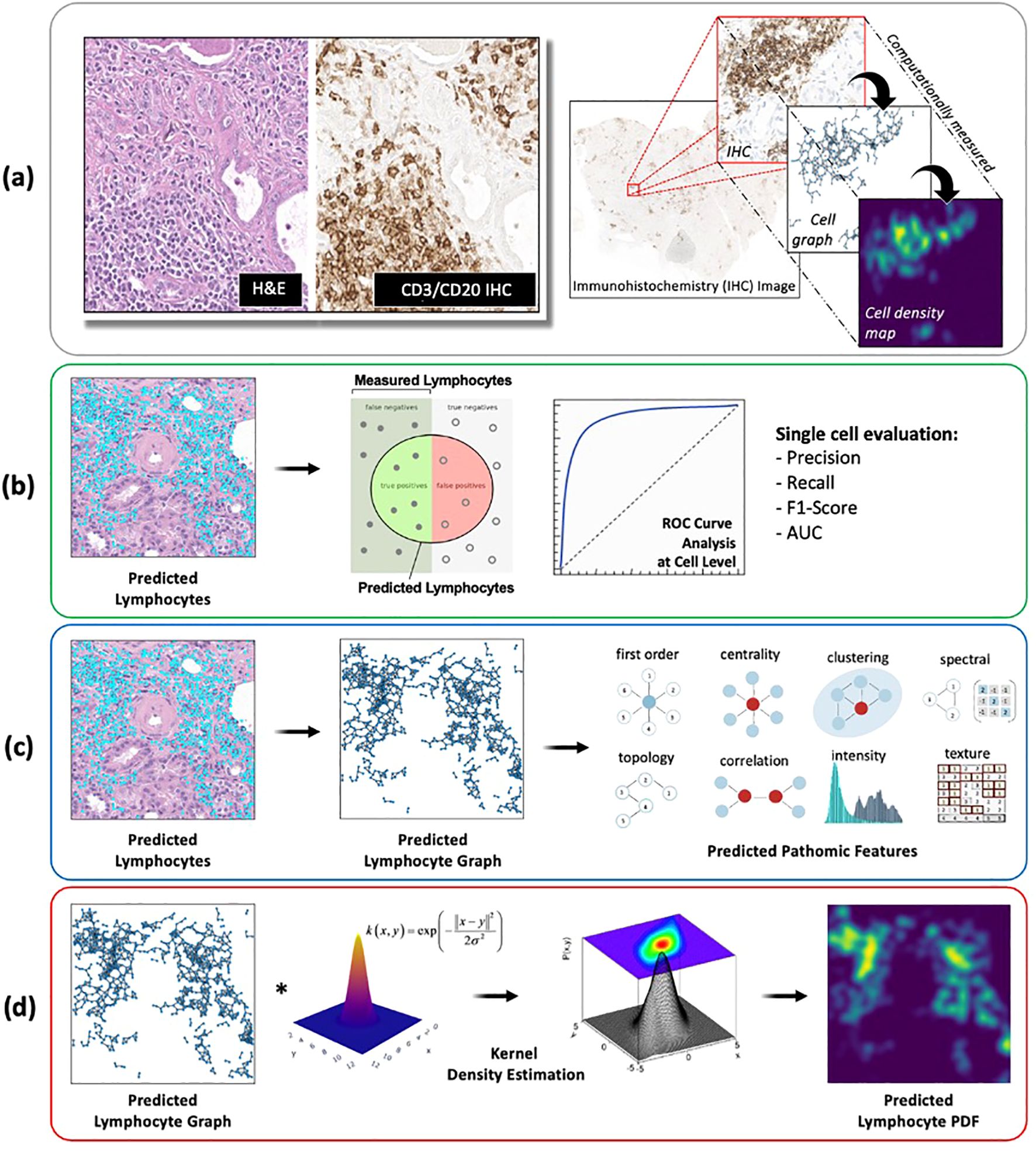
Figure 6. Deep learning model evaluation metrics at different length-scales. (A) All deep learning results were compared to IHC-based computational measurements of nuclei counts, nuclei topological graphs, and nuclei density maps. (B) Local model performance of individual nuclei predictions was based on analysis of precision-recall curves and receiver operating characteristic (ROC) curves. (C, D) Global model performance of pattern-preserving features of the immune microenvironment was based on (C) extraction of graph features, where graph nodes represent individual lymphocytes detected via deep learning, and graph edges characterize the spatial connections between different lymphocytes; and (D) structural similarity of lymphocyte probability density functions (PDF), estimated based on a kernel density estimation of the predicted lymphocyte graphs using a radial basis function.
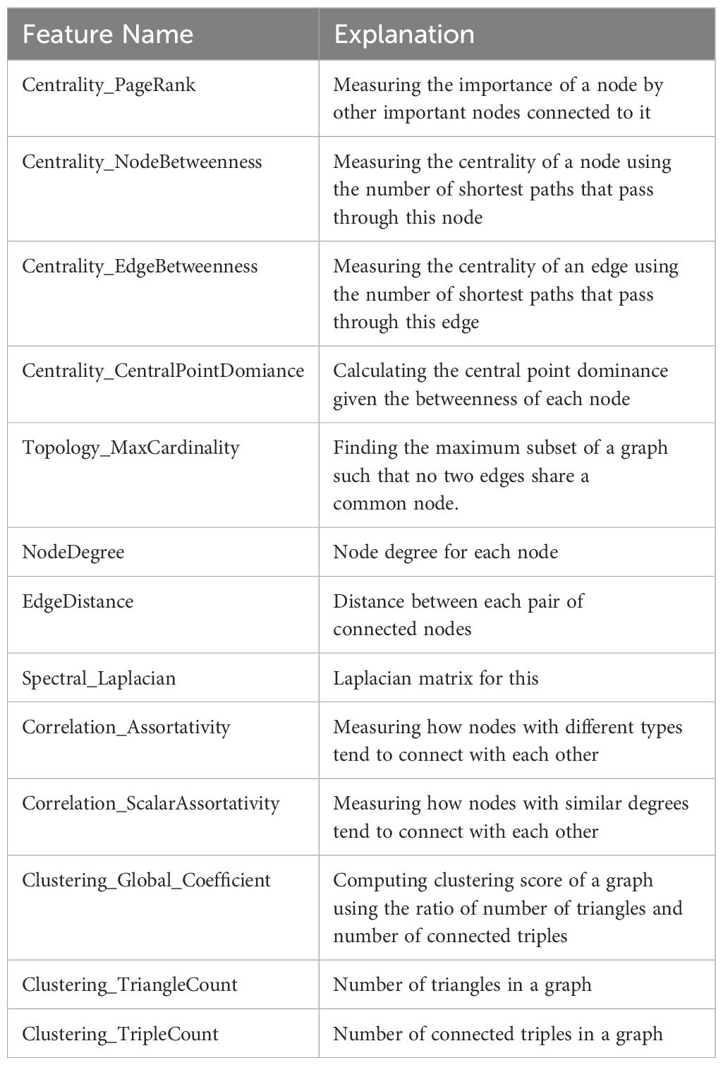
Table 2. Feature definition and explanation for graph features.
2.6 Independent testing of lymphocyte deep learning model on an external dataset of diverse human tissues
To independently evaluate the generalization capacity of the developed lymphocyte deep learning model, we applied it to a publicly-available, external dataset. Specifically, we analyzed testing data from the previously completed Multi-organ Nuclei Segmentation and Classification (MoNuSAC) Grand Challenge (20), which is designed to systematically evaluate deep learning detection and classification of different cell types on digital pathology. Since MoNuSAC data consists of four different human tissues (breast, kidney, lung, and prostate), it allows us to explore whether our lymphocyte model performs consistently across different human tissues and diverse biological contexts. In total, we applied our model to 146 H&E regions-of-interest (ROI) from 46 TCGA patients, where manual annotation of lymphocytes is available as ground-truth labels as part of the MoNuSAC Grand Challenge. On inference, we calculated precision, recall, and F1 score, as well as Panoptic Quality (PQ), which was the evaluation metric used in the MoNuSAC Grand Challenge.
2.7 Experimental confirmation of lymphocyte deep learning model in a genetically engineered mouse model
To experimentally confirm our lymphocyte model, we conducted a preclinical experiment (Figure 7) on mice genetically engineered to lack mature lymphocytes as a negative control. Here, the immune microenvironment of the mice was systematically modulated under experimental conditions (i.e., by genotype) to verify that our deep learning model accurately and reliably identifies lymphocytes. Essentially, we asked the simple question, “what would happen if a deep learning algorithm designed to detect lymphocytes experiences an organism genetically engineered not to have lymphocytes?” To address this question, we evaluated spleens and thymuses from mice genetically engineered to lack Rag2 (i.e., a gene required for lymphocyte maturation). Homozygous knockout mice (Rag2-/-) were used as a negative control because they do not produce mature lymphocytes and thus lack the biological basis of the deep learning positive class label. Meanwhile, a heterozygous littermate mouse with one intact allele (Rag2+/-) was used as a positive control.
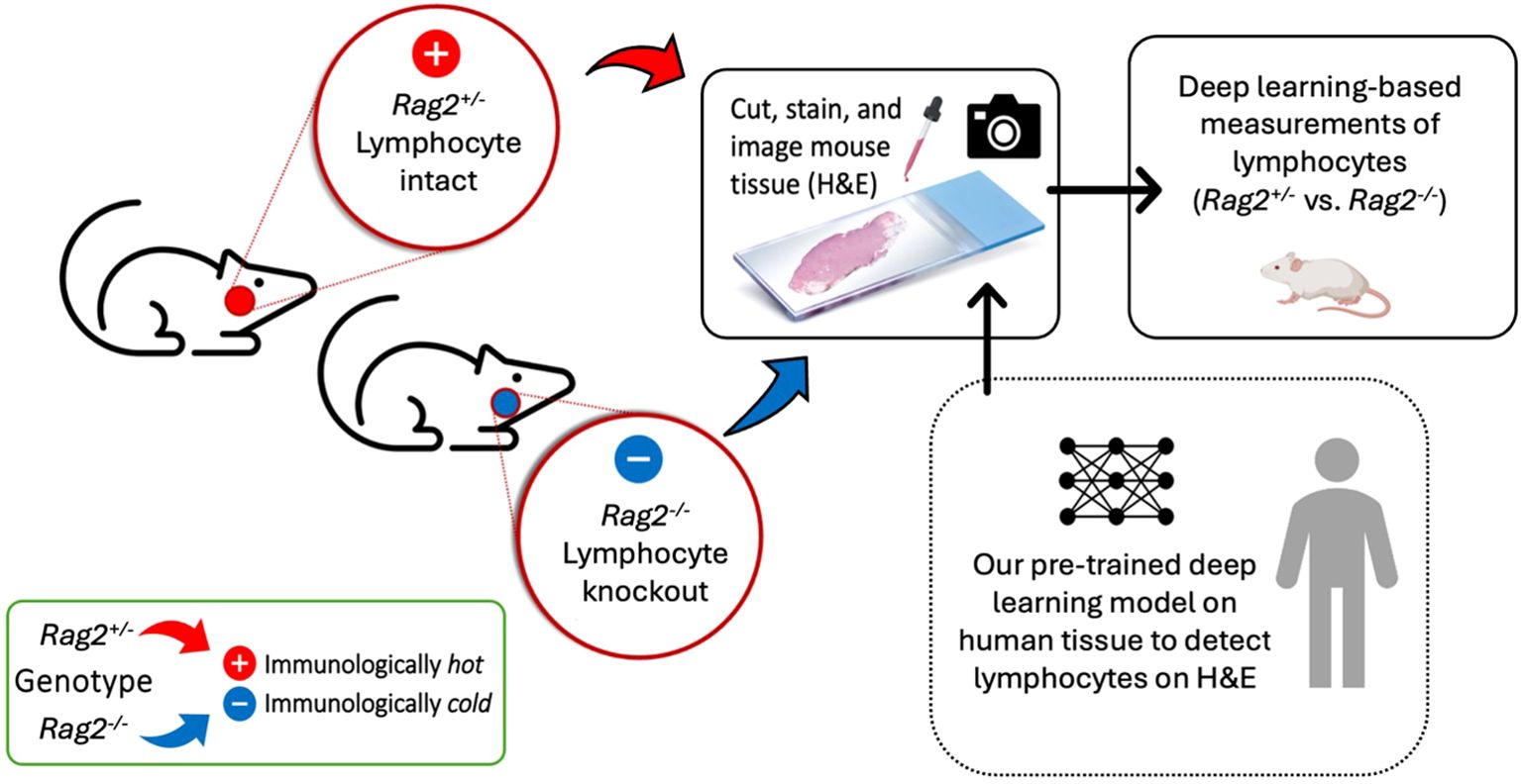
Figure 7. Experimental confirmation of lymphocytic identification by deep learning in a genetically engineered mouse model. A preclinical experiment was performed using mice that have been genetically engineered to lack mature lymphocytes as a negative control. Spleens and thymuses from Rag2-/- or littermate control Rag2+/- mice were formalin-fixed, paraffin-embedded, cut, stained with H&E, and imaged on a whole slide digital pathology scanner. A pre-trained deep learning algorithm, independently developed on human tissue to detect lymphocytes on H&E images, was applied to the mouse tissue to measure differences in lymphocytic infiltrate between Rag2-/- and Rag2+/- mice. Homozygous knockout mice (Rag2-/-) were used as negative controls because they lack mature lymphocytes. A heterozygous mouse (Rag2+/-) with one intact allele was used as a positive control.
2.7.1 Animal model
All animal studies were performed in accordance with protocols approved by the Duke University Institutional Animal Care and Use Committee (IACUC) and adhered to the NIH Guide for the Care and Use of Laboratory Animals. Rag2+/- and Rag2-/- mice were bred at Duke University. The Rag2 gene is essential for B and T cell maturation. Homozygous knockout mice (Rag2-/-) were used as negative controls because they do not contain mature lymphocytes. A heterozygous mouse with one intact Rag2 allele (Rag2+/-) was used as a positive control with mature lymphocytes present.
2.7.2 Experimental measurements of lymphocytes in genetically engineered mice
Mice were euthanized via CO2 inhalation. Their spleen and thymus were excised, formalin-fixed, and paraffin-embedded. Tissue sections were cut at 2 microns, stained with H&E, and scanned on the same Leica Aperio AT2 imaging system utilized in Section 2.1 above. Our pre-trained deep learning model, developed on human data per Section 2.4 above, was directly applied to the H&E images to calculate lymphocytic density. Lymphocytic density measurements were compared between Rag2+/- and Rag2-/- mice. Importantly, the deep learning model was directly applied to this experimental data without any re-training, fine-tuning, or transfer learning. Therefore, the mouse tissues serve as a final testing set for the deep learning model, where the biology is systematically controlled (i.e., by mouse genotype) to evaluate the performance of this model under defined experimental conditions.
3 Results
3.1 Image registration of H&E and IHC at single-cell resolution and corresponding lymphocyte training labels
We generated a total of 320 matched pairs of H&E/IHC tiles from 18 patient cases, evenly distributed among minimal, sparse, and dense lymphocytic inflammation. Following image registration of H&E to IHC, the average normalized cross correlation coefficient was 0.89 ± 06, suggesting minimal error from shift artifacts. In total, 111,110 nuclei were automatically segmented on the H&E via StarDist. Of the segmented nuclei, 45,611 nuclei were labelled as lymphocytes based on the co-registered CD3/CD20 IHC reference.
3.2 Evaluation of lymphocyte deep learning model on internal testing data
On the internal testing set, model precision, recall, and f1 score were 0.74 ± 11, 0.73 ± 10, and 0.73 ± 11, respectively, with an area under the curve (AUC) of 0.78 ± 15. Figure 8 shows illustrating examples of lymphocyte predictions on H&E testing data relative to corresponding ground-truth CD3/CD20 IHC images. These results demonstrate a strong concordance between the predicted lymphocytes (i.e., the computationally derived blue signal) and the shift-invariant ground-truth CD3/CD20 IHC (i.e., the measured brown signal). The average time to process a 1,024×1,024 image tile was 18 seconds on a single RTX A6000 with 48 GB RAM. As illustrated in Figure 9, the average structural similarity between the predicted lymphocytic PDFs and the measured lymphocytic PDFs was 0.86 ± 06. We found that 19.5% of pathomic features demonstrated a mean error of <10%, and 56.8% of features demonstrated a mean error of >30%. The calculated errors of all pathomic graph features and texture features are reported in the Supplementary Material.
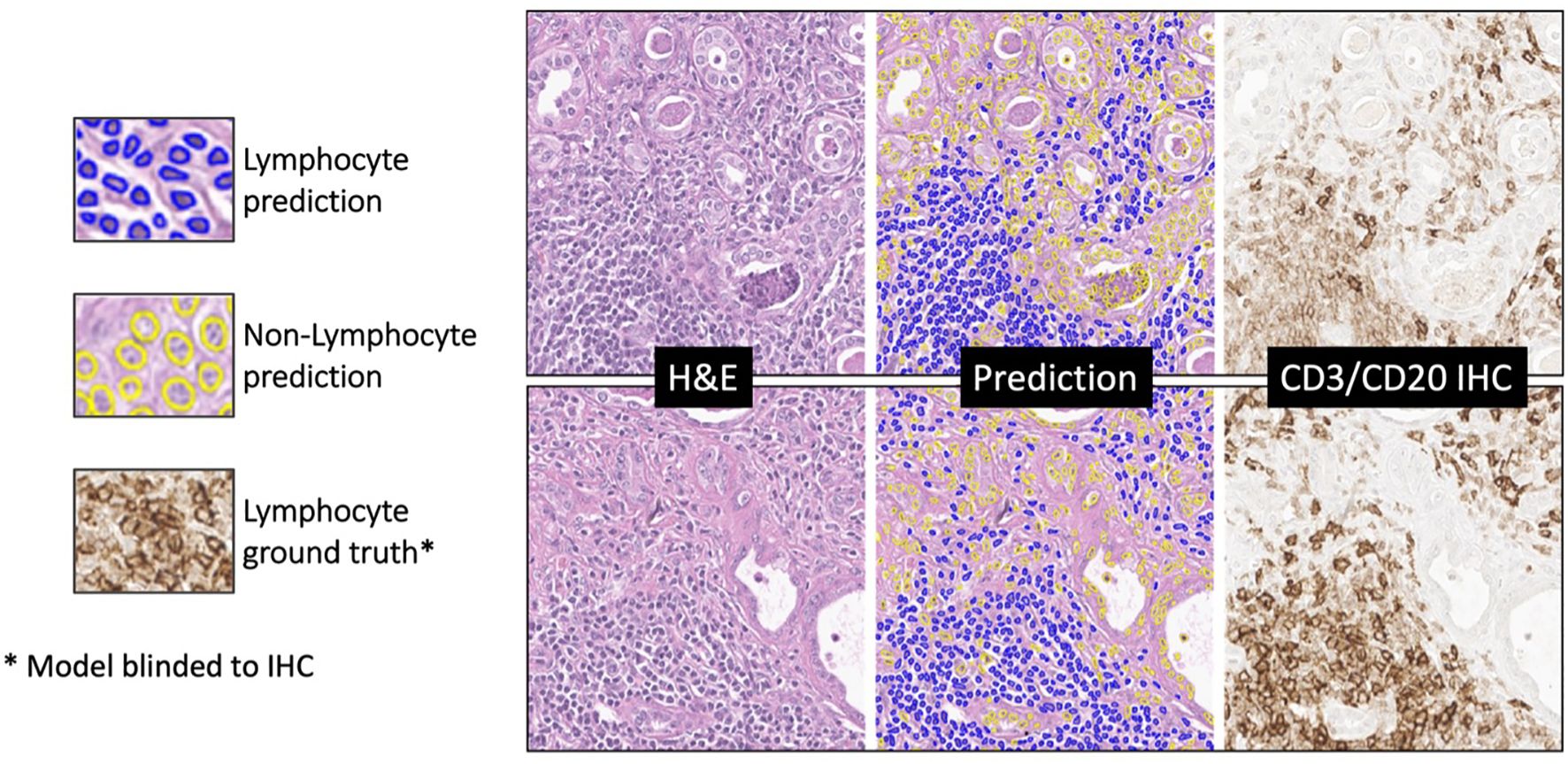
Figure 8. Deep learning lymphocyte prediction examples. From left to right are input H&E patch (1024x1024), model prediction, and CD3/CD20 IHC reference. Predicted lymphocytes are highlighted in blue, while other cells are in yellow. Notably, the model’s predictions closely resemble the brown regions in the IHC reference, indicating its ability to accurately identify lymphocytes in H&E-stained histopathology images.
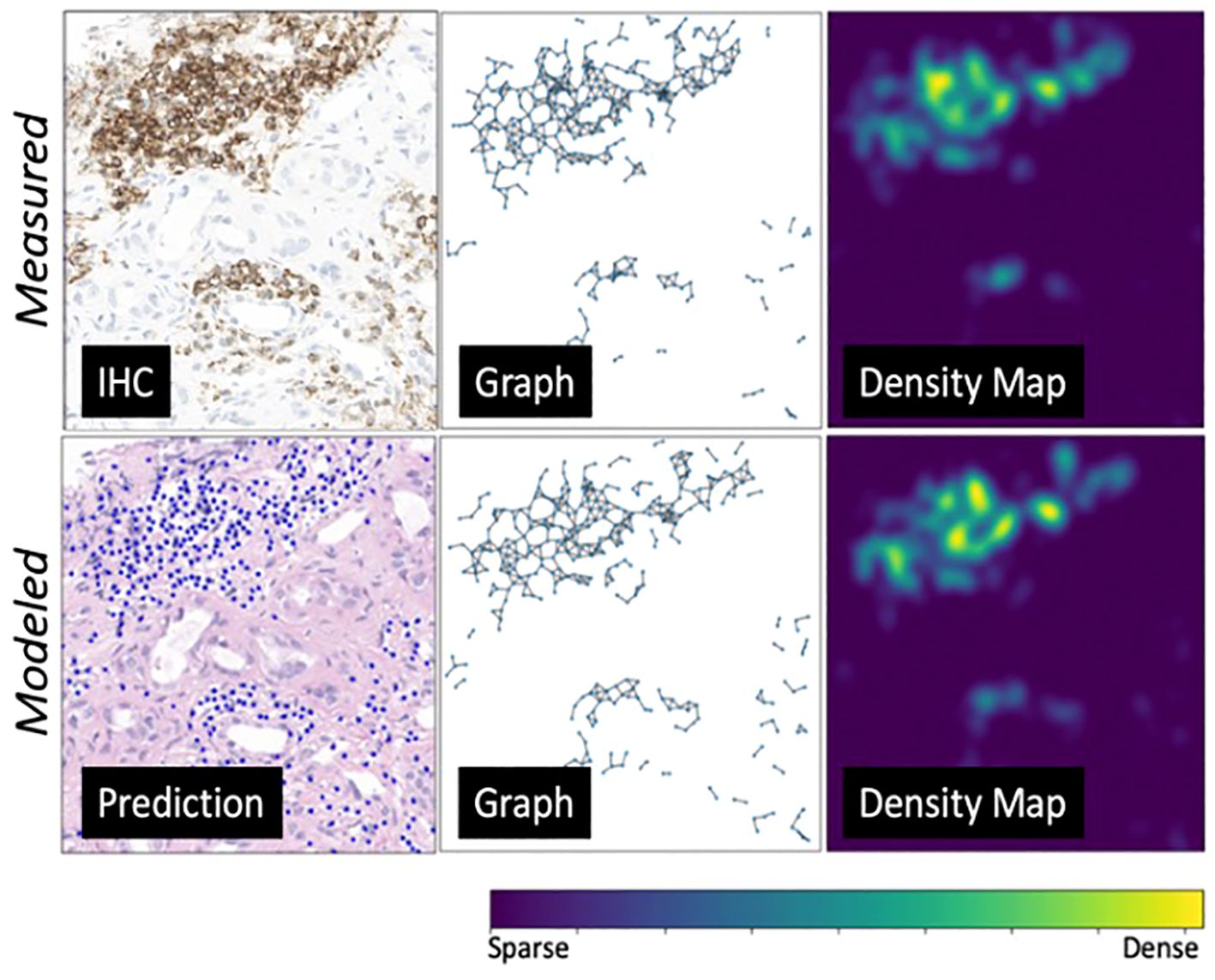
Figure 9. An environmental-level modeling system example. From left to right are predicted lymphocytes, cell graphs showing spatial relationship among lymphocytes, and cell probability density maps (transformed from the cell graphs). The top row showcases the ground truth measurements, while the bottom row presents the modeled measurements. This figure illustrates a strong alignment between the ground truth and prediction model.
On feature extraction from the predicted lymphocytic graphs vs. the measured lymphocytic graphs, we found that the graph features with the lowest errors were: First Order Edge Distance Mean (3.3%), Laplace Matrix Spectrum Standard Deviation (10.1%), Topology Max Cardinality Matching Probability (11.1%), Topology Max Cardinality Not Matching Probability (11.7%), and First Order Vertex Degree Mean (15.3%). Meanwhile, the graph features demonstrating the highest errors were Betweenness of Vertex Mean (75.1%), Betweenness of Vertex Standard Deviation (73.2%), Betweenness of Edge Standard Deviation (71.1%), Central Point Dominance (70.4%), and Betweenness of Edge Mean (60.0%).
On feature extraction from the predicted lymphocytic PDFs vs. the measured lymphocytic PDFs, we found that the texture features with the lowest errors were Gray Level Co-occurrence Matrix Inverse Difference (0.1%), Gray Level Dependence Matrix Large Dependence Emphasis (0.2%), Gray Level Co-occurrence Matrix Correlation (7.1%), Gray Level Co-occurrence Matrix Maximal Correlation Coefficient (7.1%), and Gray Level Co-occurrence Matrix Information Measure of Correlation 1 (13.3%). Meanwhile, the texture features demonstrating the highest errors were Gray Level Co-occurrence Matrix Cluster Prominence (71.3%), Gray Level Co-occurrence Matrix Cluster Shade (65.5%), Neighborhood Gray Tone Difference Matrix Complexity (59.9%), Neighborhood Gray Tone Difference Matrix Contrast (54.3%), and First Order Robust Mean Absolute Deviation (32.4%).
3.3 Independent testing of lymphocyte deep learning model on an external dataset of diverse human tissues
On the external human testing set, model precision, recall, and f1 score were 0.45 ± 33, 0.87 ± 24, and 0.53 ± 31, respectively, with an AUC of 0.71 ± 0.15. A summary of external testing results, including individual performance metrics of specific tissue types, are reported in Table 3. The illustrating examples shown in Figure 10 demonstrate that the model was able to generalize well across diverse human tissues and different biological contexts. Finally, the model achieved a PQ score of 0.43, which is consistent with the published MoNuSAC Grand Challenge leaderboard statistics (average PQ = 0.39 ± 13; range of PQ = [0.10, 0.56]; N=13).
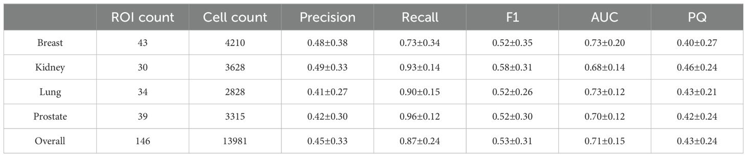
Table 3. External validation of lymphocyte detection on diverse human tissues from the public MoNuSAC dataset.

Figure 10. Illustrating examples of lymphocyte detection via deep learning on an external dataset of diverse human tissues. The external MoNuSAC dataset consists of four different human tissues (breast, kidney, lung, and prostate). Each ROI in the figure is paired with a ground truth lymphocyte mask (binary mask, where white represents ground truth lymphocytes) on the right. Blue and red contours represent model-detected lymphocytes and other cells, respectively.
3.4 Experimental confirmation of lymphocyte deep learning model in a genetically engineered mouse model
On pre-clinical interrogation of our deep learning model, we were able to reliably measure lymphocytes in lymphoid tissues from genetically engineered mice. Experimental results are shown in Figure 11, where an average lymphocyte density of 96.5 ± %1% was measured in the Rag2+/- (i.e., lymphocyte-intact) genotype compared to 16.2 ± %5% in the Rag2-/- (i.e., lymphocyte knockout) genotype (p<0.0001, ANOVA). Qualitative interpretation showed distinct differences in lymphocyte density on H&E when comparing the spleen (Figure 12A) and thymus (Figure 12B) in Rag2+/- versus Rag2-/- mice.
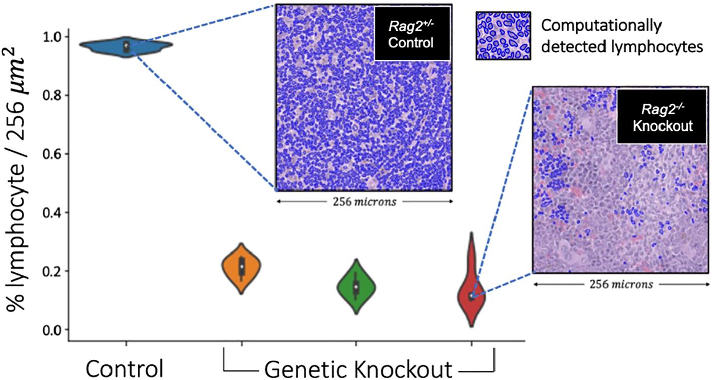
Figure 11. Validation of lymphocyte identification by a deep learning model in a genetically engineered mouse model. The violin plots depict the distribution of lymphocyte percentage in the spleens across 256x256 image patches of different mice. The Rag2-/- genetic knockout mice demonstrated an average lymphocyte density of 16.2%, which was significantly smaller compared to the Rag2+/- experimental control mouse of >90% (p<0.0001, ANOVA). These experimental data suggest that our deep learning model generalized across species and tissue types, capturing at least the basic aspects of lymphocytic immune response.
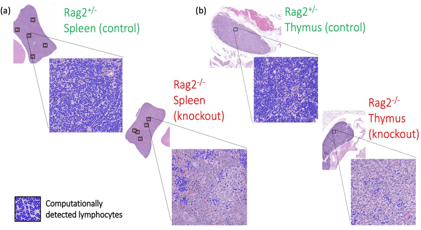
Figure 12. Differences in tissue immune phenotype in genetically engineered mice. Computationally-detected lymphocytes via deep learning inference are shown in blue for both the (A) spleen and (B) thymus. Qualitative differences in lymphocytic architecture were identified by deep learning in the Rag2-/- genetic knockout mice compared to the Rag2+/- experimental control.
4 Discussion
This study introduced an integrated research design to characterize lymphocytic infiltration on H&E WSIs across different tissue types and species. Our approach extends the capability of lymphocyte quantification to archival digital images of H&E-stained tissue without requiring IHC. Furthermore, it leverages powerful computational analysis tools to capture spatial characteristics of lymphocytic inflammation in tissues. By combining computational image analysis with novel tissue processing procedures (matched H&E and IHC on the same slide) and genetically engineered mouse models, we demonstrated a rigorous approach to deep learning algorithm development and evaluation under well-controlled laboratory conditions. Although image-based characterization of lymphocytes identified by IHC staining has shown success in various cancer studies, our approach extends this capability to digitized H&E-stained slides, overcoming limitations associated with the cost and feasibility of IHC in routine pathology and retrospective research. Furthermore, deep learning algorithms that identify different cell types on H&E WSIs offer the opportunity to simultaneously analyze the topology of those cells and to capture crucial structural details of the tissue, enabling examination of a broader spectrum of cell types and tissue organization.
A key innovation of our approach is the tissue processing and image registration procedure to generate efficient class labeling on H&E images with high accuracy and precision. This in turn enables robust deep learning model development. In biomedical imaging applications of deep learning, generation of reliable ground-truth labels remains a major challenge (21, 22). To help address this issue, we optimized training label fidelity through image registration of H&E and IHC staining performed on the same slide at single-cell resolution, achieving pixel-perfect, ground-truth labels. As the knowledge base of our model is IHC antibodies with high specificity for lymphocytes – and not manual annotation by pathologists – the training data do not suffer from intra- or inter-observer variability.
Furthermore, our approach is fundamentally different than clinical pathology workflows and other studies that typically employ H&E and IHC staining on sequential slides cut from a tissue block. While this approach generates both stains on the same specimen, the information content is not shift-invariant, making it non-trivial for deep learning applications that resolve data at the length-scale of individual immune cells. Unlike staining sequential slides, performing H&E and IHC staining on the same tissue enables data curation at single-cell resolution and is thus well-suited for deep learning applications at the physical length-scales of individual cells.
Same-tissue processing also enables error propagation of downstream pathomic features derived from the ensemble of detected cells. This is important because the spatial interplay of cells, as captured by representative pathomic features, is essential to spatially characterizing tissue microenvironments. For example, spatial cell-to-cell interplay has been shown to be linked with therapeutic responses and tumor biology (23–25), as well as broader applications from intracellular to extracellular conditions and associations with various tissue microenvironments (26–29). Many of these topological characteristics can be computationally measured in various ways, including cell geographic clusters, cell density distributions (30), cell topological graphs (31), cell clouds (32), and graph neural networks (GNNs) (33), potentially leading to deeper insights into the mechanisms at play in different pathological and immunological states.
Our experiment comparing predicted pathomic features on H&E to measured pathomic features on IHC suggest that the error-rate of pathomic extraction is largely feature-specific. That is, certain pathomic features are more sensitive than others to the same errors in the initial deep learning inference. This has major implications on pathomic biomarker models that rely on an upstream deep learning framework for initial cell detection. For example, variation induced by deep learning errors, which leads to inconsistencies in pathomic feature extraction, may consequently result in variability in the computational biomarkers derived from these features. Same-tissue staining at single-cell resolution enables characterization of this effect, such that propagation of error for individual features can be better understood. This may be a useful feature selection strategy to remove highly variable features that would otherwise introduce noise into downstream biomarker models. Our results demonstrate that even with modest deep learning performance, there are pathomic features which still preserve the global topological patterns of the immune microenvironment. To the best of our knowledge, there is limited prior research studying such pathomic feature propagation of error.
Our external testing results on diverse human tissues were similar to the published MoNuSAC Grand Challenge results (PQ = 0.43 vs. PQ = 0.39 ± 13), implying that the performance of our model is comparable to other lymphocyte deep learning models in the literature. Our lymphocyte model thus demonstrated reasonable generalization when applied to an independent testing dataset of different human tissues, suggesting that it can perform consistently across diverse biological contexts. Furthermore, these external data were of variable stain quality, tissue processing, and image acquisition, which is important because laboratory conditions cannot always be easily replicated across institutions.
However, there are several factors that may contribute to differences in model performance between the internal and external datasets. First, the manual lymphocyte annotation of the MoNuSAC Grand Challenge dataset is fundamentally different than the IHC-based measurements of our internal dataset, potentially leading to imprecise definitions of ground-truth. Second, the diverse tissue types of the MoNuSAC Grand Challenge data – which were not observed in our internal data – may contribute to a biological domain shift that requires fine-tuning of models to specific pathologies. Our external testing results support this concept, where high recall scores indicate stable lymphocyte morphology detection in different tissue conditions, yet lower precision scores suggest variation in tissue content not present in training data, where objects with similar characteristics as lymphocytes were mis-recognized by the model.
Finally, the major novelty of our paper was the cross-species validation of our lymphocyte model, where we trained our model on human tissues, and externally validated it using a genetically engineering mouse model. This is significant, because while deep learning models demonstrate remarkable proficiency in pattern recognition, their limitations in mechanistic understanding are evident. Often, these models fall short in capturing fundamental biological characteristics beyond surface-level image representation. This gap underscores a critical challenge, where purely data driven solutions alone may not sufficiently elucidate the intricate mechanisms underlying immune responses. Consequently, there is a pressing need for complementary experimental testing to validate and refine these models under controlled conditions. We also note that data assimilation methods (i.e., where mechanistic models from either physics or biology are integrated with data-driven solutions, such as deep learning (34–36)), may provide a more rigorous description of the imaging phenotype and a better understanding of deep learning (37, 38). Our mouse model results suggest that integrating computationally-derived spatial analysis with traditional basic science approaches could enhance our understanding of complex biological systems.
Although our research demonstrates several technological innovations and novel findings, our study is not without limitations. First, our tissue processing scheme may result in tissue deformations during the re-staining procedure, which could result in errors during image registration. However, these deformations are minimal when compared to those from cutting the tissue. Because we only cut the tissue once, our rigid registration errors should be smaller than with sequential cuts, where deformable image registration is required. Second, while total cell counts were substantial in our data, case level variability was limited to only 18 unique patient samples used in model development. This is unfortunately a consequence of our prospective data curation and intricate tissue processing procedure, which emphasized data quality over data quantity. We partially addressed this limitation by employing case level partitioning for training, validation, and internal testing, as well as independent external testing on both human and murine datasets. Future work needs to apply these strategies to larger, more diverse data sets. Third, the current paper only focuses on lymphocytes, but immune responses are more diverse and include non-lymphocytic pathologies (e.g., neutrophils, macrophages, etc.). Since our proposed tissue processing pipeline can be generalized to other antibodies, future work should focus on development of additional models for different immune cell types. This would enable a more comprehensive analysis of the immune microenvironment.
In summary, our results demonstrate that deep learning can reliably identify lymphocytes on H&E slides and capture differences in lymphocyte spatial architecture. We plan to continue mechanistic interrogation of this technique in preclinical animal models to obtain a deeper, more holistic understanding of underlying processes that drive disease. This approach will help to ensure that computational predictions are biologically relevant and scientifically robust with the goal of ultimately developing clinically actionable biomarkers.
Data availability statement
The raw data supporting the conclusions of this article will be made available by the authors, without undue reservation.
Ethics statement
The studies involving humans were approved by Duke University School of Medicine IRB. The studies were conducted in accordance with the local legislation and institutional requirements. The human samples used in this study were acquired from a retrospective research protocol. Written informed consent for participation was not required from the participants or the participants’ legal guardians/next of kin in accordance with the national legislation and institutional requirements. The animal study was approved by Duke University School of Medicine IACUC. The study was conducted in accordance with the local legislation and institutional requirements.
Author contributions
XL: Writing – original draft, Writing – review & editing, Methodology, Software, Visualization, Conceptualization, Data curation, Formal Analysis, Validation. CH: Writing – review & editing, Data curation, Methodology, Visualization, Formal Analysis. AR: Writing – review & editing, Data curation. GS: Writing – review & editing, Data curation. RC: Writing – review & editing, Data curation. TA: Writing – review & editing, Data curation. JE: Writing – review & editing, Validation. JH: Writing – review & editing, Validation. TW: Writing – review & editing, Validation. AJ: Writing – review & editing, Conceptualization, Methodology. YM: Writing – review & editing, Conceptualization, Funding acquisition, Methodology, Resources, Supervision. LB: Writing – review & editing, Conceptualization, Funding acquisition, Methodology, Resources, Supervision. KL: Writing – original draft, Writing – review & editing, Conceptualization, Funding acquisition, Methodology, Resources, Supervision.
Funding
The author(s) declare that financial support was received for the research, authorship, and/or publication of this article. This work was supported by DoD/CDMRP under grant number W81XWH2110248, NIH/NCI under grant number R01CA289261, NIH/NIAID under grant number U01AI163065, NIH/NIDDK under grant number R01DK118431, NIH/NIDCR under grant number K08DE029887, and a Damon Runyon Clinical Investigator Award under grant number 712041.
Conflict of interest
The authors declare that the research was conducted in the absence of any commercial or financial relationships that could be construed as a potential conflict of interest.
Publisher’s note
All claims expressed in this article are solely those of the authors and do not necessarily represent those of their affiliated organizations, or those of the publisher, the editors and the reviewers. Any product that may be evaluated in this article, or claim that may be made by its manufacturer, is not guaranteed or endorsed by the publisher.
Supplementary material
The Supplementary Material for this article can be found online at: https://www.frontiersin.org/articles/10.3389/fimmu.2024.1451261/full#supplementary-material
References
1. Chen L, Deng H, Cui H, Fang J, Zuo Z, Deng J, et al. Inflammatory responses and inflammation-associated diseases in organs. Oncotarget. (2017) 9:7204–18. doi: 10.18632/oncotarget.23208
2. Swiderska-Chadaj Z, Pinckaers H, van Rijthoven M, Balkenhol M, Melnikova M, Geessink O. Learning to detect lymphocytes in immunohistochemistry with deep learning. Med Image Analysis. (2019) 58:101547. doi: 10.1016/j.media.2019.101547
3. Eriksen AC, Andersen JB, Kristensson M, dePont Christensen R, Hansen TF, Kjær-Frifeldt S, et al. Computer-assisted stereology and automated image analysis for quantification of tumor infiltrating lymphocytes in colon cancer. Diagn Pathol. (2017) 12:65. doi: 10.1186/s13000-017-0653-0
4. Bussola N, Papa B, Melaiu O, Castellano A, Fruci D, Jurman G. Quantification of the immune content in neuroblastoma: deep learning and topological data analysis in digital pathology. Int J Mol Sci. (2021) 22:8804. doi: 10.3390/ijms22168804
5. Garcia E, Hermoza R, Castanon CB, Cano L, Castillo M, Castanneda C, et al. (2017). Automatic lymphocyte detection on gastric cancer IHC images using deep learning, in: 2017 IEEE 30th International Symposium on Computer-Based Medical Systems (CBMS), (New York City, New York, United States: IEEE). doi: 10.1109/cbms.2017.94
6. Negahbani F, Sabzi R, Pakniyat Jahromi B, Firouzabadi D, Movahedi F, Shirazi K, et al. PathoNet introduced as a deep neural network backend for evaluation of Ki-67 and tumor-infiltrating lymphocytes in breast cancer. Sci Rep. (2021) 11:8489. doi: 10.1038/s41598-021-86912-w
7. Aprupe L, Litjens GJS, Brinker TJ, van der Laak J, Grabe N. Robust and accurate quantification of biomarkers of immune cells in lung cancer micro-environment using deep convolutional neural networks. PeerJ. (2019 ) 7:e6335. doi: 10.7717/peerj.6335
8. Sarker MK, Makhlouf Y, Craig SG, Humphries MP, Loughrey MB, James J, et al. A means of assessing deep learning-based detection of ICOS protein expression in colon cancer. Cancers. (2021) 13(15):3825. doi: 10.3390/cancers13153825
9. Kreiss L, Jiang S, Li X, Xu S, Zhou KC, Mühlberg A. Digital staining in optical microscopy using deep learning – a review. PhotoniX (2023) 4:34. doi: 10.48550/arXiv.2303.08140
10. Acs B, Ahmed FS, Gupta S, Wong PF, Gartrell RD, Sarin Pradhan J, et al. An open source automated tumor infiltrating lymphocyte algorithm for prognosis in melanoma. Nat Commun. (2019) 10:5440. doi: 10.1038/s41467-019-13043-2
11. Amgad M, Sarkar A, Srinivas C, Redman R, Ratra S, Bechert CJ, et al. Joint region and nucleus segmentation for characterization of tumor infiltrating lymphocytes in breast cancer. Proc SPIE Int Soc Opt Eng. (2019) 10956:109560M. doi: 10.1117/12.2512892
12. Le H, Gupta R, Hou L, Abousamra S, Fassler D, Torre-Healy L, et al. Utilizing automated breast cancer detection to identify spatial distributions of tumor-infiltrating lymphocytes in invasive breast cancer. Am J Pathol. (2020) 190:1491–504. doi: 10.1016/j.ajpath.2020.03.012
13. Budginaitė E, Morkūnas M, Laurinavicius A, Treigys P. Deep learning model for cell nuclei segmentation and lymphocyte identification in whole slide histology images. Informatica (Lithuanian Acad Sciences). (2021) 32:23–40. doi: 10.15388/20-infor442
14. Linder N, Taylor JC, Colling R, Pell R, Alveyn E, Joseph J, et al. Deep learning for detecting tumour-infiltrating lymphocytes in testicular germ cell tumours. J Clin Pathol. (2019) 72:157–64. doi: 10.1136/jclinpath-2018-205328
15. Schmidt U, Weigert M, Broaddus C, Myers G. Cell detection with star-convex polygons. In: Frangi AF, Schnabel JA, Davatzikos C, Alberola-López C, Fichtinger G, editors. Medical Image Computing and Computer Assisted Intervention – MICCAI 2018. Berlin, Germany: Springer International Publishing (2018). p. 265–73. doi: 10.1007/978-3-030-00934-2_30
16. Graham S, Vu QD, Raza SEA, Azam A, Tsang YW, Kwak JT, et al. Hover-Net: Simultaneous segmentation and classification of nuclei in multi-tissue histology images. Med Image Anal. (2018) 58:101563. doi: 10.1016/j.media.2019.101563
17. Kim S, Kim H. A new metric of absolute percentage error for intermittent demand forecasts. Int J Forecasting. (2016) 32:669–79. doi: 10.1016/j.ijforecast.2015.12.003
18. Wang Z, Bovik AC, Sheikh HR, Simoncelli EP. Image quality assessment: from error visibility to structural similarity. IEEE Trans Image Processing. (2004) 13:600–12. doi: 10.1109/TIP.2003.819861
19. Zwanenburg A, Vallières M, Abdalah MA, Aerts HJWL, Andrearczyk V, Apte A, et al. The image biomarker standardization initiative: standardized quantitative radiomics for high-throughput image-based phenotyping. Radiology. (2020) 295:328–38. doi: 10.1148/radiol.2020191145
20. Verma R, Kumar N, Kumar N, Kurian NC, Rane S, Graham S, et al. MoNuSAC2020: A multi-organ nuclei segmentation and classification challenge. IEEE Trans Med Imaging. (2021) 40:3413–23. doi: 10.1109/tmi.2021.3085712
21. Aroyo L, Welty C. Truth is a lie: crowd truth and the seven myths of human annotation. AI Magazine. (2015) 36:15–24. doi: 10.1609/aimag.v36i1.2564
22. Albarqouni S, Baur C, Achilles F, Belagiannis V, Demirci S, Navab N. AggNet: deep learning from crowds for mitosis detection in breast cancer histology images. IEEE Trans Med Imaging. (2016) 35:1313–21. doi: 10.1109/tmi.2016.2528120
23. Nawaz S, Nawaz S, Heindl A, Koelble K, Yuan Y. Beyond immune density: critical role of spatial heterogeneity in estrogen receptor-negative breast cancer. Modern Pathol. (2015) 28:766–77. doi: 10.1038/modpathol.2015.37
24. Ho WW, Pittet MJ, Fukumura D, Jain RK. The local microenvironment matters in preclinical basic and translational studies of cancer immunology and immunotherapy. Cancer Cell. (2022) 40:701–2. doi: 10.1016/j.ccell.2022.05.016
25. Hsieh WC, Budiarto BR, Wang YF, Lin CY, Gwo MC, So DK, et al. Spatial multi-omics analyses of the tumor immune microenvironment. J Biomed Science. (2022) 29:96. doi: 10.1186/s12929-022-00879-y
26. Li Y, He X, Li Q, Lai H, Zhang H, Hu Z, et al. EV-origin: Enumerating the tissue-cellular origin of circulating extracellular vesicles using exLR profile. Comput Struct Biotechnol J. (2020) 18:2851–9. doi: 10.1016/j.csbj.2020.10.002
27. Li Y, Yu S, Qian L, Chen K, Lai H, Zhang H, et al. Circulating EVs long RNA-based subtyping and deconvolution enable prediction of immunogenic signatures and clinical outcome for PDAC. Mol Ther Nucleic Acids. (2021) 26:488–501. doi: 10.1016/j.omtn.2021.08.017
28. Guo TA, Lai HY, Li C, Li Y, Li YC, Jin YT, et al. Plasma extracellular vesicle long RNAs have potential as biomarkers in early detection of colorectal cancer. Front Oncol. (2022) 12:829230. doi: 10.3389/fonc.2022.829230
29. Lai H, Li Y, Zhang H, Hu J, Liao J, Su Y, et al. exoRBase 2.0: an atlas of mRNA, lncRNA and circRNA in extracellular vesicles from human biofluids. Nucleic Acids Res. (2022) 50:D118–28. doi: 10.1093/nar/gkab1085
30. Yuan Y. Modelling the spatial heterogeneity and molecular correlates of lymphocytic infiltration in triple-negative breast cancer. J R Soc Interface. (2015) 12:20141153. doi: 10.1098/rsif.2014.1153
31. Yener B. Cell-graphs: image-driven modeling of structure-function relationship. Commun ACM. (2016) 60:74–84. doi: 10.1145/2960404
32. Bull JA, Macklin PS, Quaiser T, Braun F, Waters SL, Pugh CW, et al. Combining multiple spatial statistics enhances the description of immune cell localisation within tumours. Sci Rep. (2020) 10:18624. doi: 10.1038/s41598-020-75180-9
33. Ahmedt-Aristizabal D, Armin MA, Denman S, Fookes C, Petersson L. A survey on graph-based deep learning for computational histopathology. Comput Med Imag Graph. (2021) 95:102027. doi: 10.48550/arXiv.2107.00272
34. Lafata K, Zhou Z, Liu JG, Yin FF. Data clustering based on Langevin annealing with a self-consistent potential. Quart Appl Math. (2019) 77:591–613. doi: 10.1090/qam/1521
35. Stevens JB, Riley BA, Je J, Gao Y, Wang C, Mowery YM, et al. Radiomics on spatial-temporal manifolds via Fokker–Planck dynamics. Med Physics. (2024) 51:3334–47. doi: 10.1002/mp.16905
36. Wang Y, Li X, Konanur M, Konkel B, Seyferth E, Brajer N, et al. Towards optimal deep fusion of imaging and clinical data via a model-based description of fusion quality. Med Phys. (2023) 50:3526–37. doi: 10.1002/mp.16181
37. Ji H, Lafata K, Mowery Y, Brizel D, Bertozzi AL, Yin FF, et al. Post-radiotherapy PET image outcome prediction by deep learning under biological model guidance: A feasibility study of oropharyngeal cancer application. Front Oncol. (2022) 12:895544. doi: 10.3389/fonc.2022.895544
Keywords: Rag2 knockout (KO) mouse, inflammatory response, lymphocytes, digital pathology, pathomics, deep learning, experimental validation
Citation: Li X, Heirman CC, Rickard AG, Sotolongo G, Castillo R, Adanlawo T, Everitt JI, Hodgin JB, Watts TL, Janowczyk A, Mowery YM, Barisoni L and Lafata KJ (2024) Computational staining of CD3/CD20 positive lymphocytes in human tissues with experimental confirmation in a genetically engineered mouse model. Front. Immunol. 15:1451261. doi: 10.3389/fimmu.2024.1451261
Received: 18 June 2024; Accepted: 18 September 2024;
Published: 28 October 2024.
Edited by:
Qiang Wen, Shandong Provincial Hospital, ChinaCopyright © 2024 Li, Heirman, Rickard, Sotolongo, Castillo, Adanlawo, Everitt, Hodgin, Watts, Janowczyk, Mowery, Barisoni and Lafata. This is an open-access article distributed under the terms of the Creative Commons Attribution License (CC BY). The use, distribution or reproduction in other forums is permitted, provided the original author(s) and the copyright owner(s) are credited and that the original publication in this journal is cited, in accordance with accepted academic practice. No use, distribution or reproduction is permitted which does not comply with these terms.
*Correspondence: Kyle J. Lafata, a3lsZS5sYWZhdGFAZHVrZS5lZHU=; Laura Barisoni, bGF1cmEuYmFyaXNvbmlAZHVrZS5lZHU=