- Department of Dermatology, General Hospital of Northern Theater Command, Shenyang, China
Dermatomyositis (DM) is a type of inflammatory myopathy with unknown causes. It is characterized by distinct skin lesions, weakness in the muscles close to the body, and the potential to affect multiple organs. Additionally, it may be associated with the presence of malignancies. The development of DM is influenced by genetic susceptibility, autoimmune response, and various external factors like cancer, drugs, and infectious agents. Psoriasis is a chronic, recurring, inflammatory, and systemic condition. Scaly erythema or plaque is the typical skin manifestation. The etiology of psoriasis involves genetic, immune, environmental and other factors. It is uncommon for a patient to have both of these diseases simultaneously, although individuals with DM may occasionally exhibit symptoms similar to those of psoriasis. Our patient was diagnosed with psoriasis in his 50s because of scalp squamous plaques, but he did not receive standard treatment. Ten years later, he developed symptoms of muscle pain and limb weakness. He was diagnosed with psoriasis complicated with dermatomyositis in our department and received corresponding treatment. Moreover, we reviewed the relevant literature to evaluate similarities and differences in clinical manifestation and treatment to other cases.
1 Introduction
Dermatomyositis (DM), an idiopathic inflammatory condition involving muscles and skin, is characterized by varying degrees of skin, muscle, and visceral organ involvement. The laboratory assay findings of DM patients revealed an elevation in muscle enzyme levels, while the electromyogram indicated damage to the muscles. Approximately 1 to 6 out of every 100,000 adults in the United States are believed to suffer from DM (1). DM has a considerable genetic component, and some human leukocyte antigen (HLA) alleles are related to DM (2). For example, HLA-B∗08:01 has a significant association with adult-onset DM, and HLA-DRB1∗03:01 is associated with juvenile-onset DM (3). Additionally, the interferon (IFN) pathway has been demonstrated to play a role in DM (4), and cutaneous activity in adult DM is connected with a type I IFN gene signature (4).
Psoriasis is an immune-mediated chronic, recurrent, inflammatory, and systemic disease. Common clinical signs include localized or widely distributed scaly erythema or plaques. Psoriasis is caused by a combination of genetic, immune, and environmental factors (5). Genetic factors are the main risk factors for psoriasis development (6). HLA-C*06:02 is associated with the earlier-onset age of psoriasis (7). HLA-B27 may contribute to the susceptibility of psoriatic arthritis(PsA) (8), And the T helper (Th)17/interleukin (IL)-23 pathway is considered to be the primary pathway in psoriasis (9). Even HLA alleles play an important role in both diseases, there seems to be no report indicating that these two diseases share similar HLA haplotypes.
From a clinical perspective, psoriasis can occur alongside autoimmune conditions like autoimmune bullous diseases, vitiligo, alopecia, and thyroiditis (10), the concurrence of DM and psoriasis. Herein, we reported a case of psoriasis combined with DM, and a literature review was performed to speculate on the possible pathogenesis and treatment.
2 Case description
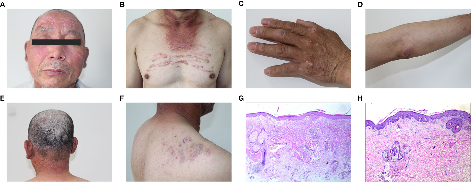
In 2021, a 63-year-old Chinese male presented with a four-month history of infiltrative erythema on his face, neck, and upper chest, accompanied by muscle soreness and weakness in his limbs. Additionally, he had a 10-year history of psoriasis that only had topical therapies, and the symptoms were often recurrent. The physical examination revealed facial and periorbital edematous violaceous erythema (Figure 1A), erythema on the neck and upper chest (Figure 1B), Gottron’s papules (Figure 1C), Gottron’s sign (Figure 1D), scaly plaques on his scalp (Figure 1E) and back (Figure 1F). There was no arthralgia or nail involvement. Both the upper and lower limbs exhibited a grade 4 muscle strength accompanied by muscle tenderness.

Figure 1 (A) Edematous violaceous erythema on the face and periorbital tissues. (B) Erythema on the neck and upper chest. (C) Left hand displaying Gottron’s papules. (D) The upper limb displays Gottron’s sign. (E) Scaly plaques on the scalp. (F) Scaly plaques on the back. (G) Histology from the facial lesion revealed liquefaction of basal cells, edema of the superficial dermis, infiltration of lymphocytes around small vessels with nuclear dust. (H) Skin biopsy from a scaly erythema lesion on the shoulder showed hyperkeratosis, parakeratosis, neutrophil accumulation, disappearance or thinning of the granular layer, mild thickening of the spinous layer, infiltration of lymphocytes and histiocytes around small vessels in the superficial dermis and deposition of mucinous substances in the interstitium.
Laboratory assay results revealed increased levels of lactate dehydrogenase (LDH) [417 U/L, normal range (NR) 120–246], aspartate aminotransferase (AST) (55 U/L, NR 15–46) and creatine kinase (CK) (229 U/L, NR 55–170). Biomarkers for lung cancer such as cytokeratin-19 fragment (CYFRA21-1) (27.1 ng/ml, NR 0–16.3) and neuron-specific enolase (NSE) (3.89 ng/ml, NR 0–3.3) were increased. However, his myositis-specific antibodies (MSAs) and autoantibody profiles were negative. Myogenic injuries to the right deltoid muscle and the biceps brachii muscle were seen on the electromyogram (EMG). A CT scan revealed chronic inflammation in the middle lobe on the right side and upper lobe on the left side of the lung, without any additional irregularities. Inflammation was detected in the lateral muscle groups of both shins through muscle MRI, along with the presence of effusion. A skin biopsy from the facial lesion revealed liquefaction of basal cells, edema of the superficial dermis, infiltration of lymphocytes around small vessels with nuclear dust (Figure 1G). Another skin biopsy from a scaly erythema lesion on his shoulder showed hyperkeratosis, parakeratosis, neutrophil accumulation, disappearance or thinning of the granular layer, mild thickening of the spinous layer, infiltration of lymphocytes and histiocytes around small vessels in the superficial dermis and deposition of mucinous substances in the interstitium (Figure 1H). The patient was diagnosed with dermatomyositis according to the Bohan and Peter’s criteria (11) and received treatment consisting of 24 mg/day methylprednisolone (MPSL), 400 mg/day hydroxychloroquine (HCQ), and 15 mg/week methotrexate(MTX). One month later, there was a notable relief in symptoms, with CK, LDH, and AST levels falling within the normal range. Additionally, the dosages of MTX and HCQ were reduced to 10 mg per week and 200 mg per day respectively. Subsequently, the doses of the three medications were gradually tapered, and now he receives 8 mg/day MPSL, 200 mg/day HCQ, and 5 mg/week MTX for treatment without any signs of recurrence. During the follow-up period, no clinical or radiological evidence of malignancy was observed in our patient.
3 Discussion
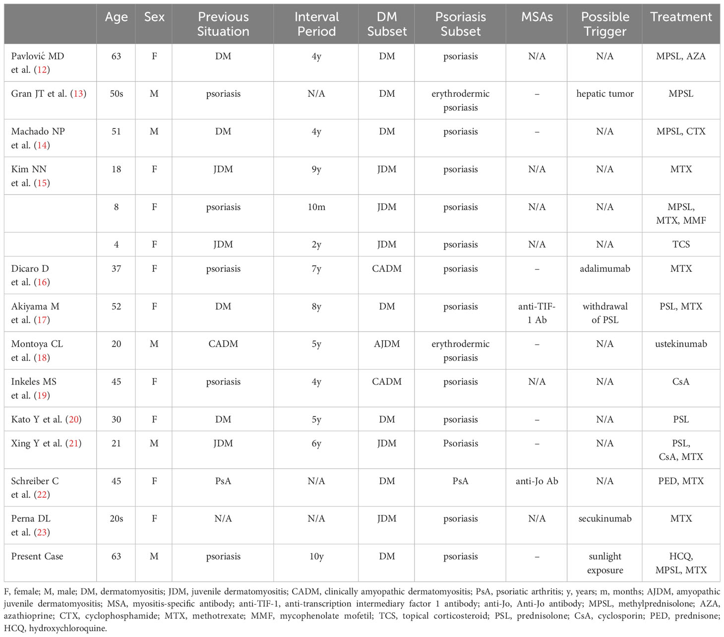
Autoimmune diseases like systemic lupus erythematosus (SLE), systemic sclerosis, and rheumatoid arthritis may coexist with psoriasis (10). Cases of psoriasis concurrent with dermatomyositis are also occasionally seen in daily clinical practice. As far as we know, there have been limited instances documented in the literature (Table 1) (12–23). In these cases, five were male and ten were female, of which six patients were under thirty years old, and they were all diagnosed with juvenile dermatomyositis. Six cases developed psoriasis prior to dermatomyositis, and dermatomyositis preceded in other cases. Among these cases, one had diabetes, hypertension, and anti-glomerular basement membrane disease, another had a hepatic tumor, a third had Hashimoto’s thyroiditis and Sjögren syndrome, and a fourth had interstitial lung disease.
Regarding the possible triggers, three cases might be associated with medications, including adalimumab, secukinumab, and withdrawal of prednisolone. One case might be linked to a hepatic tumor. As for our case, he had no medication history or underlying disease. He was a farmer who had been exposed to prolonged sunlight without any protection in this summer. We suspected that sunlight exposure might be the possible trigger.
Mechanistically, both DM and psoriasis are autoimmune diseases. Previous evidence has shown that these two diseases share some signaling pathways and cytokines (24, 25). For example, IFN can induce apoptosis and cause vascular damage directly (25), while TNF-α may also play a direct role in causing muscle inflammation in DM patients (25). In psoriasis, IFN can activate myeloid dendritic cells to secrete IL-12 and IL-23, and induce the activation and proliferation of Th1, Th17, and Th22 cells, resulting in the secretion of cytokines such as TNF-α, IL-17, and IL-22. These cytokines further stimulate keratinocytes, which produce related cytokines and chemokines to form inflammatory circuits and promote the characteristic changes in psoriasis (5). However, although psoriasis and dermatomyositis share some signaling pathways and cytokines, the mechanisms of their co-occurrence are still unclear. Based on previous studies, we speculate that there seems to be a complex, interacting, and self-sustaining inflammatory circuit among these cytokines in psoriasis (5). However, drugs such as adalimumab and secukinumab can disrupt the balance of the inflammatory circuit, leading to the accumulation or dominance of certain cytokines within inflammatory circuit and ultimately resulting in the appearance of clinical symptoms of DM, even though this condition is relatively rare (16, 23). As mentioned above, sunlight exposure may be a possible trigger for our patient. On one hand, ultraviolet (UV) radiation in sunlight is a proposed trigger for DM (26). On the other hand, UV radiation could suppress the IL-23/IL-17 axis, resulting in the inhibition of the production of IL-17 (27). This immune response results in a reduction of IL-17-mediated inflammation in skin lesions, which is similar to the effect of IL-17 inhibitors, and can also disrupt the inflammatory circuit. Accordingly, we think that UV radiation present in sunlight might play a significant role in the pathogenesis of the co-existence of dermatomyositis and psoriasis. However, further investigation is needed to confirm this point.
As for the treatment, 9 patients received corticosteroid treatment, 8 patients received MTX, other treatments included cyclosporin (CsA), cyclophosphamide (CTX), azathioprine (AZA), mycophenolate mofetil (MMF), HCQ, ustekinumab and topical corticosteroid (TCS). While two diseases share some inflammatory pathways and some therapy options could apply simultaneously, it could also be seen that treatment of one disease may exacerbate another one. For instance, UV phototherapy is safe and effective for psoriasis (28), but it is also a trigger for DM and even exacerbates the symptoms of DM (26). TNF-α inhibitors such as adalimumab, IL-17 inhibitors such as secukinumab, are effective for psoriasis, and are frequently used worldwide. However, there are studies reporting dermatomyositis or psoriasis occurring or exacerbating after receiving these two therapies (16, 23). And while TNF-α inhibitors may be a potential therapy for DM, the worsening of the disease can also be seen during treatment (29). Further observation is necessary when using TNF-α inhibitors to treat either both diseases simultaneously or only DM. Furthermore, corticosteroids are considered the preferred first-line therapy for DM-associated myopathy (30), but their use in psoriasis is not advised unless the situation is highly critical and the symptoms cannot be controlled by other therapies. Thus, when treating dermatomyositis accompanied by psoriasis, corticosteroids need to be used carefully. Clinically, immunosuppressive agents such as MTX are commonly given with corticosteroids to reduce the doses and side effects of corticosteroids (30). It seems to be most commonly combined with corticosteroid in treating dermatomyositis accompanied by psoriasis. Intravenous immunoglobulin (IVIG) was not mentioned in the treatment of the co-existence of dermatomyositis and psoriasis. On one hand, although IVIG shows efficacy in treating dermatomyositis (31), the evidence for the treatment of psoriasis is limited and may even lead to the aggravation of psoriasis (32). On the other hand, economic burden is a common reason for patients not to receive IVIG treatment. Additional drugs such as CsA (19), CTX (14) and ustekinumab (18) have shown efficacy in treating the concurrent presence of dermatomyositis and psoriasis. But these treatments are case reports, lacking high-quality evidence, and should only be for reference. Currently, the management of the simultaneous occurrence of dermatomyositis and psoriasis is still in the preliminary stage of investigation.
Limitations associated with this case report warrant mention. For example, the muscle biopsy and examination of the specific HLA alleles were not performed as consent was not obtained. Moreover, there is a lack of results for nail-fold capillary capillaroscopy, which is characteristic in dermatomyositis and could reflect the ongoing disease activity (33).
While dermatomyositis coexistence with psoriasis has been reported in various instances globally, this case stands out as the initial one to have achieved successful treatment through the combination of corticosteroids, methotrexate, and hydroxychloroquine. Herein, we report this case to provide some experience for clinical practice.
Data availability statement
The original contributions presented in the study are included in the article/supplementary material. Further inquiries can be directed to the corresponding author.
Ethics statement
Written informed consent was obtained from the individual(s), and minor(s)’ legal guardian/next of kin, for the publication of any potentially identifiable images or data included in this article.
Author contributions
DC: Conceptualization, Writing – original draft. WY: Methodology, Writing – original draft, Writing – review & editing. JN: Investigation, Writing – review & editing.
Funding
The author(s) declare financial support was received for the research, authorship, and/or publication of this article. The funding for this study were provided by the Technological Innovation Planned Project of Liaoning Province (Grant number 2022JH2/101500012).
Conflict of interest
The authors declare that the research was conducted in the absence of any commercial or financial relationships that could be construed as a potential conflict of interest.
Publisher’s note
All claims expressed in this article are solely those of the authors and do not necessarily represent those of their affiliated organizations, or those of the publisher, the editors and the reviewers. Any product that may be evaluated in this article, or claim that may be made by its manufacturer, is not guaranteed or endorsed by the publisher.
References
1. Furst DE, Amato AA, Iorga ŞR, Gajria K, Fernandes AW. Epidemiology of adult idiopathic inflammatory myopathies in a U.S. managed care plan. Muscle Nerve (2012) 45:676–83. doi: 10.1002/mus.23302
2. Miller FW, Cooper RG, Vencovský J, Rider LG, Danko K, Wedderburn LR, et al. Genome-wide association study of dermatomyositis reveals genetic overlap with other autoimmune disorders. Arthritis Rheumatism (2013) 65:3239–47. doi: 10.1002/art.38137
3. Rothwell S, Cooper RG, Lundberg IE, Miller FW, Gregersen PK, Bowes J, et al. Dense genotyping of immune-related loci in idiopathic inflammatory myopathies confirms HLA alleles as the strongest genetic risk factor and suggests different genetic background for major clinical subgroups. Ann Rheum Dis (2016) 75:1558–66. doi: 10.1136/annrheumdis-2015-208119
4. Huard C, Gullà SV, Bennett DV, Coyle AJ, Vleugels RA, Greenberg SA. Correlation of cutaneous disease activity with type 1 interferon gene signature and interferon β in dermatomyositis. Br J Dermatol (2017) 176:1224–30. doi: 10.1111/bjd.15006
5. Griffiths CEM, Armstrong AW, Gudjonsson JE, Barker JNWN. Psoriasis. Lancet (2021) 397:1301–15. doi: 10.1016/S0140-6736(20)32549-6
6. Gottlieb AB, Krueger JG. HLA region genes and immune activation in the pathogenesis of psoriasis. Arch Dermatol (1990) 126:1083–6.
7. Nair RP, Stuart PE, Nistor I, Hiremagalore R, Chia NVC, Jenisch S, et al. Sequence and haplotype analysis supports HLA-C as the psoriasis susceptibility 1 gene. Am J Hum Genet (2006) 78:827–51. doi: 10.1086/503821
8. FitzGerald O, Haroon M, Giles JT, Winchester R. Concepts of pathogenesis in psoriatic arthritis: genotype determines clinical phenotype. Arthritis Res Ther (2015) 17:115. doi: 10.1186/s13075-015-0640-3
9. Ghoreschi K, Balato A, Enerbäck C, Sabat R. Therapeutics targeting the IL-23 and IL-17 pathway in psoriasis. Lancet (2021) 397:754–66. doi: 10.1016/S0140-6736(21)00184-7
10. Sticherling M. Psoriasis and autoimmunity. Autoimmun Rev (2016) 15:1167–70. doi: 10.1016/j.autrev.2016.09.004
11. Bohan A, Peter JB. Polymyositis and dermatomyositis: (First of two parts). N Engl J Med (1975) 292:344–7. doi: 10.1056/NEJM197502132920706
12. Pavlović MD, Zecević RD, Zolotarevski L. Psoriasis in a patient with dermatomyositis. Vojnosanit Pregl (2004) 61:557–9. doi: 10.2298/vsp0405557p
13. Gran JT, Gunnarsson R, Mørk N-J. [A man with erythrodermia, muscle weakness and weight loss]. Tidsskr Nor Laegeforen (2009) 129:2240–1. doi: 10.4045/tidsskr.09.0184
14. MaChado NP, Camargo CZ, Oliveira ACD, Buosi ALP, Pucinelli MLC, de Souza AWS. Association of anti-glomerular basement membrane antibody disease with dermatomyositis and psoriasis: case report. Sao Paulo Med J (2010) 128:306–8. doi: 10.1590/s1516-31802010000500012
15. Kim NN. Double trouble: therapeutic challenges in patients with both juvenile dermatomyositis and psoriasis. Arch Dermatol (2011) 147:831. doi: 10.1001/archdermatol.2011.49
16. Dicaro D, Bowen C, Dalton SR. Dermatomyositis associated with anti-tumor necrosis factor therapy in a patient with psoriasis. J Am Acad Dermatol (2014) 70:e64–65. doi: 10.1016/j.jaad.2013.11.012
17. Akiyama M, Ueno T, Kanzaki A, Kuwana M, Nagao M, Saeki H. Association of psoriasis with Hashimoto’s thyroiditis, Sjögren’s syndrome and dermatomyositis. J Dermatol (2016) 43:711–2. doi: 10.1111/1346-8138.13265
18. Montoya CL, Gonzalez ML, Ospina FE, Tobón GJ. A rare case of amyopathic juvenile dermatomyositis associated with psoriasis successfully treated with ustekinumab. J Clin Rheumatol (2017) 23:129–30. doi: 10.1097/RHU.0000000000000430
19. Inkeles MS, No D, Wu JJ. Clinical improvement of a patient with both amyopathic dermatomyositis and psoriasis following treatment with cyclosporine. Dermatol Online J (2017) 23:13030/qt5qh0n741.
20. Kato Y, Yamamoto T. Development of psoriasis with relapse of dermatomyositis-associated interstitial lung disease. Int J Rheum Dis (2017) 20:660–1. doi: 10.1111/1756-185X.13084
21. Xing Y, Xie J, Jiang S, Upasana M, Song J. Co-existence of Juvenile dermatomyositis and psoriasis vulgaris with fungal infection: A case report and literature review. J Cosmetic Dermatol (2019) 18:1560–3. doi: 10.1111/jocd.12869
22. Schreiber C, Khamlong M, Raza N, Huynh BQ. Overlap of psoriatic arthritis and dermatomyositis. J Invest Med High Impact Case Rep (2021) 9:232470962110577. doi: 10.1177/23247096211057702
23. Perna DL, Callen JP, SChadt CR. Association of treatment with secukinumab with exacerbation of dermatomyositis in a patient with psoriasis. JAMA Dermatol (2022) 158:454–6. doi: 10.1001/jamadermatol.2021.6011
24. Rendon A, Schäkel K. Psoriasis pathogenesis and treatment. IJMS (2019) 20:1475. doi: 10.3390/ijms20061475
25. Kao L, Chung L, Fiorentino DF. Pathogenesis of dermatomyositis: role of cytokines and interferon. Curr Rheumatol Rep (2011) 13:225–32. doi: 10.1007/s11926-011-0166-x
26. DeWane ME, Waldman R, Lu J. Dermatomyositis: Clinical features and pathogenesis. J Am Acad Dermatol (2020) 82:267–81. doi: 10.1016/j.jaad.2019.06.1309
27. Johnson-Huang LM, Suárez-Fariñas M, Sullivan-Whalen M, Gilleaudeau P, Krueger JG, Lowes MA. Effective narrow-band UVB radiation therapy suppresses the IL-23/IL-17 axis in normalized psoriasis plaques. J Invest Dermatol (2010) 130:2654–63. doi: 10.1038/jid.2010.166
28. Li Y, Cao Z, Guo J, Li Q, Zhu W, Kuang Y, et al. Assessment of efficacy and safety of UV-based therapy for psoriasis: a network meta-analysis of randomized controlled trials. Ann Med (2022) 54:159–69. doi: 10.1080/07853890.2021.2022187
29. Dastmalchi M, Grundtman C, Alexanderson H, Mavragani CP, Einarsdottir H, Helmers SB, et al. A high incidence of disease flares in an open pilot study of infliximab in patients with refractory inflammatory myopathies. Ann Rheum Dis (2008) 67:1670–7. doi: 10.1136/ard.2007.077974
30. Waldman R, DeWane ME, Lu J. Dermatomyositis: diagnosis and treatment. J Am Acad Dermatol (2020) 82:283–96. doi: 10.1016/j.jaad.2019.05.105
31. Aggarwal R, Charles-Schoeman C, Schessl J, Bata-Csörgő Z, Dimachkie MM, Griger Z, et al. Trial of intravenous immune globulin in dermatomyositis. N Engl J Med (2022) 387:1264–78. doi: 10.1056/NEJMoa2117912
32. Ettler J, Arenberger P, Arenbergerova M, Gkalpakiotis S. Severe exacerbation of psoriasis after intravenous immunoglobulin in patient with multiple sclerosis that started during biologic therapy. Acad Dermatol Venereol (2016) 30:355–6. doi: 10.1111/jdv.12767
33. Christen-Zaech S, Seshadri R, Sundberg J, Paller AS, Pachman LM. Persistent association of nailfold capillaroscopy changes and skin involvement over thirty-six months with duration of untreated disease in patients with juvenile dermatomyositis. Arthritis Rheumatism (2008) 58:571–6. doi: 10.1002/art.23299
Keywords: psoriasis, dermatomyositis, pathogenesis, corticosteroid, case report
Citation: Chu D, Yang W and Niu J (2024) Concurrence of dermatomyositis and psoriasis: a case report and literature review. Front. Immunol. 15:1345646. doi: 10.3389/fimmu.2024.1345646
Received: 28 November 2023; Accepted: 12 January 2024;
Published: 29 January 2024.
Edited by:
Mattia Bellan, University of Eastern Piedmont, ItalyReviewed by:
Giusto Trevisan, University of Trieste, ItalyAlbert E. Zhou, UCONN Health, United States
Copyright © 2024 Chu, Yang and Niu. This is an open-access article distributed under the terms of the Creative Commons Attribution License (CC BY). The use, distribution or reproduction in other forums is permitted, provided the original author(s) and the copyright owner(s) are credited and that the original publication in this journal is cited, in accordance with accepted academic practice. No use, distribution or reproduction is permitted which does not comply with these terms.
*Correspondence: Jun Niu, bml1anVuMDZAMTI2LmNvbQ==
†These authors have contributed equally to this work and share first authorship
 Dan Chu
 Wei Yang
 Jun Niu*