
94% of researchers rate our articles as excellent or good
Learn more about the work of our research integrity team to safeguard the quality of each article we publish.
Find out more
CORRECTION article
Front. Immunol., 05 March 2024
Sec. Microbial Immunology
Volume 15 - 2024 | https://doi.org/10.3389/fimmu.2024.1330357
 Quang Vinh Ngo1,2
 Larissa Faass1,3
 Aline Sähr1Dagmar Hildebrand1
 Tatjana Eigenbrod1Klaus Heeg1,2
 Dennis Nurjadi1,2*
A Corrigendum on
Inflammatory response against Staphylococcus aureus via intracellular sensing of nucleic acids in keratinocytes
By Ngo QV, Faass L, Sähr A, Hildebrand D, Eigenbrod T, Heeg K and Nurjadi D (2022) Front. Immunol. 13:828626. doi: 10.3389/fimmu.2022.828626
In the published article, there was an error in the published Figure 5. In the original published version of Figure 5, we separated the Western blot (panel B) of the whole bacterial cells and the transfected bacterial RNA for illustrative purposes. As a result, the negative control was duplicated as the Western blot was run as a single blot for ease of comparison/illustration. However, this was not clearly stated in the caption or in the figure, which may have given the impression that the blot could have been ‘manipulated’. To avoid further misunderstanding, we have decided to replace the Western blot figure in panel B with an untrimmed and identical version of the same blot. For transparency, we have included the original blots and other raw data as additional supplementary files. The corrected Figure 5 and its caption appear below.
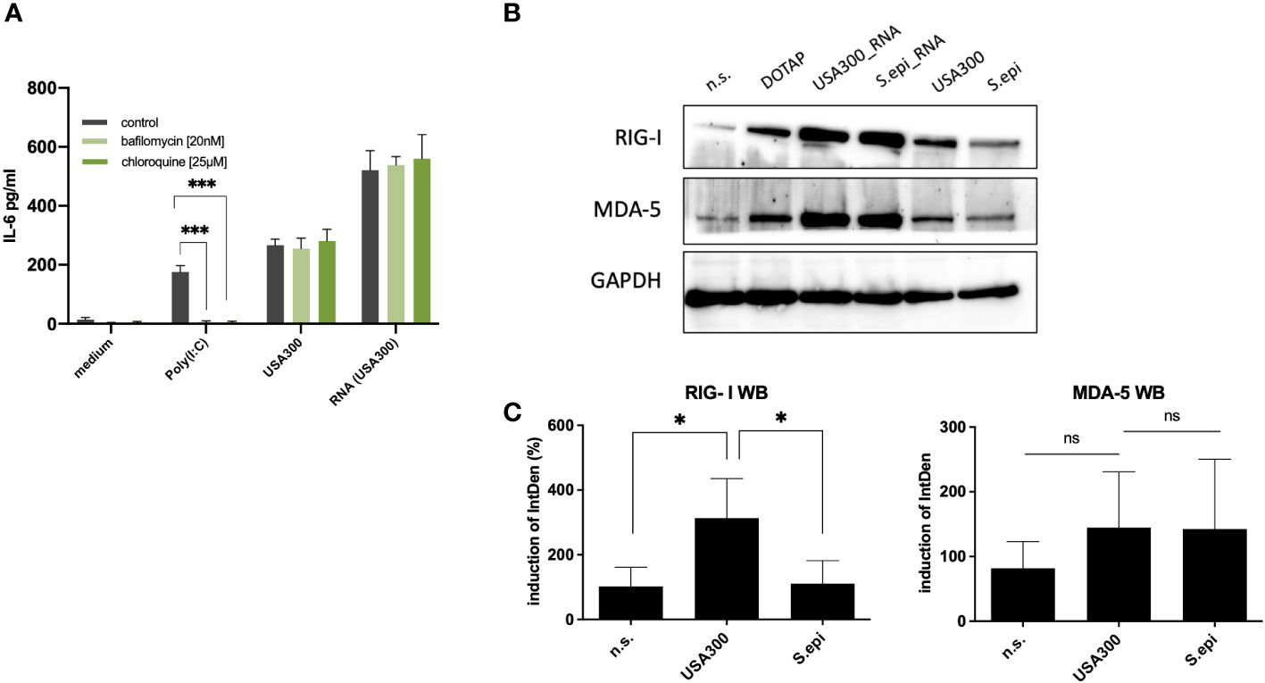
Figure 5 Cytoplasmic RNA sensors may be responsible for the induction of IL-6 by invasive staphylococci. (A) Inhibition with the endosomal TLR inhibitors bafilomycin and chloroquine did not affect the IL-6 response at MOI 100. (B) Both MDA-5 and RIG-I may be involved in the recognition of bacterial RNA following bacterial invasion, as quantified by western blot. Viable S. aureus USA300 induces more RIG-I and MDA5 in keratinocytes compared to viable S. epidermidis at MOI of 100. Transfection of S. aureus USA300 and S. epidermidis induced similar levels of RIG-I and MDA5. All experiments were performed as independent experiments in biological triplicates, each in technical duplicates, except for Western Blot (one representative experiment of three experiments). Raw file of the blot is provided in the supplementary. (C) Quantification of RIG-I and MDA-5 western blots of keratinocytes stimulated with viable S. aureus and S. epidermidis at MOI of 100. Quantification of protein bands were performed via ImageJ software. Data were analyzed by Graphpad Prism v9 (USA). The IntDen of protein band of interest was divided through IntDen of the respective houskeeping protein band. For calculation of induction mean of data of three unstimulated samples were set to 100 percentage. Statistically significant differences are indicated by * (*p < 0.5, ***p ≤ 0.001, ns, not significant).
The authors apologize for this error and state that this does not change the scientific conclusions of the article in any way. The original article has been updated.
All claims expressed in this article are solely those of the authors and do not necessarily represent those of their affiliated organizations, or those of the publisher, the editors and the reviewers. Any product that may be evaluated in this article, or claim that may be made by its manufacturer, is not guaranteed or endorsed by the publisher.
Keywords: Staphylococcus aureus, Staphylococcus epidermidis, keratinocyte, skin immune response, bacterial RNA, host-pathogen interaction
Citation: Ngo QV, Faass L, Sähr A, Hildebrand D, Eigenbrod T, Heeg K and Nurjadi D (2024) Corrigendum: Inflammatory response against Staphylococcus aureus via intracellular sensing of nucleic acids in keratinocytes. Front. Immunol. 15:1330357. doi: 10.3389/fimmu.2024.1330357
Received: 30 October 2023; Accepted: 13 February 2024;
Published: 05 March 2024.
Edited and Reviewed by:
Sasha Shafikhani, Rush University Medical Center, United StatesCopyright © 2024 Ngo, Faass, Sähr, Hildebrand, Eigenbrod, Heeg and Nurjadi. This is an open-access article distributed under the terms of the Creative Commons Attribution License (CC BY). The use, distribution or reproduction in other forums is permitted, provided the original author(s) and the copyright owner(s) are credited and that the original publication in this journal is cited, in accordance with accepted academic practice. No use, distribution or reproduction is permitted which does not comply with these terms.
*Correspondence: Dennis Nurjadi, ZGVubmlzLm51cmphZGlAdW5pLWhlaWRlbGJlcmcuZGU=
Disclaimer: All claims expressed in this article are solely those of the authors and do not necessarily represent those of their affiliated organizations, or those of the publisher, the editors and the reviewers. Any product that may be evaluated in this article or claim that may be made by its manufacturer is not guaranteed or endorsed by the publisher.
Research integrity at Frontiers
Learn more about the work of our research integrity team to safeguard the quality of each article we publish.