 Ri Zhang
 Yujia Wei
 Tingmei Wang
 Xiaoqi Nie
 Dong Li
94% of researchers rate our articles as excellent or good
Learn more about the work of our research integrity team to safeguard the quality of each article we publish.
Find out more
MINI REVIEW article
Front. Immunol., 01 December 2023
Sec. Cytokines and Soluble Mediators in Immunity
Volume 14 - 2023 | https://doi.org/10.3389/fimmu.2023.1307455
This article is part of the Research TopicThe immunological regulation of extracellular vesicles on chronic diseasesView all 12 articles
Exosomes, bilaterally phospholipid-coated small vesicles, are produced and released by nearly all cells, which comprise diverse biological macromolecules, including proteins, DNA, RNA, and others, that participate in the regulation of their biological functions. An increasing number of studies have revealed that the contents of exosomes, particularly microRNA(miRNA), play a significant role in the pathogenesis of various diseases, including autoimmune skin diseases. MiRNA is a class of single-stranded non-coding RNA molecules that possess approximately 22 nucleotides in length with the capability of binding to the untranslated as well as coding regions of target mRNA to regulate gene expression precisely at the post-transcriptional level. Various exosomal miRNAs have been found to be significantly expressed in some autoimmune skin diseases and involved in the pathogenesis of conditions via regulating the secretion of crucial pathogenic cytokines and the direction of immune cell differentiation. Thus, exosomal miRNAs might be promising biomarkers for monitoring disease progression, relapse and reflection to treatment based on their functions and changes. This review summarized the current studies on exosomal miRNAs in several common autoimmune skin diseases, aiming to dissect the underlying mechanism from a new perspective, seek novel biomarkers for disease monitoring and lay the foundation for developing innovative target therapy in the future.
MicroRNA (miRNA), first discovered within nematodes by Ambro et al. (1) in 1993, is a class of single-stranded non-coding RNA molecules typically consisting of approximately 22 nucleotides in length. MiRNA binds to the 3’ untranslated region (UTR) and coding region of target mRNA through base pairing, leading to either degradation or inhibition of translation of the target mRNA (2), enabling precise control of gene expression at the post-transcriptional level (3). MiRNA is involved in various cellular activities, including proliferation, differentiation, apoptosis, and metabolism (4). It has emerged as a potential biomarker and therapeutic target for numerous diseases, making it a prominent focus of research in the medical field in recent years (5).
Exosomes, vesicular structures with a diameter ranging from 40 to 160nm enclosed within a bilayer phospholipid membrane, contain diverse biological molecules, including nucleic acids, lipids and proteins. They are widely distributed and can be generated by almost all cells and detected in various body fluids (6–9).
Unlike freely circulating miRNA in body fluids, miRNA within exosomes resists RNA degradation by ribonucleases, making it more stable (10). Furthermore, exosomal miRNA exhibits potential homing properties (11) and selective enrichment, enabling disease occurrence, progression and relapse monitoring (12), which is critical for clinical management of autoimmune diseases.
Autoimmune skin diseases are defined as autoimmune diseases characterized by excessive activation of the immune system leading to abnormal immune reactions and responses against self-antigens that are normally tolerated, including but not limited to skin involvement, such as vitiligo, psoriasis, systemic lupus erythematosus and dermatomyositis. In recent years, there has been a steady increase in the incidence and prevalence of autoimmune skin diseases, accounting for an essential part of the global disease mortality and economic burden. The uncertainty of etiology, the complexity of pathogenesis, and the unpredictable nature of disease progression have posed significant challenges to patients treating and managing autoimmune skin diseases, significantly impacting patients’ quality of life. Therefore, searching for new therapeutic targets and identifying biomarkers that can predict disease progression and treatment effectiveness is crucial. As mentioned above, exosomal miRNA can be a promising choice for its function and nature. A comprehensive map illustrating how exosomal miRNAs are involved in autoimmune skin diseases by regulating transcripts, pathways, immune system differentiation, and their interactions with terminal cells such as keratinocytes, fibroblasts, and immune cells is no doubt important yet still lacking. Here, we reviewed recent evidence on the role of exosomal miRNAs within autoimmune skin diseases and discussed their impact on these diseases (Table 1), aiming to facilitate a better understanding of the pathogenetic mechanisms of autoimmune skin diseases and clinical management.
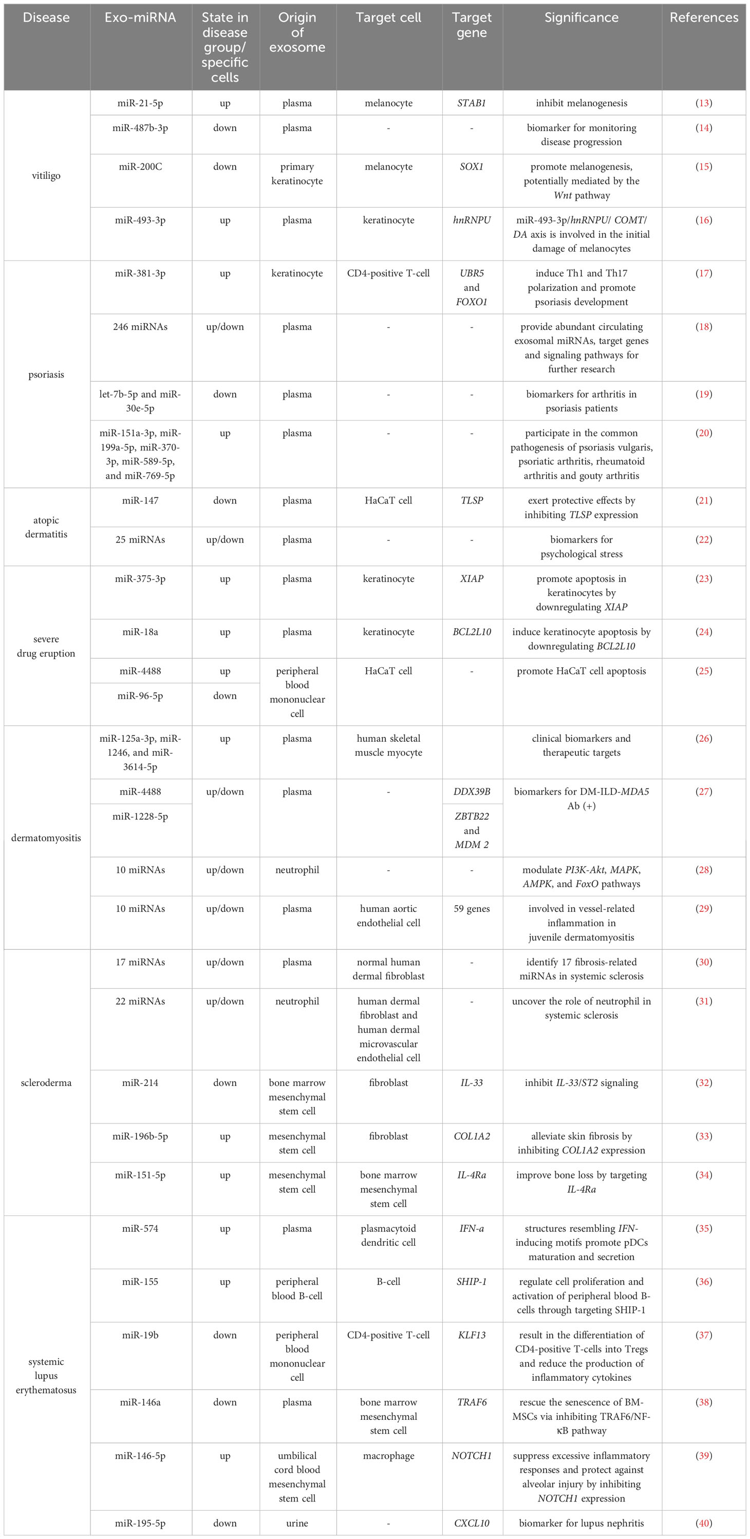
Table 1 Summary of exosomal miRNAs profiles of indicated autoimmune skin diseases.
Vitiligo is an acquired pigmentary skin disorder that involves the participation of various innate and adaptive immune cells, leading to damage to melanocytes in the skin and hair follicles (41), resulting in depigmented patches.
Several researches have yielded positive results that circulating exosomal miRNAs contribute to vitiligo’s pathogenesis. Zhang et al. (13) co-cultured circulating exosomes from vitiligo patients with the human melanocyte cell line PIG1. They observed the inhibition of melanogenesis, decreased tyrosinase activity, and altered expression of genes related to melanogenesis in melanocytes. Furthermore, they detected significantly higher expression of miR-21-5p in exosomes from vitiligo patients and confirmed that miR-21-5p inhibits melanogenesis, as evidenced by changes in the levels of tyrosinase and tyrosinase-related protein 1 (TYP1) and tyrosinase-related protein 2 (TYP2). Luo et al. (14) found that circulating exosomal miR-487b-3p in advanced-stage vitiligo patients was significantly downregulated before glucocorticoid treatment but recovered to normal levels after intervention. Enrichment analysis suggested that this miRNA primarily affects metabolic pathways.
Additionally, skin keratinocytes (42) and fibroblasts (43) are involved in this disease’s abnormal immune environment that promotes local T-cell infiltration by secreting CXCL9, CXCL10, and various cytokines. Moreover, melanocytes appear to be not solely victims of the abnormal immune response but also participants in initiating the immune dysfunction (44). Zhao et al. (15) found that melanocytes exhibited a significant decrease in melanin content and tyrosinase activity after being cultured with exosomes from vitiligo patients’ skin lesional keratinocytes. Furthermore, they discovered that miR-200C, downregulated in these exosomes, could promote melanogenesis, potentially mediated by the inhibition of SOX1 to activate the Wnt pathway. Li et al. (16) performed high-throughput sequencing and correlation analysis of circulating exosomal miRNAs from segmental vitiligo patients and healthy controls with disease progression and staging, screened and expanded the specimens to verify that miR-493-3p, which was highly expressed in circulating exosomes of the patients as well as keratinocytes of the lesions. They subsequently demonstrated in vitro the miR-493-3p-hnRNPU-COMT-DA axis on the initial damage of melanocytes. However, there are no current studies on exosomal miRNAs referring to the role of fibroblasts or melanocytes initiating abnormal immunity, which might be worth investigating in the future to uncover the mechanism of vitiligo from multidimensional perspectives.
Psoriasis is a chronic inflammatory skin disease characterized by abnormal activation and infiltration of T-cells and excessive proliferation of keratinocytes, clinically manifesting red plaques and papules covered with thick silver-white scales. Th17 cells and IL-17A/IL-23 play a crucial role in the immune dysfunction in psoriasis (45). Jiang et al. (17) discovered that exosomes derived from keratinocytes treated with psoriasis cytokines (IL-17A, IL-22, IFN-γ, TNF-α) can induce the differentiation of CD4-positive T-cells into Th1 and Th17 cells and upregulate the expression of various cytokines including IL-17A, IL-17F, IL-22, IL-23, IL-36, IFN-γ, and TNF-α. MiR-381-3p was found to upregulate the expression of the indicated cytokines in CD4-positive T-cells. UBR5 and FOXO1 were identified as critical downstream target genes, playing essential roles in the immune response in psoriasis.
Chen et al. (18) sequenced circulating exosomal miRNAs in 15 patients with psoriasis vulgaris and 15 healthy controls and identified 246 differentially expressed miRNAs. Subsequently, they discovered that enrichment analysis could enrich some target genes in inflammatory metabolic pathways. This study facilitated other researchers to select circulating exosomal miRNAs and target genes of interest at the subsequent step of investigating in psoriasis.
In addition to skin involvement, joints can also be affected, known as psoriatic arthritis (PsA). Pasquali et al. (19) identified 15 circulating exosomal miRNAs from 14 patients with psoriasis vulgaris (PsV) and 15 PsA patients. After expanding the sample size for validation along with regression analysis, they found that the expression levels of let-7b-5p and miR-30e-5p were negatively correlated with PsA group, which might be possible biomarkers for PsA. Chen et al. (20) extracted plasma exosomes from 15 PsV patients, 30 PsA patients, 15 patients with rheumatoid arthritis, 15 patients with gouty arthritis, and 15 healthy controls and identified five miRNAs (hsa-miR-151a-3p, hsa-miR-199a-5p, hsa-miR-370-3p, hsa-miR-589-5p, and hsa-miR-769-5p) that mainly participate in the common pathogenesis of these four diseases by affecting inflammation and bone metabolism.
Atopic dermatitis (AD) is a chronic inflammatory skin disease characterized by intense itching, alternating acute episodes, and remissions, significantly affecting patients’ quality of life. Crosstalk between keratinocytes and various immune cells results in skin barrier impairment via secreting diversified cytokines, such as TNF-α and IFN-γ (46). Shi et al. (21) detected significantly downregulated levels of miR-147 in the plasma, lesional tissues of a mouse model of AD and HaCaT cell models compared to the negative control group. The expression of miR-147 was negatively correlated with TLSP and VEGFA, two vascular growth-related factors considered important in the pathogenesis of AD (47). Treatment with TNF-α/IFN-γ decreased the viability of HaCaT cells, upregulated TLSP and VEGFA expression, and promoted cell apoptosis. However, overexpression of miR-147 reversed these damaging processes in HaCaT cells. Furthermore, they found that exosomes from adipose-derived stem cells overexpressing miR-147 exerted similar protective effects and inhibited TLSP expression (21). These results indicate exosomal miR-147 as a potential target for AD therapy.
Besides, some studies have indicated a correlation between the onset of atopic dermatitis and psychological stress. Moreover, it has been demonstrated that psychotherapy can alleviate some patients’ symptoms (48). Sung et al. (22) performed sequencing and differential analysis of plasma-derived neuronal exosomes from AD mouse models, identifying 9 significantly upregulated and 16 considerably downregulated miRNAs, which can be utilized to unveil mechanism of AD regarding psychological factors.
Drug eruption, one of the most common adverse drug reactions, manifests as inflammatory skin and mucosal lesions with possible systemic involvement. Stevens-Johnson Syndrome (SJS), Toxic Epidermal Necrolysis (TEN), Acute Generalized Exanthemata’s Pustulosis (AGEP) and Drug Reaction with Eosinophilia and Systemic Symptoms (DRESS) are collectively known as severe drug eruptions characterized by large-scale death of keratinocytes (49). High mortality rates of severe drug eruptions urge dermatologists to excavate disease mechanisms and develop improved therapies. exosomes miRNA helps better understand the pathogenesis of severe drug eruptions that remain unclear.
Zhang et al. (23) identified upregulation of miR-375-3p in exosomes from patients with SJS and TEN. Overexpression of miR-375-3p in primary human keratinocytes reduced cell viability and promoted apoptosis via downregulating XIAP. Furthermore, they observed a positive correlation between the expression levels of miR-375-3p and the affected body surface area and epidermal necrosis score in patients with severe drug eruptions. Salinas et al. (24) investigated circulating exosomal miRNAs of patients with severe drug eruptions (9 cases of DRESS, 8 cases of SJS/TEN) and identified 24 significantly upregulated miRNAs, which were predominantly involved in T-cell activation, cell apoptosis, and inflammation processes. From these findings, they verified differential overexpression of miR-18a, consistent with previous research (50) that showed significant upregulation of miR-18a in the skin lesions and plasma of TEN patients, and it was found to induce keratinocyte apoptosis by downregulating BCL2L10. Suthumchai et al. (25) observed cytotoxic effects on HaCaT cells by exosomes secreted by peripheral blood mononuclear cells from 12 patients with SJS/TEN and identified upregulated miR-4488 and downregulated miR-486-5p, miR-96-5p, and miR-132-3p. Furthermore, both overexpression of miR-4488 and reduced expression of miR-96-5p promoted HaCaT cells apoptosis.
Dermatomyositis (DM) is an autoimmune disease characterized by edematous purplish-red patches on the upper eyelids, as well as flat brownish-red papules on exposed areas.
Weakness and myalgia are the main manifestations of muscle involvement. Li et al. (26) confirmed the upregulation of has-miR-125a-3p, has-miR-1246, and has-miR-3614-5p in circulating exosomes from untreated DM patients and in human skeletal muscle myoblasts cells stimulated by these exosomes, which returned to normal levels after antirheumatic treatment. Furthermore, these three miRNAs were related to the cellular autophagy pathway and positively correlated with specific indicators in DM patients, such as serum creatine kinase levels and myositis antibody titers, suggesting them as potential biomarkers for DM.
The existence of anti-MDA5 antibodies was related to interstitial lung disease and a worse prognosis in DM patients (51). Zhong et al. (27) sequenced circulating exosomal miRNAs from 5 patients with DM accompanied by interstitial lung disease and positive anti-MDA5 antibody, 5 patients with DM without myositis antibodies, and 5 healthy controls and found significant differences in has-miR-4488 and hsa-miR-1228-5p in all three comparisons. H2AFX and MDM2 were identified as two essential hub genes that may be involved in the pathogenesis of DM. Nonetheless, this study didn’t focus on the mechanism underlying the distinction between MDA5 positive and MDA5 negative DM patients.
Neutrophil/lymphocyte ratio in peripheral blood was found associated with disease activity, lung involvement, and overall survival in DM patients (52). Additionally, proteases within neutrophil cytoplasm seem to participate in muscle inflammation (53). Li et al. (28) validated 10 differentially expressed miRNAs in neutrophils-derived exosomes from DM patients’ peripheral blood and determined PI3K-Akt, MAPK, AMPK, and FoxO as the main downstream signaling pathways, demonstrated and partially explained the role of neutrophil in DM.
In addition, evidences indicate a close relationship between the pathogenesis of DM and vascular changes (54). Jiang et al. (29) identified 10 differentially expressed circulating exosomal miRNAs in adolescent DM patients and 59 differentially expressed genes after co-culturing exosomes with human aortic endothelial cells. Through reciprocal prediction, they found specific downregulated genes in the patient group corresponding to upregulated miRNAs in the exosomal sequencing data, providing some miRNA-target gene axes for future research.
Scleroderma, a chronic autoimmune skin disease characterized by abnormal activation of fibroblasts leading to progressive skin and visceral fibrosis, can be classified into two types: localized cutaneous scleroderma and systemic scleroderma, also known as systemic sclerosis (SSc) (55).
Wermuth et al. (30) validated the upregulation of 9 pro-fibrotic miRNAs and the downregulation of 8 anti-fibrotic miRNAs in exosomes derived from plasma of SSc patients and detected an upregulation of type I collagen fibers and fibronectin in normal human dermal fibroblasts stimulated by these exosomes, indicating the pro-fibrotic function of circulating exosomal miRNAs in SSc.
It has been found that neutrophil-derived exosomes from the peripheral blood of patients can inhibit endothelial cell proliferation and migration (56). Li et al. (31) discovered 22 differentially expressed miRNAs in neutrophil-derived exosomes from the peripheral blood of SSc patients and identified the involvement of the Wnt, IL-23, NOTCH and AMPK pathways. Further co-culturing the exosomes above with primary human dermal fibroblasts and human dermal microvascular endothelial cells from healthy individuals validated the presence of negative correlations between specific miRNAs and target genes associated with these molecular pathways, suggesting neutrophil as one of culprits in SSc.
Mesenchymal stem cells (MSCs) are a type of pluripotent stem cell derived from various tissues such as bone marrow, adipose tissue, placenta, and umbilical cord (57). These cells have demonstrated beneficial effects in improving the manifestations of scleroderma through their anti-inflammatory and anti-fibrotic properties (58), and it is believed that these effects are primarily mediated through the production of extracellular vesicles including exosomes (59). Xie et al. (32) identified downregulation of miR-214 in the peripheral blood of the SSc patients and discovered that bone marrow- MSC-derived exosomes could transport miR-214 to fibroblasts and inhibit their proliferation, migration, and expression of fibrosis-related genes via inhibiting IL-33/ST2 signaling. Baral et al. (33) found that injection of exosomes derived from MSCs could alleviate skin fibrosis in bleomycin-induced SSc mice. They further identified upregulated miR-196b-5p and overexpression of this miRNA in mouse fibroblasts downregulated COL1A2 expression, speculating that miR-196b-5p may play a role in the anti-fibrotic effects of MSC-derived exosomes. Chen et al. (34) discovered that MSCs transplantation could improve bone loss in systemic sclerosis mice by modulating the differentiation of recipient bone marrow-MSCs probably attributed to miR-151-5p derived from MSC-derived exosomes by targeting IL-4Ra, consistent with a previous study identifying IL-4 as a suspicious pathway (60).
These findings exhibited therapeutic potential of MSC-derived exosomal miRNAs for SSc.
Systemic lupus erythematosus (SLE) is a complex autoimmune skin disease involving multiple systems. The characteristic skin lesions of SLE include edematous butterfly rash on the face, discoid rash, vasculitis-like lesions in the distal limbs, oral ulcers, and easily breakable hair at the frontal hairline.
Type I interferon plays a crucial role in the pathogenesis of SLE (61). Salvi et al. (35) demonstrated that circulating exosomal miRNAs with structures resembling interferon-inducing motifs from SLE patients can promote the maturation of human peripheral blood plasmacytoid dendritic cells(pDCs) and their secretion of type I interferon and other pro-inflammatory factors.
In addition to SSc, MSCs were also found therapeutic in SLE by modulating adaptive immunity. Patients exist various autoantibodies in their bodies, exemplified by anti-double-stranded DNA and anti-Sm antibodies (62), demonstrating the dominance of humoral immunity in SLE. Zhao et al. (36) found significantly higher numbers of peripheral blood B-cells in untreated SLE patients compared to healthy individuals, validated the upregulation of miR-155 and confirmed the binding relation between miR-155 and SHIP-1. Additionally, exosomes derived from umbilical cord blood-derived MSCs could upregulate SHIP-1 expression in B-cells and inhibit cell proliferation, activation, and promotion of apoptosis.
However, cellular immunity shouldn’t be easily neglected in SLE. Tu et al. (37) determined that miR-19b was downregulated while KLF13 was upregulated in peripheral blood mononuclear cells of patients with SLE and verified the binding and negative correlation between miR-19b and KLF13. They further discovered that umbilical cord blood-derived MSCs could enrich miR-19b in exosomes, resulting in the differentiation of CD4-positive T-cells into Tregs and reducing the production of inflammatory cytokines.
On the contrary, MSCs might be victims surrounded by abnormal immune microenvironment in SLE. Dong et al. (38) discovered that plasma and plasma exosomes derived from SLE patients could promote the senescence of bone marrow MSCs, accelerate the degradation of IκBα, phosphorylation and translocation of p65, and exosomal miR-146a was downregulated. Based on previous studies and prediction websites, miR-146a is predicted to target and inhibit the expression of TRAF6, indicating that plasma-derived exosomal miR-146a may regulate the senescence of bone marrow MSCs through negative regulation of the TRAF6/NF-κB pathway.
Studies of exosomal miRNAs also focus on lupus nephritis and lung damage in SLE. Chen et al. (39) found that exosomes derived from umbilical cord blood-derived MSCs could alleviate diffuse alveolar hemorrhage in SLE mice and demonstrated that exosomes from umbilical cord blood-derived MSCs could transport miR-146-5p to target and inhibit the expression of NOTCH1, thus promoting the polarization of M2 macrophages, leading to the suppression of excessive inflammatory responses and protection against alveolar injury. Cheng et al. (40) demonstrated downregulation of miR-195-5p in the urine of lupus nephritis patients and its negative modulation of CXCL10, consistent with their initial result based on online available data. Furthermore, this miRNA was negatively correlated with urinary protein, renal damage, serum complement levels, and disease severity.
Autoimmune skin diseases impose significant burdens on patients, families, and society. Despite the availability of various treatment options, the lack of a clear understanding of the underlying mechanisms of the diseases, coupled with their tendency for progression and relapse, often leads to unsatisfactory outcomes in terms of treatment and management. Exosomes, extracellular vesicles that transport important molecules such as miRNA to recipient cells, play a crucial role in modulating gene expression and altering cellular functions. Studies of exosomal miRNA can provide insights into the intrinsic mechanisms of diseases, facilitate the identification of potential biomarkers and molecular targets, and lay the foundation for disease monitoring and the development of novel therapies and drugs from a new perspective. However, there is limited foundational research on exosomal miRNA in autoimmune skin diseases, with a scarcity of clinical studies and low levels of evidence. Therefore, more standardized research designs and larger-scale studies to explore the role of exosomal miRNA in autoimmune skin diseases are warranted in the future.
RZ: Writing - original draft, Data curation. YW: Writing - original draft, Writing - review & editing. TW: Writing - review & editing. XN: Writing - review & editing. ZS: Writing - review & editing. YD: Writing - review & editing. DL: Conceptualization, Writing - review & editing.
The author(s) declare that no financial support was received for the research, authorship, and/or publication of this article.
The authors declare that the research was conducted in the absence of any commercial or financial relationships that could be construed as a potential conflict of interest.
All claims expressed in this article are solely those of the authors and do not necessarily represent those of their affiliated organizations, or those of the publisher, the editors and the reviewers. Any product that may be evaluated in this article, or claim that may be made by its manufacturer, is not guaranteed or endorsed by the publisher.
1. Lee RC, Feinbaum RL, Ambros V. The C. Elegans heterochronic gene lin-4 encodes small rnas with antisense complementarity to lin-14. Cell (1993) 75(5):843–54. doi: 10.1016/0092-8674(93)90529-y
2. Iwakawa HO, Tomari Y. Life of risc: formation, action, and degradation of rna-induced silencing complex. Mol Cell (2022) 82(1):30–43. doi: 10.1016/j.molcel.2021.11.026
3. Wu L, Fan J, Belasco JG. Micrornas direct rapid deadenylation of mrna. Proc Natl Acad Sci U.S.A. (2006) 103(11):4034–9. doi: 10.1073/pnas.0510928103
4. Cai Y, Yu X, Hu S, Yu J. A brief review on the mechanisms of mirna regulation. Genomics Proteomics Bioinf (2009) 7(4):147–54. doi: 10.1016/s1672-0229(08)60044-3
5. He B, Zhao Z, Cai Q, Zhang Y, Zhang P, Shi S, et al. Mirna-based biomarkers, therapies, and resistance in cancer. Int J Biol Sci (2020) 16(14):2628–47. doi: 10.7150/ijbs.47203
6. Dixon CL, Sheller-Miller S, Saade GR, Fortunato SJ, Lai A, Palma C, et al. Amniotic fluid exosome proteomic profile exhibits unique pathways of term and preterm labor. Endocrinology (2018) 159(5):2229–40. doi: 10.1210/en.2018-00073
7. Vojtech L, Woo S, Hughes S, Levy C, Ballweber L, Sauteraud RP, et al. Exosomes in human semen carry a distinctive repertoire of small non-coding rnas with potential regulatory functions. Nucleic Acids Res (2014) 42(11):7290–304. doi: 10.1093/nar/gku347
8. Pisitkun T, Shen RF, Knepper MA. Identification and proteomic profiling of exosomes in human urine. Proc Natl Acad Sci U.S.A. (2004) 101(36):13368–73. doi: 10.1073/pnas.0403453101
9. Akers JC, Ramakrishnan V, Kim R, Skog J, Nakano I, Pingle S, et al. Mir-21 in the extracellular vesicles (Evs) of cerebrospinal fluid (Csf): A platform for glioblastoma biomarker development. PloS One (2013) 8(10):e78115. doi: 10.1371/journal.pone.0078115
10. Siles-Lucas M, Morchon R, Simon F, Manzano-Roman R. Exosome-transported micrornas of helminth origin: new tools for allergic and autoimmune diseases therapy? Parasite Immunol (2015) 37(4):208–14. doi: 10.1111/pim.12182
11. Pegtel DM, Gould SJ. Exosomes. Annu Rev Biochem (2019) 88:487–514. doi: 10.1146/annurev-biochem-013118-111902
12. Turpin D, Truchetet ME, Faustin B, Augusto JF, Contin-Bordes C, Brisson A, et al. Role of extracellular vesicles in autoimmune diseases. Autoimmun Rev (2016) 15(2):174–83. doi: 10.1016/j.autrev.2015.11.004
13. Zhang C, Guo W, Wang S, Di Y, Wu D. Peripheral blood of vitiligo patients-derived exosomal mir-21-5p inhibits melanocytes melanogenesis via targeting satb1. Iranian J Public Health (2022) 51(12):2706–16. doi: 10.18502/ijph.v51i12.11461
14. Luo H, Xie B, Xu J, Zhu Y, Sun J, Shen Y, et al. Differential Expression of Serum Exosomal Hsa-Mir-487b-3p in Progressive Vitiligo before and after Systemic Corticosteroid Treatment. Clinical cosmetic investigational Dermatol (2022) 15:1377–86. doi: 10.2147/ccid.S372112
15. Zhao C, Wang D, Wang X, Mao Y, Xu Z, Sun Y, et al. Down-regulation of exosomal mir-200c derived from keratinocytes in vitiligo lesions suppresses melanogenesis. J Cell Mol Med (2020) 24(20):12164–75. doi: 10.1111/jcmm.15864
16. Li D, Zhou T, She Q, Nie X, Liu Z, Pan R, et al. Circulating exosomal mir-493-3p affects melanocyte survival and function by regulating epidermal dopamine concentration in segmental vitiligo. J Invest Dermatol (2022) 142(12):3262–73.e11. doi: 10.1016/j.jid.2022.05.1086
17. Jiang M, Fang H, Dang E, Zhang J, Qiao P, Yu C, et al. Small extracellular vesicles containing mir-381-3p from keratinocytes promote T helper type 1 and T helper type 17 polarization in psoriasis. J Invest Dermatol (2021) 141(3):563–74. doi: 10.1016/j.jid.2020.07.009
18. Chen XM, Yao DN, Wang MJ, Wu XD, Deng JW, Deng H, et al. Deep sequencing of serum exosomal microrna level in psoriasis vulgaris patients. Front Med (2022) 9:895564. doi: 10.3389/fmed.2022.895564
19. Pasquali L, Svedbom A, Srivastava A, Rosén E, Lindqvist U, Ståhle M, et al. Circulating micrornas in extracellular vesicles as potential biomarkers for psoriatic arthritis in patients with psoriasis. J Eur Acad Dermatol Venereology JEADV (2020) 34(6):1248–56. doi: 10.1111/jdv.16203
20. Chen XM, Zhao Y, Wu XD, Wang MJ, Yu H, Lu JJ, et al. Novel findings from determination of common expressed plasma exosomal micrornas in patients with psoriatic arthritis, psoriasis vulgaris, rheumatoid arthritis, and gouty arthritis. Discovery Med (2019) 28(151):47–68.
21. Shi C, Pei S, Ding Y, Tao C, Zhu Y, Peng Y, et al. Exosomes with overexpressed mir 147a suppress angiogenesis and infammatory injury in an experimental model of atopic dermatitis. Sci Rep (2023) 13(1):8904. doi: 10.1038/s41598-023-34418-y
22. Sung M, Sung SE, Kang KK, Choi JH, Lee S, Kim K, et al. Serum-derived neuronal exosomal micrornas as stress-related biomarkers in an atopic dermatitis model. Biomedicines (2021) 9(12):1764. doi: 10.3390/biomedicines9121764
23. Zhang C, Zhu Z, Gao J, Yang L, Dang E, Fang H, et al. Plasma exosomal mir-375-3p regulates mitochondria-dependent keratinocyte apoptosis by targeting xiap in severe drug-induced skin reactions. Sci Trans Med (2020) 12(574):eaaw6142. doi: 10.1126/scitranslmed.aaw6142
24. Salinas-Jaramillo O, Monroy-Arreola A, Herrera-Noreña S, Guzmán-Ortiz AL, Hernández-Hernández A, Méndez-Flores S, et al. Extracellular vesicles from human plasma show a distinctive proteome and mirnome profile in patients with severe cutaneous adverse reactions. Chem Res Toxicol (2021) 34(7):1738–48. doi: 10.1021/acs.chemrestox.1c00047
25. Suthumchai N, Buranapraditkun S, Thantiworasit P, Rerknimitr P, Wongpiyabovorn J, Khanaraksombat S, et al. Exosomal micrornas from pbmcs stimulated with culprit drugs enhanced keratinocyte cell death in stevens-johnson syndrome/toxic epidermal necrolysis. J Eur Acad Dermatol Venereology JEADV (2023) 37(7):1375–84. doi: 10.1111/jdv.19000
26. Li L, Zuo X, Liu D, Luo H, Zhang H, Peng Q, et al. Plasma exosomal rnas have potential as both clinical biomarkers and therapeutic targets of dermatomyositis. Rheumatol (Oxford England) (2022) 61(6):2672–81. doi: 10.1093/rheumatology/keab753
27. Zhong D, Wu C, Xu D, Bai J, Wang Q, Zeng X. Plasma-derived exosomal hsa-mir-4488 and hsa-mir-1228-5p: novel biomarkers for dermatomyositis-associated interstitial lung disease with anti-melanoma differentiation-associated protein 5 antibody-positive subset. BioMed Res Int (2021) 2021:6676107. doi: 10.1155/2021/6676107
28. Li L, Zuo X, Liu D, Luo H, Zhu H. The functional roles of rnas cargoes released by neutrophil-derived exosomes in dermatomyositis. Front Pharmacol (2021) 12:727901. doi: 10.3389/fphar.2021.727901
29. Jiang K, Karasawa R, Hu Z, Chen Y, Holmes L, O'Neil KM, et al. Plasma exosomes from children with juvenile dermatomyositis are taken up by human aortic endothelial cells and are associated with altered gene expression in those cells. Pediatr Rheumatol Online J (2019) 17(1):41. doi: 10.1186/s12969-019-0347-0
30. Wermuth PJ, Piera-Velazquez S, Jimenez SA. Exosomes isolated from serum of systemic sclerosis patients display alterations in their content of profibrotic and antifibrotic microrna and induce a profibrotic phenotype in cultured normal dermal fibroblasts. Clin Exp Rheumatol (2017) 35 Suppl 106(4):21–30.
31. Li L, Zuo X, Liu D, Luo H, Zhu H. The profiles of mirnas and lncrnas in peripheral blood neutrophils exosomes of diffuse cutaneous systemic sclerosis. J Dermatol Sci (2020) 98(2):88–97. doi: 10.1016/j.jdermsci.2020.02.009
32. Xie L, Long X, Mo M, Jiang J, Zhang Q, Long M, et al. Bone marrow mesenchymal stem cell-derived exosomes alleviate skin fibrosis in systemic sclerosis by inhibiting the il-33/st2 axis via the delivery of microrna-214. Mol Immunol (2023) 157:146–57. doi: 10.1016/j.molimm.2023.03.017
33. Baral H, Uchiyama A, Yokoyama Y, Sekiguchi A, Yamazaki S, Amalia SN, et al. Antifibrotic effects and mechanisms of mesenchymal stem cell-derived exosomes in a systemic sclerosis mouse model: possible contribution of mir-196b-5p. J Dermatol Sci (2021) 104(1):39–47. doi: 10.1016/j.jdermsci.2021.08.006
34. Chen C, Wang D, Moshaverinia A, Liu D, Kou X, Yu W, et al. Mesenchymal stem cell transplantation in tight-skin mice identifies mir-151-5p as a therapeutic target for systemic sclerosis. Cell Res (2017) 27(4):559–77. doi: 10.1038/cr.2017.11
35. Salvi V, Gianello V, Busatto S, Bergese P, Andreoli L, D'Oro U, et al. Exosome-delivered micrornas promote ifn-A Secretion by human plasmacytoid dcs via tlr7. JCI Insight (2018) 3(10):e98204. doi: 10.1172/jci.insight.98204
36. Zhao Y, Song W, Yuan Z, Li M, Wang G, Wang L, et al. Exosome derived from human umbilical cord mesenchymal cell exerts immunomodulatory effects on B cells from sle patients. J Immunol Res (2023) 2023:3177584. doi: 10.1155/2023/3177584
37. Tu J, Zheng N, Mao C, Liu S, Zhang H, Sun L. Uc-bscs exosomes regulate th17/treg balance in patients with systemic lupus erythematosus via mir-19b/klf13. Cells (2022) 11(24):4123. doi: 10.3390/cells11244123
38. Dong C, Zhou Q, Fu T, Zhao R, Yang J, Kong X, et al. Circulating exosomes derived-mir-146a from systemic lupus erythematosus patients regulates senescence of mesenchymal stem cells. BioMed Res Int (2019) 2019:6071308. doi: 10.1155/2019/6071308
39. Chen X, Su C, Wei Q, Sun H, Xie J, Nong G. Exosomes derived from human umbilical cord mesenchymal stem cells alleviate diffuse alveolar hemorrhage associated with systemic lupus erythematosus in mice by promoting M2 macrophage polarization via the microrna-146a-5p/notch1 axis. Immunol investigations (2022) 51(7):1975–93. doi: 10.1080/08820139.2022.2090261
40. Cheng C, Guo F, Yang H, Ma J, Li H, Yin L, et al. Identification and analysis of the predictive urinary exosomal mir-195-5p in lupus nephritis based on renal mirna-mrna co-expression network. Lupus (2022) 31(14):1786–99. doi: 10.1177/09612033221133684
41. Le Poole IC, Das PK, van den Wijngaard RM, Bos JD, Westerhof W. Review of the etiopathomechanism of vitiligo: A convergence theory. Exp Dermatol (1993) 2(4):145–53. doi: 10.1111/j.1600-0625.1993.tb00023.x
42. Hlača N, Kaštelan M, Brajac I, Prpić-Massari L. Current concepts of vitiligo immunopathogenesis. Biomedicines (2022) 10(7):1639. doi: 10.3390/biomedicines10071639
43. Peng L, Lu Y, Gu Y, Liang B, Li Y, Li H, et al. Mechanisms of Action of Lycium Barbarum Polysaccharide in Protecting against Vitiligo Mice through Modulation of the Stat3-Hsp70-Cxcl9/Cxcl10 Pathway. Pharm Biol (2023) 61(1):281–7. doi: 10.1080/13880209.2022.2163406
44. Richmond JM, Frisoli ML, Harris JE. Innate immune mechanisms in vitiligo: danger from within. Curr Opin Immunol (2013) 25(6):676–82. doi: 10.1016/j.coi.2013.10.010
45. Blauvelt A, Chiricozzi A. The immunologic role of il-17 in psoriasis and psoriatic arthritis pathogenesis. Clin Rev Allergy Immunol (2018) 55(3):379–90. doi: 10.1007/s12016-018-8702-3
46. Beck LA, Cork MJ, Amagai M, De Benedetto A, Kabashima K, Hamilton JD, et al. Type 2 inflammation contributes to skin barrier dysfunction in atopic dermatitis. JID Innov skin Sci molecules to population Health (2022) 2(5):100131. doi: 10.1016/j.xjidi.2022.100131
47. Lee HJ, Hong YJ, Kim M. Angiogenesis in chronic inflammatory skin disorders. Int J Mol Sci (2021) 22(21):12035. doi: 10.3390/ijms222112035
48. Suárez AL, Feramisco JD, Koo J, Steinhoff M. Psychoneuroimmunology of psychological stress and atopic dermatitis: pathophysiologic and therapeutic updates. Acta dermato-venereologica (2012) 92(1):7–15. doi: 10.2340/00015555-1188
49. Zhang J, Lei Z, Xu C, Zhao J, Kang X. Current perspectives on severe drug eruption. Clin Rev Allergy Immunol (2021) 61(3):282–98. doi: 10.1007/s12016-021-08859-0
50. Ichihara A, Wang Z, Jinnin M, Izuno Y, Shimozono N, Yamane K, et al. Upregulation of mir-18a-5p contributes to epidermal necrolysis in severe drug eruptions. J Allergy Clin Immunol (2014) 133(4):1065–74. doi: 10.1016/j.jaci.2013.09.019
51. Wu W, Guo L, Fu Y, Wang K, Zhang D, Xu W, et al. Interstitial lung disease in anti-mda5 positive dermatomyositis. Clin Rev Allergy Immunol (2021) 60(2):293–304. doi: 10.1007/s12016-020-08822-5
52. Yang W, Wang X, Zhang W, Ying H, Xu Y, Zhang J, et al. Neutrophil-lymphocyte ratio and platelet-lymphocyte ratio are 2 new inflammatory markers associated with pulmonary involvement and disease activity in patients with dermatomyositis. Clinica chimica acta; Int J Clin Chem (2017) 465:11–6. doi: 10.1016/j.cca.2016.12.007
53. Gao S, Zuo X, Liu D, Xiao Y, Zhu H, Zhang H, et al. The roles of neutrophil serine proteinases in idiopathic inflammatory myopathies. Arthritis Res Ther (2018) 20(1):134. doi: 10.1186/s13075-018-1632-x
54. Lundberg IE, Fujimoto M, Vencovsky J, Aggarwal R, Holmqvist M, Christopher-Stine L, et al. Idiopathic inflammatory myopathies. Nat Rev Dis Primers (2021) 7(1):86. doi: 10.1038/s41572-021-00321-x
55. Denton CP, Khanna D. Systemic sclerosis. Lancet (London England) (2017) 390(10103):1685–99. doi: 10.1016/s0140-6736(17)30933-9
56. Li L, Zuo X, Xiao Y, Liu D, Luo H, Zhu H. Neutrophil-derived exosome from systemic sclerosis inhibits the proliferation and migration of endothelial cells. Biochem Biophys Res Commun (2020) 526(2):334–40. doi: 10.1016/j.bbrc.2020.03.088
57. da Silva Meirelles L, Chagastelles PC, Nardi NB. Mesenchymal stem cells reside in virtually all post-natal organs and tissues. J Cell Sci (2006) 119(Pt 11):2204–13. doi: 10.1242/jcs.02932
58. Cras A, Farge D, Carmoi T, Lataillade JJ, Wang DD, Sun L. Update on mesenchymal stem cell-based therapy in lupus and scleroderma. Arthritis Res Ther (2015) 17:301. doi: 10.1186/s13075-015-0819-7
59. Nooshabadi VT, Mardpour S, Yousefi-Ahmadipour A, Allahverdi A, Izadpanah M, Daneshimehr F, et al. The extracellular vesicles-derived from mesenchymal stromal cells: A new therapeutic option in regenerative medicine. J Cell Biochem (2018) 119(10):8048–73. doi: 10.1002/jcb.26726
60. Wynn TA. Fibrotic disease and the T(H)1/T(H)2 paradigm. Nat Rev Immunol (2004) 4(8):583–94. doi: 10.1038/nri1412
61. Rönnblom L. The importance of the type I interferon system in autoimmunity. Clin Exp Rheumatol (2016) 34(4 Suppl 98):21–4.
Keywords: exosome, miRNA, autoimmune skin disease, immunology, skin
Citation: Zhang R, Wei Y, Wang T, Nie X, Shi Z, Deng Y and Li D (2023) Exosomal miRNAs in autoimmune skin diseases. Front. Immunol. 14:1307455. doi: 10.3389/fimmu.2023.1307455
Received: 04 October 2023; Accepted: 16 November 2023;
Published: 01 December 2023.
Edited by:
Zhiwen Luo, Fudan University, ChinaReviewed by:
Giorgio Mangino, Sapienza University of Rome, ItalyCopyright © 2023 Zhang, Wei, Wang, Nie, Shi, Deng and Li. This is an open-access article distributed under the terms of the Creative Commons Attribution License (CC BY). The use, distribution or reproduction in other forums is permitted, provided the original author(s) and the copyright owner(s) are credited and that the original publication in this journal is cited, in accordance with accepted academic practice. No use, distribution or reproduction is permitted which does not comply with these terms.
*Correspondence: Dong Li, bGlkb25nXzIwMjJAaHVzdC5lZHUuY24=
Disclaimer: All claims expressed in this article are solely those of the authors and do not necessarily represent those of their affiliated organizations, or those of the publisher, the editors and the reviewers. Any product that may be evaluated in this article or claim that may be made by its manufacturer is not guaranteed or endorsed by the publisher.
Research integrity at Frontiers
Learn more about the work of our research integrity team to safeguard the quality of each article we publish.