
95% of researchers rate our articles as excellent or good
Learn more about the work of our research integrity team to safeguard the quality of each article we publish.
Find out more
HYPOTHESIS AND THEORY article
Front. Immunol. , 19 October 2023
Sec. Autoimmune and Autoinflammatory Disorders : Autoimmune Disorders
Volume 14 - 2023 | https://doi.org/10.3389/fimmu.2023.1291590
This article is part of the Research Topic Pemphigus and pemphigoid diseases: in memoriam Detlef Zillikens View all 38 articles
 Jennifer E. Hundt1
 Christian D. Sadik2
 Nina van Beek2
 Hauke Busch1
 Frédéric Caux3
 Matthias Goebeler4
 Christoph M. Hammers1,5
 Karin Hartmann6
 Takashi Hashimoto7
 Saleh Ibrahim1,8Michael Kasperkiewicz9
 Dedee F. Murrell10
 Andreas Recke2Christian Rose2,11Nina Schumacher2Iakov Shimanovich2Cassian Sitaru12
 Patrick Terheyden2
 Diamant Thaçi13
 Ralf J. Ludwig1,2
 Enno Schmidt1,2*
Detlef Zillikens, MD, director and chair of the Department of Dermatology at the University of Lübeck, Lübeck, Germany, died in September 2022, aged only 64. He dedicated his professional life to autoimmune blistering diseases (AIBDs) and built his department into one of the world’s leading centers for these diseases. Herein, his professional life and the impact on the field of AIBDs and the research landscape at the University of Lübeck are addressed. With his warm, integrative, open-minded, ever-optimistic attitude, he was a highly reliable colleague, mentor, and friend to many in the field including each of the authors. Combined with his in-depth knowledge of dermatology, interest in many fields of life science, and hard work, Detlef Zillikens initiated the founding of two independent research institutes, the Lübeck Institute of Experimental Dermatology and the Institute and Comprehensive Center for Inflammation Medicine. He was also instrumental in establishing the Center for Research on Inflammation of the Skin, where in a new research building, over 140 scientists pursue research questions related to skin inflammation. By inviting numerous researchers and clinicians to his department and hosting two large international meetings, he brought the field of AIBDs much closer together and inspired multiple national and international research initiatives. His ideas will live on and grow in many of his colleagues and mentees.
Detlef Zillikens, MD, died on September 19, 2022, aged 64, after being diagnosed with a glioblastoma in March of that year (Figure 1). Following emergency surgery, he could not return to the office. Detlef Zillikens was the director and chair of the Department of Dermatology, Allergology and Venereology at the University of Lübeck, Lübeck, Germany. He was a leading expert in autoimmune blistering diseases (AIBDs) and has established the world’s most prominent research center for these disorders in Lübeck. Detlef Zillikens was an optimistic, warm, integrative, and highly reliable colleague, mentor, teacher, and friend to us authors. He was a great motivator and an extraordinarily hard-working clinician and researcher who consistently followed his principles and goals. We will never forget his broad knowledge, passion for research, patience, enthusiasm, empathy, smiling face, and warm-hearted character. His death left all of us stunned and deeply saddened. We will now strive to continue and further develop the enormously successful path he has led us. Some part of Detlef Zillikens will always live on in our work and hearts. His life is honoured by the naming of the Clinician Scientist Academy of the Universities of Lübeck and Kiel after his person.
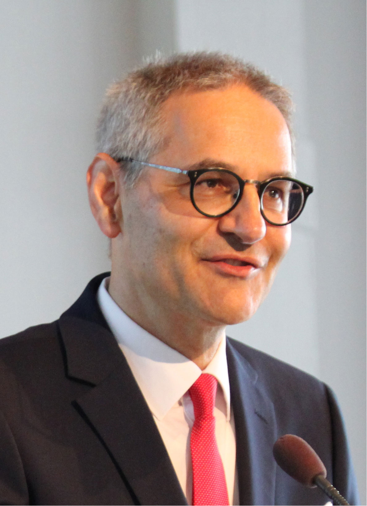
Figure 1 Detlef Zillikens, July 15, 1958, to September 19, 2022.
Several obituaries have already highlighted and valued his scientific work (1–4). Here, within the Research Topic Autoimmune Blistering Diseases: in memoriam Detlef Zillikens, numerous colleagues presented their research and how they were connected and inspired by Detlef Zillikens to focus on AIBDs. Below, we take a closer look at his life and work from different angles and perspectives.
Detlef Zillikens grew up in Wesseling near Bonn, Germany, with two sisters, his mother, who worked as a lab technician, and his father, who held a PhD in chemistry. After schooling in Wesseling and later in Bonn, he went to medical school at the Universities of Bonn and Heidelberg and, in his final year, to the University of Illinois at Champaign-Urbana, USA, where he gained his fine Midwestern American accent. His medical thesis (Dr. med.) was performed at the Institute of Clinical Chemistry, University of Heidelberg (Prof. H. Schmidt-Gayk), where he developed a radioimmunoassay to detect human parathyroid hormone. In 1986, he began a residency at the Department of General Surgery in Heppenheim, and he—fortunately—moved into Dermatology in 1988 to start his residency at the Department of Dermatology, University of Würzburg, Würzburg, Germany, under the guidance of Prof. Günther Burg. In 1992, the Department was chaired by Prof. Eva-Bettina Bröcker; just a week after passing his board examination, he became a consultant. In the same year, he published his first research papers about AIBDs (5, 6). After his board certification in allergology and phlebology, he habilitated in 1994 on “Cellular and humoral immune mechanisms in the pathogenesis of bullous pemphigoid and pemphigus”, becoming an assistant professor. With a prestigious grant from the German Research Foundation (Deutsche Forschungsgemeinschaft (DFG)), he joined the laboratory of Prof. Luis Diaz and Dr. George Giudice, Medical College of Wisconsin, Milwaukee, USA, from 1994 to 1997. During this time, Detlef Zillikens characterized the fine specificities of autoantibodies against BP180 (type XVII collagen) in different pemphigoid diseases. Back in Würzburg, he became an associate professor in 2000 and full professor in 2003.
In April 2004, Detlef Zillikens was appointed director and chair of the Dermatology Department at the University of Lübeck. In parallel, while energetically restructuring and considerably enlarging the clinical department, he implemented his major field of interest, the AIBDs, in the research agenda of the University of Lübeck. Since 2007, he acted as principal investigator and member of the steering committee of the Cluster of Excellence 303 Inflammation at Interfaces, followed since 2019 by the Cluster of Excellence 2167 Precision Medicine in Chronic Inflammation. The Clusters of Excellence are the largest funding instruments of the German Research Foundation. Through this initiative, five additional professorships were taken by R.J.L. (2008), S.I. (2009), and Manfred Kunz (2009), followed by D.T. (2013) and then H.B. (2016) and J.H. (2018). Subsequently, two independent institutes were established, the Institute and Comprehensive Center for Inflammation Medicine (CCIM; 2009–2010, director, Manfred Kunz; 2010–2013, acting director, E.S.; since 2013, director, D.T.) and, in 2014, the Lübeck Institute of Experimental Dermatology (LIED) with its four divisions: Model Systems, director, R.L. and J.H.; Genetics, director, S.I.; Translation, director, E.S. (endowed professorship 2014); and Systems Biology, director, H.B.
Detlef Zillikens initiated and led several large research consortia, initially, the Focus Program Autoimmunity by internal funding (2007–2011) being the nucleus of the DFG-funded research consortia comprising the Research Training Group 1727 Modulation of Autoimmunity (2011–2020), the Clinical Research Group 303 Pemphigoid Diseases (2016–2021), and finally, starting in 2022, the Collaborative Research Center 1526 Pathomechanisms of Antibody-mediated Autoimmunity: Insights from Pemphigoid Diseases. By the CRC 303, another professorship taken by C.S. was brought to his department in 2016.
With his kind and integrative manner, Detlef Zillikens attracted the interest in AIBDs of other institutes at the University of Lübeck, such as the Departments of Anatomy, Microbiology, Pharmacology, Nephrology, and Immunology. Currently, more than 20 clinicians and scientists work in the field of AIBDs on the Lübeck Campus outside the Department of Dermatology, CCIM, and LIED. To bring these people together under one roof, he secured funding for a 2,500-m2 research building in 2017, the Center for Research on Inflammation of the Skin (CRIS). Unfortunately, Detlef Zillikens could not enjoy the opening of this center scheduled for 2026.
Apart from basic science and clinical dermatology, two other areas were close to his heart, the development of diagnostic tools for AIBDs and the training of clinician scientists. The long-standing and very fruitful scientific cooperation with the company Euroimmun, Lübeck, resulted in the availability of various serological assays for AIBDs that have greatly facilitated the diagnosis of these disorders. For the training of clinician scientists, he acquired DFG funding that led to the foundation of the Lübeck School for Clinician Scientists and negotiated a 6-year curriculum with the board of physicians of Schleswig-Holstein combining a 4-year clinical training with a 2-year research rotation.
Detlef Zillikens served as an elected member of the review board for medicine of the DFG (2012–2020) and as vice president of the University of Lübeck responsible for scientific activities from 2013 to 2018. Detlef Zillikens received numerous prizes and honors and co-authored more than 600 articles and book chapters in the field of inflammatory dermatoses.
Detlef Zillikens leaves behind his wife Monika, a psychiatrist and psychotherapist, and two children, currently in dermatological resident training and medical school.
Under the care of 57 current physicians, the Department of Dermatology operates 62 beds on two wards and a day-care clinic that serves more than 10,000 patients per year. In 2004, when he became chair of the Department, only 17 physicians and 32 beds were present. A continuous innovative educational program based on a rotation principle through five different inpatient and outpatient teams imparts all the essential learning content of dermatology. The expansion of the research structures was accompanied by the establishment of special outpatient clinics for AIBDs (four clinics/week), skin cancer, phlebology, and chronic wounds and a section for inflammatory disease with a focus on atopic dermatitis, psoriasis, and urticaria. The clinic has a certified skin cancer center and is part of the Lübeck University Vascular and Cancer Centers. Other important pillars of clinical care are the division of allergology, the dermatopathology laboratory, and the routine autoimmune laboratory. Detlef Zillikens has particularly promoted and always supported dermatopathology, a section that constantly expanded during his guidance. Linking the autoimmune serology, direct immunofluorescence, and histopathology, the detailed histopathological features of anti-p200 and anti-laminin 332 pemphigoid were described (7, 8). The autoimmune laboratory in 2010 was the first dermatology laboratory in Germany to be ISO 15189 certified and developed into a national and international reference center for AIBDs (9). In 2022, the laboratory received AIBD samples from more than 400 sites in Germany and abroad and processed 7,000 serum and more than 2,000 tissue samples for direct immunofluorescence. Detlef Zillikens signed all reports personally and educated and supervised a team of motivated residents and junior consultants who read the immunofluorescence slides. He was in constant exchange with the dedicated technical team that valued him highly.
During the first period of the Excellence Cluster Inflammation at Interfaces, the idea of establishing a center of excellence in Lübeck was supported very early by Detlef Zillikens. In the beginning, it was organized to have a coordinating function between basic research activities and interdisciplinary scientific projects. Following the appointment of D.T. as its director in 2013 (Figure 2) under the constant support of Detlef Zillikens, CCIM grew into an interdisciplinary center of excellence in the management of patients with common chronic inflammatory skin diseases, e.g., psoriasis, hidradenitis suppurativa, or atopic dermatitis, and also more complex cases requiring highly specialized approaches in diagnosing and treatment. In interdisciplinary joined clinics, e.g., together with rheumatologists, all patients were screened for the existence of associated musculoskeletal diseases. Later, other disciplines joined like gastroenterology, nutrition medicines, and infectious diseases. Another focus of CCIM is patient-oriented clinical research including randomized controlled trials (10–12). In 2018, Detlef Zillikens supported the founding of a research institute for inflammation medicine and, within the Department of Dermatology, a section for inflammatory skin diseases. All of more than 40 coworkers of the research institute and section of dermatology considered him a strong supporter, visionary, and pioneer of modern dermatology.
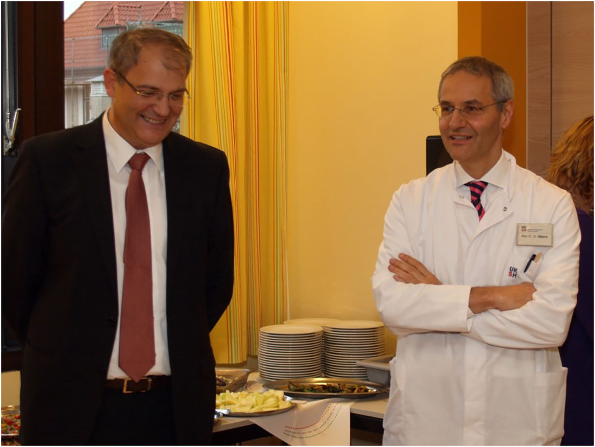
Figure 2 Inauguration of Diamant Thaci (left) to CCIM in 2013 (right, Detlef Zillikens). CCIM, Comprehensive Center for Inflammation Medicine.
Detlef Zillikens’ research agenda on AIBDs directly contributed to the scientific focus of the University of Lübeck, namely, infection and inflammation. With the acquisition of two professorships within the Excellence Cluster Inflammation at Interfaces and an endowed professorship, it became possible in 2014 to further boost research in this direction by founding this institute as a spin-off of the Department of Dermatology (Figure 3). Translational research on inflammation of the skin has been intensified ever since and extended to other inflammatory skin diseases such as psoriasis and atopic eczema. Together with two additional professorships established within the Excellence Cluster in 2016 and 2018, the LIED harbored five professorships, J.EH., H.B., S.I., R.J.L., and E.S., within its four divisions, Model Systems (R.J.L. and J.E.H.), Genetics (S.I.; until 2022), Translation (E.S.), and Systems Biology (H.B.).
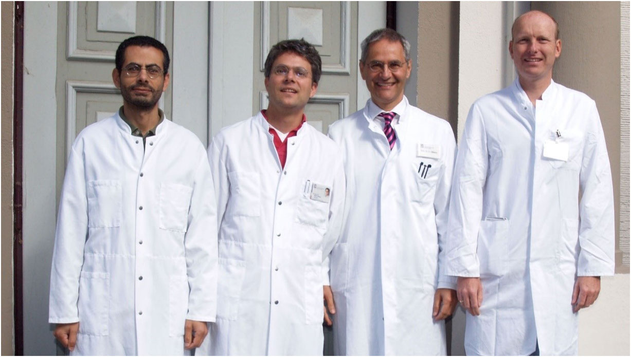
Figure 3 Founding professors and directors of the Lübeck Institute of Experimental Dermatology in 2014 (Saleh Ibrahim, Ralf Ludwig, Detlef Zillikens, and Enno Schmidt, from left to right).
Its research comprises a broad scope including various in vitro, ex vivo, and in vivo models, imaging, genetics, epidemiology, diagnostics of AIBDs, and systems biology. Research on LIED is centered around but not restricted to AIBDs.
LIED has established strong cooperation with the University of Lübeck and the Excellence Cluster Inflammation at Interfaces and its continuation Precision Medicine in Chronic Inflammation and has achieved national and international visibility. This development is reflected in the participation in various third-party funded consortia such as the Research Training Groups 1727 Modulation of Autoimmunity, 1743 Genes, Environment, and Inflammation, and 2633 Defining and Targeting Autoimmune Pre-Disease, as well as in the Clinical Research Group 303 Pemphigoid Diseases and its successor CRC 1526 Pathomechanisms of Antibody-mediated Autoimmunity. LIED also combines basic research with clinical research in close cooperation with the Department of Dermatology and the CCIM. The laboratories of LIED house more than 60 scientists, clinician scientists, PhD students, and technicians, being located at three sites on the campus. With the newly acquired CRIS building, the LIED members will be united under one roof in 2026.
The interdisciplinary projects for this Research Training Group (RTG) have evolved from the Priority Program on Autoimmunity, which was funded by the University of Lübeck from 2006 to 2011. The central scientific question of the RTG addressed the loss of immune tolerance in autoimmune diseases and the modulation of the mechanisms contributing to this loss. The projects were divided into two areas: A) the development of novel therapeutic strategies for the treatment of autoimmune diseases and B) the identification of novel therapeutic targets. The scientists involved in this RTG have been collaborating on this topic at the Lübeck campus for more than 10 years. The breadth of the spectrum of methods used in this work also served as a training program for doctoral students and is supplemented by a central qualification and supervision concept with defined training modules. This included a twice-yearly retreat where all students present their respective projects and receive feedback from the group of subproject leaders and external advisors (Figure 4). Principal investigators came from the Departments of Dermatology, LIED, Rheumatology, Immunology, Anatomy, Microbiology, and Chemistry. During the funding period of 2015–2020, three generations of 13 Dr. rer. nat./PhD students were trained. In addition, six to 12 scholarships per year were provided for medical students who pause their medical studies for 6–12 months to focus exclusively on an experimental MD thesis. In total, 36 PhD and 30 associated PhD students funded by other third-party funds as well as 80 MD and associated MD students received their degrees through this RTG.
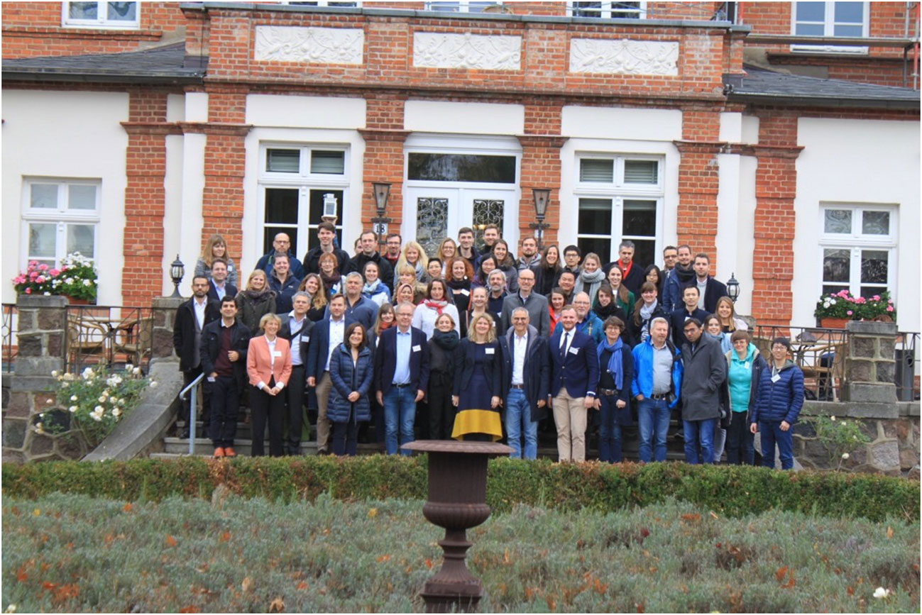
Figure 4 Meeting of the Research Training Group 1727 in Boltenhagen, Germany, in 2016.
The speaker of this RTG was Detlef Zillikens; scientific coordinators in the first funding phase were R.J.L. and in the second funding phase J.E.H. This funding instrument has also been particularly successful in getting young medical students interested in experimental research on autoimmunity. A number of PhD students have joined the Clinician Scientist Program of the clinic in their later training or continued their research as postdocs at the Lübeck campus.
In 2012, Detlef Zillikens recruited C.D.S. from the Center of Immunology and Inflammatory Diseases of the Massachusetts General Hospital/Harvard Medical School in Boston to Lübeck. He had been working at this point on the mechanisms regulating the recruitment of granulocytes in autoimmune diseases for 4 years. The mechanisms recruiting granulocytes in pemphigoid diseases thus became more of the focus of the research community at Detlef Zillikens’ Department. With Detlef Zillikens’ support, C.D.S. drove the development of a DFG-funded research consortium focused on the mechanism regulating granulocyte recruitment in the early stages of the effector phase of pemphigoid diseases as paradigm diseases for antibody-induced granulocytic tissue inflammation. This research consortium, the Clinical Research Unit (CRU) 303 Pemphigoid Diseases—Molecular Pathways and their therapeutic Potential, ran from 2015 to 2022. It consisted of nine research projects and two service projects with researchers from multiple departments of the University of Lübeck as well as from the Max Planck Institute for Evolutionary Biology, Plön. Through the translational research program of the CRU 303, the clinic and the research of the Department of Dermatology in Lübeck became more closely intertwined with the CRU, providing the resources to profile and sample patients with pemphigoid diseases at clinical and molecular levels in a highly standardized manner and giving many clinical fellows of the department the chance to engage into research rotation in the labs participating in the CRU 303.
The CRC 1526 succeeded the CRU 303. CRCs are in general larger funding instruments of the DFG than CRUs. They are meant to run for 12 years and allow more projects and research sites to engage in a concerted research effort than CRUs do. The CRC 1526 addresses pemphigoid diseases and other autoantibody-induced diseases on a broader scope. The CRC 1526 became operative in January 2022 just 2 months before Detlef Zillikens was diagnosed as terminally ill.
The CRIS is an interdisciplinary research center with a thematic focus on chronic inflammatory processes of the skin. The research program of CRIS includes the elucidation of the molecular mechanisms underlying these processes, the uncovering of differences in the mechanisms of the various types of chronic inflammation of the skin, and the development of novel, selective strategies to interrupt and reverse these mechanisms for therapeutic purposes. The CRIS research building is currently under construction on the campus of the University of Lübeck. Its completion and occupancy are planned for 2026. The building will provide 2,500 m2 of space for 140 scientists to implement the CRIS research program. In addition to state-of-the-art laboratory space, the building will house a research clinic and animal facility and several high-end equipment including a CyTOF mass cytometer and a mass spectrometry imaging system.
A total of €30 million will be made available for the construction and furnishing of the CRIS research building. The funding commitment in 2017 was made from federal and state funds on the basis of a competitive, two-stage application procedure coordinated by the Science Council in accordance with Article 91b of the German Basic Law. This application procedure promotes innovative research consortia that are to develop into international beacons. The 10-member interdisciplinary applicant group of scientists from the Lübeck campus was led by Detlef Zillikens and managed by C.D.S.
The training of clinician scientists was always paramount for Detlef Zillikens. As such, within the last 15 years, more than 25 clinician scientists including N.v.B, C.M.M, M.K., A.R., and I.S. received their dermatological education and were enabled to perform research projects on the Lübeck campus. During his time as vice president of the University of Lübeck, Detlef Zillikens initiated a competitive funding program for dedicated physicians of all clinical disciplines, which allowed several of these clinical scientists to perform their research rotations. Detlef Zillikens had negotiated the dermatological training program with the Schleswig-Holstein Medical Association. The 5-year, originally purely clinical dermatological training curriculum was extended to 6 years but integrates 2 years of protected time for research. Due to his longstanding and exceptional input in this subject and as a lasting memory, the Clinician Scientist Academy of the Universities of Lübeck and Kiel was named after him.
Soon after Detlef Zillikens started his position in Lübeck, he sought contact with Euroimmun, an aspiring and rapidly expanding company for autoimmune diagnostics situated in Lübeck. In close cooperation with Winfried Stöcker, former owner and CEO of Euroimmun, Wolfgang Schlumberger, the former CEO, and highly motivated and forward-thinking group leaders, e.g., Christian Probst, Lars Komorowski, and Cornelia Dähnrich, a variety of serological assays for the diagnosis of AIBDs were developed and became widely available. As such, employing the recombinant immunodominant fragments sensitive and specific ELISA for the detection of serum autoantibodies against major AIBD target antigens were released comprising BP180 (type VII collagen), BP230, desmoglein 1 and 3, envoplakin, type VII collagen, tissue-type transglutaminase, and deamidated gliadin (13–18). Based on the Biochip® technology, in which multiple miniature substrates are placed in a single incubation field mounted to a standard laboratory slide, additional assays were developed, e.g., the Biochip® mosaic containing monkey esophagus, primate salt-split skin, recombinant BP180 NC16A, and human HEK293 cells that recombinantly express the immunodominant regions of desmoglein 1, desmoglein 3, and BP230 on their surface (19). This Biochip® mosaic is now widely used and has further been extended by cells that express type VII collagen and laminin 332 (20–28). Future studies will address the detection of serum IgG against further target antigens such as the p200 protein, the ectodomain of BP180, and IgA autoantibodies in AIBDs. The cooperation with Euroimmun reflected Detlef Zillikens’s ability to interest others in his agenda, to build bridges, to create win–win situations, and to convey a constructive and warm atmosphere on an equal footing. This cooperation was always among his highest priorities and emphasized his strong translational thinking.
Already during residency, Detlef Zillikens became interested in AIBDs. When he was appointed head of the routine autoimmune laboratory at the Department of Dermatology in Würzburg in 1992, immediately after receiving his board certification in dermatology, he started his own research group. Without funding and experimental background, apart from his activities as a medical student, this was built on medical students and situated in the three rooms of the routine autoimmune laboratory. Here, Andreas Ambach, Achim Zentner, and E.S. were among his first students who performed their medical theses (to receive the title M.D. that in Germany is not automatically given after the final medical exam). At that time, he was mainly interested in inflammatory mediators in the sera and blister fluids of patients with the most frequent AIBDs, bullous pemphigoid. Meanwhile, Detlef Zillikens was working full time as a clinical consultant spending his evening and weekend hours with research activities. The latter was only possible through the dedicated team of the routine autoimmune laboratory that also greatly supported the medical students and highly valued Detlef Zillikens. The data of these medical students were meticulously published and, given the limited resources at that time, reached considerable attention (5, 6, 29–32).
Supported by a prestigious research grant from the DFG, he spent 3 years (1994–1997) in the laboratory of Prof. Luis Diaz, Milwaukee, WI, USA. He initially aimed to study the neonatal mouse model of bullous pemphigoid, established in this lab in 1993 (33). To his great disappointment, this did not work out, and together with Dr. George Giudice, he instead characterized the fine specificities of autoantibodies against BP180 (type XVII collagen) in different pemphigoid diseases by the use of various recombinant fragments of the immunodominant NC16A domain (34–37).
Back in Würzburg, he re-established his research group that initially included the part-time technician Heike Schömig and E.S., who decided to start residency in dermatology with a 1-year research rotation as, what today would be called a clinician scientist. At this time, anti-BP180 IgG was shown to trigger a signal-transducing event leading to the release of IL-6 and IL-8 when incubated with cultured human keratinocytes (detailed below) (38). By Detlef Zillikens’ winning, caring, and binding character, the laboratory was seen populated with a number of highly motivated and dedicated medical students, including I.S., Karin Obe, Karin Herzele, Rebeccah Döpp, Stanislav Reimer, Werner Kippes, Robert Kränsel, and Matthias Georgi (37, 39–49). These activities were soon supported by the implementation of a so-called “tandem” system. Here, Detlef Zillikens and M.G., another talented and aspiring consultant at that time and current director and chair of the Department of Dermatology, University of Würzburg, Germany, covered a single clinical position, allowing them to exclusively dedicate about 4 months per year to research. With this system, Prof. Eva-Bettina Bröcker fueled their careers and anticipated the advanced clinician scientist by nearly 20 years. When E.S. moved to the Department of Biotechnology for his PhD in 1999, C.S. took over as manager of Detlef Zillikens’ research laboratory.
Detlef Zillikens was involved as a supervisor in the DFG-funded Graduate Program Immunomodulation, a training program for students of the Graduate School of Life Sciences (GSLS) and medical students of the University of Würzburg. C.S. and later Sidonia Mihai were recruited to Detlef’s lab as fellows in this program. C.S. together with Katrin Müller-Blech as a technical assistant, Yoshi Hirako as a postdoc, and several active medical students worked on an ex vivo model of autoantibody-induced dermal–epidermal separation. This model was used in subsequent studies to address the effector phase of the autoimmune response to components of the epidermal basement membrane (50, 51). In parallel, animal models of autoimmunity to collagen VII were developed (detailed below) (52, 53).
In Lübeck, the research group considerably expanded with the postdocs and clinician scientists including I.S., Mircea Chiriac, Andreas Recke, Josep Herrero-González, Alina Sesarman, Amrei Dilling, and Bernie Gibbs. The results obtained in these projects were the basis for participation in the DFG Cluster of Excellence Inflammation at Interfaces, which started in 2007.
After R.J.L. and E.S. joined the Department of Dermatology in Lübeck in October 2007 and January 2008, respectively, Detlef Zillikens retracted from everyday research activities and focused on serving in various functions to foster his research ideas as detailed below. In addition, he initiated and steered the above-mentioned research consortia that enabled more than 70 third-party-funded clinician scientists and researchers at the campus Lübeck to work on AIBDs in 2022. Nevertheless, he attended the weekly major laboratory meetings, gave highly appreciated input in nearly every project, and reviewed the resulting manuscripts. At the same time, he fostered the work of clinician scientists represented here by M.K., A.R., and N.v.B. as further detailed in the obituary of C.M.H. (3). Detlef Zillikens personally selected them from a large number of applicants and closely mentored their careers.
Below, the authors have detailed those articles from his more than 600 publications that have been highly cited and/or are regarded as most relevant for the current research activities both in Lübeck and internationally. Among his many review articles, the three most cited were Pemphigoid diseases in Lancet 2013 (54), Pemphigus in Nature Reviews Disease Primers in 2017 (55), and Modern diagnosis of autoimmune blistering diseases in Autoimmunity Reviews in 2010 (56). He also co-authored highly cited consensus and management guidelines of AIBDs (57–68) and was instrumental in generating the guidelines for the use of immunoadsorption and rituximab. Consensus recommendations for the latter therapies, at this time, highly innovative therapeutic options for severe and/or refractory AIBDs, have greatly facilitated the reimbursement of the treatment costs in Germany (69, 70).
In 1996, Detlef Zillikens and T.H. described the first patient with anti-p200 pemphigoid (71). The patients clinically resembled bullous pemphigoid and also showed linear deposits of IgG and C3 along the dermal–epidermal junction. In contrast to bullous pemphigoid, serum IgG bound to the floor of the artificial blister by indirect IF microscopy on human salt-split skin as previously described for epidermolysis bullosa acquisita. In contrast to the latter disorder, the patient’s serum reacted with a specific 200-kDa band by immunoblotting with dermal extract. The molecular identity of the target antigen was only revealed in 2009 by the group of T.H. (72), who described laminin γ1 to be recognized by 90% of anti-p200 pemphigoid sera. Subsequently, it became clear that only 70%–90% of anti-p200 pemphigoid sera are reactive with laminin γ1 and that the pathogenicity of anti-laminin γ1 IgG in vivo could not be demonstrated (73, 74).
During his postdoc period in the lab of Dr. G. Giudice, Milwaukee, WI, USA, Detlef Zillikens revealed that approximately 90% of patients with bullous pemphigoid have circulating autoantibodies against the 76-amino-acid-long 16th non-collagenous domain just outside the transmembrane domain of BP180 (35). Based on its recombinant NC16A domain, he developed a sensitive and specific ELISA system for serum autoantibodies against NC16A in patients with bullous pemphigoid and pemphigoid gestationis (34). Applying this ELISA, he was then the first to show that serum anti-BP180 NC16A IgG correlates with disease activity in bullous pemphigoid (43). These data were later the basis for the development of a widely available commercial BP180 NC16A ELISA (13).
For the first time, an FcγR-independent effect of anti-BP180 antibodies was demonstrated when a signal-inducing event leading to the secretion of IL-6 and IL-8 was observed upon the binding of BP180-specific IgG to cultured human primary keratinocytes (38). The release of IL-8 could subsequently be shown to be significantly inhibited by dapsone but not tetracycline (75).
The initial publication was the first to describe IgE reactivity against BP180 NC16A in patients with bullous pemphigoid (42). The article by van Beek et al. confirmed the previous finding that serum levels of IgE against BP180 NC16A parallel the extent of the disease interindividually, i.e., in an individual bullous pemphigoid patient during the course of the disease (76). Unlike serum anti-BP180 NC16A IgG, IgE against this domain does not correlate with disease activity per se. In addition, while anti-BP180 NC16A IgG is associated with the clinical phenotype, i.e., with the classical picture represented by blisters and erosions, IgE against this domain is unrelated to both the classical and urticarial/erythematous phenotypes.
In this ex vivo model, autoantibodies against the NC16A domain of BP180 from patients with bullous pemphigoid bind in cryosections of human skin and activate normal human neutrophils, which results in dermal–epidermal separation (51). Subsequently, it was shown that the activation of neutrophils leads to the release of specific proteolytic enzymes, which are pathogenically involved in the autoantibody-induced granulocyte-dependent dermal–epidermal separation (77).
Detlef Zillikens and his team, initially in Würzburg and then in Lübeck, were among the first to systematically apply immunoadsorption and rituximab as novel treatment options for severe AIBDs (78). The protocol combines the short-term effects of immunoadsorption and i.v. dexamethasone pulses with the long-term effects of rituximab, in addition to azathioprine/mycophenolate mofetil, and became the new standard therapy for severe and/or refractory pemphigus patients in Lübeck and offered an improved side-effect profile compared with the former standard therapy of high-dose oral corticosteroids plus azathioprine/mycophenolate mofetil (65, 79, 80).
As another milestone in pemphigoid research, Detlef Zillikens provided in vivo proof of the autoimmune nature of epidermolysis bullosa acquisita (EBA). The story behind the paper illustrates that serendipity is sometimes a significant contributor to success: the initial aim of the project was the development of a mouse model for bullous pemphigoid. However, the murine BP180 NC15A domain was challenging to produce, while in a separate project, the production of proteins of the NC1 domain of murine collagen type VII (COL7) was successful. Hence, IgG from rabbits immunized with recombinant fragments of COL7, when injected into wild-type mice, induced clinical, histological, and immunological phenotypes duplicating human EBA (52). In parallel, similar observations were independently made by David Woodley at almost the same time (81). Ever since its initial description, this antibody transfer-induced (“passive”) EBA mouse model, or variations thereof, has been extensively used by multiple laboratories for basic and translational research in EBA and pemphigoid diseases in general (82–89).
The requirement of C5 for the induction of experimental EBA was already described in the initial description of the antibody transfer-induced EBA mouse model (see above) (52). In cooperative work within the Excellence Cluster Inflammation at Interfaces, the groups of Detlef Zillikens and Jörg Köhl identified a novel pathway blocking C5aR1-driven inflammation. In this work, they demonstrated that i) C5aR1-deficient mice, like C5-deficient mice, are almost completely protected from clinical disease manifestations in antibody transfer-induced EBA; ii) engagement of the FcγRIIB and dectin-1 by highly galactosylated immune complexes blocks signaling downstream of the C5aR1; and iii) clinical disease manifestations can be ameliorated by treatment of mice with highly galactosylated immune complexes in the antibody transfer-induced EBA mouse model (90).
Here, a multivariant indirect immunofluorescence test based on the Biochip® technology employed six miniature substrates, i.e., monkey esophagus, primate salt-split skin, recombinant BP180 NC16A, and HEK293 cells recombinantly expressing desmoglein 1, desmoglein 3, and BP230 on their cell surface in a single incubation field of a normal laboratory slide. This approach allowed the serological diagnosis of more than 90% of sera from patients with AIBDs (19). Subsequently, these findings could be corroborated using an extended Biochip® mosaic (24). Meanwhile, Biochips® with cells expressing type VII collagen and laminin 332 are widely available (20, 22).
In this study in cooperation with Prof. Eli Sprecher, Tel Aviv, the first non-HLA-related gene, ST18, associated with pemphigus was described in Israeli and Egyptian patients but not in patients of German ancestry (91).
In this collaborative study with John Baines, from the Max Planck Institute for Evolutionary Biology, Plön, Germany, the role of skin microbiota in the pathogenesis of AIBDs was first systemically studied (92). The study also provided evidence for host gene–microbiota interactions contributing to disease risk in a mouse model of EBA. The study was followed by several others (93, 94), eventually identifying multiple species involved in AIBDs and the metabolites mediating their effects.
In mouse models of bullous pemphigoid-like EBA and of bullous pemphigoid mice deficient in 5-lipoxygenase, a key enzyme in LTB4 biosynthesis, or BLT1, was resistant to anti-COL7- and anti-COL17 IgG-mediated neutrophil recruitment and, consequently, skin inflammation (95). A pronounced parallel increase in LTB4 and neutrophils in the skin was seen, while eosinophils appeared to be dispensable for blister formation in the bullous pemphigoid-like EBA mouse model. These data highlighted LTB4/BLT1 as critical drivers of antibody-induced tissue destruction in experimental bullous pemphigoid-like EBA and bullous pemphigoid.
This article describes the first mouse model for mucous membrane pemphigoid that reflects major clinical and immunological characteristics of the human disease (96). In this model, like in patients with mucous membrane pemphigoid, dapsone significantly reduced skin and oral lesions and ocular inflammation that leads to fibrosis (97, 98). This model will be instrumental in characterizing key inflammatory pathways and potential novel therapeutic agents for this devastating disease.
Based on the Biochip® technology, the first widely available detection system for serum autoantibodies against laminin 332 was developed and validated in a large multicenter study (22). This study corroborated the previous finding that anti-laminin 332 IgG in mucous membrane pemphigoid is associated with a malignancy in about a quarter of patients (99) and led to the recommendation of testing for anti-laminin IgG in all patients with mucous membrane pemphigoid in national and international guidelines (100, 101). Subsequently, N.v.B. et al. calculated a 6.8-fold higher risk of patients with anti-laminin 332 mucous membrane pemphigoid to develop malignancies than the age- and sex-matched general population (102). This association and the autoimmunity against laminin 332 are further detailed in another article on this research topic (103).
Detlef Zillikens spent several years in the laboratory of Luis Diaz at the Medical College of Wisconsin in Milwaukee, USA, where he met European postdoctoral fellows such as José Mascaro Jr. or F.C. He worked with scientists, namely, George Giudice, who cloned BP180, and Zhu Liu. At that time, he mapped epitopes on BP180 of bullous pemphigoid, pemphigoid gestationis, lichen planus pemphigoides, and linear IgA disease. He kept life-long connections with these colleagues from this period abroad.
Detlef Zillikens organized and hosted two large international meetings on AIBDs in Lübeck, the first in 2013 before the International Investigative Dermatology (IID) meeting in Edinburgh. A chartered aircraft, known as the “Detlef Zillikens Express”, flew everyone from the local airport in Lubeck to Edinburgh. With Detlef Zillikens, nothing was impossible. The second took place in 2017 with the help of the American patients’ association International Pemphigus & Pemphigoid Foundation (IPPF) (Figure 5). These reunions occurred in a friendly atmosphere, permitted many exchanges of ideas and contact between people, and are still well-remembered in the entire community.

Figure 5 The International Pemphigus & Pemphigoid Foundation (IPPF) Meeting in Lübeck, Germany, 2017.
Detlef Zillikens has always welcomed students from abroad so that they can learn or do research on AIBDs in his laboratory. He also regularly received visiting professors in his department for teaching and scientific fostering including DFM, F.C., and T.H.
From an early age, Detlef Zillikens had a passion for exploring the world and meeting people. He spent several months during his medical school at the University of Illinois in Urbana, USA, where he perfected his Midwestern American accent. He returned to the Midwest, to the University of Wisconsin, where he was mentored by another world traveler physician-scientist, Dr. Luis Diaz, originally from Peru. Through this thread, Detlef Zillikens became part of a far-reaching international team of mentees of Luis Diaz who were passionate about AIBDs. Once he had established his chairmanship in Lübeck, he was then able to draw in numerous German experts and also many of the best physician scientists from abroad with grant funding to support them including Norito Ishii (University of Kurume, Japan) Hiroaki Iwata (University of Gifu, Japan) and Kentaro Izumi (University of Sapporo, Japan).
Detlef Zillikens has always enjoyed networking and missed a few annual meetings of the German Society of Dermatological Research (ADF) and the European Society of Dermatological Research (Figure 6). He managed to find the time to lecture in far-flung corners of the world, including India and Iran, where he was constantly swamped by young dermatologists who were hoping to work with him.
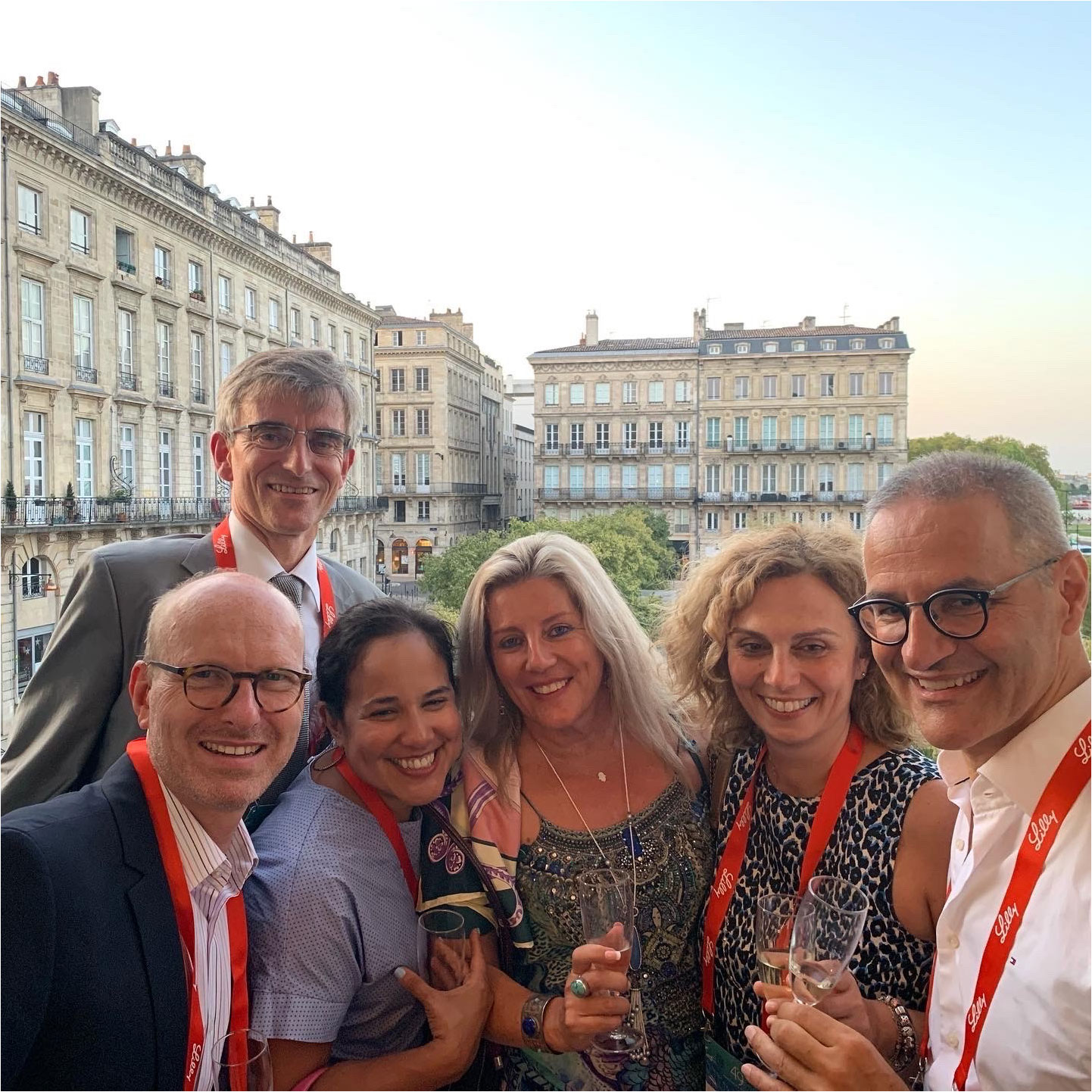
Figure 6 Detlef Zillikens (right) with Enno Schmidt, Frédéric Caux, Dolca Thomas, Dedee Murrell, and Katerina Patsatsi (from left to right) at the annual meeting of the European Society of Dermatological Research, Bordeaux, France, 2017.
Nevertheless, somehow, he also managed to fit in amazing trips to exotic locations with his family for their passion for swimming with tropical fish.
Detlef Zillikens has always been an extremely hard-working person who dedicated large parts of his spare time to his clinic and research on AIBDs. This may be exemplified when, at a time when he corrected manuscripts by hand on the printed version, he occasionally handed the reviewed manuscript back, which appeared slightly wrinkled and rippled. With an exclusive smile he added, “Oh, I am so sorry, I went to the sauna”. It was rare, even when traveling with Detlef Zillikens, that he completely relaxed and took more than a few minutes to talk about private matters. E.S. always remembered one of these rare occasions when sitting in the evening light in view of the Alhambra castle during the Autoimmune Symposium 2012 in Granada, Spain. Strangely enough, Detlef Zillikens mentioned this evening during our last encounter in the rehabilitation clinic in July 2022.
M.G. first met Detlef Zillikens in 1993 when he started his dermatology residency at the Department of Dermatology, University of Würzburg. Detlef Zillikens was his first attending: he introduced M.G. into general dermatology and stringently supervised his clinical activities and rigorously corrected his medical reports. M.G. remembers the daily medical rounds (“Mittagsvisiten”) where Detlef, together with Prof. Eva-Bettina Bröcker (at that time chair of the department), Henning Hamm, Boris Bastian (now San Francisco, CA, USA), E.S. and C.R. (now both in Lübeck), and others contributed to an extremely dynamic working atmosphere that fostered intensive clinical and scientific discourse, in which M.G., among many others, was fortunate enough to complete his training. Detlef Zillikens inspired M.G. for AIBDs, and when Detlef Zillikens went to the USA, M.G. was allowed to represent him in this field in Würzburg. Returning to Würzburg, Detlef Zillikens expanded the clinic’s autoimmunology diagnostic laboratory, introduced new therapeutic approaches for AIBDs such as immunoadsorption, and was one of the first to use rituximab for AIBDs. Detlef Zillikens and M.G. formed a clinical tandem, which gave both of us considerable freedom for our research interests. This concept was implemented by Prof. Eva-B. Bröcker, which she also introduced for her oncology senior physicians, preceded today’s demand for academic support of specialists and senior physicians (now called advanced clinician scientists) by two decades and may have inspired Detlef Zillikens to introduce something comparable in Lübeck. When M.G. became chair of the Dermatology Department in Würzburg, both not only continued their long-standing scientific cooperation with Detlef but also regularly exchanged on the promotion of young scientists and discussed information on clinic matters and key operational figures of their departments. With the passing of Detlef Zillikens, M.G. has lost a passionate teacher and close friend to whom he owes a lot.
Detlef Zillikens has been the exemplary mentor of M.K. for clinical and experimental dermatology at the University of Lübeck for over a decade. With Detlef’s most dedicated help, M.K. was able to achieve his academic goals starting from board certification in both dermatology and dermatopathology up to his current professorship at the University of Southern California, Los Angeles, CA, USA. The knowledge that M.K. gained from him concerning the diagnosis and treatment of AIBD patients has been most valuable, and M.K. feels honored to share it now with his new dermatology team including Prof. David Woodley (another well-known clinician/researcher in the field of AIBDs and close colleague of Detlef Zillikens). M.K. remembers well the moment when he started his career at the University of Lübeck. He initially asked Prof. Stefania Jabłońska, Warsaw, Poland, a pioneer in immunodermatology, for advice regarding a high-ranking academic place with a focus on immunobullous diseases. Her answer was, “Prof. Zillikens’ clinic, but you would need to work hard”. This hard work has been more than rewarding to M.K. M.K. will always mourn the loss of Detlef Zillikens and remember him as a monumental figure in his life.
Detlef Zillikens and T.H. were close friends for a long time starting in 1994 and intensified further following our landmark paper about the first case of anti-200 pemphigoid (see above) (71). Detlef Zillikens and T.H. have published 53 manuscripts together in total. T.H. still well remembers when visiting Detlef Zillikens in Würzburg, the dinner of delicious asparagus in the restaurant overlooking the city of Würzburg. Detlef Zillikens and his wife Monika also treated T.H. in their home with very nice German food. More recently, in 2017, T.H. had a 1-month sabbatical at Lübeck owing to Detlef Zillikens’ help. T.H. prays that Detlef’s soul rests in peace.
In conclusion, we have lost not only a giant in dermatology but also a wonderful and kind mentor and friend, Detlef Zillikens, to whom this Research Topic of Frontiers in Immunology is dedicated.
The original contributions presented in the study are included in the article/supplementary material. Further inquiries can be directed to the corresponding author.
Written informed consent was obtained from the individual(s) for the publication of any potentially identifiable images or data included in this article.
JEH: Conceptualization, Writing – review & editing. CDS: Writing – review & editing. NvB: Writing – review & editing. HB: Writing – review & editing. FC: Writing – review & editing. MG: Writing – review & editing. CMH: Writing – review & editing. KH: Writing – review & editing. TH: Writing – review & editing. SI: Writing – review & editing. MK: Writing – review & editing. DFM: Conceptualization, Writing – original draft, Writing – review & editing. AR: Writing – review & editing. CR: Writing – review & editing. NS: Writing – review & editing. IS: Writing – review & editing. CS: Writing – review & editing. PT: Writing – review & editing. DT: Writing – review & editing. RJL: Writing – review & editing. ES: Conceptualization, Supervision, Writing – original draft, Writing – review & editing.
The author(s) declare that no financial support was received for the research, authorship, and/or publication of this article.
ES and AR have a scientific cooperation with Euroimmun. NB, CH and ES hold patents together with Euroimmun. AR received honoraria from Euroimmun.
The remaining authors declare that the research was conducted in the absence of any commercial or financial relationships that could be construed as a potential conflict of interest.
The author(s) declared that they were an editorial board member of Frontiers, at the time of submission. This had no impact on the peer review process and the final decision.
All claims expressed in this article are solely those of the authors and do not necessarily represent those of their affiliated organizations, or those of the publisher, the editors and the reviewers. Any product that may be evaluated in this article, or claim that may be made by its manufacturer, is not guaranteed or endorsed by the publisher.
1. Schmidt E. Detlef Zillikens: Trauer um einen engagierten Dermatologen. Dtsch Arztebl (2022) 119:A–1925/B-1595.
2. Schmidt E, Goebeler M, Schoen M, Terheyden P. Nachruf: Prof. Dr. med. Detlef Zillikens, Direktor der Klinik für Dermatologie, Allergologie und Venerologie, Universität Lübeck. J Dtsch Ges Dermatol (2022) 20(1689-90). doi: 10.1111/ddg.14970_g
3. Hammers C. Obituary: Professor Detlef Zillikens 1958-2022. Br J Dermatol (2023) 188:155–6. doi: 10.1093/bjd/ljac054
4. Schmidt E, Diaz L, Amagai A, Murrell D. Obituary for Professor Detlef Zillikens- creator of a powerhouse of dermatology research at the University of Lübeck, Germany. J Invest Dermatol (2023) 143:7–8. doi: 10.1016/j.jid.2022.11.001
5. Ambach A, Zillikens D, Klingert B, Hartmann AA, Burg G. [Immune phenotyping of mononuclear infiltrate in bullous pemphigoid]. Hautarzt (1992) 43(2):81–5.
6. Zillikens D, Ambach A, Schuessler M, Dummer R, Hartmann AA, Burg G. The interleukin-2 receptor in lesions and serum of bullous pemphigoid. Arch Dermatol Res (1992) 284(3):141–5. doi: 10.1007/BF00372706
7. Rose C, Weyers W, Denisjuk N, Hillen U, Zillikens D, Shimanovich I. Histopathology of anti-p200 pemphigoid. Am J Dermatopathol (2007) 29(2):119–24. doi: 10.1097/DAD.0b013e31803326e6
8. Rose C, Schmidt E, Kerstan A, Thoma-Uszynski S, Wesselmann U, Kasbohrer U, et al. Histopathology of anti-laminin 5 mucous membrane pemphigoid. J Am Acad Dermatol (2009) 61(3):433–40. doi: 10.1016/j.jaad.2009.02.012
9. van Beek N, Knuth-Rehr D, Altmeyer P, Assaf C, Babilas P, Bayerl C, et al. Diagnostics of autoimmune bullous diseases in German dermatology departments. J Dtsch Dermatol Ges (2012) 10(7):492–9. doi: 10.1111/j.1610-0387.2011.07840.x
10. Thaci D, Simpson EL, Beck LA, Bieber T, Blauvelt A, Papp K, et al. Efficacy and safety of dupilumab in adults with moderate-to-severe atopic dermatitis inadequately controlled by topical treatments: a randomised, placebo-controlled, dose-ranging phase 2b trial. Lancet (2016) 387(10013):40–52. doi: 10.1016/S0140-6736(15)00388-8
11. Papp K, Thaci D, Marcoux D, Weibel L, Philipp S, Ghislain PD, et al. Efficacy and safety of adalimumab every other week versus methotrexate once weekly in children and adolescents with severe chronic plaque psoriasis: a randomised, double-blind, phase 3 trial. Lancet (2017) 390(10089):40–9. doi: 10.1016/S0140-6736(17)31189-3
12. Sadik CD, Rashid H, Hammers CM, Diercks GFH, Weidinger A, Beissert S, et al. Evaluation of nomacopan for treatment of Bullous pemphigoid: A phase 2a nonrandomized controlled trial. JAMA Dermatol (2022) 158(6):641–9. doi: 10.1001/jamadermatol.2022.1156
13. Sitaru C, Dahnrich C, Probst C, Komorowski L, Blocker I, Schmidt E, et al. Enzyme-linked immunosorbent assay using multimers of the 16th non-collagenous domain of the BP180 antigen for sensitive and specific detection of pemphigoid autoantibodies. Exp Dermatol (2007) 16(9):770–7. doi: 10.1111/j.1600-0625.2007.00592.x
14. Probst C, Schlumberger W, Stocker W, Recke A, Schmidt E, Hashimoto T, et al. Development of ELISA for the specific determination of autoantibodies against envoplakin and periplakin in paraneoplastic pemphigus. Clin Chim Acta (2009) 410(1-2):13–8. doi: 10.1016/j.cca.2009.08.022
15. Schmidt E, Dahnrich C, Rosemann A, Probst C, Komorowski L, Saschenbrecker S, et al. Novel ELISA systems for antibodies to desmoglein 1 and 3: correlation of disease activity with serum autoantibody levels in individual pemphigus patients. Exp Dermatol (2010) 19(5):458–63. doi: 10.1111/j.1600-0625.2010.01069.x
16. Blocker IM, Dahnrich C, Probst C, Komorowski L, Saschenbrecker S, Schlumberger W, et al. Epitope mapping of BP230 leading to a novel enzyme-linked immunosorbent assay for autoantibodies in bullous pemphigoid. Br J Dermatol (2012) 166(5):964–70. doi: 10.1111/j.1365-2133.2012.10820.x
17. Kasperkiewicz M, Dahnrich C, Probst C, Komorowski L, Stocker W, Schlumberger W, et al. Novel assay for detecting celiac disease-associated autoantibodies in dermatitis herpetiformis using deamidated gliadin-analogous fusion peptides. J Am Acad Dermatol (2012) 66(4):583–8. doi: 10.1016/j.jaad.2011.02.025
18. van Beek N, Dahnrich C, Johannsen N, Lemcke S, Goletz S, Hubner F, et al. Prospective studies on the routine use of a novel multivariant enzyme-linked immunosorbent assay for the diagnosis of autoimmune bullous diseases. J Am Acad Dermatol (2017) 76(5):889–894 e885. doi: 10.1016/j.jaad.2016.11.002
19. van Beek N, Rentzsch K, Probst C, Komorowski L, Kasperkiewicz M, Fechner K, et al. Serological diagnosis of autoimmune bullous skin diseases: Prospective comparison of the BIOCHIP mosaic-based indirect immunofluorescence technique with the conventional multi-step single test strategy. Orphanet J Rare Dis (2012) 7(1):49. doi: 10.1186/1750-1172-7-49
20. Komorowski L, Muller R, Vorobyev A, Probst C, Recke A, Jonkman MF, et al. Sensitive and specific assays for routine serological diagnosis of epidermolysis bullosa acquisita. J Am Acad Dermatol (2013) 68(3):e89–95. doi: 10.1016/j.jaad.2011.12.032
21. Marzano AV, Cozzani E, Biasin M, Russo I, Alaibac M. The use of Biochip immunofluorescence microscopy for the serological diagnosis of epidermolysis bullosa acquisita. Arch Dermatol Res (2016) 308(4):273–6. doi: 10.1007/s00403-016-1632-0
22. Goletz S, Probst C, Komorowski L, Schlumberger W, Fechner K, van Beek N, et al. A sensitive and specific assay for the serological diagnosis of antilaminin 332 mucous membrane pemphigoid. Br J Dermatol (2019) 180(1):149–56. doi: 10.1111/bjd.17202
23. Tirumalae R, Kalegowda IY. Role of BIOCHIP indirect immunofluorescence test in cutaneous vesiculobullous diseases. Am J Dermatopathol (2020) 42(5):322–8. doi: 10.1097/DAD.0000000000001471
24. van Beek N, Kruger S, Fuhrmann T, Lemcke S, Goletz S, Probst C, et al. Multicenter prospective study on multivariant diagnostics of autoimmune bullous dermatoses using the BIOCHIP technology. J Am Acad Dermatol (2020) 83(5):1315–22. doi: 10.1016/j.jaad.2020.01.049
25. Wang M, Li F, Wang X, Wang R, Chen T, Zhao J, et al. BIOCHIP mosaic for the diagnosis of autoimmune bullous diseases in Chinese patients. Eur J Dermatol (2020) 30(4):338–44. doi: 10.1684/ejd.2020.3839
26. Yang A, Xuan R, Melbourne W, Tran K, Murrell DF. Validation of the BIOCHIP test for the diagnosis of bullous pemphigoid, pemphigus vulgaris and pemphigus foliaceus. J Eur Acad Dermatol Venereol (2020) 34(1):153–60. doi: 10.1111/jdv.15770
27. Goletz S, Giurdanella F, Holtsche MM, Nijenhuis M, Horvath B, Diercks GFH, et al. Comparison of two diagnostic assays for anti-laminin 332 mucous membrane pemphigoid. Front Immunol (2021) 12:773720. doi: 10.3389/fimmu.2021.773720
28. Holtsche MM, van Beek N, Hashimoto T, Di Zenzo G, Zillikens D, Prost-Squarcioni C, et al. Diagnosis of epidermolysis bullosa acquisita: multicentre comparison of different assays for serum anti-type VII collagen reactivity. Acta Derm Venereol (2021) 101(3):adv00420. doi: 10.2340/00015555-3774
29. Schmidt E, Brocker EB, Zillikens D. High levels of soluble CD23 in blister fluid of patients with bullous pemphigoid. Arch Dermatol (1995) 131(8):966–7. doi: 10.1001/archderm.1995.01690200106030
30. Schmidt E, Ambach A, Bastian B, Brocker EB, Zillikens D. Elevated levels of interleukin-8 in blister fluid of bullous pemphigoid compared with suction blisters of healthy control subjects. J Am Acad Dermatol (1996) 34(2 Pt 1):310–2. doi: 10.1016/S0190-9622(96)80146-0
31. Schmidt E, Bastian B, Dummer R, Tony HP, Brocker EB, Zillikens D. Detection of elevated levels of IL-4, IL-6, and IL-10 in blister fluid of bullous pemphigoid. Arch Dermatol Res (1996) 288(7):353–7. doi: 10.1007/BF02507102
32. Schmidt E, Mittnacht A, Schomig H, Dummer R, Brocker EB, Zillikens D. Detection of IL-1 alpha, IL-1 beta and IL-1 receptor antagonist in blister fluid of bullous pemphigoid. J Dermatol Sci (1996) 11(2):142–7. doi: 10.1016/0923-1811(95)00435-1
33. Liu Z, Diaz LA, Troy JL, Taylor AF, Emery DJ, Fairley JA, et al. A passive transfer model of the organ-specific autoimmune disease, bullous pemphigoid, using antibodies generated against the hemidesmosomal antigen, BP180. J Clin Invest (1993) 92(5):2480–8. doi: 10.1172/JCI116856
34. Zillikens D, Mascaro JM, Rose PA, Liu Z, Ewing SM, Caux F, et al. A highly sensitive enzyme-linked immunosorbent assay for the detection of circulating anti-BP180 autoantibodies in patients with bullous pemphigoid. J Invest Dermatol (1997) 109(5):679–83. doi: 10.1111/1523-1747.ep12338088
35. Zillikens D, Rose PA, Balding SD, Liu Z, Olague-Marchan M, Diaz LA, et al. Tight clustering of extracellular BP180 epitopes recognized by bullous pemphigoid autoantibodies. J Invest Dermatol (1997) 109(4):573–9. doi: 10.1111/1523-1747.ep12337492
36. Zillikens D, Caux F, Mascaro JM, Wesselmann U, Schmidt E, Prost C, et al. Autoantibodies in lichen planus pemphigoides react with a novel epitope within the C-terminal NC16A domain of BP180. J Invest Dermatol (1999) 113(1):117–21. doi: 10.1046/j.1523-1747.1999.00618.x
37. Zillikens D, Herzele K, Georgi M, Schmidt E, Chimanovitch I, Schumann H, et al. Autoantibodies in a subgroup of patients with linear IgA disease react with the NC16A domain of BP1801. J Invest Dermatol (1999) 113(6):947–53. doi: 10.1046/j.1523-1747.1999.00808.x
38. Schmidt E, Reimer S, Kruse N, Jainta S, Brocker EB, Marinkovich MP, et al. Autoantibodies to BP180 associated with bullous pemphigoid release interleukin-6 and interleukin-8 from cultured human keratinocytes. J Invest Dermatol (2000) 115(5):842–8. doi: 10.1046/j.1523-1747.2000.00141.x
39. Chimanovitch I, Schmidt E, Messer G, Dopp R, Partscht K, Brocker EB, et al. IgG1 and IgG3 are the major immunoglobulin subclasses targeting epitopes within the NC16A domain of BP180 in pemphigoid gestationis. J Invest Dermatol (1999) 113(1):140–2. doi: 10.1046/j.1523-1747.1999.00622.x
40. Kippes W, Schmidt E, Roth A, Rzany B, Brocker EB, Zillikens D. [Immunopathologic changes in 115 patients with bullous pemphigoid]. Hautarzt (1999) 50(12):866–72. doi: 10.1007/s001050051001
41. Schmidt E, Herzele K, Schumann H, Wesselmann U, Chimanovitch I, Bruckner-Tuderman L, et al. Linear IgA disease with circulating IgA antibodies against the NC16A domain of BP180. Br J Dermatol (1999) 140(5):964–6. doi: 10.1046/j.1365-2133.1999.02838.x
42. Dopp R, Schmidt E, Chimanovitch I, Leverkus M, Brocker EB, Zillikens D. IgG4 and IgE are the major immunoglobulins targeting the NC16A domain of BP180 in Bullous pemphigoid: serum levels of these immunoglobulins reflect disease activity. J Am Acad Dermatol (2000) 42(4):577–83. doi: 10.1067/mjd.2000.103986
43. Schmidt E, Obe K, Brocker EB, Zillikens D. Serum levels of autoantibodies to BP180 correlate with disease activity in patients with bullous pemphigoid. Arch Dermatol (2000) 136(2):174–8. doi: 10.1001/archderm.136.2.174
44. Georgi M, Scheckenbach C, Kromminga A, Partscht K, Messer G, Brocker EB, et al. Mapping of epitopes on the BP180 ectodomain targeted by IgA and IgG autoantibodies in patients with the lamina lucida-type of linear IgA disease. Arch Dermatol Res (2001) 293(3):109–14. doi: 10.1007/s004030000205
45. Zillikens D, Schmidt E, Reimer S, Chimanovitch I, Hardt-Weinelt K, Rose C, et al. Antibodies to desmogleins 1 and 3, but not to BP180, induce blisters in human skin grafted onto SCID mice. J Pathol (2001) 193(1):117–24. doi: 10.1002/1096-9896(2000)9999:9999<::AID-PATH742>3.0.CO;2-W
46. Schmidt E, Skrobek C, Kromminga A, Hashimoto T, Messer G, Brocker EB, et al. Cicatricial pemphigoid: IgA and IgG autoantibodies target epitopes on both intra- and extracellular domains of bullous pemphigoid antigen 180. Br J Dermatol (2001) 145(5):778–83. doi: 10.1046/j.1365-2133.2001.04471.x
47. Schmidt E, Wehr B, Tabengwa EM, Reimer S, Brocker EB, Zillikens D. Elevated expression and release of tissue-type, but not urokinase-type, plasminogen activator after binding of autoantibodies to bullous pemphigoid antigen 180 in cultured human keratinocytes. Clin Exp Immunol (2004) 135(3):497–504. doi: 10.1111/j.1365-2249.2004.02401.x
48. Schmidt E, Kraensel R, Goebeler M, Sinkgraven R, Brocker EB, Rzany B, et al. Treatment of bullous pemphigoid with dapsone, methylprednisolone, and topical clobetasol propionate: a retrospective study of 62 cases. Cutis (2005) 76(3):205–9.
49. Schmidt E, Wehr B, Wolf K, Sitaru C, Brocker EB, Zillikens D. Localisation of bullous pemphigoid antigen 180 (BP180) in cultured human keratinocytes: functionally relevant modification by calcium. Arch Dermatol Res (2006) 298(6):283–90. doi: 10.1007/s00403-006-0690-0
50. Sitaru C, Kromminga A, Hashimoto T, Brocker EB, Zillikens D. Autoantibodies to type VII collagen mediate Fcgamma-dependent neutrophil activation and induce dermal-epidermal separation in cryosections of human skin. Am J Pathol (2002) 161(1):301–11. doi: 10.1016/S0002-9440(10)64182-X
51. Sitaru C, Schmidt E, Petermann S, Munteanu LS, Brocker EB, Zillikens D. Autoantibodies to bullous pemphigoid antigen 180 induce dermal-epidermal separation in cryosections of human skin. J Invest Dermatol (2002) 118(4):664–71. doi: 10.1046/j.1523-1747.2002.01720.x
52. Sitaru C, Mihai S, Otto C, Chiriac MT, Hausser I, Dotterweich B, et al. Induction of dermal-epidermal separation in mice by passive transfer of antibodies specific to type VII collagen. J Clin Invest (2005) 115(4):870–8. doi: 10.1172/JCI200521386
53. Sitaru C, Chiriac MT, Mihai S, Buning J, Gebert A, Ishiko A, et al. Induction of complement-fixing autoantibodies against type VII collagen results in subepidermal blistering in mice. J Immunol (2006) 177(5):3461–8. doi: 10.4049/jimmunol.177.5.3461
54. Schmidt E, Zillikens D. Pemphigoid diseases. Lancet (2013) 381(9863):320–32. doi: 10.1016/S0140-6736(12)61140-4
55. Kasperkiewicz M, Ellebrecht CT, Takahashi H, Yamagami J, Zillikens D, Payne AS, et al. Pemphigus. Nat Rev Dis Primers (2017) 3:17026. doi: 10.1038/nrdp.2017.26
56. Schmidt E, Zillikens D. Modern diagnosis of autoimmune blistering skin diseases. Autoimmun Rev (2010) 10(2):84–9. doi: 10.1016/j.autrev.2010.08.007
57. Chan LS, Ahmed AR, Anhalt GJ, Bernauer W, Cooper KD, Elder MJ, et al. The first international consensus on mucous membrane pemphigoid: definition, diagnostic criteria, pathogenic factors, medical treatment, and prognostic indicators. Arch Dermatol (2002) 138(3):370–9. doi: 10.1001/archderm.138.3.370
58. Murrell DF, Dick S, Ahmed AR, Amagai M, Barnadas MA, Borradori L, et al. Consensus statement on definitions of disease, end points, and therapeutic response for pemphigus. J Am Acad Dermatol (2008) 58(6):1043–6. doi: 10.1016/j.jaad.2008.01.012
59. Murrell DF, Daniel BS, Joly P, Borradori L, Amagai M, Hashimoto T, et al. Definitions and outcome measures for bullous pemphigoid: recommendations by an international panel of experts. J Am Acad Dermatol (2012) 66(3):479–85. doi: 10.1016/j.jaad.2011.06.032
60. Eming R, Sticherling M, Hofmann SC, Hunzelmann N, Kern JS, Kramer H, et al. S2k guidelines for the treatment of pemphigus vulgaris/foliaceus and bullous pemphigoid. J Dtsch Dermatol Ges (2015) 13(8):833–44. doi: 10.1111/ddg.12606
61. Feliciani C, Joly P, Jonkman MF, Zambruno G, Zillikens D, Ioannides D, et al. Management of bullous pemphigoid: the European Dermatology Forum consensus in collaboration with the European Academy of Dermatology and Venereology. Br J Dermatol (2015) 172(4):867–77. doi: 10.1111/bjd.13717
62. Hertl M, Jedlickova H, Karpati S, Marinovic B, Uzun S, Yayli S, et al. Pemphigus. S2 Guideline for diagnosis and treatment–guided by the European Dermatology Forum (EDF) in cooperation with the European Academy of Dermatology and Venereology (EADV). J Eur Acad Dermatol Venereol (2015) 29(3):405–14. doi: 10.1111/jdv.12772
63. Schmidt E, Goebeler M, Hertl M, Sardy M, Sitaru C, Eming R, et al. S2k guideline for the diagnosis of pemphigus vulgaris/foliaceus and bullous pemphigoid. J Dtsch Dermatol Ges (2015) 13(7):713–27. doi: 10.1111/ddg.12612
64. Prost-Squarcioni C, Caux F, Schmidt E, Jonkman MF, Vassileva S, Kim SC, et al. International Bullous Diseases Group: consensus on diagnostic criteria for epidermolysis bullosa acquisita. Br J Dermatol (2018) 179(1):30–41. doi: 10.1111/bjd.16138
65. Joly P, Horwath B, Patsatsi A, Uzun S, Bech R, Beissert S, et al. Updated S2K guidelines on the management of pemphigus vulgaris and foliaceus initiated by the european academy of dermatology and venereology (EADV). J Eur Acad Dermatol Venereol (2020) 34(9):1900–13. doi: 10.1111/jdv.16752
66. Murrell DF, Pena S, Joly P, Marinovic B, Hashimoto T, Diaz LA, et al. Diagnosis and management of pemphigus: Recommendations of an international panel of experts. J Am Acad Dermatol (2020) 82(3):575–585 e571. doi: 10.1016/j.jaad.2018.02.021
67. Schmidt E, Sticherling M, Sardy M, Eming R, Goebeler M, Hertl M, et al. S2k guidelines for the treatment of pemphigus vulgaris/foliaceus and bullous pemphigoid: 2019 update. J Dtsch Dermatol Ges (2020) 18(5):516–26. doi: 10.1111/ddg.14097
68. Rashid H, Lamberts A, Borradori L, Alberti-Violetti S, Barry RJ, Caproni M, et al. European guidelines (S3) on diagnosis and management of mucous membrane pemphigoid, initiated by the European Academy of Dermatology and Venereology - Part I. J Eur Acad Dermatol Venereol (2021) 35(9):1750–64. doi: 10.1111/jdv.17397
69. Zillikens D, Derfler K, Eming R, Fierlbeck G, Goebeler M, Hertl M, et al. Recommendations for the use of immunoapheresis in the treatment of autoimmune bullous diseases. J Dtsch Dermatol Ges (2007) 5(10):881–7. doi: 10.1111/j.1610-0387.2007.06342.x
70. Hertl M, Zillikens D, Borradori L, Bruckner-Tuderman L, Burckhard H, Eming R, et al. Recommendations for the use of rituximab (anti-CD20 antibody) in the treatment of autoimmune bullous skin diseases. J Dtsch Dermatol Ges (2008) 6(5):366–73. doi: 10.1111/j.1610-0387.2007.06602.x
71. Zillikens D, Kawahara Y, Ishiko A, Shimizu H, Mayer J, Rank CV, et al. A novel subepidermal blistering disease with autoantibodies to a 200-kDa antigen of the basement membrane zone. J Invest Dermatol (1996) 106(6):1333–8. doi: 10.1111/1523-1747.ep12349283
72. Dainichi T, Kurono S, Ohyama B, Ishii N, Sanzen N, Hayashi M, et al. Anti-laminin gamma-1 pemphigoid. Proc Natl Acad Sci USA (2009) 106(8):2800–5. doi: 10.1073/pnas.0809230106
73. Vafia K, Groth S, Beckmann T, Hirose M, Dworschak J, Recke A, et al. Pathogenicity of autoantibodies in anti-p200 pemphigoid. PloS One (2012) 7(7):e41769. doi: 10.1371/journal.pone.0041769
74. Koga H, Ishii N, Dainichi T, Tsuruta D, Hamada T, Ohata C, et al. An attempt to develop mouse model for anti-laminin gamma1 pemphigoid. J Dermatol Sci (2013) 70(2):108–15. doi: 10.1016/j.jdermsci.2013.01.001
75. Schmidt E, Reimer S, Kruse N, Brocker EB, Zillikens D. The IL-8 release from cultured human keratinocytes, mediated by antibodies to bullous pemphigoid autoantigen 180, is inhibited by dapsone. Clin Exp Immunol (2001) 124(1):157–62. doi: 10.1046/j.1365-2249.2001.01503.x
76. van Beek N, Luttmann N, Huebner F, Recke A, Karl I, Schulze FS, et al. Correlation of serum levels of igE autoantibodies against BP180 with bullous pemphigoid disease activity. JAMA Dermatol (2017) 153(1):30–8. doi: 10.1001/jamadermatol.2016.3357
77. Shimanovich I, Mihai S, Oostingh GJ, Ilenchuk TT, Brocker EB, Opdenakker G, et al. Granulocyte-derived elastase and gelatinase B are required for dermal-epidermal separation induced by autoantibodies from patients with epidermolysis bullosa acquisita and bullous pemphigoid. J Pathol (2004) 204(5):519–27. doi: 10.1002/path.1674
78. Schmidt E, Klinker E, Opitz A, Herzog S, Sitaru C, Goebeler M, et al. Protein A immunoadsorption: a novel and effective adjuvant treatment of severe pemphigus. Br J Dermatol (2003) 148(6):1222–9. doi: 10.1046/j.1365-2133.2003.05302.x
79. Kasperkiewicz M, Shimanovich I, Ludwig RJ, Rose C, Zillikens D, Schmidt E. Rituximab for treatment-refractory pemphigus and pemphigoid: a case series of 17 patients. J Am Acad Dermatol (2011) 65(3):552–8. doi: 10.1016/j.jaad.2010.07.032
80. Kasperkiewicz M, Shimanovich I, Meier M, Schumacher N, Westermann L, Kramer J, et al. Treatment of severe pemphigus with a combination of immunoadsorption, rituximab, pulsed dexamethasone and azathioprine/mycophenolate mofetil: a pilot study of 23 patients. Br J Dermatol (2012) 166(1):154–60. doi: 10.1111/j.1365-2133.2011.10585.x
81. Woodley DT, Chang C, Saadat P, Ram R, Liu Z, Chen M. Evidence that anti-type VII collagen antibodies are pathogenic and responsible for the clinical, histological, and immunological features of epidermolysis bullosa acquisita. J Invest Dermatol (2005) 124(5):958–64. doi: 10.1111/j.0022-202X.2005.23702.x
82. Kasperkiewicz M, Muller R, Manz R, Magens M, Hammers CM, Somlai C, et al. Heat-shock protein 90 inhibition in autoimmunity to type VII collagen: evidence that nonmalignant plasma cells are not therapeutic targets. Blood (2011) 117(23):6135–42. doi: 10.1182/blood-2010-10-314609
83. Hirose M, Vafia K, Kalies K, Groth S, Westermann J, Zillikens D, et al. Enzymatic autoantibody glycan hydrolysis alleviates autoimmunity against type VII collagen. J Autoimmun (2012) 39(4):304–14. doi: 10.1016/j.jaut.2012.04.002
84. Kopecki Z, Ruzehaji N, Turner C, Iwata H, Ludwig RJ, Zillikens D, et al. Topically applied flightless I neutralizing antibodies improve healing of blistered skin in a murine model of epidermolysis bullosa acquisita. J Invest Dermatol (2013) 133(4):1008–16. doi: 10.1038/jid.2012.457
85. Yu X, Akbarzadeh R, Pieper M, Scholzen T, Gehrig S, Schultz C, et al. Neutrophil adhesion is a prerequisite for antibody-mediated proteolytic tissue damage in experimental models of epidermolysis bullosa acquisita. J Invest Dermatol (2018) 138(9):1990–8. doi: 10.1016/j.jid.2018.03.1499
86. Kasprick A, Hofrichter M, Smith B, Ward P, Bieber K, Shock A, et al. Treatment with anti-neonatal Fc receptor (FcRn) antibody ameliorates experimental epidermolysis bullosa acquisita in mice. Br J Pharmacol (2020) 177(10):2381–92. doi: 10.1111/bph.14986
87. Hiroyasu S, Zeglinski MR, Zhao H, Pawluk MA, Turner CT, Kasprick A, et al. Granzyme B inhibition reduces disease severity in autoimmune blistering diseases. Nat Commun (2021) 12(1):302. doi: 10.1038/s41467-020-20604-3
88. Seiler DL, Kleingarn M, Kahler KH, Gruner C, Schanzenbacher J, Ehlers-Jeske E, et al. C5aR2 deficiency ameliorates inflammation in murine epidermolysis bullosa acquisita by regulating FcgammaRIIb expression on neutrophils. J Invest Dermatol (2022) (10):2715–23.e2. doi: 10.1016/j.jid.2021.12.029
89. Szilveszter KP, Vikar S, Horvath AI, Helyes Z, Sardy M, Mocsai A. Phospholipase Cgamma2 is essential for experimental models of epidermolysis bullosa acquisita. J Invest Dermatol (2022) 142(4):1114–25. doi: 10.1016/j.jid.2021.09.019
90. Karsten CM, Pandey MK, Figge J, Kilchenstein R, Taylor PR, Rosas M, et al. Anti-inflammatory activity of IgG1 mediated by Fc galactosylation and association of FcgammaRIIB and dectin-1. Nat Med (2012) 18(9):1401–6. doi: 10.1038/nm.2862
91. Sarig O, Bercovici S, Zoller L, Goldberg I, Indelman M, Nahum S, et al. Population-specific association between a polymorphic variant in ST18, encoding a pro-apoptotic molecule, and pemphigus vulgaris. J Invest Dermatol (2012) 132(7):1798–805. doi: 10.1038/jid.2012.46
92. Srinivas G, Moller S, Wang J, Kunzel S, Zillikens D, Baines JF, et al. Genome-wide mapping of gene-microbiota interactions in susceptibility to autoimmune skin blistering. Nat Commun (2013) 4:2462. doi: 10.1038/ncomms3462
93. Miodovnik M, Kunstner A, Langan EA, Zillikens D, Glaser R, Sprecher E, et al. A distinct cutaneous microbiota profile in autoimmune bullous disease patients. Exp Dermatol (2017) 26(12):1221–7. doi: 10.1111/exd.13357
94. Belheouane M, Hermes BM, Van Beek N, Benoit S, Bernard P, Drenovska K, et al. Characterization of the skin microbiota in bullous pemphigoid patients and controls reveals novel microbial indicators of disease. J Adv Res (2022) 4471–79. doi: 10.1016/j.jare.2022.03.019
95. Sezin T, Krajewski M, Wutkowski A, Mousavi S, Chakievska L, Bieber K, et al. The leukotriene B4 and its receptor BLT1 act as critical drivers of neutrophil recruitment in murine bullous pemphigoid-like epidermolysis bullosa acquisita. J Invest Dermatol (2017) 137(5):1104–13. doi: 10.1016/j.jid.2016.12.021
96. Heppe EN, Tofern S, Schulze FS, Ishiko A, Shimizu A, Sina C, et al. Experimental laminin 332 mucous membrane pemphigoid critically involves C5aR1 and reflects clinical and immunopathological characteristics of the human disease. J Invest Dermatol (2017) 137(8):1709–18. doi: 10.1016/j.jid.2017.03.037
97. Murthy S, Schilf P, Patzelt S, Thieme M, Becker M, Kroger L, et al. Dapsone suppresses disease in preclinical murine models of pemphigoid diseases. J Invest Dermatol (2021) 141(11):2587–2595 e2582. doi: 10.1016/j.jid.2021.04.009
98. Patzelt S, Pigors M, Steenbock H, Diel L, Boch K, Chakievska L, et al. Increased fibrosis in a mouse model of anti-laminin 332 mucous membrane pemphigoid remains unaltered by inhibition of aldehyde dehydrogenase. Front Immunol (2021) 12:812627. doi: 10.3389/fimmu.2021.812627
99. Egan CA, Lazarova Z, Darling TN, Yee C, Cote T, Yancey KB. Anti-epiligrin cicatricial pemphigoid and relative risk for cancer. Lancet (2001) 357(9271):1850–1. doi: 10.1016/S0140-6736(00)04971-0
100. Schmidt E, Rashid H, Marzano AV, Lamberts A, Di Zenzo G, Diercks GFH, et al. European Guidelines (S3) on diagnosis and management of mucous membrane pemphigoid, initiated by the European Academy of Dermatology and Venereology - Part II. J Eur Acad Dermatol Venereol (2021) 35(10):1926–48. doi: 10.1111/jdv.17395
101. Hofmann SC, Gunther C, Bockle BC, Didona D, Ehrchen J, Gaskins M, et al. S2k Guideline for the diagnosis and treatment of mucous membrane pemphigoid. J Dtsch Dermatol Ges (2022) 20(11):1530–50. doi: 10.1111/ddg.14905
102. van Beek N, Kridin K, Buhler E, Kochan AS, Stander S, Ludwig RJ, et al. Evaluation of site- and autoantigen-specific characteristics of mucous membrane pemphigoid. JAMA Dermatol (2022) 158(1):84–9. doi: 10.1001/jamadermatol.2021.4773
Keywords: autoimmune blistering diseases, pemphigoid, pemphigus, autoantibody, immuno-fluorescence, dermatology, clinician scientists
Citation: Hundt JE, Sadik CD, van Beek N, Busch H, Caux F, Goebeler M, Hammers CM, Hartmann K, Hashimoto T, Ibrahim S, Kasperkiewicz M, Murrell DF, Recke A, Rose C, Schumacher N, Shimanovich I, Sitaru C, Terheyden P, Thaçi D, Ludwig RJ and Schmidt E (2023) A life for autoimmune blistering diseases: in memoriam Detlef Zillikens. Front. Immunol. 14:1291590. doi: 10.3389/fimmu.2023.1291590
Received: 09 September 2023; Accepted: 22 September 2023;
Published: 19 October 2023.
Edited by:
Jeffrey J. Pu, Harvard Medical School, United StatesReviewed by:
Gilles Diercks, University of Groningen, NetherlandsCopyright © 2023 Hundt, Sadik, van Beek, Busch, Caux, Goebeler, Hammers, Hartmann, Hashimoto, Ibrahim, Kasperkiewicz, Murrell, Recke, Rose, Schumacher, Shimanovich, Sitaru, Terheyden, Thaçi, Ludwig and Schmidt. This is an open-access article distributed under the terms of the Creative Commons Attribution License (CC BY). The use, distribution or reproduction in other forums is permitted, provided the original author(s) and the copyright owner(s) are credited and that the original publication in this journal is cited, in accordance with accepted academic practice. No use, distribution or reproduction is permitted which does not comply with these terms.
*Correspondence: Enno Schmidt, ZW5uby5zY2htaWR0QHVrc2guZGU=
Disclaimer: All claims expressed in this article are solely those of the authors and do not necessarily represent those of their affiliated organizations, or those of the publisher, the editors and the reviewers. Any product that may be evaluated in this article or claim that may be made by its manufacturer is not guaranteed or endorsed by the publisher.
Research integrity at Frontiers
Learn more about the work of our research integrity team to safeguard the quality of each article we publish.