- State Key Laboratory of Cancer Biology, National Clinical Research Center for Digestive Diseases and Xijing Hospital of Digestive Diseases, Air Force Military Medical University, Xi’an, China
Objective: Behçet’s Disease (BD) is an intractable systemic vasculitis. When accompanied by intestinal symptoms, the prognosis is usually poor. 5-Aminosalicylic acid (5-ASA), corticosteroids, immunosuppressive drugs, and anti-tumor necrosis factor-α (anti-TNF-α) biologics are standard therapies to induce or maintain remission for intestinal BD. However, they might not be effective in refractory cases. Safety should also be considered when patients have an oncology history. Regarding the pathogenesis of intestinal BD and the specific targeting effect of vedolizumab (VDZ) on the inflammation of the ileum tract, previous case reports suggested that VDZ might be a potential treatment for refractory intestinal BD.
Methods: We report a 50-year-old woman patient with intestinal BD who had oral and genital ulcers, joint pain, and intestinal involvement for about 20 years. The patient responds well to anti-TNF-α biologics but not to conventional drugs. However, biologics treatment was discontinued due to the occurrence of colon cancer.
Results: VDZ was intravenously administered at a dose of 300 mg at 0, 2, and 6 weeks and then every eight weeks. At the 6-month follow-up, the patient reported significant improvement in abdominal pain and arthralgia. We observed complete healing of intestinal mucosal ulcers under endoscopy. However, her oral and vulvar ulcers remained unresolved, which disappeared after adding thalidomide.
Conclusion: VDZ may be a safe and effective option for refractory intestinal BD patients who do not respond well to conventional treatments, especially those with an oncology history.
1 Introduction
Behçet’s Disease (BD) is systemic vasculitis that is usually refractory, mainly characterized by ulcers (oral and genital) and lesions (ocular and skin), which may be accompanied by joint, intestinal, neurological, and vascular involvement (1). The intestinal BD morbidity rate ranges from 5% to 20% globally, and it is more common in the Mediterranean region and East Asian countries, especially in South Korea, Japan, and China (2). When patients with BD present with abdominal pain, diarrhea, bloody stools, and abdominal masses, an endoscopy should be performed as soon as possible to confirm the diagnosis. It is also essential to distinguish it from inflammatory bowel disease (IBD), intestinal tuberculosis, or infectious enteritis, which may also present with the above non-specific intestinal symptoms (3). In the recent guidelines (1), anti-TNF-α biologics are recommended for refractory cases that do not respond to conventional drugs, including 5-ASA, corticosteroids, and immunosuppressive drugs. However, some patients continue to respond poorly due to limitations such as primary or secondary loss of response, intolerance, or contraindications (4). TNF-α also plays a vital role in apoptosis and tumor suppression, and interference with relative pathways may increase the risk of malignancy (5). Therefore, it is necessary to consider other alternative treatments for refractory intestinal BD patients with an oncology history.
Although the pathogenesis is unknown, many factors are thought to contribute to BD, including genetics, environment, infection, microbiota, and immune status (6–9). Imbalance in the numbers of T cells (especially Th1, Th2, and Th17), natural killer cells, and inflammatory cytokines play an essential role (10, 11). The elevated levels of IL-1β, IL-6 and TNF-α in the gastrointestinal mucosa of patients with intestinal BD play a pathogenic role in the increasing inflammatory responses (12). VDZ can specifically target α4β7 integrins expressed on the surface of intestinal lymphocytes, preventing the recruitment of pro-inflammatory cells to the intestine from reducing inflammation (13–15).
As far as we know, only one report of VDZ being used in patients with intestinal BD (16). Six months after the infusion of VDZ, the patient achieved clinical remission. Here we continue to report a case of an intestinal BD patient with an insufficient response to conventional therapy who achieved a better outcome after conversion to VDZ. We also discuss the safety of VDZ in patients with a combined oncologic history.
2 Case report
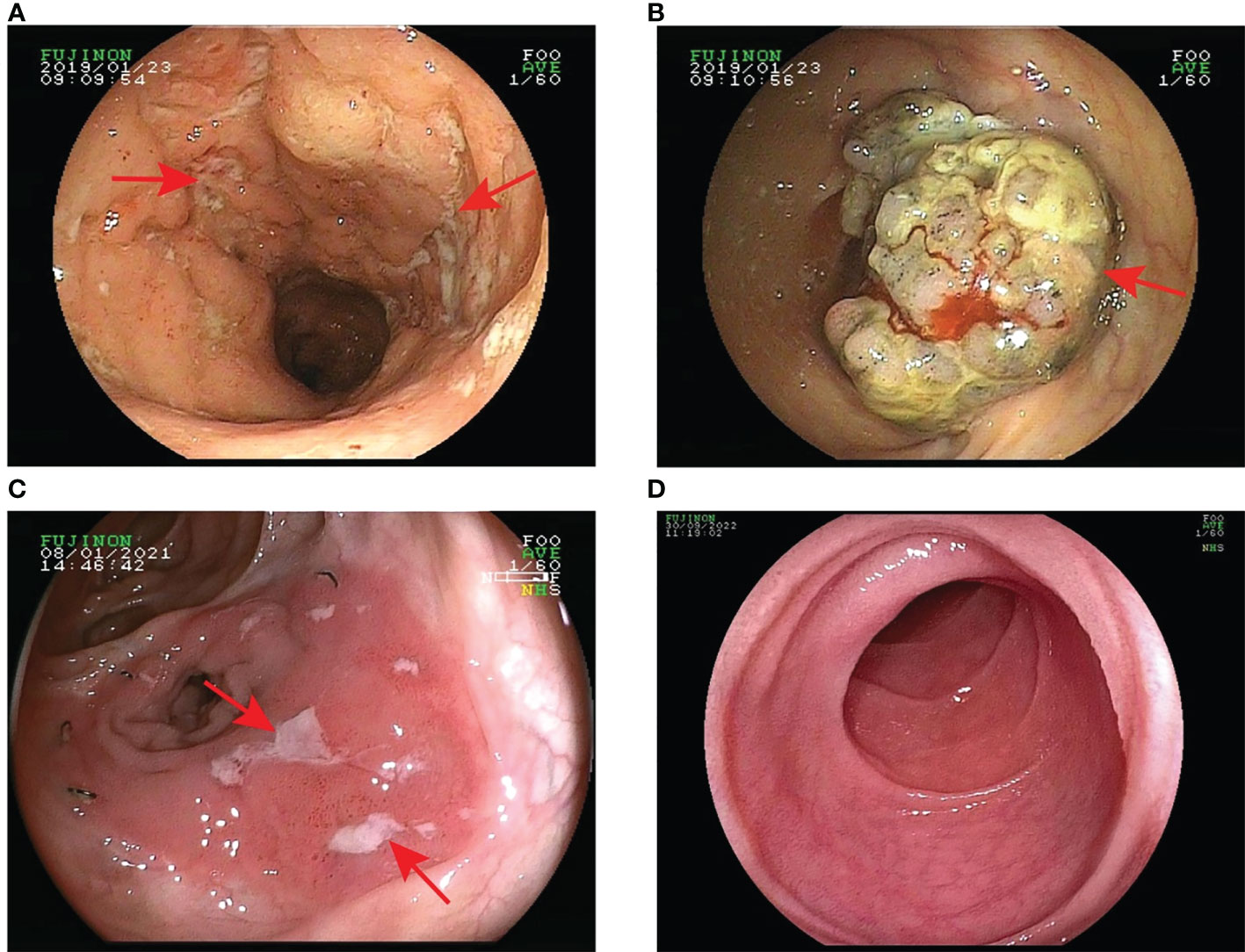
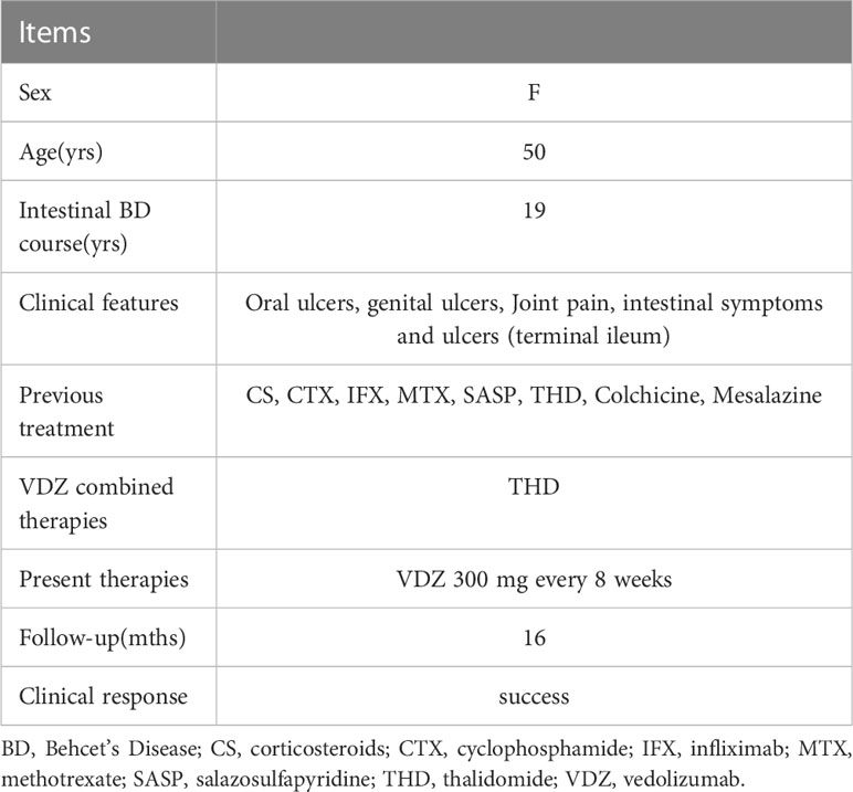
We here describe a 50-year-old female patient with intestinal BD. The clinical characteristics and treatment measures of the patient are shown in Table 1. The patient has had recurrent oral and vulvar ulcers since 2003. In March 2004, she developed nocturnal hyperthermia and dark red bloody stools. Colonoscopy showed terminal ileum ulcers and proliferative lesions. Pathological biopsies reported acute and chronic inflammation of the superficial mucosa. Antinuclear antibody (ANA) and pathergy test results were positive, and the pure protein derivative (PPD) test was negative. Indexes such as infection, tumor, and autoantibody series were normal. According to the diagnostic criteria of the International Study Group for Behçet’s Disease (17), and combined with clinical symptoms, laboratory tests, colonoscopy, and pathological findings, the patient was diagnosed with BD. Prednisone (40mg PO daily), cyclophosphamide (50mg PO daily), and mesalazine (4g PO daily) were administered to control symptoms. Prednisone and cyclophosphamide decreased gradually. In December 2005, cyclophosphamide was stopped, and 5mg of prednisone was taken orally daily to maintain symptoms. In August 2007, she developed multiple joint pains. There were no abnormalities in Anti-Streptolysin “O” (ASO), rheumatoid factor (RF), human leukocyte antigen (HLA-B27), and anti-neutrophil cytoplasmic antibody (ANCA). The treatment regimens were adjusted repeatedly according to the patient’s clinical symptoms, including prednisone (40mg PO daily), sulfasalazine (3.0mg PO daily), methotrexate (12.5mg PO weekly), thalidomide (100mg PO daily), cyclophosphamide (50mg PO daily) and colchicine (1mg PO daily), etc. In January 2019, the patient’s condition worsened with persistent right lower abdominal pain, bloody stools, frequent episodes of oral ulcers, and arthralgia. Colonoscopy showed multiple ulcers in the distal ileum and large protuberant lesions in the cecum and ascending colon (Figures 1A, B). The biopsies revealed chronic inflammatory activity of the mucosa with ulcer formation, and no evidence of neoplasia was seen. Considering the patient’s recurrent illness, no significant improvement in intestinal mucosal status, and the failure of previous medications, we decided to switch to treatment with anti-TNF-α biologics —– infliximab (IFX). We truthfully stated the benefits and risks of using biologics and obtained the informed consent of the patient. IFX was administered with a dose of 300 mg at 0, 2, 6 weeks, and then every eight weeks. The patient reported a significant reduction in oral and vulvar ulcers, abdominal pain, and arthralgia immediately after the first infusion. On August 9, 2019, a colonoscopy showed that the terminal ileal ulcer was better than before. However, there were no significant changes in ileocecal and ascending colon masses. Biopsies revealed acute mucous inflammation, small vessel dilatation and congestion, epithelial hyperplasia on the recessed surface with erosion and inflammatory exudation, and significant glandular hyperplasia and mucus secretion. There was no evidence of granuloma or neoplasia. Intestinal dual-source CT showed obvious thickening at the lower end of ascending colon, soft tissue shadow protruding into the lumen, and abundant blood supply. Endoscopists considered performing endoscopic submucosal dissection (ESD) to be risky. The gastrointestinal surgeon reported that the possibility of colon cancer could not be excluded from the ascending colonic mass and recommended surgical excision to determine its nature. Therefore, after multidisciplinary discussions, the patient underwent a right radical hemicolectomy on September 6, 2019. Post-operative pathology confirmed the mucinous tumor (Figure 2). IFX was suspended. The mFOLFOX6 chemotherapy regimen was administered for four cycles before being discontinued due to the global outbreak of the novel coronavirus. Long-term oral thalidomide (50mg PO daily) to control the disease. In January 2021, the patient’s right lower abdominal pain worsened again. Colonoscopy showed multiple large ulcers in the ileum and anastomotic orifice (Figure 1C). Enhanced CT of the abdomen showed thickening of the anastomotic wall and the proximal ileal wall of the anastomosis with significant enhancement.

Figure 1 Changes in the patient’s intestinal mucosa. (A) In January 2019, prior to the application of infliximab, colonoscopy showed multiple ulcers at the end of the ileum; (B) In August 2019, colonoscopy revealed a large bulging lesion in the cecum and ascending colon; (C) In January 2021, colonoscopy again showed multiple large ulcers in the ileum and anastomosis; (D) In September 2022, one year after vedolizumab treatment colonoscopy showed complete healing of the ileal ulcers.

Figure 2 Pathology images from the right radical hemicolectomy. H&E staining revealed an irregular glandular arrangement of the mucosal epithelium with a large amount of mucus visible in the background and infiltrative growth.
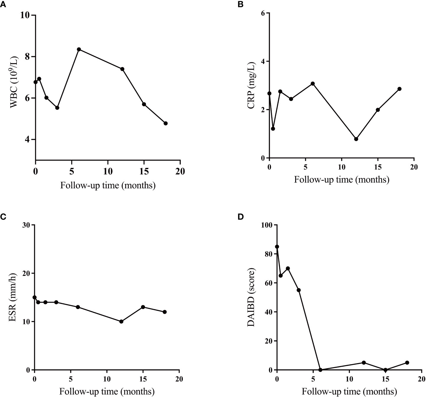
The intestinal BD recurred. The immunosuppressive effect of anti-TNF-α biologics may be risky for patients with an oncology history. Therefore, combined with the pathogenesis of intestinal BD and previous case reports, we decided to try VDZ for some clinical benefits. On October 9, 2021, the patient received an infusion of 300 mg VDZ (initially at 0, 2, and 6 weeks, then every eight weeks). After a short follow-up of 6 months, the patient reported that her lower abdominal pain and arthralgia were improved. No obvious adverse reactions occurred. The DAIBD score decreased from 85 to 0, and the WBC, ESR, and CRP levels were normal. On September 30, 2022, a colonoscopy showed a completely healed ileal ulcer with a smooth mucosal surface and well-dilated intestine (Figure 1D). However, at the 15th monthly follow-up, her oral and vulvar ulcers recurred, and the CRP concentration increased. Therefore, thalidomide was administered to alleviate systemic inflammation. As shown in Figures 3A–C, although all systemic inflammatory indices showed repeat increases, such as WBC, CRP, and ESR, these might be caused by the presence of inflammation outside of the gastrointestinal (GI) tract. As only focused on the GI tract (Figure 3D), the DAIBD showed the significant efficacy of VDZ on intestinal BD.

Figure 3 Efficacy of vedolizumab in the treatment of intestinal Behcet’s Disease. (A) Changes of white blood cell (WBC); (B) Changes of C-reactive protein (CRP); (B) Changes in erythrocyte sedimentation rate (ESR); (C) Changes in indices of disease activity in intestinal Behcet’s Disease (DAIBD).
3 Discussion and literature review
We report here a case of a refractory intestinal BD patient. She had recurrent oral and genital ulcers, joint pain, and intestinal involvement for nearly 20 years. Anti-TNF-α biologics relieved symptoms when initially applied. However, the patient developed colon cancer during BD. We performed surgical excision and chemotherapy intervention. During follow-up, her intestinal BD recurred. Considering the aggravation of the intestinal condition and the combined tumor history, we decided to try VDZ. At the 6-month follow-up, she achieved clinical remission, all laboratory test results were in normal ranges, and mucosal healing was observed under endoscopy. Several studies have demonstrated that VDZ are effective on extraintestinal manifestations of IBD (18, 19). However, oral and vulvar ulcers still occurred sporadically in the patient we report here, and the symptoms disappeared with the combination of thalidomide.
Unlike other gastrointestinal inflammatory diseases, which are primarily chronic and persistent, BD is a recurrent acute systemic vasculitis (20). Different clinical manifestations can occur individually or coexist in the same patient (21). Typical geographic distribution, infection, immunization, and environmental factors may contribute to the development of BD (10). Vascular damage, neutrophil hyperfunction, and autoimmune reactions are the key characteristics of BD. Analysis of HLA phenotype and serum IgD levels may help to make a diagnosis (20).
Intestinal involvement in BD is usually associated with poor prognosis and a risk of severe organ damage or even death. The spectrum can involve any digestive organ from the oral cavity to the anus, especially the ileocecal region, transverse colon, and ascending colon (22). In Asian countries, ileocecal involvement appears to be more common (23), and volcano type ulcers are typical endoscopic manifestations (1). Endoscopy, computed tomography, and magnetic resonance enterography are used to assess intestinal involvement and disease activity in BD (24).
The ultimate treatment goals for patients with intestinal BD are the disappearance of clinical symptoms, normalization of inflammatory indices, and intestinal mucosal healing (1). The acute phase is usually treated with corticosteroids combined with 5-ASA or azathioprine (AZA) (3). Alternative treatment options include salazosulfapyridine (SASP), mycophenolate mofetil (MMF), and methotrexate (MTX) (25). Patients with severe or refractory intestinal symptoms should be treated with anti-TNF-α biologics alone or in combination with thalidomide (THD) (1). Anti-TNF-α biologics have demonstrated promising results in inducing and maintaining remission of intestinal BD (26, 27). However, due to the role of TNF-α in NK cell- and CD8+ T cell-mediated clearance of tumor cells (28), it can trigger apoptosis through an exogenous pathway by activating caspases 8 and 10. It may also activate signaling through the NF-κB pathway, which indicates anti-TNF-α biologics may promote tumor recurrence, growth, and/or metastasis (5, 29, 30).
The patient we report here had multiple colonic mucosal biopsies, all of which indicated inflammatory lesions. In 2019, we again performed large biopsies of the ileal and ascending colon masses prior to the use of IFX. Neoplastic lesions were still excluded, suggesting chronic mucosal inflammatory activity. We obtained the patient’s informed consent for the use of IFX and the symptoms remitted. However, colonoscopy showed no significant changes in the masses. After multidisciplinary discussion, we decided to surgically resect the hyperplastic lesions, and postoperative pathology revealed local tumor. It cannot be absolutely excluding that the appearance of cancerous lesions is due to the use of IFX. However, prior to the use of IFX, we have repeatedly performed pathological biopsies of the masses to exclude malignant lesions. The current reports on anti-TNF-α biologics and tumors are mainly on skin cancers, including melanoma (31–34). ECCO guidelines recommend that infliximab is best avoided in patients with a history of malignancy (35). Therefore, it is necessary to consider other alternative therapies for patients with refractory intestinal BD with an oncology history.
Our literature review found a case report of VDZ successfully treating refractory BD (16). The patient had erythema nodosum, oro-genital ulcers, and biopsy-proven intestinal BD, which was not successfully treated with conventional immunosuppressants and several biologics agents, including anti-TNF-α biologics. Based on previous reports of the efficacy of VDZ in IBD, the authors decided to administer VDZ to treat severe intestinal involvement. The results led to a satisfactory gastrointestinal response and the concomitant disappearance of ulcerations, arthralgia, and a reversion of the skin lesions. VDZ, an intestine-selective humanized monoclonal IgG1 antibody, has been approved for treating IBD (36, 37). VDZ blocks the recruitment of pro-inflammatory cells and dendritic cells to the inflamed gut by specifically targeting α4β7 integrins expressed on the surface of intestinal lymphocytes and monocytes and inhibiting their binding to cell adhesion molecules (CAMs) (15, 38, 39), leading to alterations in the innate and acquired immune cell program to suppress inflammation without interfering with transit to other organs (40). Compared to anti-TNF-α biologics or other immunosuppressive agents, integrin receptor antagonists reduce the side effects associated with systemic immunosuppression. Moreover, studies reported no increase in malignancy and mortality among VDZ-exposed patients (41).
Growing evidence shows that IBD and BD may be closely related and are part of a typical disease spectrum (42). They have similarities in plausible pathophysiological features. We should note that VDZ had a good response in the ileum portion and not in the whole tract of intestine. That in IBD there is a heterogeneous distribution of immune cells in the enteric tract, with the presence of CD4+ memory (mem), lymphocytes, B and dendritic cells in the ileum (43). The most α4β7 positive cells are CD4+mem, CD8+mem and B cells, which can explain not only as to why VDZ works better on lesions in some parts of the intestine (as ileum) and less so in others (44), but also as too why VDZ did not work on ulcer in the other mucous as oral and vulval ones. This is probably due to the heterogeneous distribution of the α4β7 positive cells in these mucous.
The relationship between IBD and colorectal cancer has been demonstrated (45). Although BD is clinically similar to IBD. Only a tiny series of tumor-related cases had been reported, of which colon cancer is less common (46–48). There is no clear evidence that BD causes epithelial cancer or sarcoma. The patient we reported was diagnosed with colonic mucinous carcinoma 16 years after the first presentation of BD symptoms. The severity and duration of inflammation may be risk factors for cancer (45, 49). As increasing numbers of potent drugs are used to treat BD, patients may live longer than before and therefore be more likely to develop the malignant disease. In addition, extensive vasculitis and abnormal immune regulation in BD are thought to be mechanisms of increased risk of malignancy (50). Further detailed pathological studies, cytogenetic analysis, and long-term sizeable prospective cohort studies are needed to clarify the relationship between BD and tumors.
4 Conclusion
VDZ specifically blocks T-cell chemotaxis to the ileum during inflammation and inhibits inflammatory factor signaling. It is suggested that it may serve as a potential treatment drug for intestinal BD, especially involving the ileum tract. The patients with intestinal BD we reported here had poor efficacy to conventional drugs. Anti-TNF-α biologics had shown better therapeutic effects. However, due to the occurrence of a colon tumor, we attempted to switch to VDZ. The intestinal symptoms and joint pain were reduced, and intestinal mucosa ulcers healed completely. No recurrence or other side effects occurred. Thus, our example provided possible evidence for the efficacy and safety of VDZ in patients with refractory intestinal BD who have an oncology history. As an intestinal-specific antibody, VDZ has shown an inhibitory effect on ileal inflammation but is less effective in treating systemic vasculitis. During the follow-ups, the manifestations like oral and vulvar ulcers and increased CRP were further resolved by thalidomide.
Short follow-up time and only one case reported are our major limitations. Large and long-term clinical trials are needed to verify the efficacy and safety of VDZ in treating intestinal BD. In addition, in the intestinal BD the effect and mechanism of VDZ on the other tracts of intestine and on other mucous membranes need to be further explored.
Data availability statement
The original contributions presented in the study are included in the article/supplementary material. Further inquiries can be directed to the corresponding authors.
Ethics statement
Written informed consent was obtained from the individual(s) for the publication of any potentially identifiable images or data included in this article.
Author contributions
TW and JL contributed to conception and design of the study. RL organized the database. XL performed the statistical analysis. RL, XL and HZ wrote the main content of the manuscript. YS and FW provided tables and pictures. All authors contributed to the article and approved the submitted version.
Funding
Natural Science Foundation of Shaanxi Province, Key Industrial Innovation Project Fund(2023-ZDLSF-44); Airforce Aoxiang Foundation and Young Changjiang Scholars of the Ministry of Education (JL); National Natural Science Foundation of China Major Research Program Integration Project (92259302); School Science and Technology Development Fund of Air Force Military Medical University (2022XB009); Independent Funds of the Key Laboratory (CBSKL2022ZZ34, CBSKL2015Z12).
Conflict of interest
The authors declare that the research was conducted in the absence of any commercial or financial relationships that could be construed as a potential conflict of interest.
Publisher’s note
All claims expressed in this article are solely those of the authors and do not necessarily represent those of their affiliated organizations, or those of the publisher, the editors and the reviewers. Any product that may be evaluated in this article, or claim that may be made by its manufacturer, is not guaranteed or endorsed by the publisher.
References
1. Watanabe K, Tanida S, Inoue N, Kunisaki R, Kobayashi K, Nagahori M, et al. Evidence-based diagnosis and clinical practice guidelines for intestinal behcet’s disease 2020 edited by intractable diseases, the health and labour sciences research grants. J Gastroenterol (2020) 55(7):679–700. doi: 10.1007/s00535-020-01690-y
2. Cheon JH, Kim WH. An update on the diagnosis, treatment, and prognosis of intestinal behcet’s disease. Curr Opin Rheumatol (2015) 27(1):24–31. doi: 10.1097/BOR.0000000000000125
3. Hatemi G, Christensen R, Bang D, Bodaghi B, Celik AF, Fortune F, et al. Update of the EULAR recommendations for the management of behcet’s syndrome. Ann Rheum Dis (2018) 77(6):808–18. doi: 10.1136/annrheumdis-2018-213225
4. Caso F, Costa L, Rigante D, Lucherini OM, Caso P, Bascherini V, et al. Biological treatments in behcet’s disease: beyond anti-TNF therapy. Mediators Inflamm (2014) 2014:107421. doi: 10.1155/2014/107421
5. Hoentjen F, van Bodegraven AA. Safety of anti-tumor necrosis factor therapy in inflammatory bowel disease. World J Gastroenterol (2009) 15(17):2067–73. doi: 10.3748/wjg.15.2067
6. Kamada N, Seo SU, Chen GY, Nunez G. Role of the gut microbiota in immunity and inflammatory disease. Nat Rev Immunol (2013) 13(5):321–35. doi: 10.1038/nri3430
7. Consolandi C, Turroni S, Emmi G, Severgnini M, Fiori J, Peano C, et al. Behcet’s syndrome patients exhibit specific microbiome signature. Autoimmun Rev (2015) 14(4):269–76. doi: 10.1016/j.autrev.2014.11.009
8. Takeuchi M, Kastner DL, Remmers EF. The immunogenetics of behcet’s disease: a comprehensive review. J Autoimmun (2015) 64:137–48. doi: 10.1016/j.jaut.2015.08.013
9. Zeidan MJ, Saadoun D, Garrido M, Klatzmann D, Six A, Cacoub P. Behcet’s disease physiopathology: a contemporary review. Auto Immun Highlights (2016) 7(1):4. doi: 10.1007/s13317-016-0074-1
10. Chen J, Yao X. A contemporary review of behcet’s syndrome. Clin Rev Allergy Immunol (2021) 61(3):363–76. doi: 10.1007/s12016-021-08864-3
11. Castano-Nunez A, Montes-Cano MA, Garcia-Lozano JR, Ortego-Centeno N, Garcia-Hernandez FJ, Espinosa G, et al. Association of functional polymorphisms of KIR3DL1/DS1 with behcet’s disease. Front Immunol (2019) 10:2755. doi: 10.3389/fimmu.2019.02755
12. Yoshikawa K, Watanabe T, Sekai I, Takada R, Hara A, Kurimoto M, et al. Case report: a case of intestinal behcet’s disease exhibiting enhanced expression of IL-6 and forkhead box P3 mRNA after treatment with infliximab. Front Med (Lausanne) (2021) 8:679237. doi: 10.3389/fmed.2021.679237
13. Hazel K, O’Connor A. Emerging treatments for inflammatory bowel disease. Ther Adv Chronic Dis (2020) 11:2040622319899297. doi: 10.1177/2040622319899297
14. Zeissig S, Rosati E, Dowds CM, Aden K, Bethge J, Schulte B, et al. Vedolizumab is associated with changes in innate rather than adaptive immunity in patients with inflammatory bowel disease. Gut (2019) 68(1):25–39. doi: 10.1136/gutjnl-2018-316023
15. Picarella D, Hurlbut P, Rottman J, Shi X, Butcher E, Ringler DJ. Monoclonal antibodies specific for beta 7 integrin and mucosal addressin cell adhesion molecule-1 (MAdCAM-1) reduce inflammation in the colon of scid mice reconstituted with CD45RBhigh CD4+ T cells. J Immunol (1997) 158(5):2099–106. doi: 10.4049/jimmunol.158.5.2099
16. Arbrile M, Radin M, Rossi D, Menegatti E, Baldovino S, Sciascia S, et al. Vedolizumab for the management of refractory behcet’s disease: from a case report to new pieces of mosaic in a complex disease. Front Immunol (2021) 12:769785. doi: 10.3389/fimmu.2021.769785
17. Wechsler B, Davatchi F, Mizushima Y, Hamza M, Dilsen N, Kansu E, et al. Criteria for diagnosis of behcet’s disease. international study group for behcet’s disease. Lancet (1990) 335(8697):1078–80. doi: 10.1016/0140-6736(90)92643-V
18. Fleisher M, Marsal J, Lee SD, Frado LE, Parian A, Korelitz BI, et al. Effects of vedolizumab therapy on extraintestinal manifestations in inflammatory bowel disease. Dig Dis Sci (2018) 63(4):825–33. doi: 10.1007/s10620-018-4971-1
19. Hanzel J, Ma C, Casteele NV, Khanna R, Jairath V, Feagan BG. Vedolizumab and extraintestinal manifestations in inflammatory bowel disease. Drugs (2021) 81(3):333–47. doi: 10.1007/s40265-020-01460-3
20. Sakane T, Takeno M, Suzuki N, Inaba G. Behcet’s disease. N Engl J Med (1999) 341(17):1284–91. doi: 10.1056/NEJM199910213411707
21. Yazici H, Ugurlu S, Seyahi E. Behcet syndrome: is it one condition? Clin Rev Allergy Immunol (2012) 43(3):275–80. doi: 10.1007/s12016-012-8319-x
22. Naganuma M, Iwao Y, Inoue N, Hisamatsu T, Imaeda H, Ishii H, et al. Analysis of clinical course and long-term prognosis of surgical and nonsurgical patients with intestinal behcet’s disease. Am J Gastroenterol (2000) 95(10):2848–51. doi: 10.1111/j.1572-0241.2000.03198.x
23. Bayraktar Y, Ozaslan E, Van Thiel DH. Gastrointestinal manifestations of behcet’s disease. J Clin Gastroenterol (2000) 30(2):144–54. doi: 10.1097/00004836-200003000-00006
24. Cheng L, Li L, Liu C, Yan S, Li Y. Meta-analysis of anti-saccharomyces cerevisiae antibodies as diagnostic markers of behcet’s disease with gastrointestinal involvement. BMJ Open (2020) 10(10):e033880. doi: 10.1136/bmjopen-2019-033880
25. Kinoshita H, Nishioka H, Ikeda A, Ikoma K, Sameshima Y, Ohi H, et al. Remission induction, maintenance, and endoscopic outcome with oral 5-aminosalicylic acid in intestinal behcet’s disease. J Gastroenterol Hepatol (2019) 34(11):1929–39. doi: 10.1111/jgh.14690
26. Naganuma M, Sakuraba A, Hisamatsu T, Ochiai H, Hasegawa H, Ogata H, et al. Efficacy of infliximab for induction and maintenance of remission in intestinal behcet’s disease. Inflammation Bowel Dis (2008) 14(9):1259–64. doi: 10.1002/ibd.20457
27. Iwata S, Saito K, Yamaoka K, Tsujimura S, Nawata M, Suzuki K, et al. Effects of anti-TNF-alpha antibody infliximab in refractory entero-behcet’s disease. Rheumatol (Oxford) (2009) 48(8):1012–3. doi: 10.1093/rheumatology/kep126
28. Bongartz T, Sutton AJ, Sweeting MJ, Buchan I, Matteson EL, Montori V. Anti-TNF antibody therapy in rheumatoid arthritis and the risk of serious infections and malignancies: systematic review and meta-analysis of rare harmful effects in randomized controlled trials. JAMA (2006) 295(19):2275–85. doi: 10.1001/jama.295.19.2275
29. Danial NN, Korsmeyer SJ. Cell death: critical control points. Cell (2004) 116(2):205–19. doi: 10.1016/S0092-8674(04)00046-7
30. Balkwill F. Tumour necrosis factor and cancer. Nat Rev Cancer (2009) 9(5):361–71. doi: 10.1038/nrc2628
31. Long MD, Martin CF, Pipkin CA, Herfarth HH, Sandler RS, Kappelman MD. Risk of melanoma and nonmelanoma skin cancer among patients with inflammatory bowel disease. Gastroenterology (2012) 143(2):390–9 e1. doi: 10.1053/j.gastro.2012.05.004
32. Mariette X, Matucci-Cerinic M, Pavelka K, Taylor P, van Vollenhoven R, Heatley R, et al. Malignancies associated with tumour necrosis factor inhibitors in registries and prospective observational studies: a systematic review and meta-analysis. Ann Rheum Dis (2011) 70(11):1895–904. doi: 10.1136/ard.2010.149419
33. Ramiro S, Gaujoux-Viala C, Nam JL, Smolen JS, Buch M, Gossec L, et al. Safety of synthetic and biological DMARDs: a systematic literature review informing the 2013 update of the EULAR recommendations for management of rheumatoid arthritis. Ann Rheum Dis (2014) 73(3):529–35. doi: 10.1136/annrheumdis-2013-204575
34. Annese V, Beaugerie L, Egan L, Biancone L, Bolling C, Brandts C, et al. European Evidence-based consensus: inflammatory bowel disease and malignancies. J Crohns Colitis (2015) 9(11):945–65. doi: 10.1093/ecco-jcc/jjv141
35. Travis SP, Stange EF, Lemann M, Oresland T, Chowers Y, Forbes A, et al. European Evidence based consensus on the diagnosis and management of crohn’s disease: current management. Gut (2006) 55(Suppl 1):i16–35. doi: 10.1136/gut.2005.081950b
36. Sandborn WJ, Feagan BG, Rutgeerts P, Hanauer S, Colombel JF, Sands BE, et al. Vedolizumab as induction and maintenance therapy for crohn’s disease. N Engl J Med (2013) 369(8):711–21. doi: 10.1056/NEJMoa1215739
37. Feagan BG, Rutgeerts P, Sands BE, Hanauer S, Colombel JF, Sandborn WJ, et al. Vedolizumab as induction and maintenance therapy for ulcerative colitis. N Engl J Med (2013) 369(8):699–710. doi: 10.1056/NEJMoa1215734
38. Soler D, Chapman T, Yang LL, Wyant T, Egan R, Fedyk ER. The binding specificity and selective antagonism of vedolizumab, an anti-alpha4beta7 integrin therapeutic antibody in development for inflammatory bowel diseases. J Pharmacol Exp Ther (2009) 330(3):864–75. doi: 10.1124/jpet.109.153973
39. Fedyk ER, Wyant T, Yang LL, Csizmadia V, Burke K, Yang H, et al. Exclusive antagonism of the alpha4 beta7 integrin by vedolizumab confirms the gut-selectivity of this pathway in primates. Inflammation Bowel Dis (2012) 18(11):2107–19. doi: 10.1002/ibd.22940
40. Haanstra KG, Hofman SO, Lopes Estevao DM, Blezer EL, Bauer J, Yang LL, et al. Antagonizing the alpha4beta1 integrin, but not alpha4beta7, inhibits leukocytic infiltration of the central nervous system in rhesus monkey experimental autoimmune encephalomyelitis. J Immunol (2013) 190(5):1961–73. doi: 10.4049/jimmunol.1202490
41. Novak G, Hindryckx P, Khanna R, Jairath V, Feagan BG. The safety of vedolizumab for the treatment of ulcerative colitis. Expert Opin Drug Saf (2017) 16(4):501–7. doi: 10.1080/14740338.2017.1300251
42. TV A, Haikarainen T, Raivola J, Silvennoinen O. Selective JAKinibs: prospects in inflammatory and autoimmune diseases. BioDrugs (2019) 33(1):15–32. doi: 10.1007/s40259-019-00333-w
43. Bai X, Liu W, Chen H, Zuo T, Wu X. Immune cell landscaping reveals distinct immune signatures of inflammatory bowel disease. Front Immunol (2022) 13:861790. doi: 10.3389/fimmu.2022.861790
44. Luzentales-Simpson M, Pang YCF, Zhang A, Sousa JA, Sly LM. Vedolizumab: potential mechanisms of action for reducing pathological inflammation in inflammatory bowel diseases. Front Cell Dev Biol (2021) 9:612830. doi: 10.3389/fcell.2021.612830
45. Jess T, Loftus EV Jr., Velayos FS, Harmsen WS, Zinsmeister AR, Smyrk TC, et al. Risk of intestinal cancer in inflammatory bowel disease: a population-based study from olmsted county, Minnesota. Gastroenterology (2006) 130(4):1039–46. doi: 10.1053/j.gastro.2005.12.037
46. Cengiz M, Altundag MK, Zorlu AF, Gullu IH, Ozyar E, Atahan IL. Malignancy in behcet’s disease: a report of 13 cases and a review of the literature. Clin Rheumatol (2001) 20(4):239–44. doi: 10.1007/s100670170036
47. Kaklamani VG, Tzonou A, Kaklamanis PG. Behcet’s disease associated with malignancies. report of two cases and review of the literature. Clin Exp Rheumatol (2005) 23(4 Suppl 38):S35–41.
48. Na SY, Shin J, Lee ES. Morbidity of solid cancer in behvarsigmaet’s disease: analysis of 11 cases in a series of 506 patients. Yonsei Med J (2013) 54(4):895–901. doi: 10.3349/ymj.2013.54.4.895
49. Rutter M, Saunders B, Wilkinson K, Rumbles S, Schofield G, Kamm M, et al. Severity of inflammation is a risk factor for colorectal neoplasia in ulcerative colitis. Gastroenterology (2004) 126(2):451–9. doi: 10.1053/j.gastro.2003.11.010
Keywords: Behcet’s disease, intestinal disease, vedolizumab, tumor, case report
Citation: Li R, Li X, Zhou H, Shi Y, Wang F, Wu T and Liang J (2023) Successful treatment of a refractory intestinal Behcet’s disease with an oncology history by Vedolizumab: a case report and literature review. Front. Immunol. 14:1205046. doi: 10.3389/fimmu.2023.1205046
Received: 13 April 2023; Accepted: 10 May 2023;
Published: 23 May 2023.
Edited by:
Irma Convertino, University of Pisa, ItalyReviewed by:
Ibrahim Hatemi, Istanbul University-Cerrahpasa, TürkiyeToshiyuki Matsui, Fukuoka University, Japan
Copyright © 2023 Li, Li, Zhou, Shi, Wang, Wu and Liang. This is an open-access article distributed under the terms of the Creative Commons Attribution License (CC BY). The use, distribution or reproduction in other forums is permitted, provided the original author(s) and the copyright owner(s) are credited and that the original publication in this journal is cited, in accordance with accepted academic practice. No use, distribution or reproduction is permitted which does not comply with these terms.
*Correspondence: Tong Wu, d3V0b25nNzI1QDEyNi5jb20=; Jie Liang, Y29uY2lzZWxqQDEyNi5jb20=
†These authors have contributed equally to this work and share first authorship
 Ruixia Li
 Xiaofei Li†
 Yanting Shi
 Tong Wu