- 1Department of Immunology, Graduate School of Medical Sciences, Kumamoto University, Kumamoto, Japan
- 2Precursory Research for Embryonic Science and Technology (PRESTO), Japan Science and Technology Agency (JST), Kumamoto, Japan
- 3Department of Microbiology and Immunology, Graduate School of Medicine, Hokkaido University, Sapporo, Hokkaido, Japan
Type I interferon (IFN) induces many antiviral factors in host cells. RIG-I-like receptors (RLRs) are cytoplasmic viral RNA sensors that trigger the signal to induce the innate immune response that includes type I IFN production. RIG-I and MDA5 are RLRs that form nucleoprotein filaments along viral double-stranded RNA, resulting in the activation of MAVS adaptor molecule. The MAVS protein forms a prion-like aggregation structure, leading to type I IFN production. RIG-I and MDA5 undergo post-translational modification. TRIM25 and Riplet ubiquitin ligases deliver a K63-linked polyubiquitin moiety to the RIG-I N-terminal caspase activation and recruitment domains (CARDs) and C-terminal region; the polyubiquitin chain then stabilizes the two-CARD tetramer structure required for MAVS assembly. MDA5 activation is regulated by phosphorylation. RIOK3 is a protein kinase that phosphorylates the MDA5 protein in a steady state, and PP1α/γ dephosphorylate this protein, resulting in its activation. RIG-I and MDA5 require cytoplasmic RNA helicases for their efficient activation. LGP2, another RLR, is an RNA helicase involved in RLR signaling. This protein does not possess N-terminal CARDs and, thus, cannot trigger downstream signaling by itself. Recent studies have revealed that this protein modulates MDA5 filament formation, resulting in enhanced type I IFN production. Several other cytoplasmic RNA helicases are involved in RLR signaling. DDX3, DHX29, DHX36, and DDX60 RNA helicases have been reported to be involved in RLR-mediated type I IFN production after viral infection. However, the underlying mechanism is largely unknown. Future studies are required to reveal the role of RNA helicases in the RLR signaling pathway.
Introduction
The innate immune system is the first line of defense against viral infection. RIG-I-like receptors (RLRs) are cytoplasmic viral RNA sensors that recognizes viral double-stranded (ds) RNA and trigger antiviral innate immune responses (1). The RIG-I and MDA5 proteins, which are members of RLRs, comprise two caspase activation and recruitment domains (CARDs), a helicase domain, and a C-terminal domain (CTD) (2). The helicase domain and CTD bind to viral RNA, and CTD is essential for the recognition of viral RNA (3, 4). After the recognition of viral RNA, two N-terminal CARDs forms a two-CARD tetramer structure, which acts as a core of the MAVS prion-like aggregation structure (5, 6). The MAVS protein is a solo adaptor of RLRs and activates kinases and ubiquitin ligases, leading to the activation of transcription factors, such as IRF-3 and NF-κB (7–10). The transcription factors induce type I interferon (IFN) and pro-inflammatory cytokine production.
RIG-I recognizes relatively short dsRNA (<1 kbp) with a 5′ tri- or di-phosphate group (11–13), whereas another RLR member, MDA5, recognizes long dsRNA (>1 kbp) with or without a 5′ phosphate group (12). Influenza A virus, Sendaivirus, hepatitis C virus (HCV), and vesicular stomatitis virus (VSV) are mainly recognized by RIG-I, whereas encephalomyocarditis virus (EMCV) and poliovirus are recognized by MDA5 (14, 15). West Nile virus and Japanese encephalitis virus are recognized by both RIG-I and MDA5 (14, 15).
Post-Translational Modification of RLRs
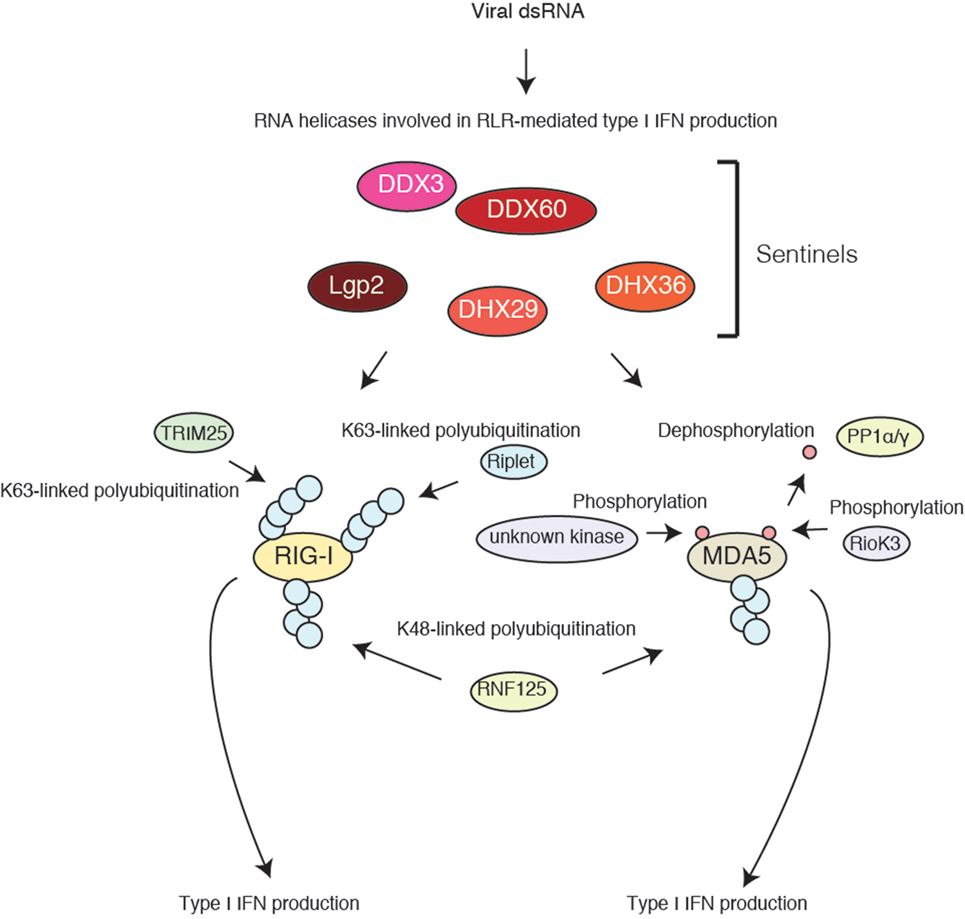
Recent studies have revealed the post-translational modification of RLRs (Figure 1). Gack and colleagues first reported the K63-linked polyubiquitination of RIG-I CARDs by TRIM25 ubiquitin ligase, which is essential for their activation (16). Later studies showed that the non-covalent binding of the K63-linked polyubiquitin chain is sufficient to activate RIG-I signaling (17). A covalent and/or non-covalent K63-linked polyubiquitin chain stabilizes the two-CARD tetramer structure (6). Another ubiquitin ligase, Riplet (also called RNF135 or REUL), mediates K63-linked polyubiquitination of RIG-I C-terminal region, which promotes the binding of TRIM25 to RIG-I (18, 19). The knockout (KO) of each ubiquitin ligase has been shown to markedly reduce RIG-I-mediated type I IFN production (16, 20). These ubiquitin ligases are targeted by several viral proteins, such as NS-1 of influenza A virus and NS3-4A of HCV, resulting in the attenuation of RIG-I-mediated type I IFN production (19, 21). These findings indicate the importance of both ubiquitin ligases in RIG-I activation. RIG-I also undergoes K48-linked polyubiquitination by RNF125, leading to its proteasomal degradation (22). TRIM25 itself undergoes linear polyubiquitination by the linear ubiquitin assembly complex (23). These observations indicate that the ubiquitin chain plays a critical role in the cytoplasmic antiviral innate immune response (24).

Figure 1. Accessory factors of RIG-I and MDA5. RIG-I and MDA5 undergo post-translational modifications. TRIM25 and Riplet ubiquitin ligases deliver a K63-linked polyubiquitin moiety to RIG-I N-terminal CARDs and C-terminal regions, respectively, resulting in type I IFN production. MDA5 is phosphorylated in resting cells. RIOK3 is a protein kinase involved in MDA5 phosphorylation; however, there seem to be other kinases targeting MDA5. PP1α/γ dephosphorylates MDA5, leading to type I IFN production. Several cytoplasmic RNA helicases are involved in RLR-mediated type I IFN production, but their biochemical activities are largely unknown.
MDA5 assembles along viral long dsRNA and forms a nucleoprotein filament structure required for MAVS activation (25). MDA5 activation is regulated by phosphorylation (26). In resting cells, the MDA5 protein undergoes phosphorylation. PP1 α/γ are required for MDA5 dephosphorylation, leading to its activation. Recently, we reported that RIOK3 is a protein kinase that mediates MDA5 phosphorylation at Ser-828, which impairs MDA5 assembly and attenuates its activation (27). Considering that Gack and colleagues have shown that the N-terminal region of MDA5 is phosphorylated (26), other protein kinases that phosphorylate MDA5 are required for the attenuation of the signaling.
The phosphorylation of RIG-I and ubiquitination of MDA5 have also been reported and are required for the regulation of RLR signaling (24).
RNA Helicases Involved in RLRs-Mediated Type I IFN Production Pathway
Several RNA helicases lacking CARDs are involved in RLR-mediated signaling (28). Bowie and colleagues first reported that a non-RLR helicase, DDX3, is involved in RIG-I-mediated type I IFN production. They showed that DDX3 is required for the activation of TBK1 and IKK-ɛ, which are downstream factors of MAVS (29, 30). Later, we reported that DDX3 associates with RIG-I and promotes RIG-I–RNA binding (31).
LGP2 is a member of RLRs but lacks N-terminal CARDs (2). Therefore, LGP2 by itself cannot trigger the signal to induce type I IFN. Initial studies reported that LGP2 attenuates RLR signaling in response to polyI:C in mouse embryonic fibroblasts (MEFs) (2, 32), whereas later studies reported a positive role of LGP2 in RLR signaling during viral infection (33). DHX36 is a cytoplasmic RNA helicase and does not comprise CARDs. DHX36 protein complexed with DDX1 and DDX21 recognizes viral RNA and triggers the signal to induce type I IFN production via TRIF/TICAM-1 adaptor in some kinds of dendritic cells (DCs) (34). DHX36 also functions upstream of RIG-I and is required for the formation of antiviral stress granules where viral RNA is recognized by RLRs (35). DHX29 RNA helicase directly binds to polyI:C and polydA:dT and also associates with RIG-I. The protein exhibits a cell-type-specific expression pattern and is required for type I IFN production only in human respiratory epithelial cells (36).
DDX60 is another cytoplasmic RNA helicase, which does not contain N-terminal CARDs as RNA helicases described above. The DDX60 protein binds to dsRNA and associates with RLRs (37). DDX60 promotes RIG-I–RNA binding, which triggers type I IFN production (37). Previously, we generated DDX60 KO mice and reported that DDX60 KO moderately reduced RIG-I-mediated type I IFN production from peritoneal macrophages and MEFs but not from bone-marrow-derived cells, suggesting the cell-type specificity (38). DDX60 exhibited antiviral activities against only specific viruses (39). Another group also generated a DDX60 gene trapping mouse (called DDX60 KO first) and a DDX60 KO mouse (called DDX60 KO full) (40), whose constructs are different from our DDX60 KO mouse construct (38, 40). They confirmed that bone-marrow-derived cells of DDX60 KO first and KO full mice normally produced type I IFN as we observed in our DDX60 KO mice (40). Although DDX60 KO first seems to reduce type I IFN production after stimulation with a low concentration of a RIG-I ligand, type I IFN production from DDX60 KO first MEFs were comparable to wild-type MEFs in other experimental conditions (40). These data implied that DDX60 is not a general factor for RIG-I activation and plays a role in RIG-I signaling only when cells are infected with specific viruses or stimulated with specific or low concentration of RIG-I ligands in a cell-type-specific manner (38–40).
Recently, we identified another role of DDX60 in the antiviral response. The DDX60 protein exhibits the similarity to SKI2 RNA helicase, a component of the RNA exosome, which degrades host and viral RNA (37, 40). We found that DDX60 associates with the core components of the RNA exosome and is involved in a viral RNA degradation pathway (37, 38). DDX60-mediated viral RNA degradation plays an important role in the antiviral response when the RIG-I pathway is blocked (38).
Perspective
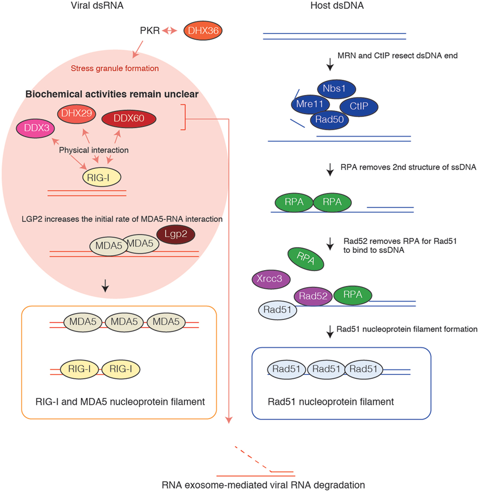
RIG-I and MDA5 forms a nucleoprotein filament (41). It is well known that the Rad51 protein, which is involved in DNA homologous recombination, forms a nucleoprotein filament along single-stranded (ss) DNA (42–45). For nucleoprotein filament formation, Rad51 requires several accessory factors. The Mre11 protein complex produces an ssDNA region (46), and then the RPA and Rad52 cooperatively produce the platform for Rad51 nucleoprotein filament formation as described in Figure 2 (47–49). There are several other factors involved in the Rad51 pathway, which occasionally compensates for a defect of other factors (50, 51). The role of each factor has been revealed by intensive biochemical studies. By contrast, the biochemical activities of accessory factors for RIG-I and MDA5 are largely unknown (Figure 2). Recently, Horvath and colleagues have revealed that LGP2 regulates MDA5 filament assembly (52).

Figure 2. Nucleoprotein filament formation. Rad51 assembles along the single-stranded (ss) DNA region and forms a nucleoprotein filament. Mre11/Rad50/Nbs1 protein complex resects DNA double-stranded (ds) DNA together with CtIP, leading to the production of the ssDNA region. First, RPA binds to the ssDNA region to prevent the formation of secondary structure. Next, Rad52 and other proteins remove RPA from DNA for Rad51 to assemble along the ssDNA. RIG-I and MDA5 assemble along viral dsRNA and form nucleoprotein filaments. LGP2 modulates MDA5 nucleoprotein filament formation, resulting in type I IFN production. DHX36 is required for PKR-mediated antiviral stress granule formation. DDX3, DHX27, and DDX60 bind to RIG-I. The biochemical activities of DDX3, DHX29, DHX36, and DDX60 RNA helicases in nucleoprotein filament formation are largely unknown.
The LGP2 protein increases the initial rate of MDA5–RNA interaction, resulting in the formation of numerous shorter MDA5 filaments (52). These numerous shorter filaments augment the signaling activity compared with that when there are fewer long MDA5 filaments. This supports the previous conclusion that LGP2 is not a negative factor but a positive factor for MDA5 signaling (33). Other accessory factors, such as DDX3, DHX29, DHX36, and DDX60, are expected to be involved in nucleoprotein filament formation. Biochemical analysis is required to clarify the role of accessory factors in RIG-I signaling.
Author Contributions
All authors listed have made substantial, direct, and intellectual contribution to the work and approved it for publication.
Conflict of Interest Statement
The authors declare that the research was conducted in the absence of any commercial or financial relationships that could be construed as a potential conflict of interest.
Funding
This work was supported in part by Grants-in-Aid from Ministry of Education, Science, and Culture and Ministry of Health, Labor, and Welfare of Japan, and also supported by tGrant-in-Aid from Japan Agency for Medical Research and Development, PRESTO JST, Mochida Memorial foundation, and Japan Diabetes Foundation.
References
1. Loo YM, Gale M Jr. Immune signaling by RIG-I-like receptors. Immunity (2011) 34:680–92. doi:10.1016/j.immuni.2011.05.003
2. Yoneyama M, Kikuchi M, Matsumoto K, Imaizumi T, Miyagishi M, Taira K, et al. Shared and unique functions of the DExD/H-box helicases RIG-I, MDA5, and LGP2 in antiviral innate immunity. J Immunol (2005) 175:2851–8. doi:10.4049/jimmunol.175.5.2851
3. Cui S, Eisenacher K, Kirchhofer A, Brzozka K, Lammens A, Lammens K, et al. The C-terminal regulatory domain is the RNA 5’-triphosphate sensor of RIG-I. Mol Cell (2008) 29:169–79. doi:10.1016/j.molcel.2007.10.032
4. Takahasi K, Yoneyama M, Nishihori T, Hirai R, Kumeta H, Narita R, et al. Nonself RNA-sensing mechanism of RIG-I helicase and activation of antiviral immune responses. Mol Cell (2008) 29:428–40. doi:10.1016/j.molcel.2007.11.028
5. Hou F, Sun L, Zheng H, Skaug B, Jiang QX, Chen ZJ. MAVS forms functional prion-like aggregates to activate and propagate antiviral innate immune response. Cell (2011) 146:448–61. doi:10.1016/j.cell.2011.06.041
6. Peisley A, Wu B, Xu H, Chen ZJ, Hur S. Structural basis for ubiquitin-mediated antiviral signal activation by RIG-I. Nature (2014) 509:110–4. doi:10.1038/nature13140
7. Kawai T, Takahashi K, Sato S, Coban C, Kumar H, Kato H, et al. IPS-1, an adaptor triggering RIG-I- and Mda5-mediated type I interferon induction. Nat Immunol (2005) 6:981–8. doi:10.1038/ni1243
8. Meylan E, Curran J, Hofmann K, Moradpour D, Binder M, Bartenschlager R, et al. Cardif is an adaptor protein in the RIG-I antiviral pathway and is targeted by hepatitis C virus. Nature (2005) 437:1167–72. doi:10.1038/nature04193
9. Seth RB, Sun L, Ea CK, Chen ZJ. Identification and characterization of MAVS, a mitochondrial antiviral signaling protein that activates NF-kappaB and IRF 3. Cell (2005) 122:669–82. doi:10.1016/j.cell.2005.08.012
10. Xu LG, Wang YY, Han KJ, Li LY, Zhai Z, Shu HB. VISA is an adapter protein required for virus-triggered IFN-beta signaling. Mol Cell (2005) 19:727–40. doi:10.1016/j.molcel.2005.08.014
11. Hornung V, Ellegast J, Kim S, Brzozka K, Jung A, Kato H, et al. 5’-Triphosphate RNA is the ligand for RIG-I. Science (2006) 314:994–7. doi:10.1126/science.1132505
12. Kato H, Takeuchi O, Mikamo-Satoh E, Hirai R, Kawai T, Matsushita K, et al. Length-dependent recognition of double-stranded ribonucleic acids by retinoic acid-inducible gene-I and melanoma differentiation-associated gene 5. J Exp Med (2008) 205:1601–10. doi:10.1084/jem.20080091
13. Goubau D, Schlee M, Deddouche S, Pruijssers AJ, Zillinger T, Goldeck M, et al. Antiviral immunity via RIG-I-mediated recognition of RNA bearing 5’-diphosphates. Nature (2014) 514:372–5. doi:10.1038/nature13590
14. Kato H, Takeuchi O, Sato S, Yoneyama M, Yamamoto M, Matsui K, et al. Differential roles of MDA5 and RIG-I helicases in the recognition of RNA viruses. Nature (2006) 441:101–5. doi:10.1038/nature04734
15. Loo YM, Fornek J, Crochet N, Bajwa G, Perwitasari O, Martinez-Sobrido L, et al. Distinct RIG-I and MDA5 signaling by RNA viruses in innate immunity. J Virol (2008) 82:335–45. doi:10.1128/JVI.01080-07
16. Gack MU, Shin YC, Joo CH, Urano T, Liang C, Sun L, et al. TRIM25 RING-finger E3 ubiquitin ligase is essential for RIG-I-mediated antiviral activity. Nature (2007) 446:916–20. doi:10.1038/nature05732
17. Zeng W, Sun L, Jiang X, Chen X, Hou F, Adhikari A, et al. Reconstitution of the RIG-I pathway reveals a signaling role of unanchored polyubiquitin chains in innate immunity. Cell (2010) 141:315–30. doi:10.1016/j.cell.2010.03.029
18. Oshiumi H, Matsumoto M, Hatakeyama S, Seya T. Riplet/RNF135, a RING finger protein, ubiquitinates RIG-I to promote interferon-beta induction during the early phase of viral infection. J Biol Chem (2009) 284:807–17. doi:10.1074/jbc.M804259200
19. Oshiumi H, Miyashita M, Matsumoto M, Seya T. A distinct role of Riplet-mediated K63-linked polyubiquitination of the RIG-I repressor domain in human antiviral innate immune responses. PLoS Pathog (2013) 9:e1003533. doi:10.1371/journal.ppat.1003533
20. Oshiumi H, Miyashita M, Inoue N, Okabe M, Matsumoto M, Seya T. The ubiquitin ligase Riplet is essential for RIG-I-dependent innate immune responses to RNA virus infection. Cell Host Microbe (2010) 8:496–509. doi:10.1016/j.chom.2010.11.008
21. Rajsbaum R, Albrecht RA, Wang MK, Maharaj NP, Versteeg GA, Nistal-Villan E, et al. Species-specific inhibition of RIG-I ubiquitination and IFN induction by the influenza A virus NS1 protein. PLoS Pathog (2012) 8:e1003059. doi:10.1371/journal.ppat.1003059
22. Arimoto K, Takahashi H, Hishiki T, Konishi H, Fujita T, Shimotohno K. Negative regulation of the RIG-I signaling by the ubiquitin ligase RNF125. Proc Natl Acad Sci U S A (2007) 104:7500–5. doi:10.1073/pnas.0611551104
23. Inn KS, Gack MU, Tokunaga F, Shi M, Wong LY, Iwai K, et al. Linear ubiquitin assembly complex negatively regulates RIG-I- and TRIM25-mediated type I interferon induction. Mol Cell (2011) 41:354–65. doi:10.1016/j.molcel.2010.12.029
24. Oshiumi H, Matsumoto M, Seya T. Ubiquitin-mediated modulation of the cytoplasmic viral RNA sensor RIG-I. J Biochem (2012) 151:5–11. doi:10.1093/jb/mvr111
25. Wu B, Peisley A, Richards C, Yao H, Zeng X, Lin C, et al. Structural basis for dsRNA recognition, filament formation, and antiviral signal activation by MDA5. Cell (2013) 152:276–89. doi:10.1016/j.cell.2012.11.048
26. Wies E, Wang MK, Maharaj NP, Chen K, Zhou S, Finberg RW, et al. Dephosphorylation of the RNA sensors RIG-I and MDA5 by the phosphatase PP1 is essential for innate immune signaling. Immunity (2013) 38:437–49. doi:10.1016/j.immuni.2012.11.018
27. Takashima K, Oshiumi H, Takaki H, Matsumoto M, Seya T. RIOK3-mediated phosphorylation of MDA5 interferes with its assembly and attenuates the innate immune response. Cell Rep (2015) 11:192–200. doi:10.1016/j.celrep.2015.03.027
28. Desmet CJ, Ishii KJ. Nucleic acid sensing at the interface between innate and adaptive immunity in vaccination. Nat Rev Immunol (2012) 12:479–91. doi:10.1038/nri3247
29. Schroder M, Baran M, Bowie AG. Viral targeting of DEAD box protein 3 reveals its role in TBK1/IKKepsilon-mediated IRF activation. EMBO J (2008) 27:2147–57. doi:10.1038/emboj.2008.143
30. Soulat D, Burckstummer T, Westermayer S, Goncalves A, Bauch A, Stefanovic A, et al. The DEAD-box helicase DDX3X is a critical component of the TANK-binding kinase 1-dependent innate immune response. EMBO J (2008) 27:2135–46. doi:10.1038/emboj.2008.126
31. Oshiumi H, Sakai K, Matsumoto M, Seya T. DEAD/H BOX 3 (DDX3) helicase binds the RIG-I adaptor IPS-1 to up-regulate IFN-beta-inducing potential. Eur J Immunol (2010) 40:940–8. doi:10.1002/eji.200940203
32. Venkataraman T, Valdes M, Elsby R, Kakuta S, Caceres G, Saijo S, et al. Loss of DExD/H box RNA helicase LGP2 manifests disparate antiviral responses. J Immunol (2007) 178:6444–55. doi:10.4049/jimmunol.178.10.6444
33. Satoh T, Kato H, Kumagai Y, Yoneyama M, Sato S, Matsushita K, et al. LGP2 is a positive regulator of RIG-I- and MDA5-mediated antiviral responses. Proc Natl Acad Sci U S A (2010) 107:1512–7. doi:10.1073/pnas.0912986107
34. Zhang Z, Kim T, Bao M, Facchinetti V, Jung SY, Ghaffari AA, et al. DDX1, DDX21, and DHX36 helicases form a complex with the adaptor molecule TRIF to sense dsRNA in dendritic cells. Immunity (2011) 34:866–78. doi:10.1016/j.immuni.2011.03.027
35. Yoo JS, Takahasi K, Ng CS, Ouda R, Onomoto K, Yoneyama M, et al. DHX36 enhances RIG-I signaling by facilitating PKR-mediated antiviral stress granule formation. PLoS Pathog (2014) 10:e1004012. doi:10.1371/journal.ppat.1004012
36. Sugimoto N, Mitoma H, Kim T, Hanabuchi S, Liu YJ. Helicase proteins DHX29 and RIG-I cosense cytosolic nucleic acids in the human airway system. Proc Natl Acad Sci U S A (2014) 111:7747–52. doi:10.1073/pnas.1400139111
37. Miyashita M, Oshiumi H, Matsumoto M, Seya T. DDX60, a DEXD/H box helicase, is a novel antiviral factor promoting RIG-I-like receptor-mediated signaling. Mol Cell Biol (2011) 31:3802–19. doi:10.1128/MCB.01368-10
38. Oshiumi H, Miyashita M, Okamoto M, Morioka Y, Okabe M, Matsumoto M, et al. DDX60 is involved in RIG-I-dependent and independent antiviral responses, and its function is attenuated by virus-induced EGFR activation. Cell Rep (2015) 11:1193–207. doi:10.1016/j.celrep.2015.04.047
39. Schoggins JW, Wilson SJ, Panis M, Murphy MY, Jones CT, Bieniasz P, et al. A diverse range of gene products are effectors of the type I interferon antiviral response. Nature (2011) 472:481–5. doi:10.1038/nature09907
40. Goubau D, van der Veen AG, Chakravarty P, Lin R, Rogers N, Rehwinkel J, et al. Mouse superkiller-2-like helicase DDX60 is dispensable for type I IFN induction and immunity to multiple viruses. Eur J Immunol (2015) 45(12):3386–403. doi:10.1002/eji.201545794
41. Peisley A, Wu B, Yao H, Walz T, Hur S. RIG-I forms signaling-competent filaments in an ATP-dependent, ubiquitin-independent manner. Mol Cell (2013) 51:573–83. doi:10.1016/j.molcel.2013.07.024
42. Bishop DK, Park D, Xu L, Kleckner N. DMC1: a meiosis-specific yeast homolog of E. coli recA required for recombination, synaptonemal complex formation, and cell cycle progression. Cell (1992) 69:439–56. doi:10.1016/0092-8674(92)90446-J
43. Shinohara A, Ogawa H, Ogawa T. Rad51 protein involved in repair and recombination in S. cerevisiae is a RecA-like protein. Cell (1992) 69:457–70. doi:10.1016/0092-8674(92)90447-K
44. Ogawa T, Yu X, Shinohara A, Egelman EH. Similarity of the yeast RAD51 filament to the bacterial RecA filament. Science (1993) 259:1896–9. doi:10.1126/science.8456314
45. Zhu J, Ghosh A, Sarkar SN. OASL-a new player in controlling antiviral innate immunity. Curr Opin Virol (2015) 12:15–9. doi:10.1016/j.coviro.2015.01.010
46. Deng SK, Chen H, Symington LS. Replication protein A prevents promiscuous annealing between short sequence homologies: implications for genome integrity. Bioessays (2015) 37:305–13. doi:10.1002/bies.201400161
47. Benson FE, Baumann P, West SC. Synergistic actions of Rad51 and Rad52 in recombination and DNA repair. Nature (1998) 391:401–4. doi:10.1038/34937
48. New JH, Sugiyama T, Zaitseva E, Kowalczykowski SC. Rad52 protein stimulates DNA strand exchange by Rad51 and replication protein A. Nature (1998) 391:407–10. doi:10.1038/34950
49. Shinohara A, Ogawa T. Stimulation by Rad52 of yeast Rad51-mediated recombination. Nature (1998) 391:404–7. doi:10.1038/34943
50. Sung P. Yeast Rad55 and Rad57 proteins form a heterodimer that functions with replication protein A to promote DNA strand exchange by Rad51 recombinase. Genes Dev (1997) 11:1111–21. doi:10.1101/gad.11.9.1111
51. Fujimori A, Tachiiri S, Sonoda E, Thompson LH, Dhar PK, Hiraoka M, et al. Rad52 partially substitutes for the Rad51 paralog XRCC3 in maintaining chromosomal integrity in vertebrate cells. EMBO J (2001) 20:5513–20. doi:10.1093/emboj/20.19.5513
Keywords: type I IFN, RIG-I-like receptors, innate immune response, viruses, signaling pathway
Citation: Oshiumi H, Kouwaki T and Seya T (2016) Accessory Factors of Cytoplasmic Viral RNA Sensors Required for Antiviral Innate Immune Response. Front. Immunol. 7:200. doi: 10.3389/fimmu.2016.00200
Received: 02 March 2016; Accepted: 09 May 2016;
Published: 25 May 2016
Edited by:
Amy Rasley, Lawrence Livermore National Laboratory, USACopyright: © 2016 Oshiumi, Kouwaki and Seya. This is an open-access article distributed under the terms of the Creative Commons Attribution License (CC BY). The use, distribution or reproduction in other forums is permitted, provided the original author(s) or licensor are credited and that the original publication in this journal is cited, in accordance with accepted academic practice. No use, distribution or reproduction is permitted which does not comply with these terms.
*Correspondence: Hiroyuki Oshiumi, b3NoaXVtaUBrdW1hbW90by11LmFjLmpw
 Hiroyuki Oshiumi
 Takahisa Kouwaki1