 Jianyang Wang
 Xinyu Gao
 Yong Yang
94% of researchers rate our articles as excellent or good
Learn more about the work of our research integrity team to safeguard the quality of each article we publish.
Find out more
REVIEW article
Front. Hum. Neurosci., 14 September 2023
Sec. Brain Imaging and Stimulation
Volume 17 - 2023 | https://doi.org/10.3389/fnhum.2023.1233499
This article is part of the Research TopicReviews In Brain Imaging and StimulationView all 4 articles
Accurate evaluation of patients with disorders of consciousness (DoC) is crucial for personalized treatment. However, misdiagnosis remains a serious issue. Neuroimaging methods could observe the conscious activity in patients who have no evidence of consciousness in behavior, and provide objective and quantitative indexes to assist doctors in their diagnosis. In the review, we discussed the current research based on the evaluation of consciousness rehabilitation after DoC using EEG, fMRI, PET, and fNIRS, as well as the advantages and limitations of each method. Nowadays single-modal neuroimaging can no longer meet the researchers` demand. Considering both spatial and temporal resolution, recent studies have attempted to focus on the multi-modal method which can enhance the capability of neuroimaging methods in the evaluation of DoC. As neuroimaging devices become wireless, integrated, and portable, multi-modal neuroimaging methods will drive new advancements in brain science research.
Disorders of consciousness (DoC) generally refers to changes in arousal or awareness caused by severe brain injury (Zhu et al., 2019). DoC mainly includes coma, unresponsive wakefulness syndrome/vegetative state (UWS/VS) (Laureys et al., 2010), and minimally conscious state (MCS). Coma refers to a state characterized by the complete absence of arousal or awareness. UWS/VS is defined as arousal without awareness and MCS is defined as minimal, reproducible but inconsistent awareness (Edlow et al., 2021). MCS can be further classified into MCS+ and MCS-, depending on the absence or presence of high-level behavioral responses such as command-following (Aubinet et al., 2020). The consciousness state of most DoC patients is difficult to alter and will remain in a particular state for months or years, which places a considerable burden on families and society. Accurate evaluation of consciousness rehabilitation is crucial for personalized and precise treatment on DoC patients.
Behavioral assessment scales are widely used in clinical settings and the coma recovery scale revised (CRS-R) has become the gold standard for evaluation the consciousness rehabilitation of DoC patients (Giacino et al., 2004; Arico et al., 2016). The CRS-R includes 29 items which are categorized into six subscales that assess auditory, visual, motor, oro-motor, communication, and arousal processes. A score of ≤2 on the auditory, motor, and oro-motor/verbal subscales, ≤1 on the visual subscale, and 0 on the communication subscale is indicative of a diagnosis of UWS/VS. Higher scores suggest a condition of MCS, whereas reaching a score of 6 in the motor subscale and 2 in the communication subscale signifies a transition out of the MCS state.
However, misdiagnosis remains a serious issue, a study (Kondziella et al., 2016) shows that approximately 40% of DoC patients are misdiagnosed as UWS due to their inability to communicate, since patients’ level of consciousness could fluctuate due to the passage of time and influence of external stimuli, and the scoring of the scale is susceptible to subjective factors. An increasing number of studies indicated that neuroimaging methods can show residual brain activity in patients who have no evidence of consciousness in behavioral or clinical assessments. These patients consciously regulate their brain activity in response to commands and even answer “yes” or “no” questions by performing mental imagery tasks (Scolding et al., 2021; Thibaut et al., 2021). Neuroimaging methods for consciousness rehabilitation evaluation is divided into two categories: resting-state and task-based paradigms (Figure 1). The resting-state paradigms detect spontaneous brain activity in patients without any stimulation, while the task-based paradigms detect brain activity changes in response to external stimuli or by having patients perform specific tasks. Neuroimaging methods do not rely on subjective judgment and assist doctors with objective, quantifiable characteristics to evaluate patients’ consciousness rehabilitation. Hopefully, neuroimaging methods will improve the survival rate of DoC patients.
Figure 1. Various neuroimaging methods.
The paper reviews the application of neuroimaging methods in the evaluation of consciousness rehabilitation of DoC patients, including traditional electroencephalography (EEG), functional magnetic resonance imaging (fMRI), positron emission computed tomography (PET), and burgeoning functional near-infrared spectroscopy (fNIRS). A search in PubMed, ScienceDirect and Web of Science was conducted using search terms (disorders of consciousness OR vegetative state OR unresponsive wakefulness syndrome OR minimally conscious state) AND (neuroimaging OR EEG OR fMRI OR PET OR fNIRS OR multi-modal). Scientific conference documents, study protocols are excluded. After removing duplicates and irrelevant works, the search results were limited to the last 10 years as far as possible. The final search results in 43 relevant works that were available for full-text download (Figure 2). More information about these studies is summarized in Table 1. The advantages and limitations of each method are discussed and the potential research directions are provided. Due to the inherent limitations of single techniques, an increasing number of studies are adopting multi-modal methods to evaluate consciousness rehabilitation in DoC patients. The review paper also discusses the potential and challenges faced by multi-modal neuroimaging methods, which offers new perspectives for the clinical assessment of DoC patients using neuroimaging methods.
Figure 2. Flow chart of study selection process.
Table 1. Summary of included studies.
With the advantage of being economical and portable, EEG has been applied earlier in the evaluation of consciousness rehabilitation among various neuroimaging methods (Ballanti et al., 2022). The brain’s neurons communicate with each other through electrical impulses, which generate small electrical currents. These electrical currents can be detected on the scalp as voltage fluctuations using the EEG electrodes. EEG studies in DoC patients can be mainly divided into two main methods: linear and nonlinear analysis (Wu et al., 2020). The linear analysis reflects brain function by extracting information in the time or frequency domain of EEG signals, such as event-related potential (ERP), power spectral density (PSD), coherence, etc. Nonlinear dynamics features can represent the intensity of neuronal activity in brain waves and brain regions, which allows the brain to be viewed as a very complex chaotic system (Theiler et al., 1992). The nonlinear analysis uses analytical methods such as sample entropy (SampEn), fuzzy entropy (FE), permutation entropy (PE), Lempel-Ziv complexity (LZC), detrended fluctuation analysis (DFA), etc (Figure 3).
Figure 3. EEG acquisition and analysis.
Early task-based EEG studies mostly applied event-related potentials (ERP) to evaluate the consciousness rehabilitation of DoC patients. ERP shows the potential changes induced in specific brain regions when the body receives stimuli associated with specific awareness or cognitive events. In 1990 O’Mahony et al. used EEG to evaluate comatose patients and found that auditory-related P300 potentials were associated with consciousness recovery (O’Mahony et al., 1990). Kempny et al. used DoC patients’ names, others’ names and reversed other names as ERP stimuli and detected statistically significant differences in patients’ ERP latencies to their own names versus other people’s names (Kempny et al., 2018). Wu et al. applied quantitative electroencephalography (QEEG) to monitor DoC patients’ responses to music, their own names and noise. The study showed that brain activation of patients was highest when stimulated by their own names, and QEEG index in specific stimulation states may be an indicator of consciousness rehabilitation in DoC patients (Wu et al., 2018). Li et al. (2018) found that patients’ hobby stimuli evoked stronger changes in EEG characteristics compared to music stimuli, and the changes were more pronounced in MCS patients than UWS patients. Sinitsyn et al. compared the perturbational complexity index (PCI) of DoC patients receiving TMS-EEG stimulation and found that high PCI value was related to well consciousness rehabilitation of DoC patients (Sinitsyn et al., 2020).
Resting-state EEG detects the electrophysiological signals of the brain in DoC patients without any stimulation and acquires information about the spontaneous physiological activity of the brain. During the acquisition EEG in resting-state, the patient is usually asked to close his eyes and remain awake and relaxed (Schorr et al., 2016). Lehembre et al. (2012) found that EEG power spectrum of UWS patients increased in the delta band but decreased in the alpha band. And the connectivity (including features such as coherence and phase lag index) in theta and alpha bands was significantly lower in UWS patients than that in MCS patients. Schorr et al. further investigated the differences in EEG coherence among different brain regions of DoC patients, which provides new theoretical support for EEG coherence as a potential marker of consciousness rehabilitation (Schorr et al., 2016). Dynamic functional connectivity (DFC) is commonly used in previous fMRI analysis, Naro et al. introduced DFC into the resting-state EEG study, and found that EEG-based DFC was significantly correlated with behavioral scale scores (Naro et al., 2018). Stefan et al. applied multiple resting-state EEG analysis methods (entropy, PSD, coherence, complex network analysis, etc.) on the same dataset and combined multiple features by using a generalized linear model to classify UWS and MCS patients. The results showed that combining these features seemed to afford high prediction power and the value of AUC reached 92 ± 4% (Stefan et al., 2018).
EEG records electrophysiological signals from the human scalp and has a higher temporal resolution compared to other metabolism-based techniques (fMRI, PET, fNIRS) (Burle et al., 2015). However, EEG has a lower spatial resolution than other methods because it is susceptible to volume conduction effects (Dong et al., 2014). Compared to fMRI, EEG allows minor movements and does not require special handling of patients, thus acquisition of EEG becomes more convenient (Jain and Ramakrishnan, 2020). EEG technology was applied earlier and therefore EEG-related analytical methods are well-established. It has been demonstrated that EEG correlates with the consciousness rehabilitation of DoC patients, and task-based EEG signals exhibit better performance in characterizing the state of consciousness and cognitive abilities of the brain (Zhang et al., 2022). Part of the studies (Stefan et al., 2018; Di Gregorio et al., 2022) have turned to fuse multiple EEG features to improve the evaluating ability of DoC patients, which requires a larger number of samples. TMS-EEG provides a new means for the diagnosis and assessment of DoC patients, and the study of TMS-EEG mapping would be a potential direction for the localization of awareness in DoC patients (Wang et al., 2022; Liu et al., 2023b).
The development of fMRI can be traced back to 1990 and it reflects the internal brain function based on the neurovascular coupling mechanism by detecting the blood oxygenation level dependent (BOLD) signal (Figure 4). Currently, it is primarily employed in investigating cognition and mental disorders (Power et al., 2011).
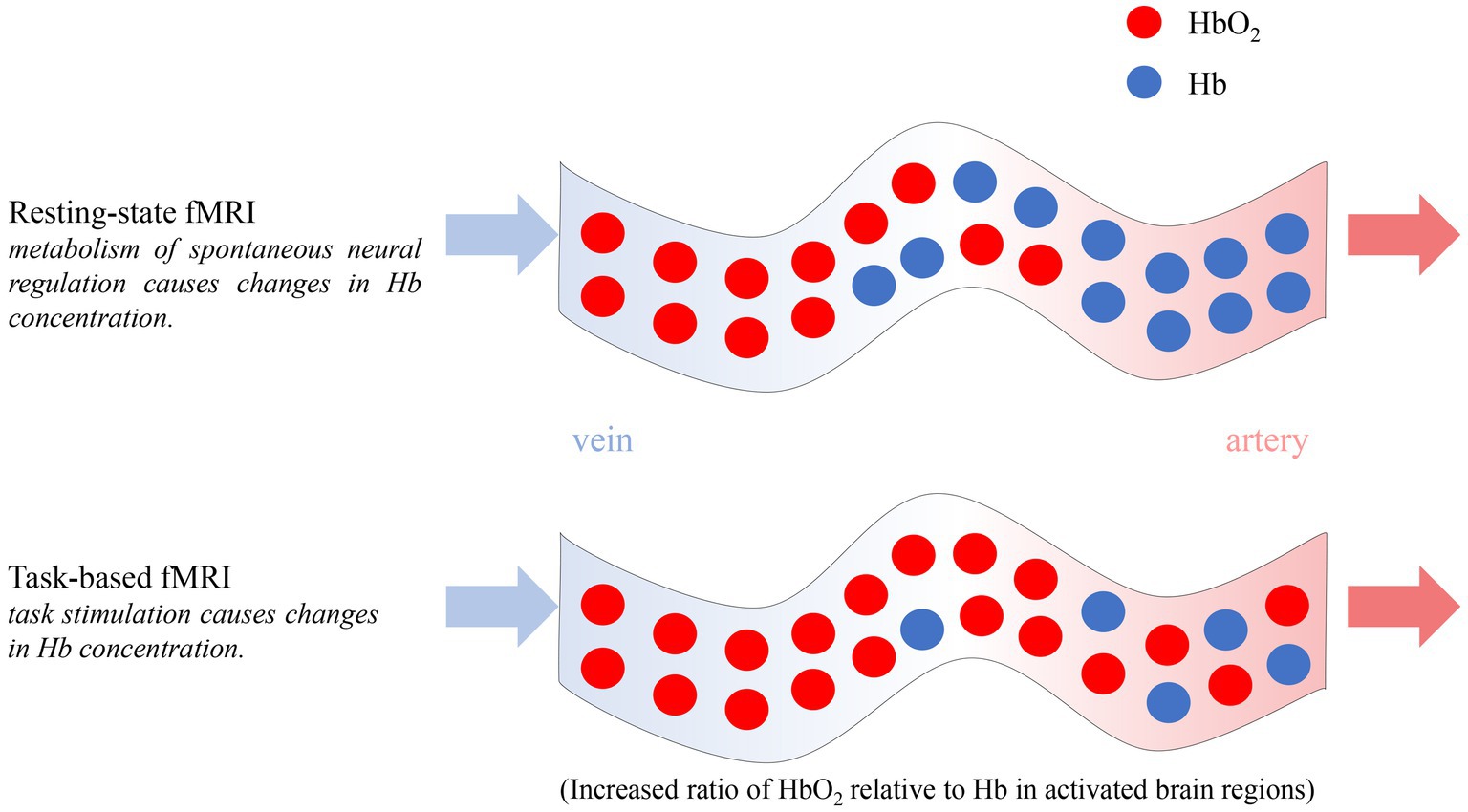
Figure 4. Principle of resting-state and task-based fMRI.
Task-based fMRI studies using auditory stimuli have been widely applied in DoC patients, as they are easier to achieve. Di et al. (2007) analyzed the brain signals using fMRI when DoC patients hearing their own name called by a familiar voice (SON-FV), and the study found a significant activation in their brains. Further studies have demonstrated that UWS/VS and MCS patients are capable of retaining brain responses to language and auditory stimuli, and fMRI is able to identify these responses (Fernandez-Espejo et al., 2008). Okumura et al. (2014) evaluated UWS/VS patients after music stimulation using fMRI and found that activation in bilateral temporal lobes was related to the recovery of consciousness. Wang et al. (2015) found that the types and volumes of auditory cortex activation induced by SON-FV were significantly correlated with the prognosis of UWS/VS patients, which suggested that repetitive use of the simple fMRI task may provide more reliable prognostic information. In a recent study, Boltzmann et al. (2021) scanned UWS/VS patients with scanner noise, preferred music and aversive auditory stimulation, and found that UWS/VS patients showed more significant responses to preferred music and aversive auditory stimulation, finally, the study indicated that UWS/VS patients require strong stimuli to elicit cerebral responses. Researches based on active fMRI paradigms have also made some progress. Liang et al. (2014) considered four imagery tasks (imagine navigating home, imagine playing tennis, imagine familiar faces, mental counting and rest) and found that different tasks activated the brain differently. Wang et al. found that the simple active fMRI “hand-raising” task can elicit brain activation in patients with DoC. Activity of the motor-related network may be taken as an indicator of high-level cognition that cannot be discerned through conventional behavioral assessment (Wang et al., 2019). Clinical implementation of fMRI motor imagery paradigms for detection of consciousness will require further development, validation, and optimization of standardized approaches to fMRI data acquisition, analysis and interpretation (Bodien et al., 2017).
Resting-state fMRI is a popular and advanced technique for studying the functional structure and static networks of the brain, because interaction with DoC patients and difficult experimental set-ups are not required (Jain and Ramakrishnan, 2020). Luppi et al. (2019) found that human consciousness relies on the spatiotemporal interactions between brain integration and functional diversity by comparing resting-state fMRI data of awake volunteers, propofol-anesthetized volunteers and DoC patients. Early studies have shown that the resting-state functional connectivity within the default mode network is decreased and proportional to the degree of consciousness impairment, from locked-in syndrome to MCS, VS, and coma patients (Vanhaudenhuyse et al., 2010). Huang et al. (2020) further analyzed resting-state fMRI signals of DoC patients and identified that human consciousness primarily depends on the default mode and the dorsal attention networks. Qin et al. (2021) analyzed fMRI data of patients with varying degrees of consciousness impairment and demonstrated that the supplementary motor area, bilateral supramarginal gyrus (part of inferior parietal lobule), supragenual/dorsal anterior cingulate cortex, and left middle temporal gyrus play important roles in maintaining brain consciousness.
fMRI is a non-invasive neuroimaging method with a high spatial resolution for accurate functional localization so it can effectively detect covert consciousness that cannot be demonstrated through clinical behavior. However, fMRI is susceptible to motion artifacts, thus has the limitations of low temporal resolution and high costs. Additionally, fMRI is not suitable for DoC patients in the Intensive Care Unit (ICU). Currently, fMRI has entered the era of “big data” with the establishment of large-scale brain datasets and the accumulation of research findings (Xia and He, 2017). The quality of fMRI data plays a critical role in the accuracy and reproducibility of research. Li et al. proposed a new denoising method called linked independent component analysis, which helps improve the accuracy and reproducibility of fMRI studies (Li et al., 2020). In addition, traditional field strengths (1.5 T and 3 T) have reached their limits in terms of spatiotemporal resolution and signal-to-noise ratio balance. However, Ultrahigh magnetic field strengths (7 T and above) allow functional imaging with even higher functional contrast-to-noise ratios for improved spatial resolution and specificity compared to traditional field strengths (1.5 T and 3 T), Which offers improved sensitivity and functional specificity for fMRI applications (Vu et al., 2017). In the future, fMRI will focus on personalized applications, standard acquisition of data and fusion of different techniques.
PET, as a more established technique for studying consciousness, use different markers to measure glucose metabolism, oxygen consumption, focal cerebral blood flow and the distribution of specific neurotransmitters (Hirschberg and Giacino, 2011). These markers can be used to assess the degree of residual brain function in patients with impaired consciousness (Jox et al., 2012).
PET includes two main imaging methods: cerebral perfusion imaging and cerebral metabolic imaging. Cerebral perfusion imaging employs imaging agents that are capable of traversing the blood–brain barrier and gaining entry into cerebral tissue where they exhibit stability and concentrate. Subsequently, cerebral perfusion images can be acquired by nuclear medicine imaging equipment. Patients with disorders of consciousness are severely ischemic in localized brain regions, and therefore cerebral perfusion imaging shows localized hypoperfusion or even no perfusion, which is manifested by low concentration or without concentration of the imaging agent.
Task-based DoC studies mostly use H215O-based cerebral perfusion imaging methods which assist experts to further predict the possibility of recovery in DoC patients by evaluating the specific and directional responses produced by DoC patients under external stimuli. Under the stimuli of auditory, visual and nociceptive, a dissociation occurs between the low-level activation of the primary sensory cortex and the activation of higher cortical networks in DoC patients (de Jong et al., 1997; Menon et al., 1998; Laureys et al., 2002; Bruno et al., 2010). Although task-based PET studies have yielded some results, there is still a demand for more extensive clinically responsive neuroimaging studies due to small number of studies and single cases, which make the task-based PET become a reliable tool for clinical assessment and treatment decisions in DoC patients.
In the studies of resting-state PET, brain metabolic imaging techniques are more widely used. Part of the study based on γ-aminobutyric acid receptor imaging agent observed the retention of injured brain neurons, while ligand 11C-labeled flumazenil (11C-FMZ) was used to assess neuronal integrity and activity (Schiff et al., 2002). Rudolf et al. used 11C-FMZ to observe the local cerebral glucose metabolism of 9 VS patients caused by hypoxia and found that 11C-FMZ PET is important in the prognostic assessment of VS. More scholars have studied resting state DoC using 18F-labeled FDG-PET/CT (Rudolf et al., 2002). As the main imaging agent for current PET/CT imaging, 18FDG can accurately reflect the glucose metabolism of organs or tissues in vivo. Zhao Jing et al. analyzed glucose metabolism in various brain regions of 40 VS patients, 12 MCS patients and 11 DoC patients with regained consciousness, and the results showed that standard uptake values in all brain regions except the brainstem were significantly different among the three groups of patients (Zhao et al., 2018). Usami et al. further found that functional preservation in the left occipital region in patients with chronic DoC might reflect an awareness of external environments, whereas extensive functional preservation in the right cerebral hemisphere might reflect communication motivation (Usami et al., 2022). Yamaki et al. investigated whether regional brain glucose metabolism assessed by 18F-FDG-PET in resting-state could predict voluntary movement in patients with DoC following severe traumatic brain injury. The research showed that glucose uptake in the left proximal and right proximal limb of the primary motor cortex may reflect contralateral voluntary movements (Yamaki et al., 2023); Another study found a significant difference in glucose metabolism in the thalamus, brain stem, and cerebellum between comatose and noncomatose patients acutely after TBI. The metabolic rate of glucose in these regions significantly correlated with the level of consciousness at the time of PET (Hattori et al., 2003). All these studies demonstrated the correlation between the rate of cerebral glucose metabolism and the level of consciousness.
Being sensitive to damaged brain cells, PET can accurately and objectively reflect the functional state of brain cells to assess the degree of brain cell damage. However, PET has a certain degree of radioactive damage, although the application of short-half-life radioisotopes could mitigate the adverse effects, it increases the imaging time and has a low spatial resolution, all these reasons greatly limit the application of PET/CT. In the future, taking into account both safety and accuracy, more suitable tracers will be explored to improve the effectiveness of PET in consciousness evaluation of DoC patients. Fusion with other methods is another potential direction for PET studies. PET is less sensitive to rapid and transient changes (Hermann et al., 2021), whereas EEG records brain activity in the millisecond range. Therefore, the combination of both methods may obtain greater performance in the evaluation of DoC patients.
fNIRS is a non-invasive optical technique based on the different absorption spectral properties of oxyhemoglobin (HbO2) and deoxyhemoglobin (HbR) in human tissues. By calculating the concentration variation of both proteins, fNIRS can be used to evaluate the recovery from DoC and thus reflect the functional state of the brain (Ekkekakis, 2009).
Due to the portability, fNIRS devices were widely used for task-based studies (Pinti et al., 2020). fNIRS identifies patients with potential consciousness by detecting brain activity during command-driven tasks and assesses their residual awareness. Existing studies mainly use active paradigms (including motor imagery and mental arithmetic tasks). Kempny et al. evaluated the brain function of DoC patients by recording their fNIRS signals under motor imagery tasks (Kempny et al., 2016). Research has shown that MCS patients prefer to experience “typical” fNIRS responses, and their hemodynamic responses were similar to those of the control group. It confirmed the feasibility of using fNIRS to evaluate the brain function of pDoC patients through motor imagery tasks for the first time. Li et al. (2021) used fNIRS combined with an active motor imagery paradigm and demonstrated the feasibility of the paradigm to study the functional brain activity of MCS patients. The study provided new method to objectively evaluate the consciousness and residual awareness in MCS patients. The mental arithmetic task, another fNIRS research paradigm which requires higher degree of active consciousness in subjects, has been conducted (Kurz et al., 2018), however, the related studies is limited and the probability yielding positive results is relatively low, thus requiring an expanded sample study for further validation.
The fNIRS studies applying with passive paradigm have also achieved notable outcomes. Current researches focus on spinal cord stimulation (SCS). Si et al. (2018) found that when high frequencies of 70 and 100 Hz were selected for SCS, patients showed significantly enhanced cerebral hemodynamic responses in the prefrontal region and stronger functional connectivity between the prefrontal and occipital lobes. Zhang et al. found a significant increase in cerebral blood volume in the prefrontal cortex at an interstimulus interval of 30 s for SCS (Zhang et al., 2018). Part of the studies used non-traumatic stimuli. Bicciato et al. first used a frequency domain approach to investigate the variations in the oscillatory signal of fNIRS in the low frequency (LFO) and very low frequency oscillation (VLFO) spectral regions in subjects under musical stimulation (Bicciato et al., 2022). The study suggests that the use of fNIRS to identify typical patterns of brain responses to specific environmental stimulation may be a potential way to detect covert awareness in clinically unresponsive patients.
There is still a relatively small amount of research based on resting-state fNIRS. Liu et al. (2023a) acquired resting-state fNIRS from 23 DoC patients and found that MCS and UWS have different patterns of topological architecture and short- and long-distance connectivity in prefrontal cortex, which confirmed that fNIRS is remarkable in distinguishing MCS and UWS. Shu et al. (2023) studied that resting-state fNIRS based functional connectivity analysis of brain networks can quantify brain communication efficiency, which would be a potential functional indicator for DoC rehabilitation.
Compared with fMRI and PET, fNIRS has the advantages of higher temporal resolution, lower cost, and high portability, as well as being less susceptible to head movements and metal implants inside the body, thus facilitating follow-up measurements and clinical application. Compared with EEG, fNIRS possesses relatively high spatial resolution, resistance to motion interference and resistance to electromagnetic interference. Since fNIRS only detects in the cerebral cortex, it has weaker sensitivity toward the deep regions of the brain and therefore cannot detect information in the deeper part of the brain; moreover, the temporal and spatial characteristics make its signal processing and data analysis algorithms lack uniform standards.
In the future, fNIRS is expected to be fused with other neuroimaging methods to achieve multimodal neuro-detection for more accurate and comprehensive evaluation. In addition, the use of time-domain-based functional near-infrared spectroscopy (TD-fNIRS) and frequency-domain-based functional near-infrared spectroscopy (FD-fNIRS) can improve the sensitivity of fNIRS toward the deep brain (Figure 5). More sophisticated machine learning approaches such as artificial neural networks could also help improve the sensitivity of fNIRS (Abdalmalak et al., 2021). Finally, researchers should establish a unified and effective method for fNIRS data analysis for the promotion and application of fNIRS.
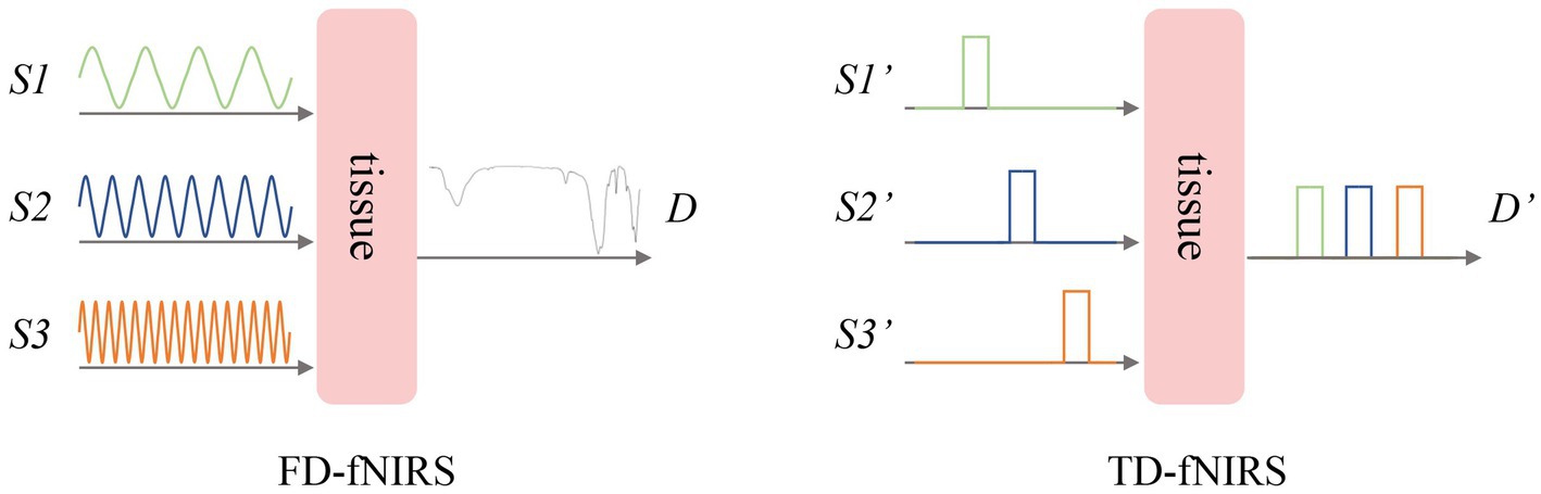
Figure 5. Frequency-domain and time-domain fNIRS.
Different neuroimaging methods have distinct characteristics, yet no single method combines all the advantages (Table 2). Neuroimaging methods using a single modality can no longer meet the requirements of researchers and multi-modal methods have been gradually applied in recent research. Multi-modal methods can achieve complementary advantages of different methods, thereby uncovering significant relationships that cannot be detected by employing a single modality alone.

Table 2. Characteristics of different neuroimaging methods.
PET/MRI can evaluate brain structure and metabolism at the same time, helping doctors to increase the probability of detecting (spared) functional connectivity (Cavaliere et al., 2018). Luppi et al. (2022) leveraged a neurobiologically realistic dynamic mean field model informed by multimodal neuroimaging including fMRI, diffusion MRI and PET. The study provides a new idea for the evaluation of DoC recovery using PET/MRI (Figure 6). Hermann et al. (2021) discovered that the combination of FDG-PET and EEG accurately identified almost all MCS patients and patients with 6-month recovery of command-following, outperforming each technique taken in isolation. The fusion application of fMRI and EEG can precisely locate brain activity in time and space due to the complementary nature of their spatiotemporal resolution (Chatzichristos et al., 2022). Based on combined EEG and fMRI features, Amiri et al. (2023) found that models which use machine-learning algorithms (SVM and Random Forest) predicted consciousness levels with improved positive predictive value and sensitivity. Despite the high spatial resolution of fMRI, the bulky size of fMRI equipment makes it difficult to collect data in ICU. While maintaining portability, fNIRS can be collected simultaneously with EEG to improve its sensitivity, which makes multi-modal detection more convenient. EEG measures an electrical process which does not interfere with the light signals measured by fNIRS. The improved spatial resolution offered by fNIRS can provide information regarding the active source location, thus complementing EEG findings (Rupawala et al., 2018). Othman et al. (2021) found that neurovascular coupling between NIRS oxyhemoglobin (0.07–0.13 Hz) and EEG band-power (1–12 Hz) signals at frontal areas was sensitive and prognostic to changing consciousness levels. The study suggests that NIRS–EEG may be worth exploring as an add-on to multimodal neuromonitoring and recovery of neurovascular coupling after acute brain injury may herald recovery of consciousness.
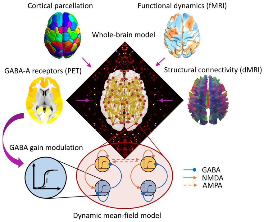
Figure 6. Multimodal study based on PET and MRI.
The human brain entertains rich spatiotemporal dynamics, and adopting multimodal methods allows for the integration of both temporal and spatial characteristics of brain signals, thus facilitating a more comprehensive understanding of the brain. However, the application of multimodal neuroimaging methods requires substantial data, which increases the workload of researchers. The development of more integrated and portable acquisition devices holds the potential to ameliorate the situation. Furthermore, Recent research based on multimodal neuroimaging methods primarily focuses on the resting-state paradigm, future studies could consider incorporating task-based paradigms to detect the temporal coupling of different neural signals.
Although there is still wide disagreement about what exactly consciousness is, neuroimaging methods have been proven to be effective in consciousness rehabilitation evaluation. Especially in patients without command following or other signs of consciousness at the bedside, neuroimaging methods are useful to detect covert consciousness (if present), avoid misdiagnosis and help patients to communicate with the outside world. PET can accurately and objectively reflect the functional status of brain cells. However, compared with other methods, it has a certain degree of radioactive damage, which limits the development of related studies. Considering both safety and accuracy, exploring tracers with greater performance is a potential research direction. Among lots of neuroimaging methods, EEG and fMRI are more widely used. EEG has made great progress in the evaluation and classification of DoC patients in task-based paradigms due to its high temporal resolution, whereas fMRI has a unique advantage in functional brain networks due to its high spatial resolution. EEG has a lower spatial resolution than other methods because it is susceptible to volume conduction effects. High-density EEG paradigms appear to have a high specificity but very low sensitivity for the detection of covert consciousness. fNIRS has a moderate spatial and temporal resolution, and has become a burgeoning tool for the evaluation of DoC because it is more portable and economical. Multi-modal methods can achieve complementary advantages of different methods. However, the challenges associated with data acquisition has hindered its development to some extent. As neuroimaging devices become wireless, integrated and portable, multi-modal neuroimaging method is expected to have a broader application in the evaluation of consciousness rehabilitation and will drive new advancements in brain science research.
JW, XG, and ZX: writing – original draft preparation and visualization. FS and YY: writing – review and editing, supervision, project administration, and funding acquisition. All authors have read and agreed to the published version of the manuscript.
This research was funded by the Key R&D Program of Zhejiang, grant number 2023C03081.
The authors declare that the research was conducted in the absence of any commercial or financial relationships that could be construed as a potential conflict of interest.
All claims expressed in this article are solely those of the authors and do not necessarily represent those of their affiliated organizations, or those of the publisher, the editors and the reviewers. Any product that may be evaluated in this article, or claim that may be made by its manufacturer, is not guaranteed or endorsed by the publisher.
Abdalmalak, A., Milej, D., Norton, L., Debicki, D. B., Owen, A. M., St, K., et al. (2021). The potential role of FNIRS in evaluating levels of consciousness. Front. Hum. Neurosci. 15:703405. doi: 10.3389/fnhum.2021.703405
Amiri, M., Fisher, P. M., Raimondo, F., Sidaros, A., Cacic Hribljan, M., Othman, M. H., et al. (2023). Multimodal prediction of residual consciousness in the intensive care unit: the CONNECT-ME study. Brain 146, 50–64. doi: 10.1093/brain/awac335
Arico, I., Naro, A., Pisani, L. R., Leo, A., Muscara, N., De Salvo, S., et al. (2016). Could combined sleep and pain evaluation be useful in the diagnosis of disorders of consciousness (DOC)? Preliminary findings. Brain Inj. 30, 159–163. doi: 10.3109/02699052.2015.1089595
Aubinet, C., Cassol, H., Gosseries, O., Bahri, M. A., Larroque, S. K., Majerus, S., et al. (2020). Brain metabolism but not gray matter volume underlies the presence of language function in the minimally conscious state (MCS): MCS plus versus MCS- neuroimaging differences. Neurorehabil. Neural Repair 34, 172–184. doi: 10.1177/1545968319899914
Ballanti, S., Campagnini, S., Liuzzi, P., Hakiki, B., Scarpino, M., Macchi, C., et al. (2022). EEG-based methods for recovery prognosis of patients with disorders of consciousness: a systematic review. Clin. Neurophysiol. 144, 98–114. doi: 10.1016/j.clinph.2022.09.017
Bicciato, G., Keller, E., Wolf, M., Brandi, G., Schulthess, S., Friedl, S. G., et al. (2022). Increase in low-frequency oscillations in FNIRS as cerebral response to auditory stimulation with familiar music. Brain Sci. 12:42. doi: 10.3390/brainsci12010042
Bodien, Y. G., Giacino, J. T., and Edlow, B. L. (2017). Functional MRI motor imagery tasks to detect command following in traumatic disorders of consciousness. Front. Neurol. 8:688. doi: 10.3389/fneur.2017.00688
Boltzmann, M., Schmidt, S. B., Gutenbrunner, C., Krauss, J. K., Stangel, M., Höglinger, G. U., et al. (2021). Auditory stimulation modulates resting-state functional connectivity in unresponsive wakefulness syndrome patients. Front. Neurosci. 15:554194. doi: 10.3389/fnins.2021.554194
Bruno, M.-A., Vanhaudenhuyse, A., Schnakers, C., Boly, M., Gosseries, O., Demertzi, A., et al. (2010). Visual fixation in the vegetative state: an observational case series PET study. BMC Neurol. 10:35. doi: 10.1186/1471-2377-10-35
Burle, B., Spieser, L., Roger, C., Casini, L., Hasbroucq, T., and Vidal, F. (2015). Spatial and temporal resolutions of EEG: is it really black and white? A scalp current density view. Int. J. Psychophysiol. 97, 210–220. doi: 10.1016/j.ijpsycho.2015.05.004
Cavaliere, C., Kandeepan, S., Aiello, M., Ribeiro de Paula, D., Marchitelli, R., Fiorenza, S., et al. (2018). Multimodal neuroimaging approach to variability of functional connectivity in disorders of consciousness: a PET/MRI pilot study. Front. Neurol. 9:861. doi: 10.3389/fneur.2018.00861
Chatzichristos, C., Kofidis, E., Van Paesschen, W., De Lathauwer, L., Theodoridis, S., and Van Huffel, S. (2022). Early soft and flexible fusion of electroencephalography and functional magnetic resonance imaging via double coupled matrix tensor factorization for multisubject group analysis. Hum. Brain Mapp. 43, 1231–1255. doi: 10.1002/hbm.25717
de Jong, B. M., Willemsen, A. T. M., and Paans, A. M. J. (1997). Regional cerebral blood flow changes related to affective speech presentation in persistent vegetative state. Clin. Neurol. Neurosurg. 99, 213–216. doi: 10.1016/S0303-8467(97)00024-3
Di, H. B., Yu, S. M., Weng, X. C., Laureys, S., Yu, D., Li, J. Q., et al. (2007). Cerebral response to patient’s own name in the vegetative and minimally conscious states. Neurology 68, 895–899. doi: 10.1212/01.wnl.0000258544.79024.d0
Dong, L., Gong, D., Valdes-Sosa, P. A., Xia, Y., Luo, C., Peng, X., et al. (2014). Simultaneous EEG-FMRI: trial level spatio-temporal fusion for hierarchically reliable information discovery. NeuroImage 99, 28–41. doi: 10.1016/j.neuroimage.2014.05.029
Edlow, B. L., Claassen, J., Schiff, N. D., and Greer, D. M. (2021). Recovery from disorders of consciousness: mechanisms, prognosis and emerging therapies. Nat. Rev. Neurol. 17, 135–156. doi: 10.1038/s41582-020-00428-x
Ekkekakis, P. (2009). Illuminating the black box: investigating prefrontal cortical hemodynamics during exercise with near-infrared spectroscopy. J. Sport Exerc. Psychol. 31, 505–553. doi: 10.1123/jsep.31.4.505
Fernandez-Espejo, D., Junque, C., Vendrell, P., Bernabeu, M., Roig, T., Bargallo, N., et al. (2008). Cerebral response to speech in vegetative and minimally conscious states after traumatic brain injury. Brain Inj. 22, 882–890. doi: 10.1080/02699050802403573
Giacino, J. T., Kalmar, K., and Whyte, J. (2004). The JFK coma recovery scale-revised: measurement characteristics and diagnostic utility. Arch. Phys. Med. Rehabil. 85, 2020–2029. doi: 10.1016/j.apmr.2004.02.033
Di Gregorio, F., La Porta, F., Petrone, V., Battaglia, S., Orlandi, S., Ippolito, G., et al. (2022). Accuracy of EEG biomarkers in the detection of clinical outcome in disorders of consciousness after severe acquired brain injury: preliminary results of a pilot study using a machine learning approach. Biomedicine 10:1897. doi: 10.3390/biomedicines10081897
Hattori, N., Huang, S.-C., Hsiao-Ming, W., Yeh, E., Glenn, T. C., Vespa, P. M., et al. (2003). Correlation of regional metabolic rates of glucose with glasgow coma scale after traumatic brain injury. J. Nucl. Med. 44, 1709–1716.
Hermann, B., Stender, J., Habert, M.-O., Kas, A., Denis-Valente, M., Raimondo, F., et al. (2021). Multimodal FDG-PET and EEG assessment improves diagnosis and prognostication of disorders of consciousness. NeuroImage 30:102601. doi: 10.1016/j.nicl.2021.102601
Hirschberg, R., and Giacino, J. T. (2011). The vegetative and minimally conscious states: diagnosis, prognosis and treatment. Neurol. Clin. 29, 773–786. doi: 10.1016/j.ncl.2011.07.009
Huang, Z., Zhang, J., Jinsong, W., Mashour, G. A., and Hudetz, A. G. (2020). Temporal circuit of macroscale dynamic brain activity supports human consciousness. Sci. Adv. 6:eaaz0087. doi: 10.1126/sciadv.aaz0087
Jain, R., and Ramakrishnan, A. G. (2020). Electrophysiological and neuroimaging studies – during resting state and sensory stimulation in disorders of consciousness: a review. Front. Neurosci. 14:555093. doi: 10.3389/fnins.2020.555093
Jox, R. J., Bernat, J. L., Laureys, S., and Racine, E. (2012). Disorders of consciousness: responding to requests for novel diagnostic and therapeutic interventions. Lancet Neurol. 11, 732–738. doi: 10.1016/S1474-4422(12)70154-0
Kempny, A. M., James, L., Yelden, K., Duport, S., Farmer, S. F., Diane Playford, E., et al. (2018). Patients with a severe prolonged disorder of consciousness can show classical EEG responses to their own name compared with others’ names. Neuroimage Clin. 19, 311–319. doi: 10.1016/j.nicl.2018.04.027
Kempny, A. M., James, L., Yelden, K., Duport, S., Simon Farmer, E., Playford, D., et al. (2016). Functional near infrared spectroscopy as a probe of brain function in people with prolonged disorders of consciousness. Neuroimage Clin. 12, 312–319. doi: 10.1016/j.nicl.2016.07.013
Kondziella, D., Friberg, C. K., Frokjaer, V. G., Fabricius, M., and Moller, K. (2016). Preserved consciousness in vegetative and minimal conscious states: systematic review and meta-analysis. J. Neurol. Neurosurg. Psychiatry 87, 485–492. doi: 10.1136/jnnp-2015-310958
Kurz, E.-M., Wood, G., Kober, S. E., Schippinger, W., Pichler, G., Mueller-Putz, G., et al. (2018). Toward using FNIRS recordings of mental arithmetic for the detection of residual cognitive activity in patients with disorders of consciousness (DOC). Brain Cogn. 125, 78–87. doi: 10.1016/j.bandc.2018.06.002
Laureys, S., Celesia, G. G., Cohadon, F., Lavrijsen, J., León-Carrión, J., Sannita, W. G., et al. (2010). Unresponsive wakefulness syndrome: a new name for the vegetative state or apallic syndrome. BMC Med. 8:68. doi: 10.1186/1741-7015-8-68
Laureys, S., Faymonville, M. E., Peigneux, P., Damas, P., Lambermont, B., del Fiore, G., et al. (2002). Cortical processing of noxious somatosensory stimuli in the persistent vegetative state. NeuroImage 17, 732–741. doi: 10.1006/nimg.2002.1236
Lehembre, R., Bruno, M.-A., Vanhaudenhuyse, A., Chatelle, C., Cologan, V., Leclercq, Y., et al. (2012). Resting-state EEG study of comatose patients: a connectivity and frequency analysis to find differences between vegetative and minimally conscious states. Funct. Neurol. 27, 41–47.
Li, J., Shen, J., Liu, S., Chauvel, M., Yang, W., Mei, J., et al. (2018). Responses of patients with disorders of consciousness to habit stimulation: a quantitative EEG study. Neurosci. Bull. 34, 691–699. doi: 10.1007/s12264-018-0258-y
Li, H., Smith, S. M., Gruber, S., Lukas, S. E., Silveri, M. M., Hill, K. P., et al. (2020). Denoising scanner effects from multimodal MRI data using linked independent component analysis. NeuroImage 208:116388. doi: 10.1016/j.neuroimage.2019.116388
Li, M., Yang, Y., Zhang, Y., Gao, Y., Jing, R., Dang, Y., et al. (2021). Detecting residual awareness in patients with prolonged disorders of consciousness: an FNIRS study. Front. Neurol. 12:618055. doi: 10.3389/fneur.2021.618055
Liang, X., Kuhlmann, L., Johnston, L. A., Grayden, D. B., Vogrin, S., Crossley, R., et al. (2014). Extending communication for patients with disorders of consciousness. J. Neuroimaging 24, 31–38. doi: 10.1111/j.1552-6569.2012.00744.x
Liu, Y., Kang, X.-g., Chen, B.-b., Song, C.-g., Liu, Y., Hao, J.-m., et al. (2023a). Detecting residual brain networks in disorders of consciousness: a resting-state FNIRS study. Brain Res. 1798:148162. doi: 10.1016/j.brainres.2022.148162
Liu, Y., Li, Z., and Bai, Y. (2023b). Frontal and parietal lobes play crucial roles in understanding the disorder of consciousness: a perspective from electroencephalogram studies. Front. Neurosci. 16:1024278. doi: 10.3389/fnins.2022.1024278
Luppi, A. I., Craig, M. M., Pappas, I., Finoia, P., Williams, G. B., Allanson, J., et al. (2019). Consciousness-specific dynamic interactions of brain integration and functional diversity. Nat. Commun. 10, 1–12. doi: 10.1038/s41467-019-12658-9
Luppi, A. I., Mediano, P. A. M., Rosas, F. E., Allanson, J., Pickard, J. D., Williams, G. B., et al. (2022). Whole-brain modelling identifies distinct but convergent paths to unconsciousness in anaesthesia and disorders of consciousness. Communicat. Biol. 5:384. doi: 10.1038/s42003-022-03330-y
Menon, D. K., Owen, A. M., Boniface, S. J., and Pickard, J. D. (1998). Cortical processing in persistent vegetative state. Lancet 352, 1148–1149. doi: 10.1016/S0140-6736(05)79795-6
Naro, A., Bramanti, A., Leo, A., Cacciola, A., Manuli, A., Bramanti, P., et al. (2018). Shedding new light on disorders of consciousness diagnosis: the dynamic functional connectivity. Cortex 103, 316–328. doi: 10.1016/j.cortex.2018.03.029
O’Mahony, D., Rowan, M., Walsh, J. B., and Coakley, D. (1990). P300 as a predictor of recovery from coma. Lancet 336, 1265–1266. doi: 10.1016/0140-6736(90)92887-N
Okumura, Y., Asano, Y., Takenaka, S., Fukuyama, S., Yonezawa, S., Kasuya, Y., et al. (2014). Brain activation by music in patients in a vegetative or minimally conscious state following diffuse brain injury. Brain Inj. 28, 944–950. doi: 10.3109/02699052.2014.888477
Othman, M. H., Bhattacharya, M., Moller, K., Kjeldsen, S., Grand, J., Kjaergaard, J., et al. (2021). Resting-state NIRS-EEG in unresponsive patients with acute brain injury: a proof-of-concept study. Neurocrit. Care. 34, 31–44. doi: 10.1007/s12028-020-00971-x
Pinti, P., Tachtsidis, I., Hamilton, A., Hirsch, J., Aichelburg, C., Gilbert, S., et al. (2020). The present and future use of functional near-infrared spectroscopy (FNIRS) for cognitive neuroscience. Ann. N. Y. Acad. Sci. 1464, 5–29. doi: 10.1111/nyas.13948
Power, J. D., Cohen, A. L., Nelson, S. M., Wig, G. S., Barnes, K. A., Church, J. A., et al. (2011). Functional network organization of the human brain. Neuron 72, 665–678. doi: 10.1016/j.neuron.2011.09.006
Qin, P., Wu, X., Wu, C., Wu, H., Zhang, J., Huang, Z., et al. (2021). Higher-order sensorimotor circuit of the brain’s global network supports human consciousness. NeuroImage 231:117850. doi: 10.1016/j.neuroimage.2021.117850
Rudolf, J., Sobesky, J., Ghaemi, M., and Heiss, W.-D. (2002). The correlation between cerebral glucose metabolism and benzodiazepine receptor density in the acute vegetative state. Eur. J. Neurol. 9, 671–677. doi: 10.1046/j.1468-1331.2002.00468.x
Rupawala, M., Dehghani, H., Lucas, S. J. E., Tino, P., and Cruse, D. (2018). Shining a light on awareness: a review of functional near-infrared spectroscopy for prolonged disorders of consciousness. Front. Neurol. 9:350. doi: 10.3389/fneur.2018.00350
Schiff, N. D., Ribary, U., Moreno, D. R., Beattie, B., Kronberg, E., Blasberg, R., et al. (2002). Residual cerebral activity and behavioural fragments can remain in the persistently vegetative brain. Brain 125, 1210–1234. doi: 10.1093/brain/awf131
Schorr, B., Schlee, W., Arndt, M., and Bender, A. (2016). Coherence in resting-state EEG as a predictor for the recovery from unresponsive wakefulness syndrome. J. Neurol. 263, 937–953. doi: 10.1007/s00415-016-8084-5
Scolding, N., Owen, A. M., and Keown, J. (2021). Prolonged disorders of consciousness: a critical evaluation of the new UK guidelines. Brain 144, 1655–1660. doi: 10.1093/brain/awab063
Shu, Z., Jingchao, W., Li, H., Liu, J., Jiewei, L., Lin, J., et al. (2023). FNIRS-based functional connectivity signifies recovery in patients with disorders of consciousness after DBS treatment. Clin. Neurophysiol. 147, 60–68. doi: 10.1016/j.clinph.2022.12.011
Si, J., Dang, Y., Zhang, Y., Li, Y., Zhang, W., Yang, Y., et al. (2018). Spinal cord stimulation frequency influences the hemodynamic response in patients with disorders of consciousness. Neurosci. Bull. 34, 659–667. doi: 10.1007/s12264-018-0252-4
Sinitsyn, D. O., Poydasheva, A. G., Bakulin, I. S., Legostaeva, L. A., Iazeva, E. G., Sergeev, D. V., et al. (2020). Detecting the potential for consciousness in unresponsive patients using the perturbational complexity index. Brain Sci. 10:917. doi: 10.3390/brainsci10120917
Stefan, S., Schorr, B., Lopez-Rolon, A., Kolassa, I.-T., Shock, J. P., Rosenfelder, M., et al. (2018). Consciousness indexing and outcome prediction with resting-state EEG in severe disorders of consciousness. Brain Topogr. 31, 848–862. doi: 10.1007/s10548-018-0643-x
Theiler, J., Eubank, S., Longtin, A., Galdrikian, B., and Doyne Farmer, J. (1992). Testing for nonlinearity in time series: the method of surrogate data. Physica D 58, 77–94. doi: 10.1016/0167-2789(92)90102-S
Thibaut, A., Panda, R., Annen, J., Sanz, L. R. D., Naccache, L., Martial, C., et al. (2021). Preservation of brain activity in unresponsive patients identifies MCS star. Ann. Neurol. 90, 89–100. doi: 10.1002/ana.26095
Usami, N., Asano, Y., Ikegame, Y., Takei, H., Yamada, Y., Yano, H., et al. (2022). Cerebral glucose metabolism in patients with chronic disorders of consciousness. Canad. J. Neurol. Sci., 50, 1–11. doi: 10.1017/cjn.2022.301
Vanhaudenhuyse, A., Noirhomme, Q., Tshibanda, L. J.-F., Bruno, M.-A., Boveroux, P., Schnakers, C., et al. (2010). Default network connectivity reflects the level of consciousness in non-communicative brain-damaged patients. Brain 133, 161–171. doi: 10.1093/brain/awp313
Vu, T., An, K. J., Glasser, M. F., Smith, S. M., Coalson, T., Moeller, S., et al. (2017). Tradeoffs in pushing the spatial resolution of FMRI for the 7T human connectome project. Neuroimage 154, 23–32. doi: 10.1016/j.neuroimage.2016.11.049
Wang, F., Di, H., Xiaohua, H., Jing, S., Thibaut, A., Di Perri, C., et al. (2015). Cerebral response to subject’s own name showed high prognostic value in traumatic vegetative state. BMC Med. 13:83. doi: 10.1186/s12916-015-0330-7
Wang, F., Hu, N., Hu, X., Jing, S., Heine, L., Thibaut, A., et al. (2019). Detecting brain activity following a verbal command in patients with disorders of consciousness. Front. Neurosci. 13:976. doi: 10.3389/fnins.2019.00976
Wang, Y., Niu, Z., Xia, X., Bai, Y., Liang, Z., He, J., et al. (2022). Application of fast perturbational complexity index to the diagnosis and prognosis for disorders of consciousness. IEEE Trans. Neural Syst. Rehabil. Eng. 30, 509–518. doi: 10.1109/TNSRE.2022.3154772
Wu, M., Bao, W.-X., Zhang, J., Yang-Fan, H., Gao, J., and Luo, B.-Y. (2018). Effect of acoustic stimuli in patients with disorders of consciousness: a quantitative electroencephalography study. Neural Regen. Res. 13, 1900–1906. doi: 10.4103/1673-5374.238622
Wu, L., Wang, X.-Q., Yang, Y., Dong, T.-F., Lei, L., Cheng, Q.-Q., et al. (2020). Spatio-temporal dynamics of EEG features during sleep in major depressive disorder after treatment with escitalopram: a pilot study. BMC Psychiatry 20:124. doi: 10.1186/s12888-020-02519-x
Xia, M., and He, Y. (2017). Functional connectomics from a ‘big data’ perspective. Neuroimage 160, 152–167. doi: 10.1016/j.neuroimage.2017.02.031
Yamaki, T., Hatakeyama, N., Murayama, T., Funakura, M., Hara, T., Onodera, S., et al. (2023). Prediction of voluntary movements of the upper extremities by resting state-brain regional glucose metabolism in patients with chronic severe brain injury: a pilot study. Hum. Brain Mapp. 44, 3158–3167. doi: 10.1002/hbm.26270
Zhang, Y., Yang, Y., Si, J., Xia, X., He, J., and Jiang, T. (2018). Influence of inter-stimulus interval of spinal cord stimulation in patients with disorders of consciousness: a preliminary functional near-infrared spectroscopy study. NeuroImage 17, 1–9. doi: 10.1016/j.nicl.2017.09.017
Zhang, L., Zhang, R., Guo, Y., Zhao, D., Li, S., Chen, M., et al. (2022). Assessing residual motor function in patients with disorders of consciousness by brain network properties of task-state EEG. Cogn. Neurodyn. 16, 609–620. doi: 10.1007/s11571-021-09741-7
Zhao, J., Yin, J., Wang, X., Yu, R., Xie, Q., Zhang, J., et al. (2018). Assessment of the brain function with 18F-FDG PET/CT in patients with disorders of consciousness. Chin. J. Nucl. Med. Mol. Imaging 38, 97–100. doi: 10.3760/cma.j.issn.2095-2848.2018.02.005
Keywords: disorders of consciousness, EEG, fMRI, PET, fNIRS, multimodal
Citation: Wang J, Gao X, Xiang Z, Sun F and Yang Y (2023) Evaluation of consciousness rehabilitation via neuroimaging methods. Front. Hum. Neurosci. 17:1233499. doi: 10.3389/fnhum.2023.1233499
Edited by:
Pierre LeVan, University of Calgary, CanadaReviewed by:
Anirban Dutta, University of Lincoln, United KingdomCopyright © 2023 Wang, Gao, Xiang, Sun and Yang. This is an open-access article distributed under the terms of the Creative Commons Attribution License (CC BY). The use, distribution or reproduction in other forums is permitted, provided the original author(s) and the copyright owner(s) are credited and that the original publication in this journal is cited, in accordance with accepted academic practice. No use, distribution or reproduction is permitted which does not comply with these terms.
*Correspondence: Fangfang Sun, c3VuZmY1MTFAaGR1LmVkdS5jbg==
Disclaimer: All claims expressed in this article are solely those of the authors and do not necessarily represent those of their affiliated organizations, or those of the publisher, the editors and the reviewers. Any product that may be evaluated in this article or claim that may be made by its manufacturer is not guaranteed or endorsed by the publisher.
Research integrity at Frontiers
Learn more about the work of our research integrity team to safeguard the quality of each article we publish.