95% of researchers rate our articles as excellent or good
Learn more about the work of our research integrity team to safeguard the quality of each article we publish.
Find out more
ORIGINAL RESEARCH article
Front. Genet. , 06 September 2022
Sec. Computational Genomics
Volume 13 - 2022 | https://doi.org/10.3389/fgene.2022.962870
This article is part of the Research Topic Methods for Imaging and Omics Data Science: Advances, Applications, and Spatiotemporal Innovations View all 9 articles
Hepatocellular carcinoma (HCC) is a leading malignant liver tumor with high mortality and morbidity. Patients at the same stage can be defined as different molecular subtypes associated with specific genomic disorders and clinical features. Thus, identifying subtypes is essential to realize efficient treatment and improve survival outcomes of HCC patients. Here, we applied a regularized multiple kernel learning with locality preserving projections method to integrate mRNA, miRNA and DNA methylation data of HCC patients to identify subtypes. We identified two HCC subtypes significantly correlated with the overall survival. The patient 3-years mortality rates in the high-risk and low-risk group was 51.0% and 23.5%, respectively. The high-risk group HCC patients were 3.37 times higher in death risk compared to the low-risk group after adjusting for clinically relevant covariates. A total of 196 differentially expressed mRNAs, 2,151 differentially methylated genes and 58 differentially expressed miRNAs were identified between the two subtypes. Additionally, pathway activity analysis showed that the activities of six pathways between the two subtypes were significantly different. Immune cell infiltration analysis revealed that the abundance of nine immune cells differed significantly between the two subtypes. We further applied the weighted gene co-expression network analysis to identify gene modules that may affect patients prognosis. Among the identified modules, the key module genes significantly associated with prognosis were found to be involved in multiple biological processes and pathways, revealing the mechanism underlying the progression of HCC. Hub gene analysis showed that the expression levels of CDK1, CDCA8, TACC3, and NCAPG were significantly associated with HCC prognosis. Our findings may bring novel insights into the subtypes of HCC and promote the realization of precision medicine.
Hepatocellular carcinoma (HCC), a primary malignant neoplasm, accounts for approximately 90% of cases of all liver cancers (Licata et al., 2021). It has been reported to be the fastest growing cause of cancer-related death in the United States, and is expected to be the third leading cause of cancer-related death by 2030, if the trends remain (Rahib et al., 2014). The current therapy and management of HCC is based on the expected returns of the main interventions and tumor grades following the Barcelona Clinic Liver Cancer (BCLC) staging system (European Association for the Study of the Liver, 2018; Llovet et al., 2021). So far, the prognosis of HCC patients remains poor (Forner et al., 2018). Actually, patients at the same stage can be defined as different molecular subtypes according to major molecular drivers and pathways involved (Villanueva, 2019). Several works have been done to identify the HCC subtypes using mRNA gene expression data (Boyault et al., 2007; Hoshida et al., 2009; Goossens et al., 2015). However, any individual omics data can only reveal the intrinsic molecular characteristics of a tumor marginally. High throughput technology has enabled the acquisition of multi-omics data more easily. The joint analysis of multi-omics data types is being increasingly emphasized. Multi-omics data integrative analysis can offer insights into the crucial links between different types of omics data and further provide a thorough comprehension of the potential biological processes (Lock et al., 2013). Three HCC molecular subtypes were identified from 183 TCGA samples by integrating five data sources (DNA copy number, DNA methylation, mRNA expression, miRNA expression and RPPA) (Cancer Genome Atlas Research Network, 2017). These molecular subtypes associated with specific genomic disorders and clinical features, allow researchers to discover targets used as drug design and biomarkers for predicting response. However, the high heterogeneity and complex etiologic factors of HCC make the prognosis prediction very challenging (Colagrande et al., 2016). HCC still has a relatively high incidence of recurrence and low 5-year survival rate. Therefore, identifying accurate molecular subtypes and biomarkers is essential for developing new effective therapies to improve the prognosis of HCC patients.
Some multi-omics integrative clustering methods have been proposed using multiple data types for subtyping. They can be divided into four categories (Rappoport and Shamir, 2018): 1) Early integration methods that input a single large dataset obtained by merging multi-omics data for clustering; 2) Late integration methods that apply a two-step clustering method which first clusters each omics data and then integrates them; 3) Methods applying statistical modeling that assume a particular data distribution (sensitive to feature selections); and 4) Similarity-based methods (e.g. regularized Multiple Kernel Learning with Locality Preserving Projections, or rMKL-LPP) (Speicher and Pfeifer, 2015) that first create similarity matrices based on each data type, then integrate them for clustering. An advantage of these methods is that they allow incorporating diverse omics data types, such as categorical and ordinal data. rMKL-LPP was extended from a multiple kernel learning based dimensionality reduction method. Based on the input data, it conducts dimension reduction such that similarities between samples and their nearest neighbors are remained in a low dimensional space. rMKL-LPP uses multiple kernel matrices to preserve the degrees of similarity within each omics data. Considering the differences of matrices, rMKL-LPP upweight the matrices with high information content and assign low weights to those with low information content. Moreover, it added a regularization term in the optimization problem to avoid overfitting. Rappoport et al. (Rappoport and Shamir, 2018) provided a comprehensive comparison of different multi-omics clustering algorithms spanning ten different cancer types and pointed out that rMKL-LPP has superior performance in terms of clinical enrichment.
Epigenetic dysregulation such as modifications in DNA methylation or changes in levels of microRNAs, plays a critical role in HCC (Rebouissou and Nault, 2020). Gene expression analysis also revealed differentiation patterns among HCC (Hoshida et al., 2009). In this work, we proposed to use rMKL-LPP method to integrate DNA methylation, miRNA and mRNA expression data to obtain subtypes of HCC. Focusing on the subtypes of HCC, downstream analyses were performed to explore the molecular features and pathways with potential prognostic value to prolong patient survival time and further promote the realization of precision diagnosis and treatment.
We downloaded mRNA expression data, miRNA expression data, DNA methylation data and clinical data from The Cancer Genome Atlas (TCGA) for HCC using TCGAbiolinks R package (Zhou et al., 2015). The DNA methylation data were measured using Illumina Human Methylation 450 Beadchip.
We performed the same data preprocessing steps for the mRNA and miRNA expression data. Features with more than 30% missing rate were removed. The rest of the missing data were imputed applying K-nearest neighbor (KNN) method (Troyanskaya et al., 2001), followed by
rMKL-LPP proposed by Speicher and Pfeifer (2015), is an extension of MKL-DR method that can perform dimensionality reduction and data integration simultaneously. To make the work complete, we briefly introduce the algorithm here.
Given M datasets
where
For a given set of input kernel matrices, we use the dimension reduction of Locality Preserving Projections (LPP) method (Rong et al., 2017) to maintain similarities between each sample and its nearest neighbors in a low dimensional space. The projection vector v is optimized according to the graph-preserving criterion:
where
The constrained optimization problem in (2) can be achieved by an implicit mapping of the features to a high-dimensional Hilbert space
where
After mapping the similarities between each sample and its nearest neighbors to a low-dimensional space, k-means was used for clustering, and the optimal subtyping number was selected based on the silhouette coefficient (Rousseeuw et al., 1987).
We carried out survival analysis to explore whether the subtyping results correlated with patient survival outcomes and evaluate the clinical significance on survival rate of the identified subgroups. The Kaplan-Meier survival curve provides an intuitive measure of the survival risk for different subtypes, followed by the log-rank test to examine the difference of survival curves. Cox regression analysis was subsequently conducted on the HCC subtypes. A prognostic model based on the selected data was established after controlling for clinically relevant covariates.
Differentially expressed mRNAs (DEmRNAs), miRNAs (DEmiRNAs) and differentially methylated genes (DMGs) between subtypes were further explored. Specifically, the DEmRNAs satisfying the log2 fold change (FC)>1 &
To explore the biological changes that lead to survival differences between subtypes, pathway activity analysis was performed using PROGENy (Schubert et al., 2018). Non-parametric tests were used to identify biological pathways that were activated differently between subtypes with the threshold set as
Gene modules that affect the prognosis of HCC patients were identified with the weighted gene co-expression network analysis (WGCNA). In this work, the top 5,000 genes were selected according to the median absolute deviation to construct an mRNA co-expression network using WGCNA R package (Langfelder and Horvath, 2008). The brief implementation was as follows: setting the power of
The candidate genes were defined as genes correlated with the ME and clinical traits. The ME is the most important component of a gene module and represents the gene module expression profile. The module membership (MM) of a gene represents the correlation of its gene expression profile with a specific ME. Candidate genes were defined as those correlated with the ME (cor. MM> 0.85) and clinical traits (cor. gene Trait Significance >0.30). The Maximal Clique Centrality (MCC) algorithm was then used to obtain highly connected genes from candidate genes using the cytoHubba plugin in Cytoscape software (v3.7.2) (Ceccarelli et al., 2020). The top 15 highly correlated genes were used as hub genes for further analysis.
We evaluated the prognostic value of hub genes by dividing patients into two groups according to the median value of hub gene expression. Patients equal or above the median value were categorized as the high-level group and those below the median were categorized as the low-level group. The statistical significance of survival outcomes in the two groups was assessed by survival analysis, screening for genes associated with prognosis (p-value < 0.05).
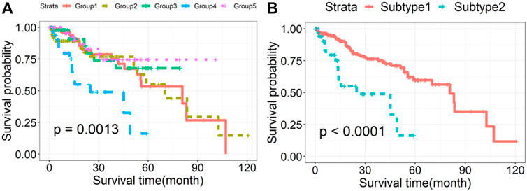
The survival curves of HCC subtypes identified by rMKL-LPP method were given in Figure 1A. The survival probability between groups 1, 2, 4 and 5 was not statistically significant. Thus, we combined patients in the four groups into one group named as Subtype1, while named the group 3 as Subtype2. Thirty-nine patients (13.6%) in Subtype2 had 3-years mortality rate of 51%; while those in the Subtype1 had 3-years mortality rate of 23.5%. The basic characteristics of the two subtypes are shown in Table 1, and the survival curves are presented in Figure 1B. It can be seen that the two subtypes differed significantly in survival outcomes. Compared to Subtype1, Subtype2 had a significantly lower survival probability (p = 4.14E-05).

FIGURE 1. Kaplan-Meier survival curves of HCC subtypes identified by rMKL-LPP method. (A) The survival curves drawn based on the initial subtypes in HCC and (B) the survival curves of the regrouped Subtype 1 and Subtype 2.
To study the prognosis of different subtypes, the Cox regression model was constructed after controlling for age, gender and pathologic stage. The regression results are presented in Table 2. Patients in Subtype2 were 3.369 times higher in risk of death than Subtype1. Therefore, Subtype 1 was named the low-risk group and Subtype 2 was named the high-risk group. The only significant covariate is pathologic stage (p = 0.003).
Focusing on the two subtypes, differential expression analysis was conducted for each omics data type. Based on the pre-set threshold (see Section 2.3.2), a total of 196 DEmRNAs were selected, where 132 were upregulated and 64 were downregulated; 58 DEmiRNAs were selected, among which 56 were upregulated and 2 were downregulated. A total of 2,151 DMGs were also identified, where 1,254 were hypermethylated and 897 were hypomethylated. Figure 2A shows a heatmap of differentially expressed profiles of different omics data between the two subtypes. The heatmap showed that the expression profiles among the three omics data types are different between high- and low-risk groups of HCC. A total of 458 genes targeted by 58 DEmiRNAs were predicted. Then we performed the comprehensive analysis of differentially expressed genes (DEGs) in different omics data. As shown in Figure 2B, 32 genes were observed to be differentially expressed in mRNA, along with abnormal methylation; 22 genes were abnormally methylated and differentially expressed in miRNA, and 2 genes were differentially expressed in mRNA and their corresponding miRNA.

FIGURE 2. (A) Heatmap of DEmRNAs, DMGs and DEmiRNAs between the two subtypes. Each column corresponds to a patient and each row indicates an individual feature. The relatively high and low expression of genes are shown in red and green color respectively. (B) Venn diagram of differentially expressed gene analysis results in different omics data.
We further merged these 56 genes into a core set for KEGG pathway and GO enrichment analysis. KEGG analysis indicated that these genes were enriched in 10 pathways (see Figure 3). In addition, these genes were mainly enriched in 31 GO terms (see Figure 4).
We performed pathway activity and immune cell infiltration analysis to further explore the biological and clinical meaning of the two subtypes. As shown in Figure 5, the activities of 6 pathways between two subtypes were significantly different. Specifically, the activities of Hypoxia, MAPK, EGFR, NF-kβ, and TNFα pathways was found to be significantly higher in the high-risk group than in the low-risk group; while VEGF pathway activity showed higher in the low-risk group.
Figure 6 revealed that the abundance of 9 immune cells differed significantly between two subtypes. The abundance of monocytic lineage, CD8+T cell, T cell, myeloid dendritic cell, and cytotoxicity score was found to be significantly higher in the high-risk group than in the low-risk group. Tumor-infiltrating immune cells are closely related to clinical outcomes of patients in many types of tumors and are likely to serve as target spots in cancer-targeting drug delivery systems. This provides ideas for a targeted therapeutic strategy of HCC.
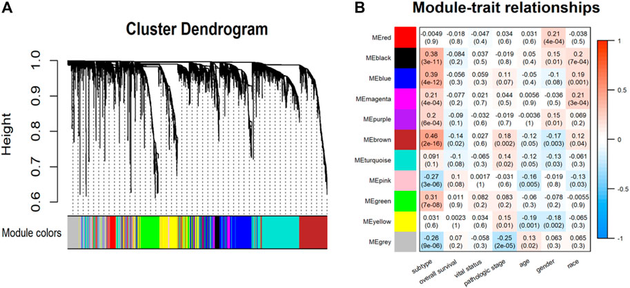
In the WGCNA analysis, 11 co-expression modules were identified (Figure 7A). Among them, the brown module that is significantly related to subtype (r = 0.46, p < 0.0001) was selected for subsequent analysis (Figure 7B). Then, 59 candidate genes were screened from the brown module that included a total of 758 genes based on the preset criteria. The CytoHubba plugin in Cytoscape software was employed to measure the MCC score of candidate genes to identify hub genes. Finally, the top 15 genes were selected as hub genes for further analysis by sorting the MCC score. They were TPX2, KIFC1, MYBL2, TOP2A, NUSAP1, ARHGAP11A, LMNB1, CDK1, CDCA8, TACC3, NUF2, NCAPG, HJURP, NCAPH, and CENPA. The interaction between hub genes and candidate genes was visualized using Cytoscape software. Shown in Figure 8, each candidate gene is less connected to all other candidate genes and more connected to hub genes.

FIGURE 7. (A) Hierarchical clustering dendrogram of identified co-expression modules. (B) Heatmaps of the correlation between modules and clinical traits. Each row represents a module and each column represents a clinical feature. Each cell consists of the correlation and p-value (in parenthesis).
We further investigated the links between hub genes and the prognosis of patients in Subtype2 separately using the Kaplan-Meier method. Four of the 15 hub genes (CDK1, CDCA8, TACC3, and NCAPG) significantly correlated with prognosis (p < 0.05). As shown in Figure 9, these genes with high expression were accompanied by a poor prognosis in Subtype2 patients, indicating the role of these genes in the high-risk group.
In this study, we applied rMKL-LPP method to integrate three omics data types (mRNA expression, miRNA expression and DNA methylation) with 287 patients for HCC. Coupling with the survival analysis, these patients were further classified into two subtypes which show significant association with overall survival. The high-risk group had a higher 3-years mortality rate of 51.0% while the low-risk group had a 3-years mortality rate of 23.5%. Furthermore, the death risk of HCC patients in the high-risk group was found to be 3.37 times higher than that in the low-risk group. Focusing on the two subtypes, potential diagnostic biomarkers (genes or signaling pathways) were identified through bioinformatics analysis. The present results provided an important reference for future precision treatment of HCC patients.
We performed the differential expression analysis and pathway activity analysis to reveal the biological changes that contribute to survival differences between two subtypes. Abnormal expression of DNA methylation and miRNA can occur at all stages of HCC development and play a cancer-promoting or carcinostatic role through several mechanisms (Zheng et al., 2019). When analyzing the interactive relationship among DEmiRNAs, DEmRNAs and DMGs, 56 DEGs were selected. Some of the DEGs may serve as potential biomarkers of HCC. For example, CLEC4M and CYP2C8 have been reported as potential prognostic biomarkers in patients with HCC (Li et al., 2019; Luo et al., 2020). Both PPOX and HMBS play key roles as tumor suppressors in the hepatocarcinogenesis (Schneider-Yin et al., 2015). APLN can be used as an independent prognostic factor for HCC (Chen et al., 2019). ANXA2 and C8orf33 have been reported as key genes to distinguish poorly differentiated HCC and well-differentiated HCC (Shao et al., 2017). Different pathways play different roles in multiple biochemical and pathological mechanisms of hepatocarcinogenesis. The activity of the six pathways: Hypoxia, MAPK, EGFR, NF-kβ, TNFα and VEGF pathway varied significantly across subtypes. Pathways such as the TNFα and NF-kβ were found to have a procardiogenic effect on the liver (Pikarsky et al., 2004; Ramakrishna et al., 2013). Recently, (Liu et al., 2020), found that combination therapy, involving anti-VEGF and ICBs, could potentially benefit patients with HCC. This suggests that pathway-blocking therapy can provide new opportunities for precise treatment of HCC.
We also performed WGCNA analysis to identify gene modules and genes affecting the prognosis of HCC patients. The results demonstrated that the brown module was most strongly associated with prognosis. This indicated that the critical genes in the brown module may serve as potential biomarkers affecting the progression of HCC. Further analysis found that 4 out of 15 hub genes were closely correlated with the prognosis of Subtype2 patients. These 4 genes have also been reported in the occurrence and development of HCC. NCAPG plays a substantial role in genetic factors that modulate fetal growth (Eberlein et al., 2009) and is associated with vascular invasion in HCC (Guo and Zhu, 2021). Some studies have reported that NCAPG dysregulation is associated with cancers, including gliomas and melanomas (Ryu et al., 2007). CDCA8, a key component of the chromosome passenger complex, regulates cell dynamic localization during mitosis (Cui et al., 2021). High expression of CDCA8 may lead to poor prognosis in patients with lung and gastric cancer. CDK1, a catalytic subunit of the highly conserved protein kinase complex, may serve as a potential target for lycorine against HCC (Yin et al., 2021). TACC3 is responsible for cell mitosis and transcriptional functions. Its high expression is positively associated with poor overall survival (Zhou et al., 2015).
In summary, the present research integrated HCC multi-omics data and effectively identified subtypes using rMKL-LPP method, which provides novel strategies and ideas for the subtyping study of HCC. In addition, the selected potential pathogenic genes, pathways and tumor-infiltrating immune cells can be used as references to control related gene expression or interfere with their target signal transduction pathways to provide potential opportunities for the treatment of HCC. For future research, the limitations of the present study must be acknowledged. More adequate experiments are needed to confirm the role of potential biomarkers and further validation of the HCC subtypes identified in this study is needed.
Publicly available datasets were analyzed in this study. This data can be found here: https://www.cancer.gov/about-nci/organization/ccg/research/structural-genomics/tcga.
Ethical review and approval was not required for the study on human participants in accordance with the local legislation and institutional requirements. Written informed consent for participation was not required for this study in accordance with the national legislation and the institutional requirements. Ethical review and approval was not required for the animal study because The data were publicly available and individuals are de-identified.
LL, YW, YC, HC conceived and designed the project; JW, LL, YW collected the data; JW analyzed and interpreted the data; YM, YR drafted the manuscript; YC, HC revised the manuscript; All authors read and approved the final manuscript.
This work was supported in part by grants from the NSFC (71403156,8210053813), Applied Basic Research Project of Shanxi Province (201901D111204), Open Fund from Shanxi Medical University-Yidu Cloud Institute of Medical Data Science (IMDS/SXMU-2020), and Startup Foundation for Doctors of Shanxi Medical University (BS201722).
We thank TCGA for maintaining the critical public databases and services. The expression matrices were obtained from TCGA (http://cancergenome.nih.gov/).
The authors declare that they have no conflict of interest.
All claims expressed in this article are solely those of the authors and do not necessarily represent those of their affiliated organizations, or those of the publisher, the editors and the reviewers. Any product that may be evaluated in this article, or claim that may be made by its manufacturer, is not guaranteed or endorsed by the publisher.
Ashburner, M., Ball, C. A., Blake, J. A., Botstein, D., Butler, H., Cherry, J. M., et al. (2000). Gene ontology: Tool for the unification of biology. The gene Ontology consortium. Nat. Genet. 25 (1), 25–29. doi:10.1038/75556
Becht, E., Giraldo, N. A., Lacroix, L., Buttard, B., Elarouci, N., Petitprez, F., et al. (2016). Estimating the population abundance of tissue-infiltrating immune and stromal cell populations using gene expression. Genome Biol. 17 (1), 218. doi:10.1186/s13059-016-1070-5
Boyault, S., Rickman, D. S., de Reyniès, A., Balabaud, C., Rebouissou, S., Jeannot, E., et al. (2007). Transcriptome classification of HCC is related to gene alterations and to new therapeutic targets. Hepatology 45 (1), 42–52. doi:10.1002/hep.21467
Cancer Genome Atlas Research Network (2017). Electronic address:d2hlZWxlckBiY20uZWR1; cancer Genome Atlas research NetworkComprehensive and integrative genomic characterization of hepatocellular carcinoma. Cell 169 (7), 1327–1341.e23. doi:10.1016/j.cell.2017.05.046
Ceccarelli, F., Turei, D., Gabor, A., and Saez-Rodriguez, J. (2020). Bringing data from curated pathway resources to Cytoscape with OmniPath. Bioinformatics 36 (8), 2632–2633. doi:10.1093/bioinformatics/btz968
Chen, H., Wong, C. C., Liu, D., Go, M. Y. Y., Wu, B., Peng, S., et al. (2019). APLN promotes hepatocellular carcinoma through activating PI3K/Akt pathway and is a druggable target. Theranostics 9 (18), 5246–5260. doi:10.7150/thno.34713
Chou, C. H., Shrestha, S., Yang, C. D., Chang, N. W., Lin, Y. L., Liao, K. W., et al. (2018). miRTarBase update 2018: a resource for experimentally validated microRNA-target interactions. Nucleic Acids Res. 46 (D1), D296–d302. doi:10.1093/nar/gkx1067
Colagrande, S., Inghilesi, A. L., Aburas, S., Taliani, G. G., Nardi, C., and Marra, F. (2016). Challenges of advanced hepatocellular carcinoma. World J. Gastroenterol. 22 (34), 7645–7659. doi:10.3748/wjg.v22.i34.7645
Cui, X. H., Peng, Q. J., Li, R. Z., Lyu, X. J., Zhu, C. F., and Qin, X. H. (2021). Cell division cycle associated 8: A novel diagnostic and prognostic biomarker for hepatocellular carcinoma. J. Cell. Mol. Med. 25 (24), 11097–11112. doi:10.1111/jcmm.17032
Eberlein, A., Takasuga, A., Setoguchi, K., Pfuhl, R., Flisikowski, K., Fries, R., et al. (2009). Dissection of genetic factors modulating fetal growth in cattle indicates a substantial role of the non-SMC condensin I complex, subunit G (NCAPG) gene. Genetics 183 (3), 951–964. doi:10.1534/genetics.109.106476
European Association for the Study of the Liver (2018). Electronic address:ZWFzbG9mZmljZUBlYXNsb2ZmaWNlLmV1; European association for the study of the LiverEASL clinical practice guidelines: Management of hepatocellular carcinoma. J. Hepatol. 69 (1), 182–236. doi:10.1016/j.jhep.2018.03.019
Forner, A., Reig, M., and Bruix, J. (2018). Hepatocellular carcinoma. Lancet 391 (10127), 1301–1314. doi:10.1016/s0140-6736(18)30010-2
Goossens, N., Sun, X., and Hoshida, Y. (2015). Molecular classification of hepatocellular carcinoma: Potential therapeutic implications. Hepat. Oncol. 2 (4), 371–379. doi:10.2217/hep.15.26
Guo, Z. Y., and Zhu, Z. T. (2021). NCAPG is a prognostic biomarker associated with vascular invasion in hepatocellular carcinoma. Eur. Rev. Med. Pharmacol. Sci. 25 (23), 7238–7251. doi:10.26355/eurrev_202112_27417
Gusev, A., Lee, S. H., Trynka, G., Finucane, H., Vilhjálmsson, B. J., Xu, H., et al. (2014). Partitioning heritability of regulatory and cell-type-specific variants across 11 common diseases. Am. J. Hum. Genet. 95 (5), 535–552. doi:10.1016/j.ajhg.2014.10.004
Hoshida, Y., Nijman, S. M., Kobayashi, M., Chan, J. A., Brunet, J. P., Chiang, D. Y., et al. (2009). Integrative transcriptome analysis reveals common molecular subclasses of human hepatocellular carcinoma. Cancer Res. 69 (18), 7385–7392. doi:10.1158/0008-5472.Can-09-1089
Kanehisa, M., Goto, S., Sato, Y., Furumichi, M., and Tanabe, M. (2012). KEGG for integration and interpretation of large-scale molecular data sets. Nucleic Acids Res. 40, D109–D114. Database issue. doi:10.1093/nar/gkr988
Langfelder, P., and Horvath, S. (2008). Wgcna: an R package for weighted correlation network analysis. BMC Bioinforma. 9, 559. doi:10.1186/1471-2105-9-559
Li, B., Severson, E., Pignon, J. C., Zhao, H., Li, T., Novak, J., et al. (2016). Comprehensive analyses of tumor immunity: Implications for cancer immunotherapy. Genome Biol. 17 (1), 174. doi:10.1186/s13059-016-1028-7
Li, C., Zhou, D., Jiang, X., Liu, M., Tang, H., and Mei, Z. (2019). Identifying hepatocellular carcinoma-related hub genes by bioinformatics analysis and CYP2C8 is a potential prognostic biomarker. Gene 698, 9–18. doi:10.1016/j.gene.2019.02.062
Li, T., Fu, J., Zeng, Z., Cohen, D., Li, J., Chen, Q., et al. (2020). TIMER2.0 for analysis of tumor-infiltrating immune cells. Nucleic Acids Res. 48 (W1), W509–w514. doi:10.1093/nar/gkaa407
Licata, A., Zerbo, M., Como, S., Cammilleri, M., Soresi, M., Montalto, G., et al. (2021). The role of vitamin deficiency in liver disease: To supplement or not supplement? Nutrients 13 (11), 4014. doi:10.3390/nu13114014
Liu, Z. L., Zhu, L. L., Liu, J. H., Pu, Z. Y., Ruan, Z. P., and Chen, J. (2020). Vascular endothelial growth factor receptor-2 and its association with tumor immune regulatory gene expression in hepatocellular carcinoma. Aging (Albany NY) 12 (24), 25172–25188. doi:10.18632/aging.104119
Llovet, J. M., Villanueva, A., Marrero, J. A., Schwartz, M., Meyer, T., Galle, P. R., et al. (2021). Trial design and endpoints in hepatocellular carcinoma: AASLD consensus conference. Hepatology 73 (1), 158–191. doi:10.1002/hep.31327
Lock, E. F., Hoadley, K. A., Marron, J. S., and Nobel, A. B. (2013). Joint and individual variation explained (jive) for integrated analysis of multiple data types. Ann. Appl. Stat. 7 (1), 523–542. doi:10.1214/12-aoas597
Love, M. I., Huber, W., and Anders, S. (2014). Moderated estimation of fold change and dispersion for RNA-seq data with DESeq2. Genome Biol. 15 (12), 550. doi:10.1186/s13059-014-0550-8
Luo, L., Chen, L., Ke, K., Zhao, B., Wang, L., Zhang, C., et al. (2020). High expression levels of CLEC4M indicate poor prognosis in patients with hepatocellular carcinoma. Oncol. Lett. 19 (3), 1711–1720. doi:10.3892/ol.2020.11294
Pikarsky, E., Porat, R. M., Stein, I., Abramovitch, R., Amit, S., Kasem, S., et al. (2004). NF-kappaB functions as a tumour promoter in inflammation-associated cancer. Nature 431 (7007), 461–466. doi:10.1038/nature02924
Rahib, L., Smith, B. D., Aizenberg, R., Rosenzweig, A. B., Fleshman, J. M., and Matrisian, L. M. (2014). Projecting cancer incidence and deaths to 2030: The unexpected burden of thyroid, liver, and pancreas cancers in the United States. Cancer Res. 74 (11), 2913–2921. doi:10.1158/0008-5472.Can-14-0155
Ramakrishna, G., Rastogi, A., Trehanpati, N., Sen, B., Khosla, R., and Sarin, S. K. (2013). From cirrhosis to hepatocellular carcinoma: New molecular insights on inflammation and cellular senescence. Liver Cancer 2 (3-4), 367–383. doi:10.1159/000343852
Rappoport, N., and Shamir, R. (2018). Multi-omic and multi-view clustering algorithms: Review and cancer benchmark. Nucleic Acids Res. 46 (20), 10546–10562. doi:10.1093/nar/gky889
Rebouissou, S., and Nault, J. C. (2020). Advances in molecular classification and precision oncology in hepatocellular carcinoma. J. Hepatol. 72 (2), 215–229. doi:10.1016/j.jhep.2019.08.017
Ritchie, M. E., Phipson, B., Wu, D., Hu, Y., Law, C. W., Shi, W., et al. (2015). Limma powers differential expression analyses for RNA-sequencing and microarray studies. Nucleic Acids Res. 43 (7), e47. doi:10.1093/nar/gkv007
Rong, W., Feiping, N., Richang, H., Xiaojun, C., Xiaojun, Y., and Weizhong, Y. (2017). Fast and orthogonal locality preserving projections for dimensionality reduction. IEEE Trans. Image Process. 26 (10), 5019–5030. doi:10.1109/tip.2017.2726188
Rousseeuw, P. J., and Mathematics, A. (1987). Rousseeuw, P. J.: Silhouettes: A graphical aid to the interpretation and validation of cluster Analysis. J. Comput. Appl. Math. 20, 53–65. doi:10.1016/0377-0427(87)90125-7
Ryu, B., Kim, D. S., Deluca, A. M., and Alani, R. M. (2007). Comprehensive expression profiling of tumor cell lines identifies molecular signatures of melanoma progression. PLoS One 2 (7), e594. doi:10.1371/journal.pone.0000594
Schneider-Yin, X., van Tuyll van Serooskerken, A. M., Siegesmund, M., Went, P., Barman-Aksözen, J., Bladergroen, R. S., et al. (2015). Biallelic inactivation of protoporphyrinogen oxidase and hydroxymethylbilane synthase is associated with liver cancer in acute porphyrias. J. Hepatol. 62 (3), 734–738. doi:10.1016/j.jhep.2014.11.029
Schubert, M., Klinger, B., Klünemann, M., Sieber, A., Uhlitz, F., Sauer, S., et al. (2018). Perturbation-response genes reveal signaling footprints in cancer gene expression. Nat. Commun. 9 (1), 20. doi:10.1038/s41467-017-02391-6
Shao, P., Sun, D., Wang, L., Fan, R., and Gao, Z. (2017). Deep sequencing and comprehensive expression analysis identifies several molecules potentially related to human poorly differentiated hepatocellular carcinoma. FEBS Open Bio 7 (11), 1696–1706. doi:10.1002/2211-5463.12310
Speicher, N. K., and Pfeifer, N. (2015). Integrating different data types by regularized unsupervised multiple kernel learning with application to cancer subtype discovery. Bioinformatics 31 (12), i268–275. doi:10.1093/bioinformatics/btv244
Troyanskaya, O., Cantor, M., Sherlock, G., Brown, P., Hastie, T., Tibshirani, R., et al. (2001). Missing value estimation methods for DNA microarrays. Bioinformatics 17 (6), 520–525. doi:10.1093/bioinformatics/17.6.520
Villanueva, A. (2019). Hepatocellular carcinoma. N. Engl. J. Med. 380 (15), 1450–1462. doi:10.1056/NEJMra1713263
Xie, C., Mao, X., Huang, J., Ding, Y., Wu, J., Dong, S., et al. (2011). Kobas 2.0: A web server for annotation and identification of enriched pathways and diseases. Nucleic Acids Res. 39, W316–W322. Web Server issue. doi:10.1093/nar/gkr483
Yin, S., Yang, S., Luo, Y., Lu, J., Hu, G., Wang, K., et al. (2021). Cyclin-dependent kinase 1 as a potential target for lycorine against hepatocellular carcinoma. Biochem. Pharmacol. 193, 114806. doi:10.1016/j.bcp.2021.114806
Zheng, Y. F., Lu, X., Zhang, X. Y., and Guan, B. G. (2019). The landscape of DNA methylation in hepatocellular carcinoma. J. Cell. Physiol. 234 (3), 2631–2638. doi:10.1002/jcp.27077
Keywords: biomarkers, omics data integration, rMKL-LPP, subtype identification, multiple kernel learning
Citation: Wang J, Miao Y, Li L, Wu Y, Ren Y, Cui Y and Cao H (2022) Multi-omics data integration for hepatocellular carcinoma subtyping with multi-kernel learning. Front. Genet. 13:962870. doi: 10.3389/fgene.2022.962870
Received: 06 June 2022; Accepted: 12 August 2022;
Published: 06 September 2022.
Edited by:
Shrabanti Chowdhury, Icahn School of Medicine at Mount Sinai, United StatesReviewed by:
Himel Mallick, Merck, United StatesCopyright © 2022 Wang, Miao, Li, Wu, Ren, Cui and Cao. This is an open-access article distributed under the terms of the Creative Commons Attribution License (CC BY). The use, distribution or reproduction in other forums is permitted, provided the original author(s) and the copyright owner(s) are credited and that the original publication in this journal is cited, in accordance with accepted academic practice. No use, distribution or reproduction is permitted which does not comply with these terms.
*Correspondence: Yan Ren, cmVueWFuc3htdUBvdXRsb29rLmNvbQ==; Yuehua Cui, Y3VpeUBtc3UuZWR1; Hongyan Cao, Y2FvaHlAc3htdS5lZHUuY24=
Disclaimer: All claims expressed in this article are solely those of the authors and do not necessarily represent those of their affiliated organizations, or those of the publisher, the editors and the reviewers. Any product that may be evaluated in this article or claim that may be made by its manufacturer is not guaranteed or endorsed by the publisher.
Research integrity at Frontiers

Learn more about the work of our research integrity team to safeguard the quality of each article we publish.