 Azlan Abas
 Fateen Nabilla Rasli
- Centre for Research in Development, Social and Environment (SEEDS), Faculty of Social Sciences and Humanities, Universiti Kebangsaan Malaysia (UKM), Bangi, Malaysia
Lichens are recognized as highly efficient biological indicators of air pollution. They have been extensively used to detect various types of air pollutants, both from outdoor and indoor sources. Environmental Tobacco Smoke (ETS) is known to be hazardous not only to its consumers but also to passive consumers, particularly within buildings. This study aims to explore the detection of ETS using lichens within a building in Malaysia. The research was conducted in a public building in Selangor, Malaysia, employing the lichen species Usnea misaminensis. The transplanting technique was utilized, and the nicotine concentration and lichen vitality rate were analyzed. The study demonstrated that lichens can serve as effective biological indicators of indoor air pollutants, specifically nicotine. Additionally, a significant negative relationship (r = −0.71) was found between the lichen vitality rate and nicotine concentrations in the lichen samples placed in the building. This indicates that higher nicotine concentrations in the lichens after approximately 2 months of exposure correspond to lower lichen vitality rates. Research into nicotine detection using lichens may lead to the development of novel monitoring techniques for indoor air quality assessment. Integrating lichen-based sensors into portable monitoring devices or passive sampling systems could provide cost-effective and non-intrusive methods for continuously monitoring nicotine levels in buildings.
1 Introduction
Lichens have been widely utilized to indicate numerous air pollutants (Conti and Cecchetti, 2001) and trace elements (Kurnaz and Cobanoglu, 2017) in various environments, including both open spaces and confined areas (Abas, 2021). Lichens are composite organisms resulting from a stable association between at least two different organisms, such as fungi and algae or cyanobacteria (Nash, 2008; Abas, 2018). They are highly efficient biological indicators of air pollution due to their unique morphological characteristics, such as the absence of a cuticle layer on their thalli, which allows for the absorption of elements from the air, and their slow growth rate, which ensures their presence in the environment for extended periods (Benítez et al., 2018).
As biological indicators, lichens have been used to monitor air pollution in various settings, including forests (Root et al., 2015), mountainous areas (Klimek et al., 2015), and urban areas (Abas and Awang, 2017; Boonpeng et al., 2018). Various biomonitoring techniques have been employed, such as the transplanting technique (Boonpeng et al., 2017; Abas et al., 2020) and diversity distribution and richness analysis (Pinho et al., 2004). Several studies have been conducted in Malaysian metropolitan areas using the lichen Usnea minaminensis to detect air pollution (Abas and Awang, 2017; Abas et al., 2020; Abas, 2021; Abas et al., 2022).
Lichens, such as Evernia prunastri, have been proven to be effective detectors of nicotine from cigarettes in smokers’ cars (Paoli et al., 2019). However, this is the only known study demonstrating lichens’ ability to detect compounds produced by smoking tobacco products. According to Digiacomo et al. (2019), smoking tobacco products produces elements that adversely affect human health. These compounds can travel some distance and harm nearby non-smokers, known as passive smokers (Parida and Patel, 2023). The traveling elements produced by burning tobacco are collectively known as environmental tobacco smoke (ETS) (Krakowiak et al., 2020). ETS comprises second- and third-hand smoke (SHS and THS). SHS is a mixture of side-stream smoke from smoldering cigarettes and smoke exhaled by smokers, while THS refers to smoke that remains in the atmosphere long after active smoking has ceased. Thus, SHS exposure involves unintended inhalation of smoke near smokers or in indoor spaces recently used for smoking, whereas THS exposure occurs in enclosed spaces where tobacco was used hours or days prior (Yang et al., 2022).
ETS exposure poses serious health risks, including an increased likelihood of malignant neoplasms, non-cancer respiratory illnesses, cardiovascular diseases, and pregnancy complications. Due to these health hazards, promotional and educational actions, as well as new legislative measures, have been implemented over the years to protect non-smokers from passive ETS exposure (Akyuz, 2023).
ETS is also a major contributor to sick building syndrome (SBS) (Tanir and Mete, 2022). SBS encompasses various nonspecific symptoms experienced by building occupants, leading to increased absenteeism and decreased productivity. ETS typically travels through buildings via ventilation systems, which are often located at the ceiling due to the higher temperature and lower density of smoke, causing it to rise. Studies have shown that ETS can also travel through walls, floors, and ceilings, even in small concentrations (Marcham and Springston, 2019). Depending on weather conditions and air flow, tobacco smoke can be detected at distances of 25–30 feet away (Sheu et al., 2020). The harm of tobacco smoke is greater if multiple cigarettes are burning simultaneously and if someone is close to the smoke (Simonavicious et al., 2019).
The presence of ETS inside a building is usually determined using specific devices and instruments (Krakowiak et al., 2020), or by taking samples from human bodies, such as blood (Yingst et al., 2019) or saliva (Alqahtani et al., 2020). The use of biological indicators to detect ETS is still limited. However, a recent study by Paoli et al. (2019) demonstrated that lichens could be used as biological indicators to detect the presence of nicotine in the environment. Therefore, this study aims to explore ETS detection using lichens in buildings in Malaysia. To achieve this aim, two research objectives have been outlined: a) to measure ETS levels in the building using lichens, and b) to analyze the effects of ETS on lichen vitality.
2 Research methods
2.1 Experimental design and sampling procedures
2.1.1 Lichen sample collection
The lichen samples of Usnea misaminensis were collected from Bukit Larut in Perak, Malaysia (N4°51′ 43.6″ E100°47′ 39.5″). Bukit Larut, located in the central division of Perak, is known for being the most humid area in Malaysia, with annual rainfall reaching up to 4,000 mm. This high level of precipitation supports a diverse and lush rainforest. The temperature in Bukit Larut ranges from 15°C to 25°C during the daytime and can drop to 10°C at night (Musa and Nadarajah, 2023).
2.1.2 Sampling technique
The lichen species were collected using a convenient sampling technique, focusing on areas where the lichen grew abundantly. The lichens were carefully removed from their substrates using a clean ceramic knife and gloves to avoid contamination. The samples were then placed in labeled, sealed paper bags to prevent them from absorbing pollutants elsewhere. Approximately 500 g of lichen samples were collected and brought back to the laboratory for further processing and exposure at the selected locations (Abas et al., 2022). Figure 1 illustrates U. misaminensis found at the control site.
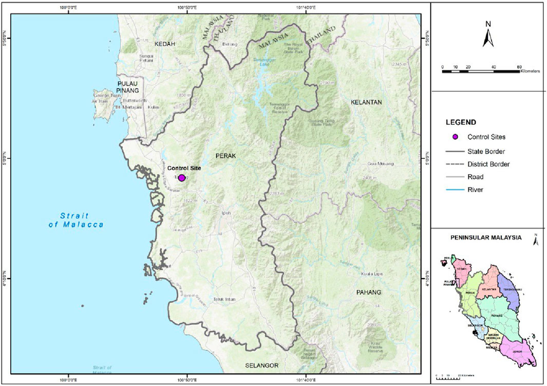
Figure 1. Map of control area (bukit larut, perak).
2.1.3 Preparation and exposure of lichen samples
Upon arrival at the lab, the lichen samples were separated into small blobs weighing around 10–20 g each. These blobs were placed in nets with holes to allow pollutants to be absorbed by the samples (Abas et al., 2020). To measure the nicotine concentration within a building, 34 sampling sites were selected, as shown in Table 1. The samples were hung from the ceiling, with some positioned near ventilation systems.
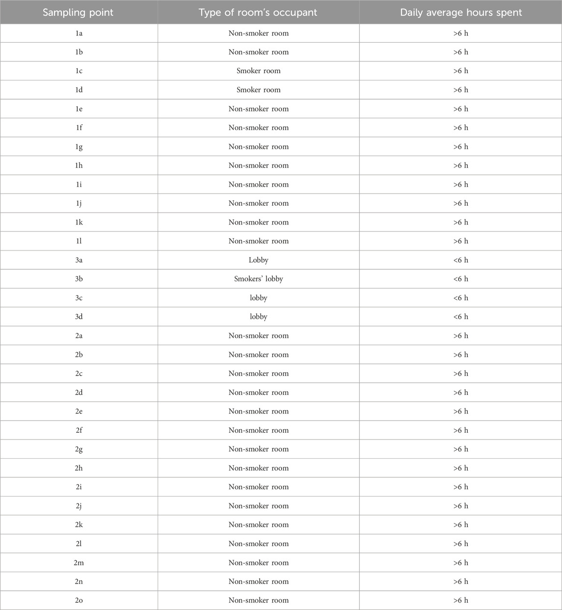
Table 1. The sampling point description.
A total of 30 sampling sites were selected on the ground floor of D block, Faculty of Social Sciences and Humanities, The National University of Malaysia, Selangor. The sampling points were determined using a stratified technique based on room segregation. The selection of sampling points was guided by three main factors: a) proximity to smokers’ rooms, b) average hours spent in the room, and c) quality of air flow and ventilation system. Figure 2 shows the location of Bukit Larut (control site), and Figure 3 details each selected sampling point.
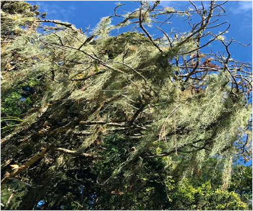
Figure 2. Lichen usnea misaminensis found at bukit barut (control area). Source: field work (2023).
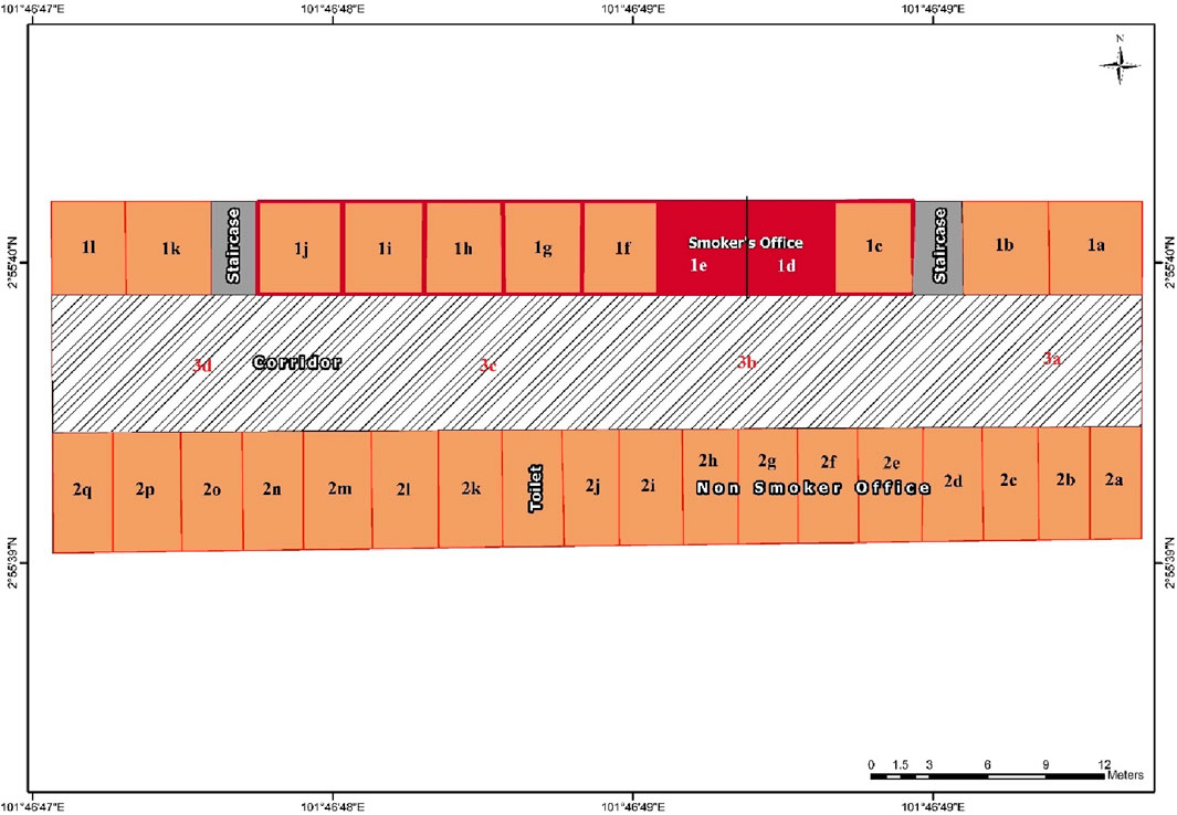
Figure 3. The map of the sampling point.
2.1.4 Exposure period
The lichen samples were exposed indoors for approximately 2 months (from 1 February 2023, to 3 April 2023). During this period, the lichens absorbed trace elements, particularly nicotine, from the indoor air and surfaces such as ceilings and walls. According to Yang et al. (2022), nicotine traces can be found in the environment up to a week after exposure from its source, making a 2-month exposure period sufficient for the lichens to absorb all traces of nicotine.
2.1.5 Post-exposure handling and analysis
After the 2-month exposure period, all lichen samples were packed in paper bags to keep them dry and then transported to the lab for further analysis. The nicotine concentration and vitality rate of the lichen samples were subsequently measured.
2.2 Analysis procedure of nicotine concentration
50 g of lichen specimens from each sampling point were desiccated and pulverized into a powdered form. Subsequently, 10 g of the powdered material underwent extraction using a Soxhlet apparatus (Barosil), employing 100 mL of various organic solvents such as methanol, acetone, benzene, and diethyl ether for 24 h at a controlled temperature of 45°C. Following 6 to 8 cycles of heat-driven extraction, the resultant solvent-laden extract was evaporated in a hot air oven maintained at a temperature range of 42°C–45°C, yielding dry extract quantities of up to 2 g per extraction.
After the extraction process, analytical scrutiny was executed via gas chromatography coupled with mass spectrometry (GC-MS) using a quadrupole mass spectrometer and an Elite-wax Capillary-column chromatography (dimensions: 60.0 m × 250 μm × 0.25 μm). Sample injection, facilitated by an autosampler, was achieved at a split ratio of 10:1. The temperature regime entailed an initial setting of 60°C for 1 min, followed by programmed increments to 200°C, with a holding period of 3 min, further augmented to a range of 10°C–300°C, sustained for a duration of 10 min. The injection port temperature was maintained at 280°C, with helium serving as the carrier gas, flowing at a rate of 1.0 mL/min.
A solvent delay of 7 min, with a corresponding transferred temperature of 160°C, was enacted, alongside a source temperature of 150°C. The compositional analysis of the extract was predicated upon the computation of percentages derived from the areas of GC peaks, with the identification of the nicotine peak conducted at 280 nm. Calibration procedures were enacted utilizing a nicotine standard, specifically (±)-Nicotine – NO267 (Sigma-Aldrich), while the limit of quantification (LOQ) was established at 5.5 μg/mL. Each sampling point was represented by triplicate measurements (Paoli et al., 2019).
2.3 Lichen’s vitality rate measurement
To assess the vitality rates among lichen samples across all sampling sites, analysis of chlorophyll (Chl.) α fluorescent emissions was employed. The evaluation of the photosynthetic components in the lichen samples was conducted based on the fundamental principle of photochemical quantum yield, specifically FV/FM, where FV represents the variable fluorescence, while FM and F0 denote the maximum and minimum Chl α fluorescence values, respectively. Additionally, the overall index of photosynthetic performance was computed using the Performance Index formula (PIABS).
Subsequently, the samples underwent a reactivation period of 24 h, followed by misting with mineral water and storage at 16°C under ambient light conditions (70 µmol m⁻2 s⁻1). After this, the samples were misted again with water and subjected to darkness for 10 min before being exposed to saturated light for a brief duration (3,000 µmol m⁻2 s⁻1), during which fluorescent emissions were recorded. Measurement procedures were conducted utilizing the Plant Efficiency Analyzer (Handy PEA, Hansatech Ltd., King’s Lynn, Norfolk, United Kingdom) (Protano et al., 2017).
2.4 Data analysis and interpretation
The analysis of nicotine concentration data and vitality rates involved using the Pearson coefficient correlation, executed via Past Software V4.03, to determine the connection between nicotine levels in lichen and their vitality post-exposure. This statistical method was crucial in identifying the relationship between the nicotine concentration absorbed by the lichen samples and their subsequent photosynthetic performance.
Additionally, the nicotine concentration data was examined through Geographical Information System (GIS) interpolation analysis using ArcGIS software version 10.8.1. Interpolation analysis constructs a surface based on values from isolated sample locations, with each sample point representing a site of data collection that includes recorded geographical coordinates. The delineation of nicotine concentration distribution used the lowest concentration (LoQ) of environmental tobacco smoke, set at 0.45 μg/mL (Alqahtani et al., 2020), as the cutoff point.
To facilitate visualization, five concentration ranges were established for the sampling points, each with distinct interpretations as detailed in Table 2. The visualization employed a color scheme: red for “Very High Concentration,” indicating a strong presence of nicotine; orange for “High Concentration,” signifying a potent but slightly lower level; yellow for “Moderate Concentration,” indicating a balanced presence; light green for “Low Concentration,” representing reduced nicotine levels; and dark green for “Ambient Concentration,” showing minimal nicotine content. This color scheme using GIS aids users in quickly interpreting and understanding the geographical distribution and strength of nicotine concentration.
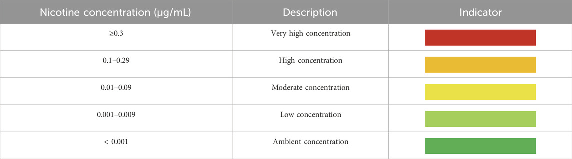
Table 2. The indication of nicotine concentration’s range.
3 Results
Table 3 shows the nicotine concentration and the vitality rate of the lichen sample after 2 months of exposure at the sampling points. The two smokers’ rooms, have the highest nicotine concentration (0.61 μg/mL and 0.57 μg/mL) and also the lowest lichen vitality’s rate (0.27 μg/mL and 0.29 μg/mL) compare to the other sampling points.
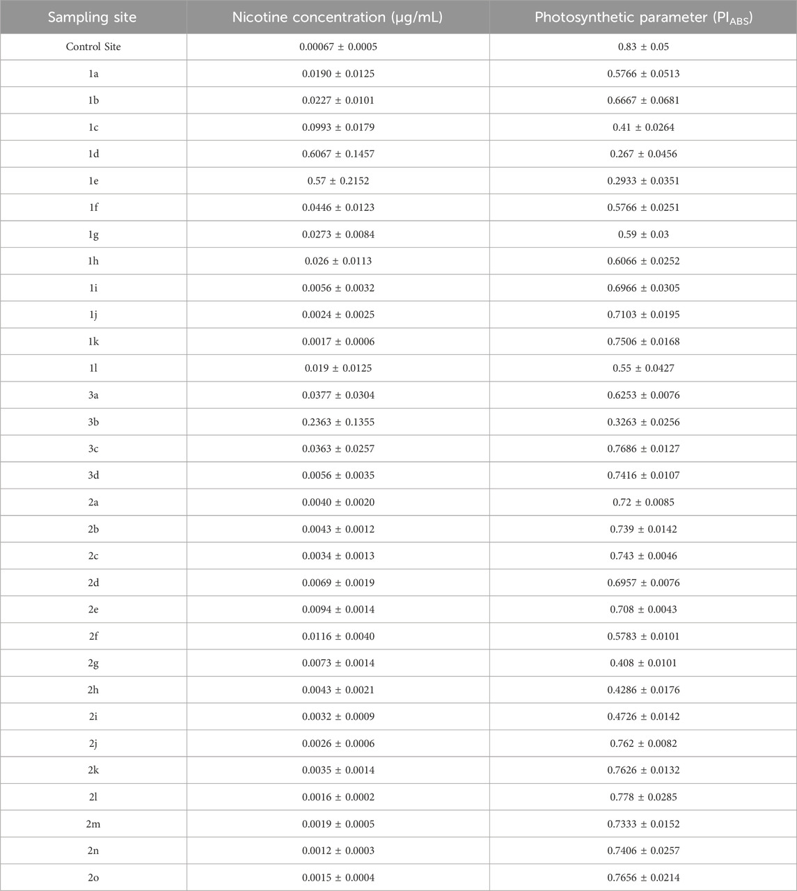
Table 3. The nicotine concentration and lichen’s vitality rate.
The analysis revealed a significant inverted relationship between nicotine concentration and the vitality rate of the lichen, with a Pearson coefficient value of −0.71 (the significant value range being −1 < r < 1). This indicates that higher nicotine concentrations are associated with lower vitality rates in the lichen samples. In other words, as the nicotine concentration increases, the vitality of the lichen decreases.
Figure 4 illustrates the spatial analysis of nicotine concentration at the sampling site. The red zones indicate areas of high nicotine concentration, while the green zones represent areas of low nicotine concentration. The analysis shows that nicotine concentrations are highest near the smokers’ rooms, with concentrations decreasing as the distance from these sources increases. However, some sampling points located far from the primary nicotine sources also exhibited traces of nicotine, indicating potential secondary distribution pathways or lingering effects in the environment.
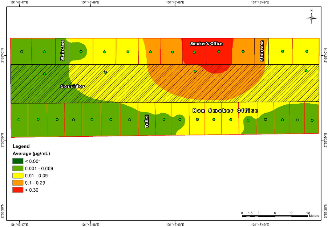
Figure 4. The map of nicotine concentration distribution.
4 Discussion
Lichens have long been recognized as sensitive indicators of environmental pollution due to their capacity to absorb and accumulate airborne pollutants (Abas, 2021). This characteristic makes them valuable bioindicators for assessing air quality. Recent studies have demonstrated that lichens can absorb and retain nicotine from the atmosphere, thereby serving as potential tools for detecting nicotine in indoor environments. Lichen thalli, the main body of lichens, possess a large surface area that efficiently absorbs airborne substances, including nicotine molecules (da Silva et al., 2021). This study confirms the efficient detection and absorption of nicotine by lichens, showing significant differences in nicotine concentrations between smokers’ and non-smokers’ rooms. Similar results were found in a study by Tao et al. (2023), which employed solid phase cartridge sampling followed by GC-MS analysis, and in other studies using conventional methods, such as those by Habibagahi et al. (2020).
The vitality of lichens is significantly influenced by air pollutants in their environment. This study also indicates that nicotine has a negative impact on lichen vitality rates, showing a significant negative correlation between nicotine concentration levels and lichen vitality. This suggests that higher nicotine concentrations lead to lower lichen vitality rates. Nicotine, known for its toxic properties, can adversely affect various organisms, including plants and fungi. According to Cheng et al. (2021), nicotine exhibits allelopathic inhibitory effects on chlorophyll and seeds, as well as hormesis effects on germinated seeds and seedlings. This underscores the potential negative effects of nicotine on lichen growth, considering that lichens also contain chlorophyll (Santos et al., 2022).
The distribution of nicotine concentrations in this study shows a noticeable trend of decreasing concentrations farther from smokers’ rooms, reflecting equilibrium effects in the vicinity. However, traces of nicotine were detected in some non-smokers’ rooms despite separation by fiber walls and ceilings. This phenomenon may be attributed to several factors, including the volatility and dispersion of nicotine, influences of air currents and ventilation, adsorption and deposition processes, and diffusion within indoor environments. Marcham and Springston (2019) noted the rapid volatilization of nicotine from cigarettes into the air, forming airborne plumes that disperse quickly, with detectable concentrations observed several meters away from the emission source. Studies by Sheu et al. (2020) on the effects of ventilation and air movement on environmental tobacco smoke (ETS) distribution indoors demonstrated that airflow and ventilation systems significantly influence the movement of ETS particles, facilitating their transport to distant locations within buildings (Jung et al., 2020). Adsorption and deposition studies have shown that nicotine particles suspended in the air can deposit onto surfaces, leading to the accumulation of detectable levels away from their source. Additionally, Wu et al. (2022) highlighted the role of diffusion in the uniform distribution of nicotine molecules throughout indoor air, emphasizing how diffusion processes contribute to the spatial distribution of nicotine within buildings.
5 Conclusion
Lichens are widely recognized as sensitive bioindicators of environmental pollution, including specific pollutants like nicotine. This study confirms that lichens can effectively indicate indoor air quality, particularly regarding nicotine levels. Moreover, it establishes a significant negative relationship (r = −0.71) between nicotine concentration and lichen vitality, indicating that higher nicotine levels correlate with reduced lichen health. The study highlights several factors contributing to nicotine’s presence in indoor environments, such as volatility, dispersion, air currents, ventilation patterns, adsorption, deposition, and diffusion processes.
However, a limitation of this study is its reliance on cumulative nicotine concentration measurements over a 2-month period, rather than continuous monitoring. Future research could benefit from real-time monitoring techniques to capture fluctuations in nicotine levels more precisely.
Research into nicotine detection using lichens could lead to innovative indoor air quality monitoring methods. Integrating lichen-based sensors into portable devices or passive sampling systems offers cost-effective and non-intrusive means to continuously monitor nicotine levels in buildings. These advancements can complement existing air quality monitoring strategies, enabling targeted interventions to reduce tobacco smoke exposure.
Understanding the spatial distribution of nicotine within indoor environments carries significant public health implications. Identifying nicotine in areas distant from smoking zones can raise awareness about the risks of second-hand smoke exposure, supporting efforts to promote smoke-free environments. Additionally, using lichens for nicotine detection can enhance environmental education and advocacy efforts. Lichens’ accessibility and visual appeal make them suitable for educational programs aimed at increasing awareness of air pollution’s impact on ecosystems and human health. Engaging the public in citizen science initiatives involving lichen monitoring can promote environmental stewardship and empower communities to advocate for cleaner air and healthier living spaces.
This study suggests several recommendations, including implementing and enforcing smoke-free policies in indoor environments, conducting regular monitoring of indoor air quality (including nicotine levels) in smoking-permitted or suspected buildings, promoting tobacco cessation programs, and launching education campaigns on the dangers of second-hand smoke. These efforts require collaboration among various stakeholders to create supportive environments that prioritize public health and wellbeing.
Data availability statement
The raw data supporting the conclusions of this article will be made available by the authors, without undue reservation.
Author contributions
AA: Funding acquisition, Investigation, Methodology, Project administration, Writing–original draft. FR: Data curation, Formal Analysis, Writing–review and editing. MJ: Conceptualization, Investigation, Writing–review and editing.
Funding
The author(s) declare that financial support was received for the research, authorship, and/or publication of this article. This study has been supported by Universiti Kebangsaan Malaysia through a research grant (SK-2023-034).
Acknowledgments
Dr. Rose Norman for her generous assistance in conducting the proofreading for this article.
Conflict of interest
The authors declare that the research was conducted in the absence of any commercial or financial relationships that could be construed as a potential conflict of interest.
Publisher’s note
All claims expressed in this article are solely those of the authors and do not necessarily represent those of their affiliated organizations, or those of the publisher, the editors and the reviewers. Any product that may be evaluated in this article, or claim that may be made by its manufacturer, is not guaranteed or endorsed by the publisher.
References
Abas, A. (2021). A systematic review on biomonitoring using lichen as the biological indicator: a decade of practices, progress and challenges. Ecol. Indic. 121, 107197. doi:10.1016/j.ecolind.2020.107197
Abas, A., Aiyub, K., and Awang, A. (2022). Biomonitoring potentially toxic elements (PTEs) using lichen transplant Usnea misaminensis: a case study from Malaysia. Sustain. Switz. 14 (12), 7254. doi:10.3390/su14127254
Abas, A., and Awang, A. (2017). Air pollution assessments using lichen biodiversity index (LBI) in kuala lumpur, Malaysia. Pollut. Res. 36 (2), 242–249.
Abas, A., Awang, A., and Aiyub, K. (2020). Analysis of heavy metal concentration using transplanted lichen Usnea misaminensis at Kota Kinabalu, Sabah (Malaysia). Appl. Ecol. Environ. Res. 18 (1), 1175–1182. doi:10.15666/aeer/1801_11751182
Akyuz, E. (2023). An environmental human rights approach to environmental tobacco smoking. Muslim World J. Hum. Rights 20, 97–120. doi:10.1515/mwjhr-2022-0024
Alqahtani, S., Cooper, B., Spears, C. A., Wright, C., and Shannahan, J. (2020). Electronic nicotine delivery system-induced alterations in oral health via saliva assessment. Exp. Biol. Med. 245 (15), 1319–1325. doi:10.1177/1535370220941258
Benítez, A., Aragón, G., González, Y., and Prieto, M. (2018). Functional traits of epiphytic lichens in response to forest disturbance and as predictors of total richness and diversity. Ecol. Indic. 86, 18–26. doi:10.1016/j.ecolind.2017.12.021
Boonpeng, C., Polyiam, W., Sriviboon, C., Sangiamdee, D., Watthana, S., Nimis, P., et al. (2017). Airborne trace elements near a petrochemical industrial complex in Thailand assessed by the lichen Parmotrema tinctorum (Despr. ex Nyl.) Hale. Environ. Sci. And Pollut. Res. 24 (13), 12393–12404. doi:10.1007/s11356-017-8893-9
Boonpeng, C., Sriviboon, C., Polyiam, W., Sangiamdee, D., Watthana, S., and Boonpragob, K. (2018). Assessing atmospheric pollution in a petrochemical industrial district using a lichen-air quality index (LiAQI). Ecol. Indic. 95, 589–594. doi:10.1016/j.ecolind.2018.08.012
Cheng, Y. D., Bai, Y. X., Jia, M., Chen, Y., Wang, D., Wu, T., et al. (2021). Potential risks of nicotine on the germination, growth, and nutritional properties of broad bean. Ecotoxicol. Environ. Saf. 209, 111797. doi:10.1016/j.ecoenv.2020.111797
Conti, M., and Cecchetti, G. (2001). Biological monitoring: lichens as bioindicators of air pollution assessment — a review. Environ. Pollut. 114 (3), 471–492. doi:10.1016/s0269-7491(00)00224-4
da Silva, I. G., de Oliveira Nunes, C. R., de Oliveira Costa, R., Pereira, E. C., and Canela, M. C. (2021). Formaldehyde exposure and atmospheric biomonitoring with lichen Cladonia verticillaris in an anatomy laboratory. Environ. Sci. Pollut. Res. 28 (35), 48569–48580. doi:10.1007/s11356-021-14036-9
Digiacomo, S. I., Jazayeri, M. A., Barua, R. S., and Ambrose, J. A. (2019). Environmental tobacco smoke and cardiovascular disease. Int. J. Environ. Res. Public Health. MDPI AG 16, 96. doi:10.3390/ijerph16010096
Habibagahi, A., Alderman, N., and Kubwabo, C. (2020). A review of the analysis of biomarkers of exposure to tobacco and vaping products. Anal. Methods. R. Soc. Chem. 12, 4276–4302. doi:10.1039/d0ay01467b
Jung, C. C., Lin, W. Y., Hsu, N. Y., Wu, C. D., Chang, H. T., and Su, H. J. (2020). Development of hourly indoor pm2.5 concentration prediction model: the role of outdoor air, ventilation, building characteristic, and human activity. Int. J. Environ. Res. Public Health 17 (16), 5906–5917. doi:10.3390/ijerph17165906
Klimek, B., Tarasek, A., and Hajduk, J. (2015). Trace element concentrations in lichens collected in the beskidy mountains, the outer western carpathians. Bull. Environ. Contam. Toxicol. 94 (4), 532–536. doi:10.1007/s00128-015-1478-8
Krakowiak, E., Sygit, K., Sygit, M., Cipora, E., and Krakowiak, J. (2020). Exposure to environmental tobacco smoke (ETS) among employees of hospitality venues in the light of changes in anti-tobacco legislation in Poland. Int. J. Environ. Res. Public Health 17 (10), 3691. doi:10.3390/ijerph17103691
Kurnaz, K., and Cobanoglu, G. (2017). Biomonitoring of air quality in istanbul metropolitan territory with epiphytic lichen physcia adscendens (FR.) H. Olivier. Fresenius Environ. Bull. 26 (12), 7296–7308.
Marcham, C. L., and Springston, J. P. (2019). Electronic cigarettes in the indoor environment. Rev. Environ. Health 34, 105–124. doi:10.1515/reveh-2019-0012
Musa, F., and Nadarajah, R. (2023). Valuing visitor’s willingness to pay for green tourism conservation: a case study of Bukit Larut Forest Recreation Area, Perak, Malaysia. Sustain. Environ. 9 (1). doi:10.1080/27658511.2023.2188767
Nash, T. H. (2008) “Lichen biology,” in Lichen biology. Second Edition. Cambridge University Press, 1–486. doi:10.1017/CBO9780511790478
Paoli, L., Maccelli, C., Guarnieri, M., Vannini, A., and Loppi, S. (2019). Lichens “travelling” in smokers’ cars are suitable biomonitors of indoor air quality. Ecol. Indic. 103, 576–580. doi:10.1016/j.ecolind.2019.04.058
Parida, L., and Patel, T. N. (2023). Systemic impact of heavy metals and their role in cancer development: a review. Environ. Monit. Assess. 195 (6), 766. doi:10.1007/s10661-023-11399-z
Protano, C., Owczarek, M., Antonucci, A., Guidotti, M., and Vitali, M. (2017). Assessing indoor air quality of school environments: transplanted lichen Pseudevernia furfuracea as a new tool for biomonitoring and bioaccumulation. Environ. Monit. Assess. 189, 358. doi:10.1007/s10661-017-6076-2
Root, H., Geiser, L., Jovan, S., and Neitlich, P. (2015). Epiphytic macrolichen indication of air quality and climate in interior forested mountains of the Pacific Northwest, USA. Ecol. Indic. 53, 95–105. doi:10.1016/j.ecolind.2015.01.029
Santos, A. M. dos., Vitorino, L. C., Cruvinel, B. G., Ávila, R. G., Vasconcelos Filho, S. de C., Batista, P. F., et al. (2022). Impacts of Cd pollution on the vitality, anatomy and physiology of two morphologically different lichen species of the genera parmotrema and Usnea, evaluated under experimental conditions. Diversity 14 (11), 926. doi:10.3390/d14110926
Sheu, R., Stönner, C., Ditto, J. C., Klüpfel, T., Williams, J., and Gentner, D. R. (2020). Human transport of thirdhand tobacco smoke: a prominent source of hazardous air pollutants into indoor nonsmoking environments. Sci. Adv. 6 (10), eaay4109. doi:10.1126/sciadv.aay4109
Simonavicius, E., McNeill, A., Shahab, L., and Brose, L. S. (2019) “Heat-not-burn tobacco products: a systematic literature review,” in Tobacco control. Tavistock Square London, United Kingdom: BMJ Publishing Group. doi:10.1136/tobaccocontrol-2018-054419
Tanir, F., and Mete, B. (2022). “Impacts of the indoor air quality on the health of the employee and protection against these impacts,” in Air quality and health (London, United Kingdom: IntechOpen). doi:10.5772/intechopen.102708
Tao, X. Y., Zhang, Y., Zhou, Y., Liu, Z. F., and Feng, X. S. (2023). Nicotine in complex samples: recent updates on the pretreatment and analysis method. Crit. Rev. Anal. Chem. 53, 1209–1238. doi:10.1080/10408347.2021.2016365
Wu, S., Kim, E., Vethanayagam, D., and Zhao, R. (2022). Indoor partitioning and potential thirdhand exposure to carbonyl flavoring agents added in e-cigarettes and hookah tobacco. Environ. Sci. Process. Impacts 24 (12), 2294–2309. doi:10.1039/d2em00365a
Yang, J., Hashemi, S., Han, W., Song, Y., and Lim, Y. (2022). Exposure and risk assessment of second-and third-hand tobacco smoke using urinary cotinine levels in South Korea. Int. J. Environ. Res. Public Health 19 (6), 3746. doi:10.3390/ijerph19063746
Keywords: biological indicator, lichens, nicotine, indoor environment, sick building syndrome
Citation: Abas A, Rasli FN and Juhari ML (2024) Lichen as the biological indicator for detection of environmental tobacco smoke (ETS) at the public office building in Selangor, Malaysia. Front. Environ. Sci. 12:1433941. doi: 10.3389/fenvs.2024.1433941
Received: 18 May 2024; Accepted: 25 June 2024;
Published: 10 July 2024.
Edited by:
Oana Cadar, INCDO INOE 2000 Research Institute for Analytical Instrumentation, RomaniaReviewed by:
Katalin Hubai, University of Pannonia, HungaryManfred Neuberger, Medical University of Vienna, Austria
Manfred Sager, Bioforschung Austria, Austria
Copyright © 2024 Abas, Rasli and Juhari. This is an open-access article distributed under the terms of the Creative Commons Attribution License (CC BY). The use, distribution or reproduction in other forums is permitted, provided the original author(s) and the copyright owner(s) are credited and that the original publication in this journal is cited, in accordance with accepted academic practice. No use, distribution or reproduction is permitted which does not comply with these terms.
*Correspondence: Azlan Abas, azlanabas@ukm.edu.my